Embed Size (px)
Citation preview
British Journal of Oral Surgery 16 (1978-79) 207-211
FLORID FOCAL EPITHELIAL HYPERPLASIA IN FANCONI’S SYNDROME
ALLAN G. FARMAN~. 2 and JOHN KATZ~, 3 *
1 Department of Oral Pathology, University of Stellenbosch and Dental Epidemiology Research Group of the South African Medical Research Council; 2 Department of Oral Medicine, University of the Western Cape; 3 Red Cross Memorial Children’s Hospital,
Rondebosch, Cape
(Received 9 August 1977; accepted 13 September 1977)
Summary. Florid focal epithelial hyperplasia of at least 5 years’ duration in a Cape Coloured boy aged 12 years and suffering from Fanconi’s syndrome is reported.
Introduction
Focal epithelial hyperplasia (I.C.D. - D.A. 528.73, 529.73) was defined as an entity by Archard et al. (1965) when they reported a condition first observed by Heck on the oral mucosa of Navajo Indian children. The clinical appearance is typically that of soft nodular elevations on the oral mucosa, usually 1 to 5 mm in diameter, but sometimes so numerous as to form larger plaques (van Wyk et al., 1977a). These nodules become less noticeable when the mucosa is stretched (Archard et al., 1965). Histologically, there is acanthosis with horizontal outgrowths and anastomosis of the rete ridges, and enlarged cells with mitosis-like degeneration are noticeable within the prickle-cell layer (Praetorius-Clausen, 1969). Recently, viral particles measuring 500 A in diameter and probably of the Papova group, were observed in this condition (Praetorius-Clausen & Willis, 1971; Hanks et al., 1972; van Wyk et al., 1977b). Whilst one such report could represent an adventitious virus, the finding of similar particles in lesions from persons residing in diverse geographic centres constitutes strong circumstantial evidence of a likely cause-and-effect relationship.
Van Wyk (1977) described 76 cases of focal epithelial hyperplasia encountered during an epidemiological survey in the Western Cape region of South Africa. All of these cases were restricted to pupils of remand schools and residents of a secluded community. In that paper it was questioned whether focal epithelial hyperplasia was a hitherto undetected manifestation of an existing virus or a disease newly introduced into South Africa. This communication outlines a particularly florid case of focal epithelial hyperplasia in a boy with Fanconi’s syndrome. Since the child was described as having a cobblestone appearance to the oral mucosa as early as 1972 it would appear that focal epithelial hyperplasia was introduced into South Africa before that date. Ignorance of the features of this disease is the probable reason for it not being commonly described previously.
Case report
A 12-year-old Cape Coloured boy was referred to the Dental Section of the Red Cross Memorial Children’s Hospital, Rondebosch, for treatment of dental caries and
* Address for correspondence: Dr J. Katz, Department of Oral Medicine, University of the Western Cape, Private Bag 17, Bellville, Cape, South Africa.
207
208 I3 It I T I S H J ( ) II It N A I, 0 I; 0 K A 1 S l! I< G I‘ I< \I’
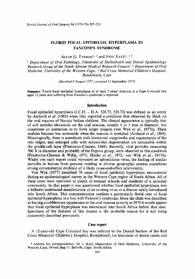
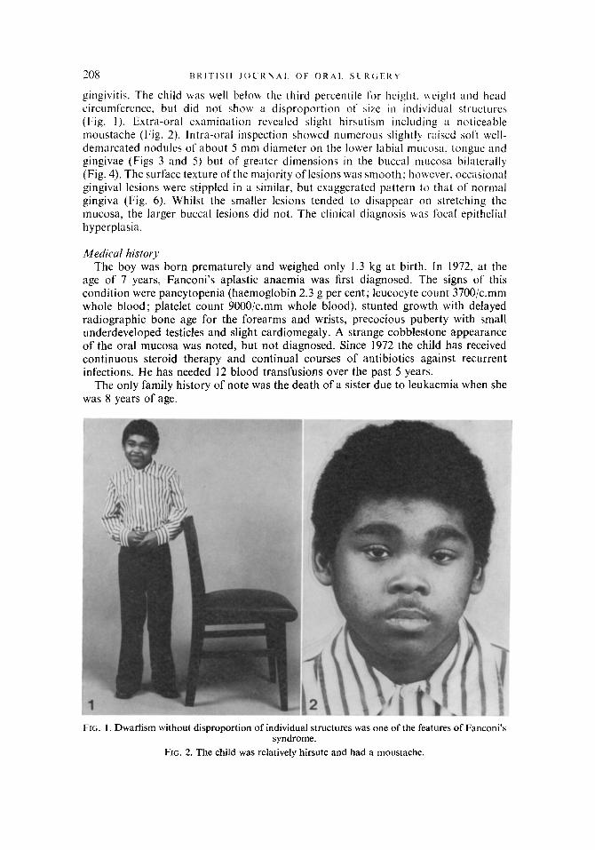
gingivitis. The child was well below the third percentile for height. height and head circumference, but did not show a disproportion of sire in individual structures (Fig. 1). Extra-oral examination revealed slight hirsutism including a noticeable moustache (Fig. 2). Intra-oral inspection showed numerous slightly raised soft well- demarcated nodules of about 5 mm diameter on the lower labial mucosa. tongue and gingivae (Figs 3 and 5) but of greater dimensions in the buccal IIILICOS~ bilaterally (Fig. 4). The surface texture ofthe majority of lesions was smooth: however, occasional gingival lesions were stippled in a similar, but exaggerated pattern to that of normal gingiva (Fig. 6). Whilst the smaller lesions tended to disappear on stretching the mucosa, the larger buccal lesions did not. The clinical diagnosis was focal epithelial hyperplasia.
Medical history The boy was born prematurely and weighed only 1.3 kg at birth. In 1972. at the
age of 7 years, Fanconi’s aplastic anaemia was first diagnosed. The signs of this condition were pancytopenia (haemoglobin 2.3 g per cent; leucocyte count 3700/c.mm whole blood; platelet count 9000/c.mm whole blood), stunted growth with delayed radiographic bone age for the forearms and wrists, precocious puberty with small underdeveloped testicles and slight cardiomegaly. A strange cobblestone appearance of the oral mucosa was noted, but not diagnosed. Since 1972 the child has received continuous steroid therapy and continual courses of antibiotics against recurrent infections. He has needed 12 blood transfusions over the past 5 years.
The only family history of note was the death of a sister due to leukaemia when she was 8 years of age.
FIG. I. Dwarfism without disproportion of individual structures was one of the features of Fanconi’s syndrome.
FIG. 2. The child was relatively hirsute and had a moustache.
FOCAL EPITHELIAL HYPERPLASIA IN FANCONI’S SYNDROME 209
FIG. 3. Flaccid nodules of approximately 5 mm diameter, typical of focal epithehal hyperplasia, were present on the tongue and lower lip.
FIG. 4. Larger lesions, possibly due to the confluence of several nodules, were present on the cheek mucosa.
FIG. 5. Gingival lesions commonly resembled the nodules on the tongue and lip. FIG. 6. Occasional gingival lesions had a granular appearance.
Histopathology In view of the florid nature of the lesions, the atypical size of the buccal lesions and
the unusual texture of several gingival lesions, it was decided to take biopsy specimens from the right cheek, lower lip and gingivae. These were obtained under local anaes- thetic and the specimens were fixed in Bouin’s fluid for 24 hours prior to selected slices being processed for routine light microscopic examination.
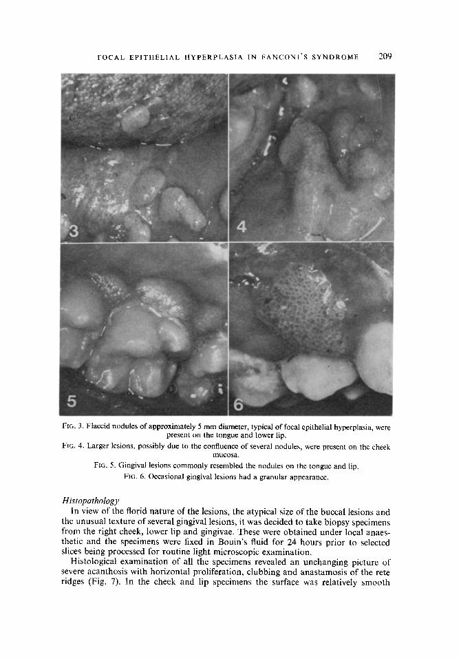
Histological examination of all the specimens revealed an unchanging picture of severe acanthosis with horizontal proliferation, clubbing and anastamosis of the rete ridges (Fig. 7). In the cheek and lip specimens the surface was relatively smooth
FIG. 7. Acanthosis with rete ridge anastomosis. (H Xr E. 35.)
FIG. 8. Shallow crenations on the surface of a granular gingival lesion (arrowed). (H & E. 50.)
FIG. 9. Mitosis-like and granular nuclear degeneration in enlarged prickle-cells. (H & E. ,: 252.)
FIG. 10. Mitosis-like nuclear degeneration. (H & E. x 400.)
FOCAL EPITHELIAL HYPERPLASIA IN FANCONI’S SYNDROME 211
whereas the gingival biopsy specimen had distinct shallow crenations (Fig. 8). A basket-weave appearance due to intra-cellular oedema was only noticeable in the specimen from the cheek mucosa, but sections from all three sites contained enlarged prickle-cells with mitosis-like and granular degenerations (Figs 9 and 10). There was no evidence of atypia and the lamina propria contained only a very slight chronic inflammatory cell infiltrate. These features are pathognomonic of focal epithelial hyperplasia.
Follow-up A 6-month review examination showed the florid focal epithelial hyperplasia to be
a chronic malady. Nevertheless, the distribution of the lesions had altered somewhat. The gingivae were free from lesions, but the tongue was much more severely affected by the disease.
Discussion
As there would appear to be no previously reported case of focal epithelial hyper- plasia in a patient with Fanconi’s syndrome it is possible that the two conditions occurred simultaneously simply by chance. It must, however, be remembered that certain viral infections (e.g. recurrent herpes simplex) are commonly related to systemic ill-health (Shafer et al., 1974). If, as seems likely, focal epithelial hyperplasia is also virally-induced this particularly severe case could have been triggered or aggravated by the pancytopenia.
Acknowledgements
We wish to thank Mr C. Maree for preparation of the histological specimens and Mr M. Jooste for photographic assistance.
References
Archard, H. O., Heck, J. W. & Stanley, H. R. (1965). Focal epithelial hyperplasia: an unusual oral mucosal lesion found in Indian children. Oral Surgery, Oral Medicine and Oral Pathology, 20, 201.
Hanks, C. T., Fischman, S. L. & de Guzman, M. N. (1972). Focal epithelial hyperplasia. A light and electron microscopic study of one case. Oral Surgery, Oral Medicine and Oral Pathology, 33, 934.
Praetorius-Clausen, F. & Willis, J. M. (1971). Papova virus-like particles in focal epithelial hyper- plasia. Scandinavian Journal of Dental Research, 79, 362.
Praetorius-Clausen, F. (1969). Histopathology of focal epithelial hyperplasia. Evidence of viral infection. TandIaegebladet, 73, 1013.
Shafer, W. G., Hine, M. K. & Levy, B. M. (1974). Textbook of Oral Pathology, 3rd Ed., pp. 328-329. Philadelphia, London, Toronto: Saunders.
Van Wyk, C. W. (1977). Focal epithelial hyperplasia of the mouth. Recently discovered in South Africa. British Journal of Dermatology, 96, 381.
Van Wyk, C. W., Staz, J. & Farman, A. G. (1977a). Focal epithelial hyperplasia in a group of South Africans. Its clinical and microscopic features. Journal of Oral Pathology, 6, 1.
Van Wyk, C. W., Staz, J. and Farman, A. G. (1977b). Focal epithelial hyperplasia in a group of South Africans. Its ultrastructure features. Journal of Oral Pathology, 6, 14.