Embed Size (px)
Citation preview

Phytochemistry, Vol. 32, No. 5, pp. 1301 1303, 1993 003 l-9422/93 S6.00 + 0.00 Printed in Great Britain. G 1993 Pergamon Press Ltd
FLAVONOIDS FROM THE TWIGS OF EUCRYPHIA GLUTINOSA
SILVIA SEPULVEDA-BOZA,* SUALAHEEK DELHVI and BRUCE K. CASSELS~
Institut fiir Physiologische Chemie der Universitlt Bonn, Bonn, F.R.G.; tDepartamento de Quimica, Facultad de Ciencias, Universidad de Chile, Casilla 653, Santiago 1, Chile
(Received in revised form 21 August 1992)
Key Word Index-Eucryphia glutinosa; Eucryphiaceae; twigs; flavonoids; dihydroquercetin 3-O-p-D- xyloside; reynoutrin; jaceidin 5-0-p-D-glucoside; caryatin 7-0-P-D-glucoside.
Abstract-The rare dihydroquercetin 3-0-fl-D-xyloside, caryatin 7-0-p-D-glucoside and the previously unknown 6-methoxylated flavonoid glycoside jaceidin 5-0-b-D-glucoside were isolated from twigs of Eucryphia glutinosa where they co-o&ur with the widespread Ieynoutrin.
INTRODUCTION
The leaf flavonoids of Eucryphia cordifolia Cav. and E. glutinosa (Poepp. et Endl.) Baillon, from south central Chile, have been shown to differ markedly from those of the three Australasian species of this small, Southern genus. Both Chilean species are characterized by the presence of quercetin-5-methyl ether (azaleatin), 3-glycos- ides of this unusual flavonol methyl ether, and the very rare quercetin 3,5-dimethyl ether (caryatin) in their leaves, as well as a number of quercetin glycosides and unidenti- fied constituents Cl]. More recently, the trunk bark of E. cordifolia was studied and shown to contain the 3-0-a- L-rhamnosides of dihydroquercetin (astilbin, the major component) and of dihydrokaempferol (engelitin), as well as a new chromone 3-0-ol+rhamnoside named eu- cryphin; no 0-methylated flavonoids were found, how- ever [2]. Eucryphia glutinosa is the only other species growing on the American continent, where it is known as ‘guindo santo’ (‘holy cherry tree’) because of its appear- ance when covered with large white blossoms. It is only found near rivers and streams west of the Andes between latitudes 36” and 38” south, north of the range of the much larger E. cordifolia [3]. We now report the results of an analysis of the butanol-soluble fraction of the twigs of E. glutinosa.
RESULTS AND DISCUSSION
The crude butanol extract was subjected to chromato- graphy on a silica gel column to give two main flavonoid- containing fractions. The larger of these, after column chromatography on Sephadex LH-20 and TLC on silica gel, afforded two pure compounds. The major constituent
*Author to whom correspondence should be addressed. Present address: Departamento de Quimica, Facultad de Ciencia, USACH, Casilla 307, Santiago 2, Chile.
was found to be dihydroquercetin 3-0-fl-D-xyloside (1) by spectroscopic methods and by acid hydrolysis and chro- matographic comparison of the products with standards. This substance had been found, but not fully characte- rized, as a component of an unresolved mixture of dihydroquercetin monoglycosides in leaves of the Tas- manian E. milliganii [1] and later reported as a consti- tuent of Leucothoe keiskei (Ericaceae) [4] and of Cordia obliqua (Boraginaceae) [S]. Its abundance in E. glutinosa twigs parallels that of the analogous dihydroquercetin rhamnoside in the bark of E. cordifolia [2]. The second known substance now reisolated from this fraction of E. glutinosa is the widely distributed quercetin 3-O-/?-D- xyloside (reynoutrin), which may be assumed to be bio- genetically related to its dihydro derivative.
The minor flavonoid fraction was further fractionated by successive chromatographic runs on Sephadex and on silica gel which afforded a small amount of the 7-O-/?-D- glucoside of caryatin (2) and the previously undescribed compound 3. The complete ‘H and “CNMR spectral assignments of 2, based on 2D experiments, are sum- marized in Tables 1 and 2.
Compound 3 was laevorotatory in pyridine. Its UV spectrum showed maxima at 254 (log E 3.52), 275 (log E 3.60), and 353 nm (log E 3.76). Upon addition of the usual reagents [6]. NaOMe produced a 42 nm bathochromic shift indicating the presence of a C-4’ free phenolic function, and AlCl, gave no change indicating the ab- sence of C-3 or C-5 hydroxyl groups or a catechol moiety. The EI mass spectrum showed the molecular ion of the aglycone at m/p 360 (100%) and large peaks at m/z 359 (84%), typical of C-3-methoxylated flavonoids [7], and at m/z 345 (68%), consistent with the presence of a methoxyl group at C-6 or C-8 [S]. In its ‘H NMR spectrum three methoxyl resonances were clearly distinguishable, and two of these appearing at relatively high fields (6 3.74 and 3.77 m) could be assigned to groups attached to C-3 and the sterically hindered C-6 or C-8. A one-proton singlet at
1301
1302 S. SFPULVEDA-BOZA et ~1.
Table 1. Assignment of the ‘H NMR and HN-COSY spectra of compound 2 (400 MHz. DMSO-d,)
H 6 @pm) H-H correlation
2 7.53 d (2.5) 7.53-7.42 6 7.42 dd (8.5, 2.5) 7.42.-6X-7.42 5’ 6.88 d (8.5) 1.42 8 6.80 d (2.4) ft.57 6 6.57 d (2.4) 6.80 3.86* 1” 5.05 d (7.4) 3.25 OMe-5 3.86 s -- 6.5?* OMe-3 3.72 s _ _-
6” 371 m--- 3.47 4” 3.47 m 3.71-3.18 5” 3.30 .-. _. 2” 3.25 m - 5.02 3” 3.18 t - 3.47-3.2s
*Long-range. Coupling constants (J in Hz) in parentheses.
66.86 was in good agreement with the chemical shift expected for this atom at C-8. Furthermore, the signal corresponding to the anomeric hydrogen atom appeared at 65.16, showing that the sugar moiety is bonded to an aromatic ring and not to C-3. A 5,7,4’-trihydroxy-3,6,3’- trimethoxytlavone (jaceidin) 5-O-hexoside structure was therefore indicated. The r3C NMR spectrum lent further support to this inte~retation, as two of the methoxyl carbon resonances appeared at ‘normal’ field values (656.1 and 56.8) and can be assigned to the groups attached to C-3’ and C-6 (but not C-8) [Breitmaier, E., personal communization], while the third appeared at 660.3, as expected for a methoxyl bonded to C-3. Upon acetylation, a hexaacetate was obtained for which a mass spectrum was recorded, showing the expected molecular ion peak at m/z 775.2. Hydrolysis with &$rcosidase gave glucose (identified chromatograp~cally) and a substance which, after purification, melted at 126-130”. This value is in agreement with that expected forjaceidin [S-l 1] and differs markedly from those of its positional isomers centaureidin (5,7,3‘-trihydroxy-3,8,~-t~methoxyflavone: 196-197”) [ll, 121 and 5,7,4’-trihydroxy-3,8,3’-tri- methoxyflavone (215-217”) [13]. The mass spectrum of the aglycone agreed well with that described for jaceidin [9], and so did the UV spectra recorded with different shift reagents [6].
The presence of caraytin 7-O-P-D-glucoside in the bark of E. ghtinosa can be related to the occurrence of its aglgcone in the leaves of both Chilean species of this genus. On the other hand, the fact that this glycoside was found in rather small amounts in comparison with the strongly dominant dihydroquercetin xyloside agrees to some extent with the apparent absence of O-methylated flavonoids in E. cordifolia bark.
EXPERIMENTAL
General. The plant material was collected around Bullileo dam (36” 17’ S, 71” 24’ W), in the month of
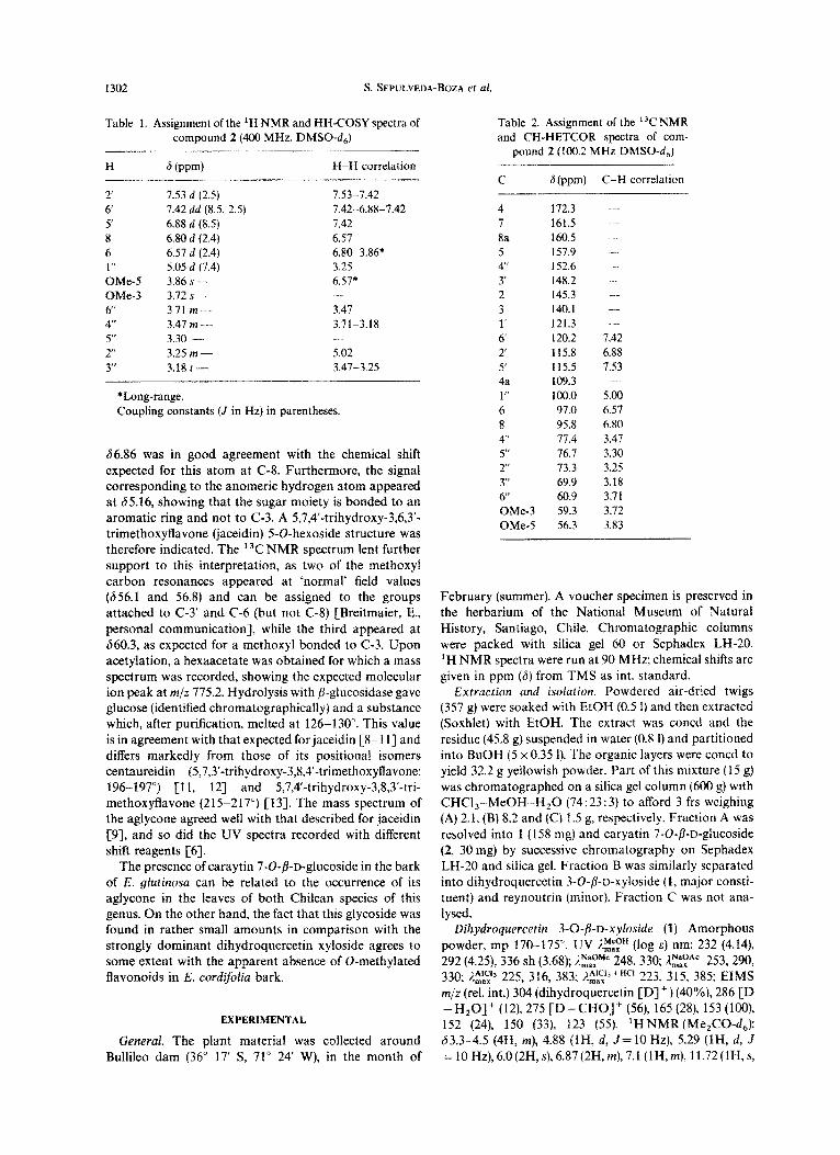
Table 2. Assignment of the 13CNMR and CH-HETCOR spectra of com-
pound 2 (100.2 MHz DMSO-d,)
C fi (ppm) C-H correlation
4 172.3 7 161.5 8a 160.5 - 5 157.9 4’ 152.6 3 148.2 ..- 2 145.3 --. 3 140.1 - 1’ 121.3 .-- 6’ 120.2 1.42 2 115.8 6.88 5 115.5 7.53 4a 109.3 1” loo.0 5.00 6 97.0 6.57 8 95.8 6.80 4” 77.4 3.47 5” 76.7 3.30 2’ 73.3 3.25 3” 69.9 3.18 6” 60.9 3.71 OMe-3 59.3 3.72 OMe-5 56.3 3.83
February (summer). A voucher specimen is preserved in the herbarium of the National Museum of Natural History, Santiago, Chile. Chromatographic columns were packed with silica gel 60 or Sephadex LH-20. ‘H NMR spectra were run at 90 MHz; chemical shifts are given in ppm (S) from TMS as int. standard.
Extraction and ~s~~utio~~. Powdered air-dried twigs (357 g) were soaked with EtOH (0.5 1) and then extracted (Soxhlet) with EtOH. The extract was coned and the residue (45.8 g) suspended in water (0.8 1) and partitioned into BuOH (5 x 0.35 I). The organic layers were coned to yield 32.2 g yellowish powder. Part of this mixture (15 g) was chromatographed on a silica gel column (600 g) wrth CHCl,-.MeOH H,O (74:23:3) to afford 3 frs weighing (A) 2.1, (B) 8.2 and (C) 1.5 g, respectively, Fraction A was resolved into 1 (158 mg) and caryatin 7-O-~-D-gIucoside (2, 30 mg) by successive chromatography on Sephadex LH-20 and silica gel. Fraction B was similarly separated into dihydroquercetin 3-O-b-D-xyloside (1, major consti- tuent) and reynout~n ~minor). Fraction C was not ana- lysed.
Dihydroquercetin 3-0-/3-D-xyloside (1) Amorphous powder, mp 170-175’. UV J~~~~” (log E) nm: 232 (4.14), 292 (4.25), 336 sh (3.68); I.:=zMe 248,330; ,E:“’ 253,290, 330; ii$:r, 225, 316, 383: ,$LafxiHC’ 223, 315, 385; EIMS m/z (rel. int.) 304 (dihydroquercetin CD]‘) (40%), 286 [D -H,O]+ (12), 275 [D-CHO]+ (56), 165 (28). 153 (lo(l), 152 (24), 150 (33), 123 (55). ‘HNMR(Me,CO-d,): 63.334.5 (4H, m), 4.88 (lH, d, J= 10 Hz), 5.29 flH, d, J =10Hz),6.0(2H,s),6.87(2H,m),7.1(1H,m),11.72(1H,s,

Flavonoids from Eucryphia glutinosa 1303
exchangeable with D,O). Acid hydrolysis gave xylose, identified chromatographically.
Caryatin 7-O-/?-n-glucoside (2). Amorphous powder, mp 287-291”, [a&- 16.7” (MeOH; c 0.12). UV ;I!$‘” nm: 253,266 sh, 348; 2:;“’ 23 1,258,396; npa:“’ 253,266,348; ~AICII 733 257 375. ~AlClx+HCI
Inax 253, 266 sh, 348, 430, (weak). l;NtiR (4&lrHz, MeOH-d,): 63.18 (lH, t), 3.25 (lH, m), 3.47 (lH, m), 3.71 (lH, m), 3.72 (lH, s), 3.86 (lH, s), 5.05 (lH, d, 5=7.4 Hz), 6.57 (lH, d, J=2.4 Hz), 6.80(1H, d, J=2.4 Hz), 6.88 (lH, d, J=8.5 Hz), 7.42 (lH, dd, J=2.4,8.5 Hz), 7.53 (lH, d, 5=2.5 Hz). Acid hydroly- sis gave glucose, identified chromatographyically, and caryatin; UV E.!$yH nm: 253, 266 sh, 298 sh, 347; A!$:“” 266,314,392; n:;“” 253,266 sh, 300,348; l&,l,c,13 257,297, 370, 475; &$$+HC’ 253, 266 sh, 304, 350, 430.
5,7,4’-Trihydroxy-3,6,3’-trimethoxy$avone (jaceidin) 5-O-@bglucoside (3). Yellow amorphous powder, mp 285-290”, [XI;“- 89” (pyridine; c 1). EIMS m/z (rel. int.) 360.0855 (jaceidin [a+, talc. C,,H,,O, 360.0860) (lOO%), 359 [J-a’ (84), 345 [J-Me]+ (68), 343 [J-OH]’ (l4), 342 [J-HzO]+ (30). ‘HNMR (DMSO-d&63.35 (4H, m), 3.74 (3H, s), 3.77 (3H, s), 3.84 (3H, s), 4.5-5.0 (2H, m), 5.16 (lH, d, 5=2Hz), 6.86 (lH, s), 6.96 (lH, d, J =S.OHz), 7.63 (lH, dd, 3=2.0, 8.0Hz), 7.71 (lH, d, J = 2.0 Hz).
Hydrolysis of compound 3 wirh fi-glucosidase. Com- pound 3 was suspended in H,O and incubated with /3- glucosidase for 48 hr at 37”. Glucose was identified chromatographically. After freeze-drying and prep. chro- matography on silica gel (CHCl,-MeOH, 10: 1) the dark yellow, gummy product was further purified by chro- matography on Sephadex LH-20 (MeOH-CHCI,, 20: 1) to afford jaceidin as an amorphous powder melting at 126-130”. EIMS m/z (rel. int.) 360 [M]’ (lOO%), 359 [M -H] * (54), 345 [M-Me]+ (68); UV nE:F” nm: 257,273, 352; &‘$yMe 273, 335, 416; l&$‘3 272, 281, 298 sh, 383; l;‘,C;+HC’ 260, 280, 299 sh, 368, 412.
5,7/l’-Trihydroxy-3,6,3’-trimethoxyjauone 5-0-/?-D- glucoside (3) hexaacetate. Compound 3 dissolved in pyri- dine and treated with Ac,O at room temp. for 48 hr, afforded a single product which crystallized as needles, mp 187-189” (CHCl,-MeOH), [a]~“-54” (CHCI,; c 1). IR vmax cm-‘: 1770,1655. UV nz:FH nm: 246,265,298 sh, 337. FAB-MS m/z (rel. int.) 775.2 [M] + (4), 46O(3), 445 (7), 331 (2), 307 (24), 290 (13), 271 (1.5), 242 (2), 229 (1.5), 169
(6), 165 (5), 155 (25), 154 (lOO), 152 (ll), 139 (12), 138 (29), 137 (53), 136(69), 135 (7), 125 (8), 120(11), 107 (22),95 (3). ‘H NMR (CDCI,): 62.02 (3H, s), 2.04 (6H, s), 2.06 (3H, s), 2.33 (3H, s), 2.35 (3H, s), 3.85 (3H, s), 3.86 (3H, s), 3.97 (3H, s), 4.15 (2H, m), 5.1-5.4 (4H, m), 6.55 (lH, s), 7.13 (lH, d, 5=8.4 Hz), 7.57 (lH, dd, J=2.0, 8.4, Hz), 7.70 (lH, d, J =2.0 Hz). 13C NMR (CDCI,): 656.1 (OMe-3’), 56.8 (OMe-6), 60.3 (OMe-3).
Acknowledgements-Thanks are due to Mr Rodrigo Urzlia for collecting and Mr Godofredo Seplilveda for preparing the plant material, and Prof. Eberhard Breitmaier and Dr Gert Eckhardt for providing the NMR and mass spectra. S.D. thanks the Friedrich Naumann Foundation for a fellowship.
REFERENCES
1. Bate-Smith, E. C., Davenport, S. M. and Harborne, J. B. (1967) Phytochemistry 6, 1407.
2. Tschesche, R., Delhvi, S., Septilveda, S. and Breit- maier, E. (1979) Phytochemistry 18, 867.
3. Mufioz Pizarro, C. (1966) Flares Siluestres de Chile. Universidad de Chile, Santiago.
4. Ogiso, A. and Kashida, I. (1972) Phytochemistry 11, 3545.
5. Srivastava, S. K. (1980) Indian .I. Pharm. Sci. 42, 95. 6. Mabry, T. J., Markham, K. R. and Thomas, M. B.
(1970) The Systematic Identijication of Flavonoids. Springer, Berlin.
7. Kingston, D. G. I. (1971) Tetrahedron 27, 2691. 8. Roitman, J. N. and James, L. F. (1985) Phyto-
chemistry 24, 835. 9. Harborne, J. B., Mabry, T. J. and Mabry, H. (1975)
The Flavonoids. Chapman & Hall, London. 10. Fukui, K., Matsumoto, T., Nakamura, S. and
Nakayama, H (1968) Bull. Chem. Sot. J 41, 1413. 11. FBrk&, L., H&hammer, L., Wagner, H., R&ler, H.
and Gurniak, R. (1964) Chem. Ber. 97, 610. 12. Fgrkis, L., H&-hammer, L., Wagner, H., Riisler, H.
and Gurniak, R. (1964) Chem. Ber. 97, 1666. 13. Horie, T., Tsukuyama, M., Kawamura, Y. and
Yamamoto, S. (1988) Phytochemistry 27, 1491.