Embed Size (px)
Citation preview
Identification of Gene Copy Number Alterations in Human Non-small Cell Lung Cancer
Elinor Velasquez San Francisco State University
& Department of Pathology
Stanford University, School of Medicine
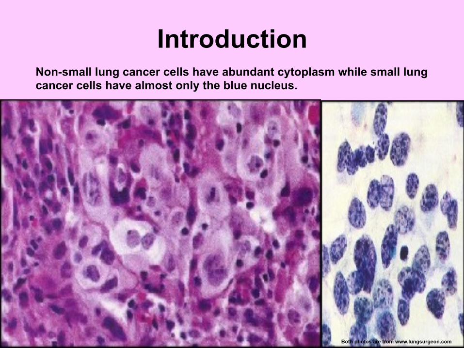
Introduction
Non-small lung cancer cells have abundant cytoplasm while small lung cancer cells have almost only the blue nucleus.
Both photos are from www.lungsurgeon.com
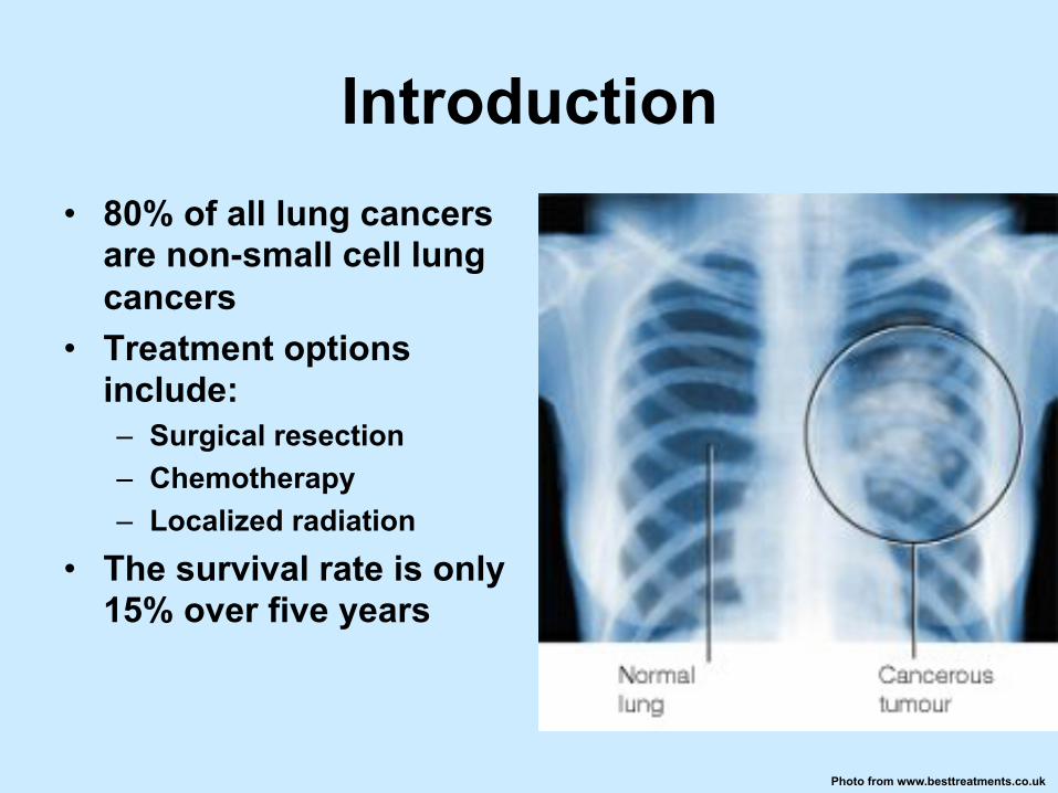
Introduction • 80% of all lung cancers
are non-small cell lung cancers
• Treatment options include: – Surgical resection – Chemotherapy – Localized radiation
• The survival rate is only 15% over five years
Photo from www.besttreatments.co.uk
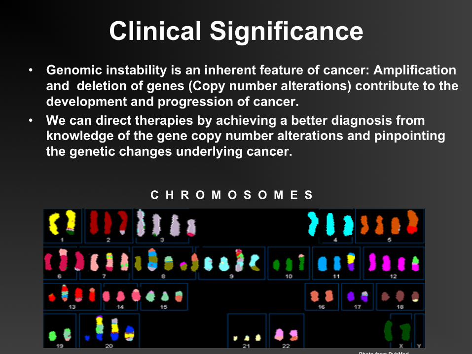
Clinical Significance • Genomic instability is an inherent feature of cancer: Amplification
and deletion of genes (Copy number alterations) contribute to the development and progression of cancer.
• We can direct therapies by achieving a better diagnosis from knowledge of the gene copy number alterations and pinpointing the genetic changes underlying cancer.
C H R O M O S O M E S
Photo from PubMed
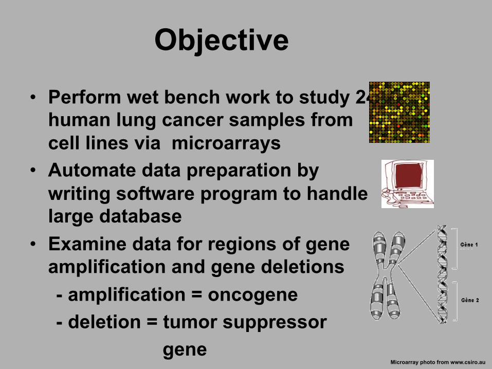
Objective
• Perform wet bench work to study 24 human lung cancer samples from cell lines via microarrays
• Automate data preparation by writing software program to handle large database
• Examine data for regions of gene amplification and gene deletions
- amplification = oncogene - deletion = tumor suppressor gene
Microarray photo from www.csiro.au
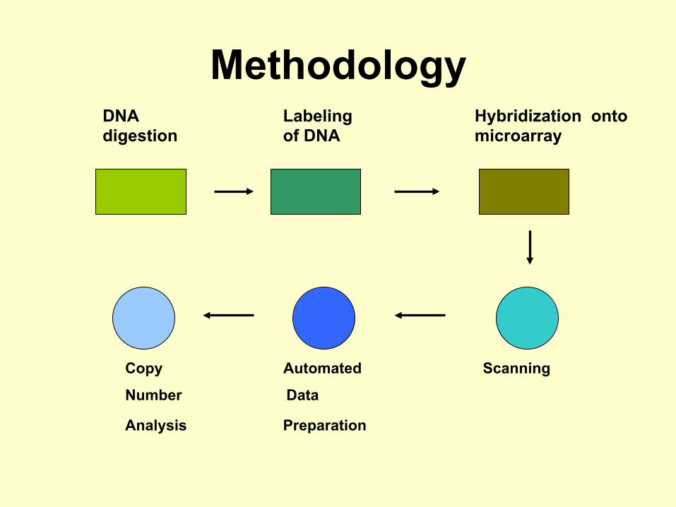
Methodology
Copy Automated Scanning
Number Data
Analysis Preparation
Labeling of DNA
Hybridization onto microarray
DNA digestion
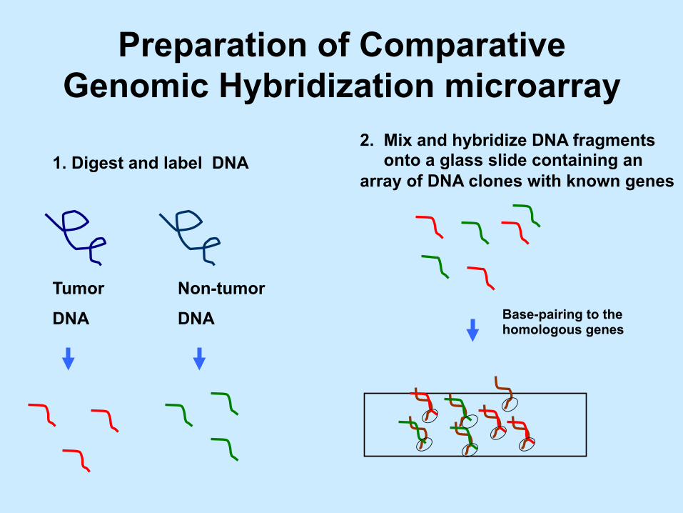
Preparation of Comparative Genomic Hybridization microarray
1. Digest and label DNA
2. Mix and hybridize DNA fragments onto a glass slide containing an array of DNA clones with known genes
Tumor
DNA
Non-tumor
DNA Base-pairing to the homologous genes
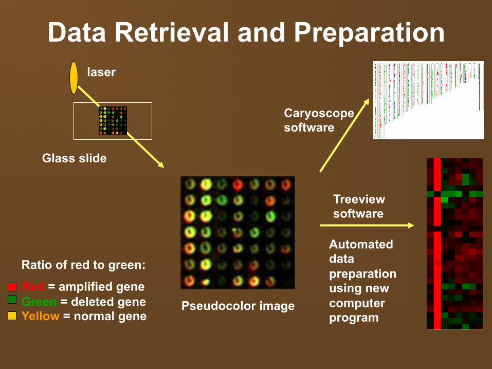
Data Retrieval and Preparation
laser
Pseudocolor image
Ratio of red to green:
Red = amplified gene Green = deleted gene Yellow = normal gene
Automated data preparation using new computer program
Treeview software
Glass slide
Caryoscope software
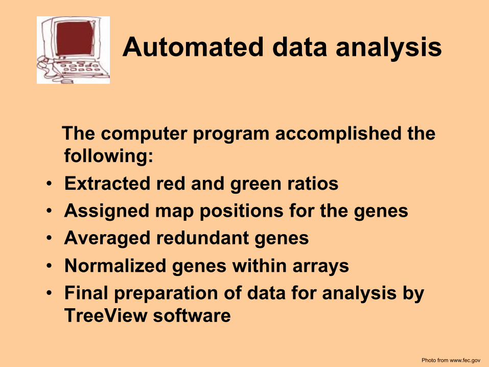
Automated data analysis
The computer program accomplished the following:
• Extracted red and green ratios • Assigned map positions for the genes • Averaged redundant genes • Normalized genes within arrays • Final preparation of data for analysis by
TreeView software
Photo from www.fec.gov
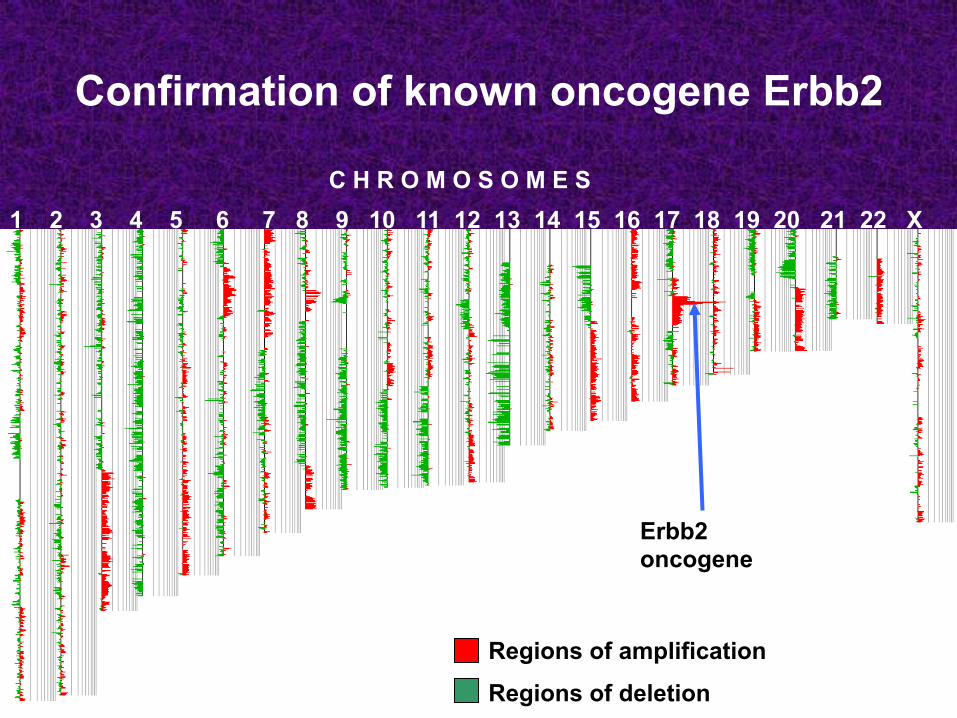
Confirmation of known oncogene Erbb2
1 2 3 4 5 6 7 8 9 10 11 12 13 14 15 16 17 18 19 20 21 22 X
Regions of deletion Regions of amplification
Regions of deletion
Erbb2 oncogene
C H R O M O S O M E S
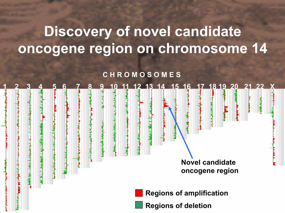
Discovery of novel candidate oncogene region on chromosome 14
1 2 3 4 5 6 7 8 9 10 11 12 13 14 15 16 17 18 19 20 21 22 X C H R O M O S O M E S
Regions of amplification
Regions of deletion
Novel candidate oncogene region
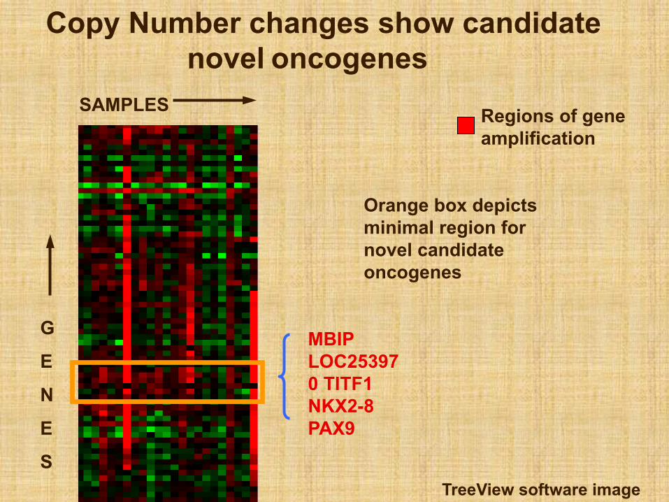
Copy Number changes show candidate novel oncogenes
G
E
N
E
S
SAMPLES Regions of gene amplification
MBIP LOC253970 TITF1 NKX2-8 PAX9
Orange box depicts minimal region for novel candidate oncogenes
TreeView software image
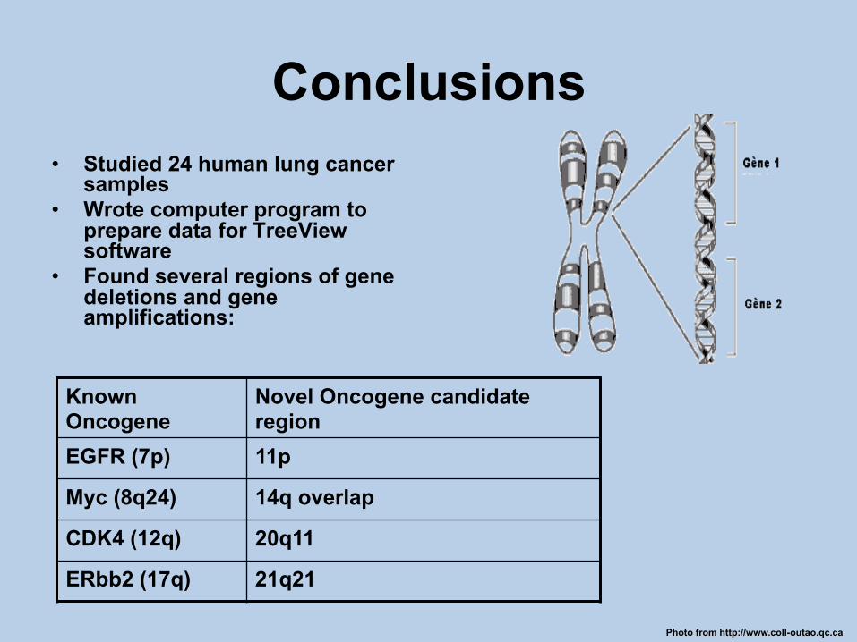
Conclusions • Studied 24 human lung cancer
samples • Wrote computer program to
prepare data for TreeView software
• Found several regions of gene deletions and gene amplifications:
Photo from http://www.coll-outao.qc.ca
Known Oncogene
Novel Oncogene candidate region
EGFR (7p) 11p
Myc (8q24) 14q overlap
CDK4 (12q) 20q11
ERbb2 (17q) 21q21
Future Studies • Narrow recurrent regions of amplifications
and deletions • Characterize candidate cancer genes such
as 14q overlap on chromosome 14. • Apply knowledge for improved diagnosis or
therapies
Photo from www1.wfubmc.edu
Acknowledgements • Jonathan Pollack • Kevin Kwei • Young Kim • The Pollack lab • Ki Goosens • Anika Green • Velessa Peairs • SSRPers


![Telecomm presentation [2005]](https://img.dokumen.tips/doc/110x75/55508844b4c9051e5b8b4b8a/telecomm-presentation-2005.jpg)