Embed Size (px)
Citation preview

Research Article
FGF23 regulates renal sodium handling andblood pressureOlena Andrukhova1, Svetlana Slavic1, Alina Smorodchenko1, Ute Zeitz1, Victoria Shalhoub2, Beate
Lanske3, Elena E Pohl1 & Reinhold G Erben1,*
Abstract
Fibroblast growth factor-23 (FGF23) is a bone-derived hormoneregulating renal phosphate reabsorption and vitamin D synthesisin renal proximal tubules. Here, we show that FGF23 directly regu-lates the membrane abundance of the Na+:Cl� co-transporter NCCin distal renal tubules by a signaling mechanism involving the FGFreceptor/aKlotho complex, extracellular signal-regulated kinase 1/2(ERK1/2), serum/glucocorticoid-regulated kinase 1 (SGK1), andwith-no lysine kinase-4 (WNK4). Renal sodium (Na+) reabsorptionand distal tubular membrane expression of NCC are reduced inmouse models of Fgf23 and aKlotho deficiency. Conversely, gain ofFGF23 function by injection of wild-type mice with recombinantFGF23 or by elevated circulating levels of endogenous Fgf23 in Hypmice increases distal tubular Na+ uptake and membrane abun-dance of NCC, leading to volume expansion, hypertension, andheart hypertrophy in a aKlotho and dietary Na+-dependent fashion.The NCC inhibitor chlorothiazide abrogates FGF23-induced volumeexpansion and heart hypertrophy. Our findings suggest that FGF23is a key regulator of renal Na+ reabsorption and plasma volume,and may explain the association of FGF23 with cardiovascular riskin chronic kidney disease patients.
Keywords aldosterone; blood pressure; fibroblast growth factor-23; heart
hypertrophy; sodium homeostasis
Subject Categories Cardiovascular System; Urogenital System
DOI 10.1002/emmm.201303716 | Received 25 November 2013 | Revised 27
February 2014 | Accepted 6 March 2014 | Published online 5 May 2014
EMBO Mol Med (2014) 6: 744–759
Introduction
Fibroblast growth factor-23 (FGF23) is a bone-derived phosphate-
and vitamin D-regulating hormone which is secreted by osteocytes
and osteoblasts in response to vitamin D and increased extracellular
phosphate (The ADHR Consortium, 2000; Saito et al, 2005; Martin
et al, 2012). In the kidney, circulating FGF23 reduces phosphate
reabsorption from urine through a direct downregulation of sodium
phosphate co-transporters in renal proximal tubular epithelial cells
(Shimada et al, 2004a,b, 2005; Andrukhova et al, 2012). In addi-
tion, FGF23 suppresses renal 1a-hydroxylase expression, the key
enzyme in vitamin D activation, in proximal tubules (Shimada et al,
2001, 2004a). At physiological concentrations, binding of FGF23 to
target cells requires co-expression of the ubiquitously expressed
FGF receptor-1c and of aKlotho (Urakawa et al, 2006), hereafter
referred to as Klotho. Klotho is a single-pass transmembrane protein
which is mainly expressed in the kidney in renal proximal and distal
convoluted tubules, in parathyroid glands, but also in other tissues
such as the brain choroid plexus (Kuro-o et al, 1997; Hu et al, 2010;
Andrukhova et al, 2012).
In chronic kidney disease (CKD), the declining glomerular filtra-
tion rate leads to decreased renal phosphate excretion and subse-
quent hyperphosphatemia. Hyperphosphatemia in turn stimulates
FGF23 secretion from the skeleton. Therefore, FGF23 serum levels
increase with CKD progression (Weber et al, 2003). It is thought
that increased circulating FGF23 helps to maximize renal phosphate
excretion during the early stages of CKD (Juppner et al, 2010).
However, prospective and cross-sectional clinical studies have
shown that circulating FGF23 is positively and dose dependently
associated with CKD progression, cardiovascular risk factors such as
left ventricular hypertrophy, vascular calcifications, and mortality in
CKD patients (Juppner et al, 2010; Faul et al, 2011), suggesting that
FGF23 may have additional biological functions which cannot be
explained by the known effects of FGF23 on mineral metabolism.
The recent report by Faul and coworkers (Faul et al, 2011)
suggested that FGF23 may induce left ventricular hypertrophy by a
direct, Klotho-independent action on cardiomyocytes. In contrast,
Xie and coworkers (Xie et al, 2012) reported that Klotho may be
cardioprotective by an FGF23-independent downregulation of stress-
induced calcium channels.
We recently discovered that FGF23 signaling in distal renal
tubules upregulates membrane expression of the epithelial calcium
channel transient receptor potential vanilloid-5 (TRPV5) by a
Klotho-dependent signaling cascade involving extracellular signal-
regulated kinase 1 and 2 (ERK1/2), serum/glucocorticoid-regulated
kinase 1 (SGK1), and with-no lysine kinase-4 (WNK4) (Andrukhova
et al, 2014). Both SGK1 and WNK4 are well known to be also
1 University of Veterinary Medicine Vienna, Vienna, Austria2 Amgen Inc., Thousand Oaks, CA, USA3 Harvard School of Dental Medicine, Boston, MA, USA
*Corresponding author. Tel: +43 1 250 77 4550; Fax: +43 1 250 77 4599; E-mail: [email protected]
EMBO Molecular Medicine Vol 6 | No 6 | 2014 ª 2014 The Authors. Published under the terms of the CC BY 4.0 license744
Published online: May 5, 2014
involved in renal sodium (Na+) handling. The Na+ and volume-
conserving hormone aldosterone increases SGK1 expression and
activity, leading to increased renal tubular Na+ reabsorption
through augmented membrane abundance of the epithelial Na+
channel (ENaC) in the distal parts of the nephron (Chen et al,
1999). Aldosterone is secreted from the adrenal cortex in response
to lowered serum Na+, increased serum potassium, and increased
circulating angiotensin II. ENaC is a heteromultimeric membrane
protein consisting of a-, b-, and c-subunits. The abundance of the
aldosterone-induced a-subunit is the rate-limiting factor in the
assembly of the ENaC complex (May et al, 1997), whereas
the b- and c-subunits are involved in ubiquitination and degradation
of ENaC (Lee et al, 2009). WNK4 is an important regulator of distal
tubular membrane abundance of the Na+:Cl� co-transporter NCC
and physically interacts with the NCC protein to regulate its
membrane trafficking (Cai et al, 2006). Patients with WNK4 muta-
tions leading to excessive NCC expression in distal tubules suffer
from volume expansion and hypertension (Wilson et al, 2001; Kahle
et al, 2003). Because FGF23 signaling leads to increased serine
phosphorylation and activation of SGK1 and WNK4 (Andrukhova
et al, 2014), we hypothesized that FGF23 may not only regulate the
membrane abundance of TRPV5 but also of ENaC and NCC in distal
renal tubules. NCC and ENaC are the two key ion channels responsi-
ble for Na+ reabsorption in the distal nephron.
Results
Fgf23- and Klotho-deficient mice show renal Na+ wasting andare hypovolemic
To test our hypothesis, we first examined Na+ homeostasis in loss-
of-function models. Because our earlier studies (Hesse et al, 2007;
Anour et al, 2012; Andrukhova et al, 2014) suggested that more
subtle effects of Fgf23 or Klotho deficiency on mineral homeostasis
might be masked by rapid growth in young mice, we first examined
renal Na+ excretion in a non-growing, 9-month-old, compound
mutant mouse model characterized by combined loss of Fgf23 or
Klotho (Kl) and of a functional vitamin D receptor (VDR). Ablation
of Fgf23 or Klotho gene function in mice is associated with early
lethality due to uncontrolled production of the active vitamin D
hormone and subsequent vitamin D intoxication. However, parallel
genetic ablation of vitamin D signaling rescues Fgf23�/� and Kl�/�
mice (Hesse et al, 2007; Anour et al, 2012), so that Fgf23�/�/VDRD/D
and Kl�/�/VDRD/D double mutant mice can be examined at older
ages (Streicher et al, 2012). To prevent hypocalcemia and severe
hyperparathyroidism in mice with a non-functioning VDR, all mice
were kept life-long on a so-called rescue diet rich in calcium, phos-
phorus, and lactose (Li et al, 1998; Erben et al, 2002).
Interestingly, both Fgf23�/�/VDRD/D and Kl�/�/VDRD/D double
mutant mice showed renal Na+ wasting relative to VDR mutants
and wild-type mice (Fig 1A), which was associated with elevated
urinary aldosterone concentrations (Fig 1B). Serum aldosterone
levels were higher in Fgf23�/�/VDRD/D but not Kl�/�/VDRD/D mice
relative to wild-type and VDRD/D mice (Fig 1B). Urinary aldosterone
excretion reflects the changes in serum aldosterone over the whole
urine sampling period (12 h in our case) and is therefore often more
sensitive than serum aldosterone concentration, which reflects only
a specific time point. Fgf23�/�/VDRD/D and Kl�/�/VDRD/D double
mutant mice showed decreased NCC but upregulated membrane
abundance of the a-subunit of ENaC relative to wild-type and single
VDR mutants as evidenced by immunoblotting of renal membrane
preparations and immunohistochemistry (Fig 1C–D). In contrast,
the membrane expression of the b- and c-subunits of ENaC was
lower in Fgf23�/�/VDRD/D and Kl�/�/VDRD/D double mutant mice
compared with single VDR mutants (Supplementary Fig S1A). We
quantified only the full-length isoforms of a-, b-, and c-ENAC.Serum Na+ was not significantly different between Fgf23�/�/VDRD/D
and Kl�/�/VDRD/D compound mutants and VDRD/D mice (Supplemen-
tary Fig S1B). Urinary volume tended to be higher in Fgf23�/�/VDRD/D
and Kl�/�/VDRD/D double mutant mice relative to wild-type mice, but
was not significantly changed relative to VDRD/D mice (Supplementary
Fig S1B). Serum potassium remained unchanged in Kl�/�/VDRD/D and
was actually lower in Fgf23�/�/VDRD/D compared with wild-type and
VDRD/D mice (Supplementary Fig S1B), ruling out hyperkalemia as the
driving force for increased aldosterone secretion in compound mutant
mice. Urinary potassium excretion, urinary volume, and urinary pH did
not differ between VDRD/D and Kl�/�/VDRD/D or Fgf23�/�/VDRD/D mice
(Supplementary Fig S1B). To rule out differences in dietary Na+ intake
as a possible cause of increased urinary Na+ excretion in Kl�/�/VDRD/D
and Fgf23�/�/VDRD/D mice, we measured food consumption over
1 week. However, mean food consumption, and thus Na+ intake, did
not differ between the groups (Supplementary Fig S1C). Rather, our
data suggest that the Fgf23 and Klotho deficiency-induced down-
regulation of NCC causes increased urinary Na+ excretion, overriding
the counter-regulatory and probably aldosterone-driven increase in
a-ENaC expression.
To examine whether similar changes would be present in Fgf23�/�
and Kl�/� mice, we examined Na+ homeostasis at 4 weeks of age,
when Fgf23�/� and Kl�/� mice are still viable. Although renal Na+
wasting was observed only in Fgf23�/�/VDRD/D mice, 4-week-old
Fgf23�/�, Kl�/�, Fgf23�/�/VDRD/D, and Kl�/�/VDRD/D mice displayed
downregulated renal NCC expression, upregulated urinary aldoste-
rone, and increased renal a-ENaC expression (Supplementary Fig S2),
in very good agreement with the data from 9-month-old mice. Consis-
tent with our findings, increased serum aldosterone in hypomorphic
Kl/Kl mice was previously reported also by other investigators
(Fischer et al, 2010). The phenotypes of the originally described hypo-
morphic Kl/Kl mouse (Kuro-o et al, 1997) and of Klotho null mice
(Kl�/�) generated later are identical (Tsujikawa et al, 2003).
Collectively, these results demonstrate that Fgf23 and Klotho
deficiency leads to decreased membrane expression of NCC in renal
distal tubules in young and aged mice, and subsequently to renal
Na+ wasting in non-growing mice despite elevated aldosterone
secretion. It is well known that activity of the NCC channel is regu-
lated by protein phosphorylation at different sites (Pacheco-Alvarez
et al, 2006). Therefore, we assessed NCC phosphorylation at serine
71 and 91 and threonine 55 by immunoblotting. As shown in
Supplementary Fig S1D, abundance of phospho-NCC was reduced in
9-month-old Fgf23�/�/VDRD/D and Kl�/�/VDRD/D mutants relative
to wild-type and single VDR mutant mice. These findings are in
accordance with the notion that loss of Fgf23 and Klotho function
leads to decreased membrane transport and activation of the NCC
channel.
It is clear that despite chronically increased urinary Na+ loss,
9-month-old Fgf23�/�/VDRD/D and Kl�/�/VDRD/D mice must be in a
ª 2014 The Authors EMBO Molecular Medicine Vol 6 | No 6 | 2014
Olena Andrukhova et al FGF23 regulates sodium metabolism EMBO Molecular Medicine
745
Published online: May 5, 2014

A
C
D
B
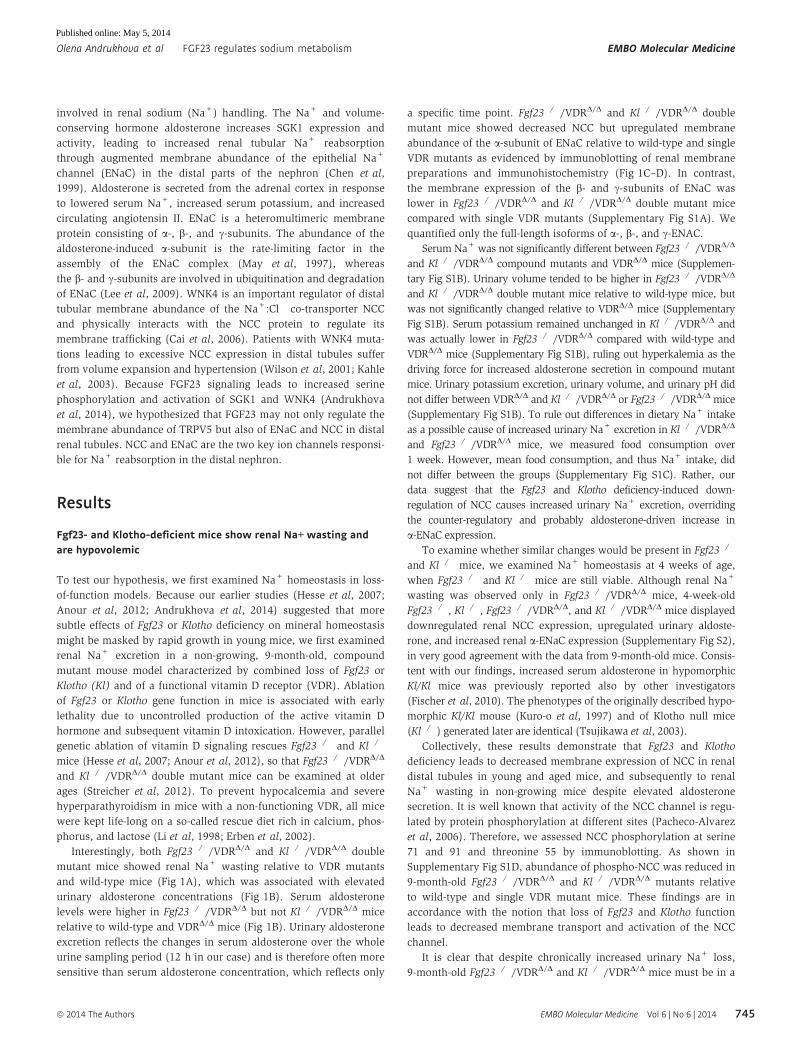
Figure 1. Fgf23 or Klotho deficiency induces renal sodium wasting caused by reduced expression of the Na+:Cl� co-transporter NCC.
A, B (A) Urinary Na+ excretion corrected by urinary creatinine (Crea) (n = 10–12, one-way ANOVA followed by SNK test, *P = 0.0114 versus WT, #P = 0.0325 versusVDRD/D for Fgf23�/�/VDRD/D, *P = 0.0218 versus WT, #P = 0.0185 versus VDRD/D for Kl�/�/VDRD/D) and (B) urinary aldosterone concentration corrected by urinarycreatinine and serum aldosterone concentration measured by ELISA (n = 8–10, one-way ANOVA followed by SNK test, *P < 0.05 versus WT, #P < 0.05 versusVDRD/D), in 9-month-old male wild-type (WT), VDRD/D, Fgf23�/�/VDRD/D, or Kl�/�/VDRD/D compound mutant mice on the rescue diet.
C, D Western blotting analysis of NCC and a-ENaC protein expression in renal cortical total membrane fractions (n = 7–9, one-way ANOVA followed by SNK test,*P < 0.005 versus WT, #P < 0.005 versus VDRD/D), and immunohistochemical detection of NCC and a-ENaC protein expression in paraffin sections ofparaformaldehyde-fixed kidneys (n = 3–5) in 9-month-old male wild-type (WT), VDRD/D, Fgf23�/�/VDRD/D, or Kl�/�/VDRD/D compound mutant mice on the rescuediet. Data represent mean � s.e.m.
Source data are available for this figure.
EMBO Molecular Medicine Vol 6 | No 6 | 2014 ª 2014 The Authors
EMBO Molecular Medicine FGF23 regulates sodium metabolism Olena Andrukhova et al
746
Published online: May 5, 2014

steady state. Our data suggest that in order to adapt to reduced NCC
expression/activation and the accompanying renal Na+ wasting,
Fgf23�/�/VDRΔ/Δ and Kl�/�/VDRΔ/Δ mutants upregulate aldosterone
to conserve Na+ in aldosterone target organs and to maintain
normal serum Na+ and osmolarity. Therefore, although food intake
was not different between the genotypes, it is likely that compound
mutants had higher intestinal Na+ absorption due to increased aldo-
sterone. In this explanatory model, the driving force behind
increased aldosterone secretion in compound mutants would be
hypovolemia, leading to activation of the renin-angiotensin-aldoste-
rone system. Therefore, we assessed blood volume and blood pres-
sure in 9-month-old Fgf23�/�/VDRD/D and Kl�/�/VDRD/D mice.
Indeed, we found lower blood pressure and volume in Fgf23�/�/VDRD/D
and Kl�/�/VDRD/D relative to VDRD/D mice (Fig 2). However, we
were unable to detect differences in plasma renin activity between
the genotypes, using a commercial assay (Supplementary Fig S1E).
We don’t have a good explanation why plasma renin activity
remained unchanged in Fgf23�/�/VDRD/D and Kl�/�/VDRD/D mice
despite hypovolemia. Because the observed increases in urinary
aldosterone excretion in Fgf23�/�/VDRD/D and Kl�/�/VDRD/D
mutants were mild, it is possible that the changes in plasma renin
activity were too small to be picked up by the assay. Taken together,
serum Na+ concentrations are maintained in Fgf23�/�/VDRΔ/Δ and
Kl�/�/VDRΔ/Δ mutants at the expense of reduced blood volume
and hypotension.
Recombinant FGF23 directly upregulates distal tubular NCC andcauses hypertension
Next, we examined gain-of-function models. As expected, intraperi-
toneal injection of 10 lg recombinant FGF23 (rFGF23) over 5 days
into 3-month-old wild-type mice caused hyperphosphaturia and
hypophosphatemia (Supplementary Fig S3). In addition, rFGF23
profoundly reduced urine volume, reduced renal Na+ excretion,
and increased blood Na+ concentration (Fig 3A). Serum and
urinary aldosterone was suppressed in rFGF23-treated mice,
whereas plasma renin activity remained unchanged (Fig 3A). NCC
was about 40% upregulated in renal membrane preparations from
rFGF23-treated wild-type mice relative to vehicle controls (Fig 3B).
Immunohistochemistry also showed increased NCC staining of the
luminal cell membranes in distal tubules after rFGF23 treatment
(Fig 3C). In addition, rFGF23 treatment increased the abundance of
phosphorylated NCC at serine 71 and 91 in renal membrane prepa-
rations (Fig 3D). After correction for total NCC expression, the
strongest effect of rFGF23 on NCC phosphorylation was observed at
serine 71 (Supplementary Fig S4). Inversely to our findings in loss-
of-function models, serum and urinary aldosterone as well as renal
a-ENaC expression was downregulated, whereas renal expression of
the full-length b- and c-subunits of ENaC was increased by rFGF23
(Fig 3A–C). In agreement with the notion that WNK4 physically
interacts with the NCC protein to regulate its membrane trafficking
(Cai et al, 2006) and that FGF23 signaling activates WNK4
(Andrukhova et al, 2014), we found higher WNK4 serine phosphoryla-
tion and an increased association between WNK4 and NCC in
kidney homogenates of rFGF23-treated mice (Fig 3E). We confirmed
the specificity of the anti-NCC and anti-WNK4 antibodies by using
extracts from kidneys of NCC- and WNK4-knockout mice, respec-
tively (Supplementary Fig S5).
To examine whether the rFGF23-induced upregulation in
membrane expression and phosphorylation of NCC is associated
with increased Na+ uptake in distal tubular epithelium, we
performed intracellular Na+ imaging in live kidney slices, using
2-photon microscopy. Three-hundred-lm-thick kidney slices were
prepared from wild-type mice treated with vehicle or rFGF23 8 h
before necropsy. The slices were stained with the fluorescent intra-
vital Na+ indicator SFBI (Harootunian et al, 1989), which was
excited at a wavelength of 820 nm. Fig 3F shows that rFGF23 treat-
ment induced an about threefold increase in fluorescence intensity
in distal tubules. SFBI fluorescence intensity in distal tubules of
rFGF23-treated mice returned to normal within 30 min after ex vivo
addition of the thiazide diuretic chlorothiazide (Fig 3F), a well-
known functional blocker of the NCC channel (Monroy et al, 2000).
In addition, we treated live SFBI-loaded kidney slices prepared from
wild-type mice with rFGF23 or vehicle in vitro. rFGF23 gradually
increased intracellular SFBI fluorescence over 105 min in distal
tubules, whereas fluorescence intensity remained unchanged in
vehicle-treated slices (Fig 3G and Supplementary Videos S1 and S2).
The rFGF23-induced increase in intracellular Na+ concentration
was reversed by chlorothiazide within 30 min (Fig 3G and Supple-
mentary Video S1). Taken together, these data show that rFGF23
increases NCC membrane abundance and phosphorylation and acti-
vates Na+ uptake in distal renal tubules in vivo and in vitro.
A B
Figure 2. Decreased mean arterial pressure and blood volume in Fgf23- or Klotho-deficient mice.
A, B Mean arterial pressure (A) and blood volume (B) (n = 6–8, one-way ANOVA followed by SNK test, *P < 0.05 versus WT, #P < 0.05 versus VDRD/D) in 9-month-old malewild-type (WT), VDRD/D, Fgf23�/�/VDRD/D, or Kl�/�/VDRD/D compound mutant mice on the rescue diet. Data represent mean � s.e.m.
ª 2014 The Authors EMBO Molecular Medicine Vol 6 | No 6 | 2014
Olena Andrukhova et al FGF23 regulates sodium metabolism EMBO Molecular Medicine
747
Published online: May 5, 2014

As a consequence of renal Na+ retention and volume expansion,
a 5-day rFGF23 treatment increased diastolic, systolic, and mean
arterial blood pressure by about 20 mm Hg (Fig 4A). The heart/
body weight ratio was increased and cross-sections of the heart
showed thickening of the ventricular septum in rFGF23-treated mice
after only 5 days of treatment (Fig 4A). In contrast to wild-type and
VDRD/D mutant mice, rFGF23 treatment of 3-month-old Kl�/�/VDRD/D double mutant mice did not result in renal Na+ retention
A
BC
E
G
F
D
Figure 3.
EMBO Molecular Medicine Vol 6 | No 6 | 2014 ª 2014 The Authors
EMBO Molecular Medicine FGF23 regulates sodium metabolism Olena Andrukhova et al
748
Published online: May 5, 2014

and heart hypertrophy (Fig 4B), showing that the effects of FGF23
on Na+ homeostasis and heart hypertrophy are Klotho dependent,
but VDR independent.
It was reported that Klotho-deficient mice develop heart hyper-
trophy caused by increased circulating Fgf23 (Faul et al, 2011).
However, we actually found a decreased heart/body weight ratio in
4-week-old Kl�/� mice, and unchanged heart/body weight ratio in
Kl�/�/VDRD/D mice compared to wild-type and VDRD/D littermates
(Supplementary Fig S6), suggesting that chronically elevated endog-
enous Fgf23 serum levels in Kl�/� mice do not cause heart hypertro-
phy in the absence of the co-receptor Klotho.
To verify that the regulation of NCC by FGF23 is a direct effect
on the distal tubule, we isolated distal tubular segments from wild-
type and Fgf23-deficient mice and treated these segments with
A
B
Figure 4. FGF23 administration induces hypertension and heart hypertrophy in a Klotho-dependent fashion.
A Representative aortic blood pressure curves, heart weight/body weight ratios, and H&E-stained paraffin cross-sections of hearts from 3-month-old male wild-typemice treated for 5 days with vehicle or 10 lg rFGF23 per mouse per day (n = 5–6, Students t-test, *P = 0.0216). Insets show systolic (BPs), diastolic (BPd), and meanarterial pressure (MAP).
B Urinary Na+ excretion per 12 h and heart weight/body weight ratio in 3-month-old male wild-type, VDRD/D, and Kl�/�/VDRD/D compound mutant mice treated for5 days with vehicle or 10 lg rFGF23 per mouse per day (n = 4–6, Students t-test, *P < 0.05 versus vehicle). Data represent mean � s.e.m.
Figure 3. Gain of FGF23 function induces renal Na+ retention through increased renal NCC expression and channel activation.
A Urine volume (n = 15–17), urinary Na+ excretion per 12 h (n = 15–17, Students t-test, *P = 0.0085), urinary Na+ excretion corrected by urinary creatinine (Crea)(n = 15–17, Students t-test, *P = 0.0251), serum Na+ concentration (n = 15–17, Students t-test, *P = 0.0308), serum and urinary aldosterone concentrationscorrected by urinary creatinine (n = 4–5, Students t-test, * serum P = 0.0040, urine P = 0.0156), and plasma renin activity (RPA) (n = 4–5) after 5 days oftreatment of 3-month-old male wild-type mice with vehicle (Veh) or recombinant FGF23 (10 lg per mouse per day).
B, C Western blotting quantification (B) of NCC, a-ENaC, b-ENaC, and c-ENaC protein expression in renal cortical total membrane fractions (n = 4–5, Students t-test,*NCC P = 0.0014, a-ENaC P = 0.0007, b-ENaC P = 0.0251, c-ENaC P = 0.0344), and immunohistochemical detection (C) of NCC and a-ENaC protein expression inkidney sections of 3-month-old wild-type mice treated for 5 days with vehicle or rFGF23 (n = 3–4).
D Western blotting quantification of NCC phosphorylation at Ser71, Ser91, and Thr58 (pNCC S71, pNCC S91, pNCC T55) in total kidney homogenates of 3-month-oldwild-type mice treated for 5 days with vehicle or rFGF23 (n = 4–5, Students t-test, *pNCC S71 P = 0.0001, pNCC S91 P = 0.0182, pNCC T55 P = 0.0056).
E Reciprocal immunoprecipitation (IP) of serine-phosphorylated (P-Ser) proteins, followed by Western blot (WB) analysis of WNK4 or vice versa from homogenizedrenal cortex protein samples of 3-month-old male wild-type mice treated for 5 days with vehicle or rFGF23 (n = 5–6, Students t-test, *WNK4-P-Ser P = 0.0057).For co-immunoprecipitation of NCC/WNK4 complexes, WNK4 or NCC were immunoprecipitated with specific antibodies (anti-NCC and anti-WNK4) fromhomogenized renal cortex protein samples of 3-month-old wild-type mice treated for 5 days with vehicle or rFGF23. Western blot analysis was performed withcorresponding anti-NCC or anti-WNK4 antibodies to identify co-precipitated NCC and WNK4 protein, respectively (n = 4–6, Students t-test, WNK4-P-NCC*P = 0.0116).
F Quantification and original images of intracellular Na+ levels in renal distal tubular cells in live 300-lm-thick kidney slices of 3-month-old WT mice treated withvehicle or rFGF23 (10 lg/mouse), 8 h before necropsy (n = 4, one-way ANOVA followed by SNK test, *P = 0.0026 versus vehicle-treated mice, #P = 0.0175 versusrFGF23-treated mice). Kidney slices were stained with the sodium-sensitive dye SBFI. Chlorothiazide (CTZ, 10 lM) was used as NCC inhibitor.
G Time-dependent changes in intracellular Na+ levels in renal distal tubules in SBFI-loaded, 300-lm-thick, live kidney slices of 3-month-old WT mice treated in vitroat time 0 with rFGF23 (100 ng/ml) or vehicle (n = 3–6). After 105 min, 10 lM CTZ or vehicle was added. Fluorescence intensity in G and H was quantified in 4–9regions of interest per image, sample, and time point from 2–3 independent experiments. Students t-test, *P < 0.05 versus vehicle-treated or versus vehicle- + CTZvehicle-treated (past 105 min). Data represent mean � s.e.m.
Source data are available for this figure.
◀
ª 2014 The Authors EMBO Molecular Medicine Vol 6 | No 6 | 2014
Olena Andrukhova et al FGF23 regulates sodium metabolism EMBO Molecular Medicine
749
Published online: May 5, 2014

rFGF23 alone or in combination with specific ERK1/2 and SGK1
inhibitors for 2 h in vitro. rFGF23 upregulated NCC protein expres-
sion in distal tubular segments from wild-type and Fgf23-deficient
mice (Fig 5A). This effect was blocked by ERK1/2 or SGK1 inhibi-
tors (Fig 5A), showing that the ERK1/2-SGK1 signaling pathway is
essential for the direct, FGF23-induced regulation of NCC expression
in the distal tubule. In addition, rFGF23 increased NCC phosphory-
lation at serine 71 in distal tubules from wild-type but not from
Klotho-deficient mice, indicating that the co-receptor Klotho is
essential for the FGF23-induced phosphorylation of NCC (Fig 5B).
rFGF23 had no effect on ENaC expression in distal tubules isolated
from wild-type mice, further supporting the notion that the
inhibitory effects of rFGF23 on ENaC expression in vivo are indirect
effects mediated through the suppression of aldosterone secretion
(Fig 5C).
The NCC inhibitor chlorothiazide abrogates the hypertensiveeffects of FGF23
Next, we reasoned that if indeed the cardiovascular effects of
rFGF23 were mediated by Na+ retention through upregulated distal
renal tubular NCC expression, an inhibitor of NCC function should
prevent the rFGF23-mediated rise in circulating blood volume, blood
pressure, and heart/body weight ratio. As expected, treatment of
wild-type mice with the NCC inhibitor chlorothiazide increased
urine volume and renal Na+ excretion, but did not change blood
volume, central venous pressure, arterial blood pressure, or heart/
body weight ratio (Fig 6). However, co-treatment of wild-type mice
with rFGF23 and chlorothiazide completely prevented the rFGF23-
induced Na+ retention, volume expansion, rise in central venous
and arterial blood pressure, heart hypertrophy, and rise in cardiac
expression of the hypertrophy-associated gene b-myosin heavy
chain (Fig 6 and Supplementary Fig S7). These results clearly indi-
cate that the cardiovascular effects of increased circulating FGF23
are mediated through upregulation of distal renal tubular NCC and
consequently higher renal tubular reabsorption of Na+.
Dietary Na+ modulates the effects of FGF23 on renal Na+
handling and blood pressure
To assess the modulatory effect of dietary Na+ on the hypertensive
effect of FGF23, we fed diets with different Na+ content to wild-type
mice and treated them for 5 days with vehicle or rFGF23. Analysis
of the data by two-way ANOVA showed a significant interaction
between the diet and the rFGF23-induced increase in blood pressure
(Fig 7A). However, much to our surprise, the rFGF23-induced
increase in arterial blood pressure was inversely related to dietary
Na+, that is, stronger on the low Na+ diet (Fig 7A). To find an
explanation for this puzzling finding, we analyzed serum and
urinary Na+ and aldosterone together with renal expression of NCC
and of a-, b-, and c-ENaC subunits. In analogy to the effects on
blood pressure, the rFGF23-induced increase in serum Na+ and the
suppression of urinary Na+ excretion (in absolute numbers) were
most pronounced on the low Na+ diet (Fig 7B). As expected, serum
and especially urinary aldosterone were inversely related to dietary
Na+ content in vehicle-treated mice (Fig 7C). The 5-day treatment
with rFGF23 suppressed serum and urinary aldosterone on the low
and normal Na+ diets (Fig 7C). However, the remaining levels of
urinary aldosterone excretion in rFGF23-treated mice were inversely
related to dietary Na+ content (Fig 7C).
rFGF23-treated mice showed increased renal NCC expression
compared with vehicle controls on all three diets, but the level of
NCC abundance was profoundly modulated by dietary Na+
(Fig 7D). NCC abundance was more than twofold higher in kidneys
of rFGF23-treated mice on low Na+ compared with those on high
Na+ diet (Fig 7D). Interestingly, rFGF23 treatment downregulated
renal expression of a-ENaC and upregulated expression of the
b- and c-ENaC subunits on the normal and high Na+ diet, but had
A B C
Figure 5. FGF23 regulates NCC expression and phosphorylation in isolated distal tubular segments in a Klotho-dependent manner.
A Western blotting quantification of NCC expression in isolated distal tubular segments from wild-type (WT) and Fgf23�/� mice treated for 2 h in vitro with vehicle orrFGF23 alone or in combination with specific ERK1/2 (iERK1/2) or SGK1 inhibitors (iSGK1) (n = 4–6, one-way ANOVA followed by SNK test, *P < 0.05 versus vehicle,#P < 0.05 versus rFGF23 alone).
B Western blotting quantification of NCC phosphorylation at Ser71 (pNCC S71) in isolated distal tubular segments from WT and Kl�/� mice treated for 2 h in vitrowith vehicle or rFGF23 alone or in combination with specific ERK1/2 or SGK1 inhibitors (n = 4, one-way ANOVA followed by SNK test, *P = 0.0002 versus vehicle,#P < 0.005 versus rFGF23 alone).
C Western blotting quantification of a-ENaC protein expression in isolated distal tubular segments from WT mice treated for 2 h in vitro with vehicle (Veh) or rFGF23(n = 3–4). Data represent mean � s.e.m.
Source data are available for this figure.
EMBO Molecular Medicine Vol 6 | No 6 | 2014 ª 2014 The Authors
EMBO Molecular Medicine FGF23 regulates sodium metabolism Olena Andrukhova et al
750
Published online: May 5, 2014

the opposite effect on the low Na+ diet (Fig 7D). These findings are
consistent with the notion that FGF23 does not directly regulate
ENaC membrane abundance, but that higher residual aldosterone
levels on the low Na+ diet interfere with the counter-regulatory
suppression of a-ENaC seen in the distal nephron of rFGF23-treated
mice on the normal and high Na+ diets. It is well known that aldo-
sterone signaling increases transcription and activation of SGK1
(Chen et al, 1999). Therefore, we hypothesized that FGF23 and
aldosterone signaling might converge on SGK1, resulting in over-
additive effects on SGK1 activation in rFGF23-treated mice on the
low Na+ diet. To test this, we analyzed phosphorylated (pSGK1)
and total SGK1 in renal homogenates by immunoblotting. We found
that the ratio of pSGK1 versus total SGK1 was inversely associated
with the dietary Na+ content in rFGF23-treated mice (Fig 7E),
corroborating the notion that higher aldosterone levels on the low
Na+ diet augmented the rFGF23-induced SGK1 activation. Collec-
tively, these results show that FGF23 and aldosterone signaling
pathways interact in the activation of SGK1 and the regulation of
Na+ reabsorption in the distal nephron.
Hyp mice show overexpression of NCC and hypertension
Finally, we assessed the cardiovascular effects of chronically
elevated endogenous Fgf23 in Hyp mice, a model of human X-linked
hypophosphatemia (XLH). Hyp mice and XLH patients are charac-
terized by loss-of-function mutations in PHEX (phosphate-regulating
gene with homologies to endopeptidases on the X-chromosome),
leading to impaired bone mineralization and subsequently increased
biosynthesis of Fgf23 (Liu et al, 2003; Barros et al, 2013). As
expected, Hyp mice showed about 20-fold increased serum levels of
intact Fgf23 (Fig 8A). In accordance with our findings in rFGF23-
treated mice, chronically elevated circulating Fgf23 in Hyp mice was
associated with increased heart-to-body weight ratio, elevated
serum Na+, and decreased urinary Na+ excretion (Fig 8B). More-
over, mean arterial blood pressure (Fig 8C), renal NCC membrane
expression (Fig 8D), and NCC phosphorylation at S71, S91, and T58
(Fig 8D) were increased in Hyp mice, relative to wild-type controls.
Similar to the findings in rFGF23-treated wild-type mice, serum and
urinary aldosterone was suppressed in Hyp mice compared with
wild-type controls (Fig 8E). Thus, Hyp mice recapitulate the changes
in Na+ homeostasis and blood pressure found in rFGF23-treated
wild-type mice.
Discussion
Our study suggests that FGF23 directly regulates NCC membrane
abundance and activity in distal renal tubules through its canonical
signaling pathway involving the FGF receptor 1c/aKlotho-ERK1/2-SGK1-WNK4 signaling axis. Thus, FGF23 is not only a phosphaturic,
but also a Na+-conserving hormone involved in volume and blood
pressure homeostasis. This new paradigm describing the novel
FGF23-mediated bone-kidney-heart axis is shown in Fig 9.
Loss-of-function mutations in NCC result in Gitelman’s
syndrome in humans (Naesens et al, 2004). Gitelman’s syndrome
is characterized by normal to low blood pressure, hypokalemia,
hypocalciuria, and metabolic alkalosis. Although Fgf23�/�/VDRΔ/Δ
and Kl�/�/VDRΔ/Δ mutants show reduced NCC expression and
increased urinary Na+ excretion, they do not develop a typical
Gitelman’s syndrome. Rather, Fgf23�/�/VDRΔ/Δ and Kl�/�/VDRΔ/Δ
mutants are characterized by hypercalciuria (Andrukhova et al,
2014), and, as shown in the current study, are not consistently
hypokalemic and have normal urinary pH. It is likely that the
reason for these discrepancies is that FGF23 signaling regulates
WNK4 activity. WNK4 is involved in the membrane transport and
activation of not only NCC, but also of other ion channels such as
TRPV5 and ROMK1 in the distal nephron (Ring et al, 2007;
Andrukhova et al, 2014). Therefore, Fgf23�/�/VDRΔ/Δ and Kl�/
�/VDRΔ/Δ compound mutants develop a more complex phenotype
than Gitelman’s syndrome.
Figure 6. Co-treatment of mice with rFGF23 and chlorothiazide abrogates the untoward cardiovascular effects of rFGF23.Urinary Na+ excretion per 12 h, urine volume, blood volume, central venous pressure, mean arterial pressure, and heart/body weight ratio in 3-month-old male wild-typemice treated for 5 days with vehicle (Veh), recombinant FGF23 (10 lg per mouse per day), or chlorothiazide (CTZ, 25 mg/kg) alone or in combination (n = 8–10). One-wayANOVA followed by SNK test, *P < 0.05 versus vehicle, #P < 0.05 versus rFGF23. Data represent mean � s.e.m.
ª 2014 The Authors EMBO Molecular Medicine Vol 6 | No 6 | 2014
Olena Andrukhova et al FGF23 regulates sodium metabolism EMBO Molecular Medicine
751
Published online: May 5, 2014

A
C
D E
B
Figure 7. Dietary Na+ modulates the effects of rFGF23 on blood pressure and renal Na+ handling.
A Mean arterial blood pressure (MAP) of 3-month-old male wild-type mice treated for 5 days with vehicle (Veh) or 10 lg rFGF23 per mouse per day on high (HighNa), normal (Normal Na), and low (Low Na) sodium diets (n = 6–7, Students t-test, * P < 0.05 versus vehicle). Inset shows results of two-way ANOVA.
B, C Urinary Na+ excretion corrected by urinary creatinine (Crea), serum Na+ concentration (n = 6–7, Students t-test * P < 0.05 versus vehicle) and urinary aldosteronecorrected by urinary creatinine (Crea) and serum aldosterone concentrations (n = 6–7, Students t-test, * urine P < 0.0005, serum P < 0.05 versus vehicle) after5 days of treatment of 3-month-old male wild-type mice with vehicle or rFGF23 (10 lg per mouse per day) on high, normal and low sodium diets.
D, E Western blotting quantification of NCC, a-ENaC, b-ENaC and c-ENaC protein expression in renal cortical total membrane fractions (n = 4–5, Students t-test,*P < 0.05 versus vehicle), and ratio of phospho-SGK1 versus total-SGK1 protein expression in kidney total homogenates of 3-month-old wild-type mice on high,normal, and low sodium diets treated for 5 days with vehicle or rFGF23 (n = 4–5, Students t-test, *P < 0.01 versus vehicle). Data represent mean � s.e.m.
Source data are available for this figure.
EMBO Molecular Medicine Vol 6 | No 6 | 2014 ª 2014 The Authors
EMBO Molecular Medicine FGF23 regulates sodium metabolism Olena Andrukhova et al
752
Published online: May 5, 2014

The physiological function of WNK4 in the regulation of distal
renal tubular NCC membrane abundance is still controversial. It
was previously thought that the WNK4 mutations found in
patients with pseudohyperaldosteronism type II (PHAII, an auto-
somal dominant disease characterized by hypertension, hyperkalemia,
and metabolic acidosis) are loss-of-function mutations and that
WNK4 activation inhibits the membrane transport of NCC (Yang
et al, 2003). However, more recent studies in mice with targeted
disruption of the Wnk4 gene suggest that WNK4 is actually a
positive regulator of NCC membrane abundance and function
(Ohta et al, 2009; Castaneda-Bueno et al, 2012). This notion is
also supported by our data which suggest that FGF23-induced
serine phosphorylation of WNK4 increases the complex formation
between NCC and WNK4, and the distal tubular membrane abun-
dance of NCC.
The current study has shown that a- versus b- and c-ENaCsubunits are reciprocally regulated in loss- and gain-of-Fgf23 func-
tion models. In addition, our data suggest that this regulation is an
indirect, aldosterone-mediated process. Aldosterone regulates the
abundance of the ENaC complex by selectively upregulating the
a-subunit (May et al, 1997; Masilamani et al, 1999). In agreement
with this notion, urinary aldosterone and renal a-ENaC expression
were higher in Fgf23�/�/VDRΔ/Δ and Kl�/�/VDRΔ/Δ compound
mutants versus VDR single mutants, whereas serum aldosterone
and renal a-ENaC expression were lower in rFGF23-treated and Hyp
mice versus vehicle-treated and wild-type mice, respectively.
Conversely, the full-length b- and c-subunits were downregulated
in loss-of-Fgf23 function models and upregulated in gain-of-Fgf23
function models. The C-terminal proline-rich motifs of the b- and
c-subunits of the ENaC complex interact with the ubiquitin ligase
Nedd-4 and are involved in ubiquitination and degradation of ENaC
(Lee et al, 2009). Aldosterone has been shown to induce proteolytic
cleavage of the c-subunit (Masilamani et al, 1999). Therefore, the
reciprocal regulation of a- versus full-length b- and c-ENaC subunits
observed in our loss- and gain-of-function models can likely be
explained by the concomitant changes in aldosterone signaling.
A surprising finding in our study was that a low Na+ diet
augmented the rFGF23-induced increase in arterial blood pressure
A B
C
E
D
Figure 8. Hyp mice show hypertension and increased NCC expression and phosphorylation.
A–E Serum intact Fgf23 concentration (A, n = 8–9, Students t-test, *P = 0.0001 versus vehicle); heart-to-body weight ratio, serum Na+ and urinary Na+ excretioncorrected by urinary creatinine (Crea) (B); mean arterial blood pressure (C, n = 8–9, Students t-test, *P < 0.05 versus vehicle); Western blotting quantification ofrenal NCC membrane expression and NCC phosphorylation at Ser71, Ser91, and Thr55 (pNCC S71, pNCC S91, pNCC T55) (n = 8–9) (D); and urinary aldosteronecorrected by urinary creatinine and serum aldosterone concentrations in 3-month-old male wild-type (WT) and Hyp mice (n = 8–9, Students t-test, *P < 0.05 versusvehicle) (E). Data represent mean � s.e.m.
Source data are available for this figure.
ª 2014 The Authors EMBO Molecular Medicine Vol 6 | No 6 | 2014
Olena Andrukhova et al FGF23 regulates sodium metabolism EMBO Molecular Medicine
753
Published online: May 5, 2014

as compared to rFGF23-treated mice on a normal or high Na+ diet.
Interestingly, urinary aldosterone excretion and renal NCC and
a-ENaC expression were inversely correlated with dietary Na+ in
rFGF23-treated mice. Therefore, we hypothesized that higher aldo-
sterone levels on the low Na+ diet might further enhance the
rFGF23-induced SGK1 activation, leading to higher distal renal tubu-
lar expression of NCC and a-ENaC and consequently higher renal
Na+ reabsorption. Indeed, we found that SGK1 phosphorylation
was inversely associated with dietary Na+ in rFGF23-treated mice.
Our finding that aldosterone and FGF23 signaling converge on SGK1
and interact in the regulation of NCC- and ENaC-driven Na+ reab-
sorption in the distal nephron may have important implications for
clinical medicine. SGK1 is a central molecule in the regulation of
renal Na+ handling and in the pathophysiology of hypertension and
renal fibrosis (Lang et al, 2009). If extrapolated to humans, our data
would predict that in situations where circulating concentrations of
both aldosterone and intact FGF23 are elevated, such as in chronic
kidney disease, aldosterone may amplify the effects of FGF23 on
Na+ retention. Activation of the renin-angiotensin-aldosterone
system (RAAS) is a typical finding in chronic kidney disease
(Lattanzio & Weir, 2010). Conversely, based on our data, RAAS
inhibition as a therapeutic intervention may also modulate the Na+-
conserving function of FGF23 in the kidney. Similar to FGF23, aldo-
sterone can activate NCC through a signaling mechanism involving
SGK1, WNK4, and STE20/SPS-1-related proline/alanine-rich kinase
(SPAK) (Rozansky et al, 2009; van der Lubbe et al, 2012; Ko et al,
2013). Therefore, aldosterone and FGF23 have synergistic effects on
NCC activation. Although ERK1/2 activation has also been impli-
cated in NCC ubiquitination and degradation in some cellular
models (Ko et al, 2007, 2010), our study has clearly established that
FGF23 increases the membrane abundance and activates NCC
through a ERK1/2-SGK1-WNK4 signaling pathway. NCC is mainly
expressed in the entire distal convoluted tubule, whereas ENaC is
expressed in the late distal convoluted tubule (DCT2), the connect-
ing tubule, and the collecting duct (Nesterov et al, 2012). Whether
the crosstalk between aldosterone and FGF23 signaling involves
only DCT2, where NCC and ENaC are co-expressed, or also other
nephron segments, is currently unclear. In addition, it is currently
unclear why SGK1 activation by FGF23 signaling does not directly
upregulate a-ENaC expression in distal tubules. It is conceivable in
this context that different SGK1 activators such as FGF23 or aldoste-
rone result in different phosphorylation patterns of downstream
molecules such as Nedd4 (Flores et al, 2003) due to specific
modulation of the activity of additional protein kinases or
phosphatases.
Hyp mice are a model of XLH in humans. Our data showed that
chronically elevated circulating levels of intact Fgf23 levels in Hyp
mice lead to Na+ retention, hypertension, and heart hypertrophy
through increased expression of NCC. To the best of our knowledge,
data about Na+ homeostasis are not available in XLH patients. Nota-
bly, XLH patients show a high incidence of left ventricular hyper-
trophy, although they are not hypertensive (Nehgme et al, 1997). In
analogy to Hyp mice, it is conceivable that chronically increased
plasma volume due to FGF23-induced Na+ retention may contribute
to the development of left ventricular hypertrophy in XLH patients.
FGF23 is a protective hormone against the untoward biological
consequences of hyperphosphatemia. Hyperphosphatemia is a
major risk factor for vascular calcification in patients with chronic
renal disease (Scialla et al, 2013) and cardiovascular disease in
normal subjects (Dhingra et al, 2007). Therefore, in a hyperphos-
phatemic situation, it may make biological sense to couple increased
phosphaturia with renal Na+ conservation and volume expansion
in order to additionally “dilute” extracellular phosphate to prevent
vascular calcification. However, the downside of this putative
protection mechanism may be that chronic gain of FGF23 function
causes volume expansion, hypertension, and heart hypertrophy
through upregulation of distal renal tubular NCC. Thus, our findings
may provide a mechanistic explanation why circulating FGF23 is
associated with cardiovascular risk and mortality in patients with
CKD, and may reposition NCC blockers such as thiazides in the ther-
apy of CKD and of other conditions characterized by elevated intact
circulating FGF23. Elevated aldosterone levels may additionally
augment the effects of FGF23 on Na+ retention in CKD patients. A
major task for the future is to determine the detailed molecular
mechanisms involved in the crosstalk between aldosterone and
FGF23 signaling in distal renal epithelium. Moreover, based on our
data, it is conceivable that high dietary phosphate intake might
predispose to the development of CKD and hypertension through
augmented FGF23-induced SGK1 activation and Na+ retention also
in the normal population and that aldosterone might modulate this
effect. Interestingly, a recent epidemiologic study in almost 14,000
US adults reported that higher dietary Na+ intake was associated
with lower odds of CKD (Sharma et al, 2013). Based on our finding
that the hypertensive effects of FGF23 are suppressed by high
Figure 9. Proposed model of FGF23-mediated bone-kidney-heart axis.Increased circulating FGF23 augments distal renal tubular NCC expression andactivity which leads to renal Na+ retention, volume expansion, hypertension,and heart hypertrophy. As a counter-regulatory mechanism, hypernatremia andincreased blood volume decrease aldosterone secretion from adrenal glands,leading to a downregulation of renal a-ENaC expression. A low sodium dietaugments the hypertensive effect of increased FGF23 signaling in this model,because it interferes with the counter-regulatory downregulation ofaldosterone. Similarly, in chronic kidney failure FGF23 and aldosteronesignaling pathways are concurrently activated, potentially leading to astimulation of both NCC and a-ENaC-driven Na+ reabsorption mechanismsin renal distal tubules, and subsequent augmentation of the FGF23-inducedvolume expansion, hypertension, and heart hypertrophy.
EMBO Molecular Medicine Vol 6 | No 6 | 2014 ª 2014 The Authors
EMBO Molecular Medicine FGF23 regulates sodium metabolism Olena Andrukhova et al
754
Published online: May 5, 2014

dietary Na+ (Fig 9), the interaction between phosphate and Na+
intake may be an important determinant of cardiovascular and
kidney health in humans.
Materials and Methods
Animals
All animal procedures were approved by the Ethical Committees of
the University of Veterinary Medicine Vienna and of the local
government authorities. Heterozygous VDR+/D (Erben et al, 2002)
were mated with heterozygous Fgf23+/� (Sitara et al, 2004), and
heterozygous Kl+/� (Lexicon Genetics, Mutant Mouse Regional
Resource Centers, University of California, Davis, CA, USA) mutant
mice to generate double heterozygous animals. Fgf23+/�/VDR+/D
and Kl+/�/VDR+/D mutant mice on C57BL/6 background were
interbred to generate WT, VDRD/D, Kl�/�, Fgf23�/� and compound
Fgf23�/�/VDRD/D and Kl�/�/VDRD/D mutant mice. Genotyping of
the mice was performed by multiplex PCR using genomic DNA
extracted from tail as described (Hesse et al, 2007; Anour et al,
2012). The mice were kept at 24°C with a 12/12-h light/dark cycle
and were allowed free access to a rescue diet and tap water. The
rescue diet (Ssniff, Soest, Germany) containing 2.0% calcium,
1.25% phosphorus, 20% lactose, and 600 IU vitamin D/kg was fed
starting from 16 days of age. This diet has been shown to normalize
mineral homeostasis in VDR-ablated mice (Li et al, 1998; Erben
et al, 2002). Hyp mice were kept on a normal mouse diet and geno-
typed by PCR analysis. For some experiments, male C57BL/6
mice on a normal mouse chow (Ssniff, Soest, Germany) were used.
In 3- and 9-month-old mice, urine was collected in metabolic cages
during a 12-h period overnight from 7 p.m. to 7 a.m. In 4-week-old
mice, spontaneous urine was collected before necropsy. Some
3-month-old mice received daily intraperitoneal injections of vehicle
(phosphate-buffered saline with 2% DMSO), 10 lg recombinant
human FGF23 R176/179Q (rFGF23, kindly provided by Amgen,
Thousand Oaks, CA, USA) per mouse, or 25 mg/kg chlorothiazide
(CTZ, Sigma) for 5 days, and were killed 8–12 h after the last injec-
tion. For the experiment with different Na+ diets, 3-month-old male
C57BL/6 mice were allowed to adapt to control (0.2%),
low (0.05%), and high (4%) Na+ diets (Ssniff, Soest, Germany) for
a 3-day period. Starting at day 4, mice on the different Na+ diets
were treated with rFGF23 (10 lg/day/mouse) or vehicle for 5 days.
At necropsy, the mice were exsanguinated from the abdominal
V. cava under anesthesia with ketamine/xylazine (67/7 mg/kg i.p.) for
serum collection. In some mice, food consumption was calculated
as the average intake of rescue diet over 7 days. Food intake and
body weight were measured every 24 h.
Histology
Hearts were fixed in 40% ethanol for 48 h, embedded in paraffin,
and routinely stained with hematoxylin/eosin.
Serum and urine biochemistry
Serum and urinary sodium, potassium, phosphorus, and creatinine
were analyzed on a Hitachi 912 Autoanalyzer (Boehringer
Mannheim) or on a Cobas c111 analyzer (Roche). Serum and
urinary aldosterone were determined by ELISA (NovaTec). Plasma
renin activity was measured by RIA (GammaCOAT, DiaSorin).
Serum intact Fgf23 was assessed by ELISA (Kainos).
Immunohistochemistry
For immunohistochemistry, 5-lm-thick paraffin sections of paraform-
aldehyde (PFA)-fixed kidneys were prepared. Before immunofluo-
rescence staining, dewaxed sections were pretreated with blocking
solution containing 5% normal goat serum in PBS with 0.1% bovine
serum albumin and 0.3% Triton X-100 for 60 min. Without rinsing,
sections were incubated with polyclonal rabbit anti-NCC (Millipore,
1:500) or anti-a-ENaC (Novus Biologicals, 1:500) antibodies at 4°C
overnight. After washing, sections were incubated for 1.5 h with
goat anti-rabbit Alexa 488 or goat anti-rabbit Alexa 568 secondary
antibodies (Invitrogen, 1:400), respectively. Controls were
performed by omitting primary antibodies. The slides were analyzed
on a Zeiss LSM 510 Axioplan 2 confocal microscope equipped with
a 63 × oil immersion lens (NA 1.3). Individual fluorochromes were
simultaneously excited by lasers at 488- and 543-nm wavelengths
with appropriate filter sets for the emitted light to avoid crosstalk.
Images were merged using Adobe Photoshop.
Total cell membrane isolation
Mouse kidney cortex was homogenized in a homogenizing buffer
[20 mM Tris (pH 7.4/HCl), 5 mM MgCl2, 5 mM NaH2PO4, 1 mM
ethylenediaminetetraacetic acid (pH 8.0/NaOH), 80 mM sucrose,
1 mM phenyl-methylsulfonyl fluoride, 10 lg/ml leupeptin, and
10 lg/ml pepstatin] and subsequently centrifuged for 15 min at
4,000 g. Supernatants were transferred to a new tube and centri-
fuged for an additional 30 min at 16,000 g.
Isolation of distal tubular segments
Renal distal tubules were isolated as reported previously (Andrukh-
ova et al, 2012, 2014). In brief, murine kidneys were perfused with
sterile culture medium (Ham’s F12; GIBCO) containing 1 mg/ml
collagenase (type II; Sigma) and 1 mg/ml pronase E (type XXV,
Sigma) at pH 7.4 and 37°C. The cortical tissue was dissected in
small pieces and placed at 37°C in sterile Ham’s F12 medium
containing 0.5 mg/ml collagenase II and 0.5 mg/ml pronase E for
15 min with vigorous shaking. After centrifugation at 3,000 rpm for
4 min, the enzyme-containing solution was removed, and tubules
were resuspended in ice-cold medium. Individual distal tubule
segments were identified based on morphology in a dissection
microscope at ×25–40 magnification by their appearance and dimen-
sions. To rule out contamination with proximal tubules, we
performed purity and quality controls, using mRNA expression of
distal (TRPV5, calbindin 28k) and proximal (NaPi-2a, NaPi-2c)
tubule-specific genes (Andrukhova et al, 2012). Distal tubular
segments from wild-type, Kl�/� and Fgf23�/� mice were incubated
with vehicle (PBS) or rFGF23 (100 ng/ml) and/or 10 ng/ml of the
specific SGK1 kinase inhibitor GSK 650394 (Axon Medchem), or
10 ng/ml of the ERK1/2 inhibitor PD184352 (Sigma) for 2 h.
Protein samples were collected for Western blotting analysis in lysis
buffer.
ª 2014 The Authors EMBO Molecular Medicine Vol 6 | No 6 | 2014
Olena Andrukhova et al FGF23 regulates sodium metabolism EMBO Molecular Medicine
755
Published online: May 5, 2014

Western blot
Kidney cortex homogenates or total cell membrane preparations
were solubilized in Laemmli sample buffer, fractionated on SDS–
PAGE (30 lg/well) and transferred to a nitrocellulose membrane
(Thermo Scientific). Immunoblots were incubated overnight at 4°C
with primary antibodies including polyclonal rabbit anti-NCC
(1:3,000, Millipore), rabbit anti-phospho-NCC Ser71 (pNCC S71;
1:1,000), anti-phospho-NCC Ser91 (pNCC S91; 1:1,000), anti-
phospho-NCC Thr 55 (pNCC T55; 1:1,000) (generous gifts of Dario
R. Alessi, University of Dundee, Dundee, UK), anti-a-ENaC (Novus
Biologicals, 1:1,000), anti-b-ENaC and anti-c-ENaC (Antikoerperon-
line.com, 1:1,500), anti-WNK4 (1:2,000, Novus Biologicals), and
monoclonal mouse anti-b-actin (1:5,000, Sigma) in 2% (w/v) bovine
serum albumin (BSA, Sigma) in a TBS-T buffer [150 mM NaCl,
10 mM Tris (pH 7.4/HCl), 0.2% (v/v) Tween-20]. After washing,
membranes were incubated with horseradish peroxidase-conjugated
secondary antibodies (Amersham Life Sciences). Specific signal was
visualized by ECL kit (Amersham Life Sciences). The protein bands
were quantified by Image Quant 5.0 software (Molecular Dynamics).
The expression levels were normalized to Ponceau S stain.
Co-immunoprecipitation
Kidney cortex homogenate protein samples (1 mg) were incubated
with 2 lg of anti-WNK4 (Novus Biologicals), anti-phosphoserine
(Alpha Diagnostics), or anti-NCC (Millipore) antibody at 4°C over-
night. The immune complexes were captured by adding 50 llProtein A or G agarose/sepharose beads (Santa Cruz Biotechnol-
ogy) and overnight incubation at 4°C with gentle rocking. The
immunoprecipitates were collected by centrifugation at 1,000 × g
for 5 min at 4°C and washed for four times in PBS, each time
repeating the centrifugation step. After the final wash, the pellets
were suspended in 40 ll of electrophoresis sample buffer and
boiled for 2–3 min. Western blot analysis was performed as
described above using a primary anti-NCC, anti-phosphoserine, or
anti-WNK4 antibody.
Central arterial and central venous pressures measurements
Central arterial pressure and central venous pressure (CVP) were
assessed using a SPR-671NR pressure catheter (1.4F, Millar Instru-
ments, Houston, TX, USA). Central arterial pressure measurements
were performed under 1.5% isoflurane anesthesia by inserting the
catheter into the ascending aorta via the carotid artery. In addition
to the central arterial pressure analysis, central venous pressure was
measured by inserting the catheter into the internal jugular vein in
experiments with 5 days of treatment with rFGF23 or vehicle. Pres-
sure was recorded over 5 min and traces were analyzed using
LabchartPro software and a blood pressure module. CVP was calcu-
lated as the average between pressure values of the ascending “a”
wave and descending “x” wave determined from at least 5 cardiac
cycles.
Blood volume measurements
The blood volume was determined from plasma volume and hemato-
crit as described (Lee & Blaufox, 1985). Plasma volume was
determined by Evans blue dye dilution (Barron et al, 1984). Briefly,
20 ll of a 0.4% (wt/vol) solution of Evans blue (Sigma) in sterile
physiological saline was injected into a tail vein. Blood samples
(10 ll) were collected at 10 min and 30 min after injection to
measure disappearance kinetics. Tubes were centrifuged and the
hematocrit was recorded. Evans blue concentration in the plasma
was measured in duplicate as optical density using the 2-wavelength
method.
RNA isolation and quantitative RT-PCR
Shock-frozen hearts were homogenized using TRI Reagent
(Molecular Research Center). Total RNA was extracted with phenol/
chloroform, precipitated using isopropanol, and then treated with
RQ1 RNase-free DNase (Promega). RNA purity and quality was
determined spectrophotometrically (BioPhotometer; Eppendorf).
After first-strand cDNA synthesis (iScript cDNA Synthesis Kit, Bio-
Rad), quantitative RT-PCR was performed on a Rotor-GeneTM 6000
(Corbett Life Science) using SsoFastTM EvaGreen PCR kit (Bio-Rad). A
melting curve analysis was done for all assays to make sure that only
a single PCR product was amplified. Primer sequences are available
on request. Efficiencies were examined by standard curve. Gene
expression data were corrected for efficiency and normalized to orni-
thine decarboxylase antizyme-1 (Oaz1) as house-keeping gene.
Intracellular Na+ imaging
Longitudinal 300-lm-thick live slices of freshly isolated kidneys
were prepared using a Leica VT1000 Vibratome (Leica Micro-
systems). For the preparation of 10 mM stock solution of the sodium-
sensitive dye SBFI (Molecular Probes), SBFI was diluted in DMSO
(Merck Millipore International) and 20% Pluronic (Merck Millipore
International). The kidney slices were incubated for 60 min at 37°C
with 2 lM SBFI (stock solution diluted 1:5,000 with cell culture
medium). Thereafter, the slices were washed two times for 20 min
each in 0.1M PBS. Some kidney slices were incubated in vitro with
rFGF23 (100 ng/ml) or vehicle (PBS). For visualization of intracellu-
lar Na+ content, SBFI was excited by a Ti:sapphire laser (Chame-
leon, Coherent Inc.) at 820 nm. Images (512 × 512 pixels) were
acquired every 30 sec at a depth of 60–80 lm. For the inhibition of
NCC activity, tissue slices were incubated with 10 lM of chlorothia-
zide (CTZ, Sigma) or vehicle (PBS + 1% ethanol) at 37°C, 5% CO2/
95% air humidified atmosphere for 30 min. Fluorescence images
were analyzed using Image J software. The whole epithelial layer of
the distal tubules was selected by manually drawing the region of
interest (ROI) to quantify intracellular Na+ levels. Fluorescence
intensity was quantified in 4–9 ROIs per image, and the ratio
between the fluorescence intensity and the ROI area was calculated
for each tubule. This ratio was used for all subsequent calculations.
Statistical analyses
Statistics were computed using SPSS for Windows 17.0. The data
were analyzed by two-sided t-test (2 groups) or one-way analysis of
variance (ANOVA) followed by Student-Newman-Keuls (SNK)
multiple comparison test (> 2 groups). In addition, arterial blood
pressure data from the Na+ diet experiment were analyzed by two-
way ANOVA, assessing the influence of the diet and of rFGF23
EMBO Molecular Medicine Vol 6 | No 6 | 2014 ª 2014 The Authors
EMBO Molecular Medicine FGF23 regulates sodium metabolism Olena Andrukhova et al
756
Published online: May 5, 2014

treatment as well as their two-way interaction. P values of less than
0.05 were considered significant. The data are presented as the
mean � s.e.m.
Supplementary information for this article is available online:
http://embomolmed.embopress.org
AcknowledgmentsWe thank C. Bergow for excellent technical assistance and William G.
Richards for critical reading of the manuscript. Recombinant FGF23 was a
generous gift of Vicky Shalhoub, Amgen Inc. Thousand Oaks, CA. The anti-
phospho-NCC antibodies were generous gifts of Dario R. Alessi, University
of Dundee, Dundee, UK. We are grateful to Manoocher Soleimani, Cincin-
nati University, OH, USA, and to Gerardo Gamba, Molecular Physiology
Unit, Mexico City, Mexico, for providing kidneys from NCC- and WNK4-
knockout mice, respectively. This work was supported by a grant from the
Austrian Science Fund (FWF P24186-B21) to R.G.E. O.A. was supported
by a postdoctoral fellowship of the University of Veterinary Medicine
Vienna.
Author contributionsOA, AS, EEP, and RGE conceived and designed the experiments; OA, SS, AS, and
UZ performed experiments and analyzed the data; OA and RGE wrote the
manuscript; VS and BL provided important tools; OA, SS, AS, VS, BL, EEP, and
RGE discussed and reviewed the manuscript.
Conflict of interestVS was an employee of Amgen, Inc. The other authors declare no conflict of
interest.
References
Andrukhova O, Zeitz U, Goetz R, Mohammadi M, Lanske B, Erben RG (2012)
FGF23 acts directly on renal proximal tubules to induce phosphaturia
through activation of the ERK1/2-SGK1 signaling pathway. Bone 51:
621 – 628
Andrukhova O, Smorodchenko A, Egerbacher M, Streicher C, Zeitz U, Goetz R,
Shalhoub V, Mohammadi M, Pohl EE, Lanske B et al (2014) FGF23
promotes renal calcium reabsorption through the TRPV5 channel. EMBO J
33: 229 – 246
Anour R, Andrukhova O, Ritter E, Zeitz U, Erben RG (2012) Klotho lacks a
vitamin D independent physiological role in glucose homeostasis,
bone turnover, and steady-state PTH secretion in vivo. PLoS ONE 7:
e31376
Barron WM, Stamoutsos BA, Lindheimer MD (1984) Role of volume in the
regulation of vasopressin secretion during pregnancy in the rat. J Clin
Invest 73: 923 – 932
Barros NM, Hoac B, Neves RL, Addison WN, Assis DM, Murshed M, Carmona
AK, McKee MD (2013) Proteolytic processing of osteopontin by PHEX and
accumulation of osteopontin fragments in Hyp mouse bone, the murine
model of X-linked hypophosphatemia. J Bone Miner Res 28: 688 – 699
Cai H, Cebotaru V, Wang YH, Zhang XM, Cebotaru L, Guggino SE, Guggino
WB (2006) WNK4 kinase regulates surface expression of the human
sodium chloride cotransporter in mammalian cells. Kidney Int 69:
2162 – 2170
Castaneda-Bueno M, Cervantes-Perez LG, Vazquez N, Uribe N, Kantesaria S,
Morla L, Bobadilla NA, Doucet A, Alessi DR, Gamba G (2012) Activation of
the renal Na+:Cl- cotransporter by angiotensin II is a WNK4-dependent
process. Proc Natl Acad Sci USA 109: 7929 – 7934
Chen SY, Bhargava A, Mastroberardino L, Meijer OC, Wang J, Buse P,
Firestone GL, Verrey F, Pearce D (1999) Epithelial sodium channel
regulated by aldosterone-induced protein sgk. Proc Natl Acad Sci USA 96:
2514 – 2519
Dhingra R, Sullivan LM, Fox CS, Wang TJ, D’Agostino RB Sr, Gaziano JM, Vasan
RS (2007) Relations of serum phosphorus and calcium levels to the
incidence of cardiovascular disease in the community. Arch Intern Med
167: 879 – 885
Erben RG, Soegiarto DW, Weber K, Zeitz U, Lieberherr M, Gniadecki R, Möller
G, Adamski J, Balling R (2002) Deletion of deoxyribonucleic acid binding
domain of the vitamin D receptor abrogates genomic and nongenomic
functions of vitamin D. Mol Endocrinol 16: 1524 – 1537
Faul C, Amaral AP, Oskouei B, Hu MC, Sloan A, Isakova T, Gutierrez OM,
Aguillon-Prada R, Lincoln J, Hare JM et al (2011) FGF23 induces left
ventricular hypertrophy. J Clin Invest 121: 4393 – 4408
The paper explained
ProblemFibroblast growth factor-23 (FGF23) is a hormone secreted by bonecells in response to increased extracellular phosphate and vitamin D.FGF23 in turn stimulates renal phosphate excretion and suppressesvitamin D hormone synthesis as part of a negative feedback loopbetween bone and kidney. In patients with chronic kidney disease(CKD), the declining kidney function leads to decreased renal phos-phate excretion, increased blood phosphate levels, and subsequentlyelevated FGF23 serum levels. Interestingly, prospective and cross-sectional clinical studies have shown that circulating FGF23 is posi-tively and dose dependently associated with CKD progression, cardio-vascular risk factors such as left ventricular hypertrophy, vascularcalcifications, and mortality in CKD patients. The molecular mecha-nism underlying these associations has so far remained elusive.
ResultsHere, we show that FGF23 is a direct regulator of the sodium-chloridechannel NCC in distal renal tubules. This channel has a crucial role inthe reabsorption of sodium from renal tubules. Mice lacking Fgf23 or itsco-receptor Klotho showed lower expression of NCC, leading to renalsodium wasting, reduced plasma volume, and lower blood pressure.Conversely, injection of recombinant FGF23 into normal mice resultedin upregulation of renal NCC expression, renal sodium retention,plasma expansion, hypertension, and heart hypertrophy. Co-treatmentwith the NCC channel blocker chlorothiazide abrogated the FGF23-induced volume expansion and increase in blood pressure. Intriguingly,a low sodium diet aggravated the hypertensive effects of recombinantFGF23 in normal mice, probably because intracellular signaling ofFGF23 and of the other major sodium-conserving hormone aldosteroneconverge on the same molecules in distal renal tubules.
ImpactOur study identifies FGF23 as a sodium-conserving hormone. Becausesodium homeostasis is tightly coupled to volume regulation andblood pressure, our paper may explain why FGF23 is associated withcardiovascular risk and mortality in CKD patients. In addition, ourstudy may reposition NCC blockers such as thiazide diuretics in thetherapy of CKD and of other conditions characterized by elevatedcirculating FGF23. The novel link between phosphate and sodiumhomeostasis may also have important implications for the generalpopulation. Based on our findings, a high dietary phosphate intakemight predispose to the development of hypertension.
ª 2014 The Authors EMBO Molecular Medicine Vol 6 | No 6 | 2014
Olena Andrukhova et al FGF23 regulates sodium metabolism EMBO Molecular Medicine
757
Published online: May 5, 2014

Fischer SS, Kempe DS, Leibrock CB, Rexhepaj R, Siraskar B, Boini KM,
Ackermann TF, Foller M, Hocher B, Rosenblatt KP et al (2010)
Hyperaldosteronism in Klotho-deficient mice. Am J Physiol Renal Physiol
299: F1171 – F1177
Flores SY, Debonneville C, Staub O (2003) The role of Nedd4/Nedd4-like
dependant ubiquitylation in epithelial transport processes. Pflugers Arch
446: 334 – 338
Harootunian AT, Kao JP, Eckert BK, Tsien RY (1989) Fluorescence ratio
imaging of cytosolic free Na+ in individual fibroblasts and lymphocytes.
J Biol Chem 264: 19458 – 19467
Hesse M, Frohlich LF, Zeitz U, Lanske B, Erben RG (2007) Ablation of vitamin
D signaling rescues bone, mineral, and glucose homeostasis in Fgf-23
deficient mice. Matrix Biol 26: 75 – 84
Hu MC, Shi M, Zhang J, Pastor J, Nakatani T, Lanske B, Razzaque MS,
Rosenblatt KP, Baum MG, Kuro-o M et al (2010) Klotho: a novel
phosphaturic substance acting as an autocrine enzyme in the renal
proximal tubule. FASEB J 24: 3438 – 3450
Juppner H, Wolf M, Salusky IB (2010) FGF-23: More than a regulator of renal
phosphate handling? J Bone Miner Res 25: 2091 – 2097
Kahle KT, Wilson FH, Leng Q, Lalioti MD, O’Connell AD, Dong K, Rapson AK,
MacGregor GG, Giebisch G, Hebert SC et al (2003) WNK4 regulates the
balance between renal NaCl reabsorption and K+ secretion. Nat Genet 35:
372 – 376
Ko B, Joshi LM, Cooke LL, Vazquez N, Musch MW, Hebert SC, Gamba G,
Hoover RS (2007) Phorbol ester stimulation of RasGRP1 regulates the
sodium-chloride cotransporter by a PKC-independent pathway. Proc Natl
Acad Sci USA 104: 20120 – 20125
Ko B, Kamsteeg EJ, Cooke LL, Moddes LN, Deen PM, Hoover RS (2010)
RasGRP1 stimulation enhances ubiquitination and endocytosis of
the sodium-chloride cotransporter. Am J Physiol Renal Physiol 299:
F300 – F309
Ko B, Mistry AC, Hanson L, Mallick R, Wynne BM, Thai TL, Bailey JL, Klein JD,
Hoover RS (2013) Aldosterone acutely stimulates NCC activity via a SPAK-
mediated pathway. Am J Physiol Renal Physiol 305: F645 – F652
Kuro-o M, Matsumura Y, Aizawa H, Kawaguchi H, Suga T, Utsugi T, Ohyama
Y, Kurabayashi M, Kaname T, Kume E et al (1997) Mutation of the mouse
klotho gene leads to a syndrome resembling ageing. Nature 390: 45 – 51
Lang F, Artunc F, Vallon V (2009) The physiological impact of the serum and
glucocorticoid-inducible kinase SGK1. Curr Opin Nephrol Hypertens 18:
439 – 448
Lattanzio MR, Weir MR (2010) Does blockade of the Renin-Angiotensin-
aldosterone system slow progression of all forms of kidney disease? Curr
Hypertens Rep 12: 369 – 377
Lee HB, Blaufox MD (1985) Blood volume in the rat. J Nucl Med 26: 72 – 76
Lee IH, Campbell CR, Song SH, Day ML, Kumar S, Cook DI, Dinudom A (2009)
The activity of the epithelial sodium channels is regulated by caveolin-1
via a Nedd4-2-dependent mechanism. J Biol Chem 284: 12663 – 12669
Li YC, Amling M, Pirro AE, Priemel M, Meuse J, Baron R, Delling G, Demay MB
(1998) Normalization of mineral ion homeostasis by dietary means
prevents hyperparathyroidism, rickets, and osteomalacia, but not alopecia
in vitamin D receptor-ablated mice. Endocrinology 139: 4391 – 4396
Liu S, Guo R, Simpson LG, Xiao ZS, Burnham CE, Quarles LD (2003) Regulation
of fibroblastic growth factor 23 expression but not degradation by PHEX.
J Biol Chem 278: 37419 – 37426
van der Lubbe N, Lim CH, Meima ME, van VR, Rosenbaek LL, Mutig K, Danser
AH, Fenton RA, Zietse R, Hoorn EJ (2012) Aldosterone does not require
angiotensin II to activate NCC through a WNK4-SPAK-dependent
pathway. Pflugers Arch 463: 853 – 863.
Martin A, David V, Quarles LD (2012) Regulation and function of the FGF23/
klotho endocrine pathways. Physiol Rev 92: 131 – 155
Masilamani S, Kim GH, Mitchell C, Wade JB, Knepper MA (1999) Aldosterone-
mediated regulation of ENaC alpha, beta, and gamma subunit proteins in
rat kidney. J Clin Invest 104: R19 –R23
May A, Puoti A, Gaeggeler HP, Horisberger JD, Rossier BC (1997) Early effect of
aldosterone on the rate of synthesis of the epithelial sodium channel
alpha subunit in A6 renal cells. J Am Soc Nephrol 8: 1813 – 1822
Monroy A, Plata C, Hebert SC, Gamba G (2000) Characterization of the
thiazide-sensitive Na(+)-Cl(-) cotransporter: a new model for ions and
diuretics interaction. Am J Physiol Renal Physiol 279: F161 – F169
Naesens M, Steels P, Verberckmoes R, Vanrenterghem Y, Kuypers D (2004)
Bartters and Gitelmans syndromes: from gene to clinic. Nephron Physiol
96: 65 – 78
Nehgme R, Fahey JT, Smith C, Carpenter TO (1997) Cardiovascular
abnormalities in patients with X-linked hypophosphatemia. J Clin
Endocrinol Metab 82: 2450 – 2454
Nesterov V, Dahlmann A, Krueger B, Bertog M, Loffing J, Korbmacher C (2012)
Aldosterone-dependent and -independent regulation of the epithelial
sodium channel (ENaC) in mouse distal nephron. Am J Physiol Renal
Physiol 303: F1289 – F1299
Ohta A, Rai T, Yui N, Chiga M, Yang SS, Lin SH, Sohara E, Sasaki S, Uchida S
(2009) Targeted disruption of the Wnk4 gene decreases phosphorylation
of Na-Cl cotransporter, increases Na excretion and lowers blood pressure.
Hum Mol Genet 18: 3978 – 3986
Pacheco-Alvarez D, Cristobal PS, Meade P, Moreno E, Vazquez N, Munoz E,
Diaz A, Juarez ME, Gimenez I, Gamba G (2006) The Na+:Cl- cotransporter
is activated and phosphorylated at the amino-terminal domain upon
intracellular chloride depletion. J Biol Chem 281: 28755 – 28763
Ring AM, Leng Q, Rinehart J, Wilson FH, Kahle KT, Hebert SC, Lifton RP (2007)
An SGK1 site in WNK4 regulates Na+ channel and K+ channel activity
and has implications for aldosterone signaling and K+ homeostasis. Proc
Natl Acad Sci USA 104: 4025 – 4029
Rozansky DJ, Cornwall T, Subramanya AR, Rogers S, Yang YF, David LL, Zhu X,
Yang CL, Ellison DH (2009) Aldosterone mediates activation of the
thiazide-sensitive Na-Cl cotransporter through an SGK1 and WNK4
signaling pathway. J Clin Invest 119: 2601 – 2612
Saito H, Maeda A, Ohtomo S, Hirata M, Kusano K, Kato S, Ogata E, Segawa H,
Miyamoto K, Fukushima N (2005) Circulating FGF-23 is regulated by
1alpha,25-dihydroxyvitamin D3 and phosphorus in vivo. J Biol Chem 280:
2543 – 2549
Scialla JJ, Lau WL, Reilly MP, Isakova T, Yang HY, Crouthamel MH, Chavkin
NW, Rahman M, Wahl P, Amaral AP et al (2013) Fibroblast growth factor
23 is not associated with and does not induce arterial calcification. Kidney
Int 83: 1159 – 1168
Sharma S, McFann K, Chonchol M, de Boer IH, Kendrick J (2013) Association
between dietary sodium and potassium intake with chronic kidney
disease in US adults: a cross-sectional study. Am J Nephrol 37: 526 – 533
Shimada T, Mizutani S, Muto T, Yoneya T, Hino R, Takeda S, Takeuchi Y,
Fujita T, Fukumoto S, Yamashita T (2001) Cloning and characterization of
FGF23 as a causative factor of tumor-induced osteomalacia. Proc Natl
Acad Sci USA 98: 6500 – 6505
Shimada T, Hasegawa H, Yamazaki Y, Muto T, Hino R, Takeuchi Y, Fujita T,
Nakahara K, Fukumoto S, Yamashita T (2004a) FGF-23 is a potent
regulator of vitamin D metabolism and phosphate homeostasis. J Bone
Miner Res 19: 429 – 435
Shimada T, Kakitani M, Yamazaki Y, Hasegawa H, Takeuchi Y, Fujita T,
Fukumoto S, Tomizuka K, Yamashita T (2004b) Targeted ablation of Fgf23
EMBO Molecular Medicine Vol 6 | No 6 | 2014 ª 2014 The Authors
EMBO Molecular Medicine FGF23 regulates sodium metabolism Olena Andrukhova et al
758
Published online: May 5, 2014

demonstrates an essential physiological role of FGF23 in phosphate and
vitamin D metabolism. J Clin Invest 113: 561 – 568
Shimada T, Yamazaki Y, Takahashi M, Hasegawa H, Urakawa I, Oshima T,
Ono K, Kakitani M, Tomizuka K, Fujita T et al (2005) Vitamin D
receptor-independent FGF23 actions in regulating phosphate and
vitamin D metabolism. Am J Physiol Renal Physiol 289: F1088 – F1095
Sitara D, Razzaque MS, Hesse M, Yoganathan S, Taguchi T, Erben RG, Juppner
H, Lanske B (2004) Homozygous ablation of fibroblast growth factor-23
results in hyperphosphatemia and impaired skeletogenesis, and reverses
hypophosphatemia in Phex-deficient mice. Matrix Biol 23: 421 –432
Streicher C, Zeitz U, Andrukhova O, Rupprecht A, Pohl E, Larsson TE, Windisch
W, Lanske B, Erben RG (2012) Long-term Fgf23 deficiency does not
influence aging, glucose homeostasis, or fat metabolism in mice with a
nonfunctioning vitamin D receptor. Endocrinology 153: 1795 – 1805
The ADHR Consortium (2000) Autosomal dominant hypophosphataemic
rickets is associated with mutations in FGF23. Nat Genet 26:
345 – 348
Tsujikawa H, Kurotaki Y, Fujimori T, Fukuda K, Nabeshima Y (2003) Klotho, a
gene related to a syndrome resembling human premature aging,
functions in a negative regulatory circuit of vitamin D endocrine system.
Mol Endocrinol 17: 2393 – 2403
Urakawa I, Yamazaki Y, Shimada T, Iijima K, Hasegawa H, Okawa K, Fujita T,
Fukumoto S, Yamashita T (2006) Klotho converts canonical FGF receptor
into a specific receptor for FGF23. Nature 444: 770 – 774
Weber TJ, Liu S, Indridason OS, Quarles LD (2003) Serum FGF23 levels in
normal and disordered phosphorus homeostasis. J Bone Miner Res 18:
1227 – 1234
Wilson FH, Disse-Nicodeme S, Choate KA, Ishikawa K, Nelson-Williams C,
Desitter I, Gunel M, Milford DV, Lipkin GW, Achard JM et al (2001) Human
hypertension caused by mutations in WNK kinases. Science 293:
1107 – 1112
Xie J, Cha SK, An SW, Kuro O, Birnbaumer L, Huang CL (2012)
Cardioprotection by Klotho through downregulation of TRPC6 channels in
the mouse heart. Nat Commun 3: 1238
Yang CL, Angell J, Mitchell R, Ellison DH (2003) WNK kinases regulate
thiazide-sensitive Na-Cl cotransport. J Clin Invest 111: 1039 – 1045
License: This is an open access article under the
terms of the Creative Commons Attribution 4.0
License, which permits use, distribution and reproduc-
tion in any medium, provided the original work is
properly cited.
ª 2014 The Authors EMBO Molecular Medicine Vol 6 | No 6 | 2014
Olena Andrukhova et al FGF23 regulates sodium metabolism EMBO Molecular Medicine
759
Published online: May 5, 2014


![Sodium Phytate Presentation.pptx [Read-Only]formulatorsampleshop.com/v/reference/Sodium Phytate Presentation.pdfLaurate (Skin Conditioning Agent), Sodium Benzoate (Preservative), Sodium](https://img.dokumen.tips/doc/110x75/5eb52012fb0f3e0d55767ea6/sodium-phytate-read-onlyformulatorsampleshopcomvreferencesodium-phytate-presentationpdf.jpg)