Embed Size (px)
Citation preview

FAMILIAL CLUSTERING OF SUICIDE AND MAJOR DEPRESSIVE DISORDER: AN OBSERVATIONAL ANALYSIS
by
Nicolette Walano
BS, DePaul University, 2010
Submitted to the Graduate Faculty of
the Department of Human Genetics
the Graduate School of Public Health in partial fulfillment
of the requirements for the degree of
Master of Science
University of Pittsburgh
2016
ii
UNIVERSITY OF PITTSBURGH
GRADUATE SCHOOL OF PUBLIC HEALTH
This thesis was presented
by
Nicolette Walano
It was defended on
April 13, 2016
and approved by
Thesis Director
Lisa Pan, MD, Assistant Professor of Psychiatry, School of Medicine, Attending Physician, Services for Teens at Risk, University of Pittsburgh
Committee Members
David Finegold, MD, Professor, Human Genetics, Graduate School of Public Health, University of Pittsburgh
Andrea Durst, MS, DrPH, LCGC, Assistant Professor, Human Genetics, Licensed Genetic Counselor, Assistant Director, Genetic Counseling Program, Graduate School
of Public Health, University of Pittsburgh
Ryan Minster, PhD, MSIS, Assistant Professor, Department of Human Genetics Graduate School of Public Health, University of Pittsburgh

iii
Copyright © by Nicolette Walano
2016
iv
ABSTRACT
Depression is considered the third most important burden of disease globally; further, it is ranked
first in middle and high-income countries. It is well understood that depression is has a
heritability of 31-43%, but no genetic associations predisposing individuals to depression have
been found. A small group of individuals have previously been identified with treatment resistant
major depressive disorder (TR-MDD), suicidality and a myriad of metabolic alterations. This is
considered a neuropsychiatric inborn error of metabolism that initially presents with psychiatric
manifestation. The most common metabolic finding is cerebral folate deficiency.
Supplementation of folinic acid has been shown to result in a reduction of symptoms.
Understanding this group of individuals, particularly with the knowledge that metabolic disease
commonly manifests with psychiatric illness, has implications for the way depression may be
diagnosed and treated in the future. This study captured and analyzed the family histories of 36
individuals in an attempt to discern whether the transmission of depression and suicide in these
families fits known Mendelian inheritance patterns. By using segregation analysis in addition to
an observational analysis this study has assessed autosomal recessive and autosomal dominant
inheritance patterns. Observation of family histories showed male-to-male transmission and thus
excluded the possibility of X-linked or mitochondrial inheritance in this group. The
Lisa Pan, MD
FAMILIAL CLUSTERING OF SUICIDE AND MAJOR DEPRESSIVE DISORDER:
AN OBSERVATIONAL ANALYSIS
Nicolette Walano, MS
University of Pittsburgh, 2016
v
observational study identified that 6/36 families (16.7%) met all criteria for autosomal dominant
inheritance and no families met all criteria for autosomal recessive inheritance. The just over
30% of families met a few, but not all of the criteria for autosomal dominant inheritance (11/36)
and 33 of 36 families or 91.67% met only one criterion for autosomal recessive inheritance. This
suggests that these families are transmitting depression and suicidality in a polygenic or
multifactorial pattern. The statistical analysis supports this conclusion finding that the families fit
a non-Mendelian pattern of inheritance. Understanding the way depression is being transmitted
in these families has significant public health relevance as it may inform our understanding and
future studies of the genetics of depression, a significant health burden.

vi
TABLE OF CONTENTS
PREFACE ..................................................................................................................................... X
1.0 INTRODUCTION ........................................................................................................ 1
1.1 BACKGROUND AND SIGNIFICANCE .......................................................... 1
1.1.1 Treatment Resistant Depression .................................................................. 1
1.1.1.1 The Effect of Depression on Family Members ................................... 3
1.1.1.2 Diagnosis of Treatment Resistance ...................................................... 4
1.1.1.3 Predictors of Treatment Response ...................................................... 6
1.1.2 Genetics of Depression .................................................................................. 9
1.1.2.1 Family Studies of Depression and Suicide ........................................ 10
1.1.2.2 Twin Studies of Depression and Suicide ........................................... 11
1.1.2.3 Linkage and Association Studies ....................................................... 12
1.1.3 Depression in Metabolic Disorders............................................................ 14
1.1.3.1 Other Metabolic Disorders ................................................................. 14
1.1.3.2 Cerebral Folate Deficiency (CFD) ..................................................... 15
1.1.4 Genetic Counseling and Testing ................................................................ 18
1.1.4.1 Psychiatric Genetic Counseling ......................................................... 18
1.1.4.2 Neuropsychiatric IEM’s and Genetic Counseling............................ 20
1.2 SPECIFIC AIMS ............................................................................................... 21

vii
1.2.1 Specific Aim 1 .............................................................................................. 21
1.2.2 Specific Aim 2 .............................................................................................. 22
2.0 MATERIALS AND METHODS .............................................................................. 23
2.1 DATA SOURCE ................................................................................................ 23
2.2 DATA ANALYSIS ............................................................................................. 25
2.3 OBSERVATIONAL ANALYSIS ..................................................................... 26
2.4 STATISTICAL ANALYSIS ............................................................................. 27
3.0 RESULTS ................................................................................................................... 30
3.1 DEMOGRAPHICS ............................................................................................ 30
3.2 OBSERVATIONAL ANALYSIS ..................................................................... 31
3.3 SEGREGATION ANALYSIS .......................................................................... 35
4.0 DISCUSSION ............................................................................................................. 37
4.1 PUBLIC HEALTH SIGNIFICANCE.............................................................. 39
4.2 LIMITATIONS .................................................................................................. 41
4.3 FUTURE STUDIES ........................................................................................... 42
5.0 CONCLUSION ........................................................................................................... 43
APPENDIX A : FIGURES ......................................................................................................... 44
APPENDIX B : IRB APPROVAL............................................................................................. 50
APPENDIX C : MTRD ADULT CONSENT FORM .............................................................. 53
BIBLIOGRAPHY ....................................................................................................................... 59
viii
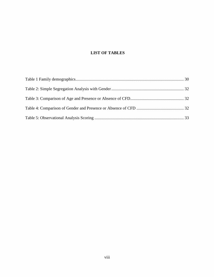
LIST OF TABLES
Table 1 Family demographics ....................................................................................................... 30
Table 2: Simple Segregation Analysis with Gender ..................................................................... 32
Table 3: Comparison of Age and Presence or Absence of CFD................................................... 32
Table 4: Comparison of Gender and Presence or Absence of CFD ............................................. 32
Table 5: Observational Analysis Scoring ..................................................................................... 33
ix
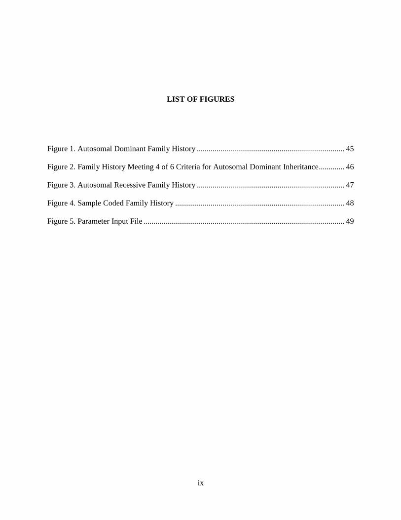
LIST OF FIGURES
Figure 1. Autosomal Dominant Family History ........................................................................... 45
Figure 2. Family History Meeting 4 of 6 Criteria for Autosomal Dominant Inheritance ............. 46
Figure 3. Autosomal Recessive Family History ........................................................................... 47
Figure 4. Sample Coded Family History ...................................................................................... 48
Figure 5. Parameter Input File ...................................................................................................... 49

x
PREFACE
Some of the results of this paper were obtained by using the software package S.A.G.E., which
was supported by the U.S. Public Health Service Resource Grant (RR03655) from the National
Center for Research Resources.
This project would not have been possible without the participants in this study and Dr.
Pan for her dedication to this research, thank you for the opportunity to participant in this project
and meet some of these incredible participants. I am very thankful to my program directors,
Robin Grubs and Andrea Durst for their help and support throughout this program. I am also
grateful to each member of my committee for their wisdom and guidance on this project, it
would not have been possible without them. My classmates have been the most amazing source
of support throughout this endeavor and I am a better human and genetic counselor for having
grown with them in this process. To the others, the teachers, mentors, supervisors, friends and
family, who have given me unconditional support along the way, you have truly made this an
opportunity to learn and grow. Thank you, I am grateful.
1
1.0 INTRODUCTION
1.1 BACKGROUND AND SIGNIFICANCE
1.1.1 Treatment Resistant Depression
Major depressive disorder (MDD) is defined as severe symptoms of depression that interfere
with one’s ability to eat, sleep, work and enjoy life1. MDD is also called unipolar major
depression, which differs from bipolar disorder in that people suffering from unipolar major
depression are particularly prone to major depressive episodes and have never experienced an
episode of mania or hypomania2. Many people experience depression, but major depression is an
episode of depression that is present every day for most of the day and lasts for at least two
weeks3. The Diagnostic and Statistical Manual (DSM) 5 defines MDD as:
“Depressed mood or loss of interest or pleasure in most activities and persisting nearly
every day accompanied by four of the following; significant weight loss, insomnia or
hypersomnia, noticeable psychomotor agitation or retardation, fatigue or loss of energy,
feelings of worthlessness or guilt, diminished ability to think or concentrate and suicidal
ideation”.
The symptoms must interfere with daily functioning and must not be caused by a medication,
medical condition or other substance. Persistent depressive disorder is a depressive episode that
2
lasts at least two years without remission3. It is estimated that 30% of depressive people suffer
from persistent depression3.
The ultimate goal of treatment for MDD is remittance; this is considered a full response
to treatment and is measured by a behavioral rating scale4,5. Partial response may require further
treatment or result in relapse or return of significant symptoms of MDD4,6. Failure to respond
may be a result of such a relapse of symptoms or it may be a complete lack of response to
treatment4,7. Treatment for MDD may include medication, cognitive behavioral therapy (CBT),
electroconvulsive therapy (ECT) or some combination of the three. New methods for treatment
include transcranial direct current stimulation and other alternative methods like folate
supplementation8–10. Typically patients with MDD begin treatment on a selective serotonin
reuptake inhibitor (SSRI). Non-response to an SSRI often results in a change to a different class
of antidepressant such as selective norepinephrine reuptake inhibitor (SNRI), a monamine
oxidase inhibitor (MAOI), or another anti-depressant in the same class11,12. There is no proven
benefit of one course of treatment over another12. Individuals who do not show adequate
response to monotherapy will require augmented or optimized treatment; typically patients are
prescribed a more traditional class of antidepressant, such as an SSRI, plus another supplemental
anti-depressant or alternative medication. These patients may also be undergoing some form of
CBT concurrently, thus treatment varies significantly for each person and changes throughout
course of treatment depending on individual response11. Lithium augmentation and ECT may be
recommended after several rounds of non-response or insufficient response to treatment; one
algorithm suggests incorporating lithium as the fourth augmentation of treatment, lithium plus a
combination of two other anti-depressant classes as the fifth and ECT as the sixth13.
3
The current operating definition of treatment resistance in depression is a poor or
unsatisfactory response to two adequate (defined as both optimal dose and duration)
monotherapy trials of two different classes of therapy6. An unsatisfactory response is measured
by before and after scores on commonly used rating scales such as the Hamilton Rating Scale for
Depression (HRSD)6. It is increasingly less likely for an individual to remit after a failed round
of treatment, and the number of failed treatments is inversely correlated with the chance of
remittance2,11. Treatment resistant depression that does not respond to multiple treatment
regimens is considered treatment refractory depression and thus, is considered highly resistant
depression.
There is a high mortality rate associated with depression, most often resulting from
suicide. Many researchers and clinicians rely on the stress-diathesis model to describe the
relationship between depression and suicide, i.e. the risk for suicide increases due to high stress
caused by the presence of both psychiatric illness and psychosocial adversity14. Therefore,
among suicidal patients, distinguishing depression from psychosocial adversity can pose a
challenge14. While over 400,000 deaths per year are suicides, there is currently no reliable way to
tell who may be at risk for committing suicide1. Among Americans ages 15-34, suicide is the
second most common cause of death regardless of gender15. There is a 3.4% lifetime risk for
suicide among individuals with major depression16. Additionally, men are six times more likely
to commit suicide than women with a 7% lifetime risk for suicide among men and a 1% lifetime
risk for women16.
1.1.1.1 The Effect of Depression on Family Members
In families with a history of psychiatric disease requests for genetic counseling regarding the
psychiatric disorders can arise for a multitude of reasons. Some people are interested in the risks
4
to future children while others are concerned about the risks to themselves based on a significant
family history17,18. A diagnosis of depression in a first-degree relative conveys a 31-42% risk for
family members to develop depression; however, little research exists regarding how having
family members living with depression affects an individual’s perceived risk of developing
depression themselves. In addition, there is also little information regarding the perception of
risks for people related to someone who has committed or attempted suicide.
The impact of living within a family unit where someone is affected by depression or has
exhibited significant suicidality has been studied in depth. Families of depressed patients exhibit
more impairment in family functioning than families of bipolar or schizophrenic patients19.
Additionally, they also exhibit more difficulties than families of patients with other types of
medical illnesses such as rheumatoid arthritis or heart disease19. It should be noted that this
impairment of functioning subsides as the patient’s episode of depression also remits. This leads
to the question of the long-term effect on families of patients with chronic, unremitting
depression. Little research has explored this area. Several studies have explored the effects of
having a parent experiencing significant suicidality, the majority of which have concluded that
having a parent who attempts or completes suicide is a risk factor for developing psychiatric
illness, and psychiatric illness develops earlier in life than those patients who do not have a
parent who has experienced suicidality19. However there is a dearth of information regarding
family members’ perceived risks to their mental health. Further there is no ability to clinically
test affected individuals or their family members to learn information for risk assessment.
1.1.1.2 Diagnosis of Treatment Resistance
Nearly 50-66% of people with depression will not recover fully in spite of treatment with anti-
depressant monotherapy and 15% of individuals with MDD will be treatment refractory11.
5
Treatment resistance is defined as an episode of depression that displays inadequate response to
two adequate rounds of two different classes of antidepressants. Inadequate response is measured
using a validated survey method11,12,20. The most common method is the Hamilton Rating Scale
for Depression (HRSD) where a score below eight is normal, a score between eight and 13
indicates mild depression, 14-18 indicates moderate depression, 19-22 indicates severe
depression and a score greater than 23 indicates severe depression. Remission is indicated by the
achievement of a normal score on a depression rating scale5.
In 25% of patients who are treatment resistant optimized or combined treatment will
result in a response. Another 50% of patients with treatment resistant major depressive disorder
(TR-MDD) will respond to switching therapy and thus remit after a second round of treatment.
The final 25% represent those who pose the biggest challenge for clinicians providing treatment6.
Predictors of treatment response are non-specific and have few clear indications to aid in
determining treatment course7,12,21 For example, demographic predictors of treatment response
include being Caucasian, female, well-educated and having a higher income2,21. However, the
demographic predictors are not consistent across studies22,2,21. In addition, some clinical
predictors of treatment non-response have also been identified, including comorbidity of a panic
or anxiety disorder, high suicide risk, melancholic features and non-response to first anti-
depressant treatment in their lifetime22. These predictors allow practitioners to understand whom
maybe more or less likely to respond to treatment, but not which courses of treatment are most
effective.
As the definition for treatment resistance states, one must fail to respond to two adequate
trials of two types of anti-depressants to be considered treatment resistant11,12,20. However, the
term adequate is not well defined. Some researchers state that the adequate time to respond is
6
between four and six weeks of medication and others define adequate as upwards of eight weeks
to response7. The definition also states that the dose must be adequate, but there is little
consensus on the adequate dose7.
There is not a single, unified method to establish a diagnosis of TR-MDD23. Further, the
DSM 5 does not recognize TR-MDD either on its own or as a subtype of depression resulting in
a lack of information for clinicians to uniformly identify TR-MDD3. The result of this lack of
uniformity is a group of pseudo-resistant individuals who have either received inadequate doses,
discontinued treatment, have a pharmacogenomic cause for their resistance, are non-compliant or
have been misdiagnosed7.
1.1.1.3 Predictors of Treatment Response
Treatment for depression can involve a number of different types of medications including:
SSRI’s, tricyclics, MAOI’s and ECT1. There have been several models for treatment algorithms
posed in the literature, many researchers have worked to determine which, if any, of these
methods for diagnosing treatment resistance in depressed patients are most effective but there is
little consensus on how treatment response should be treated and scored7,11,14,20. Studies of
treatment algorithms have found them to be beneficial to patients13.
A significant proportion of patients with depression will fail to respond fully to treatment.
Approximately 26-49% of patients with depression will fail to respond to the recommended 6
weeks of anti-depressant therapy7. A study by Rush et al. 2006, which seeks to determine the
utility of a step-wise treatment model for depression, determined that 32.9% of patients
responded to treatment in the first level11. The first level of treatment in this study was an SNRI
(serotonin-norepinephrine reuptake inhibitor) called citalopram. Participants who did not respond
were moved to the next level of treatment where citalopram was either changed or supplemented
7
with an adjunctive therapy. In the second round of treatment 30.6% remitted. Two subsequent
levels altered treatment similarly, the third level achieved 13.6% remission and the fourth level
achieved 14.7% remission. Another study found that between 66-95% of individuals treated with
up to three rounds of treatment achieved remission24. It is important to note that some research
has shown that up to 40% of remitting individuals relapse after 15 months (cite). This relapse
rate has been interpreted as the persistence of depression and serves to highlight the need for
continued treatment even after relief of symptoms25. Non-response to the first ever treatment of
depression increases the risk for being diagnosed with treatment resistant depression 1.6-fold22.
Further, remission rates are highest among those who have never been treated for depression
(42.7%)11.
There are other markers for treatment resistance, which can be used to direct the type of
anti-depressant to incur the best results. One such marker is the presence of psychiatric
comorbidity20,26. For example, people with comorbid anxiety are less likely to respond to
treatment with SSRI’s, tri-cyclic anti-depressants (TCA’s) and MAOI’s21. Atypical depression,
which is depression that mimics MDD but exhibits improved mood in response to positive events
and pleasure, is more likely to respond to an MAOI than a TCA27,21. A diagnosis of less severe
depression in addition to early response to treatment are both predictors of treatment
remittance7,21. Typically, early response to treatment is based on a neuroimaging response to
treatment rather than mood improvement21. Conversely, patients who are more severely affected
or do not show early signs of response are more likely to be deemed treatment resistant21.
There are also genotype-based predictors of treatment response. There are two categories
of genes implicated in treatment response, those that are directly related to monoamine
neurotransmitters and genes that are indirectly related. Genes associated with monoamine
8
neurotransmitters include a variant of the TPH gene and an in/del in the promoter of the SLC6A4
gene called 5-HTTLPR, but there is conflicting data about the role these genes play in treatment
resistance21,28,29. For instance, SLC6A4 was not shown to predict treatment response in all
groups, but did predict treatment response in patients with anxious depression only29,30. Receptor
genes for 5-HT, such as HTR1A, which are associated with serotonin uptake, were investigated
and were found to have a role in response among melancholic depression30,31. The gene TPH2
contains a polymorphism that has been associated with susceptibility to commit suicide, but not
with treatment response30,32. The COMT and MAOA genes have also been investigated and have
led to conflicting results as to response to treatment resistance30,33–36.
Genes not associated with the monoaminergic system BDNF, TREK1, GRIK4 and
FKBP5, contain variants that have been associated with SSRI treatment outcome21,30. The BDNF
gene, a member of the nerve growth factor super family, has been noted to be under-expressed in
depressed states30,37. It has also been implicated in treatment response to venlafaxine, but the
allele or genotype that conveys risk is still uncertain30,37. GRIK4 has been associated with
response to citalopram, but results have been inconsistent29,30.
The most reliable genes for prediction of treatment response are outside of the nervous
system and are instead are the associated with oxidation and reduction of substrates and
drugs30,38. The cytochrome P450 (CYP450) class of enzymes indicates metabolizer status based
on catalytic capacity ranging from poor to ultra rapid38. Both CYP2C19 and CYP2D6 have both
been associated with metabolizer status, in particular metabolizer status associated with CYP2D6
has been associated with anti-depressant response30,38,39.
9
1.1.2 Genetics of Depression
In addition to genetic predictors of response to depression, genetic factors are likely to play a
role in the development of depression and the subsequent clustering observed in families14,40–42.
At this time, the exact role is still a subject of much investigation. Depression is considered a
complex disease in which not only genetics, but also shared environments contribute to the
heritability43,44. Thus many family and twin studies have attempted to determine the extent of
these illnesses in families45–47.
It has been estimated that depression has a moderate heritability of 31-42%44,45. The odds
ratio for relatives of a proband to develop depression is 3.62 for families that only have
depression and no other psychiatric disease in their family history and 2.38 for families who have
depression and other psychiatric disease44. Some smaller studies have evaluated the heritability
of these more homogeneous subsets of depression48,42. For example, some studies have
determined that patients with dysthymic depression (now called pervasive depressive disorder),
which is associated with chronic depressive episodes that last at least two years, were more likely
to have a relative with dysthymic depression than those who had major depression26,42. In
addition to understanding heritability, other studies have attempted to associate environmental
factors with major depression. One such study established that factors that increase risk are
employment, marital status and alcohol intake47.
Genetic epidemiology studies and GWAS have both been utilized to determine the
specific genetic factors that may be involved in the development of depression44,49. In addition,
some evidence suggests that there are a number of pathways that lead to a common endpoint of
major depression, and many studies have suggested that depression is a heterogeneous grouping
of disease. Taken together, attempting to determine the etiology of depression is complex and

10
requires a homogeneous study population44. To elaborate this point, genetics is not simply the
concept that ‘like class begets like class’, in fact, the heredity of complex traits is likely more
varied than the heredity of single gene traits50. Family studies have been the most effective
method for the study of heredity in complex disease until the advent of sequencing the human
genome, which has given us significant ability to interrogate the genome for genetic associations
of complex diseases51.
The common disease, common variant hypothesis, which has impacted research on heart
disease, diabetes and mental illness, supposes that many susceptibility alleles exist and do not
individually convey a highly deleterious function, but synergistically interact to significantly
impact the development of disease52. As a result, much work is currently being done to tease out
the genetic contributions to complex diseases like depression51,52. Several identified genes have
also been associated with bipolar disorder, schizophrenia and autism51,53,54. Even after many
genome wide association studies only a handful of candidate genes for depression have been
identified and with limited power55. Additionally, some investigators are evaluating the role of
epigenetics in the development of depression56.
1.1.2.1 Family Studies of Depression and Suicide
Studies of depression and suicide in families are meant to tease out the extent that these
psychiatric illnesses pervade families. Family members of those completing or attempting
suicide are more likely to exhibit suicidal behavior than those who are not related to a suicidal
family member57,58. Additionally, first-degree relatives of suicide victims were more likely to
attempt suicide than those without a first-degree relative who committed suicide19,59,60. One
study has found that there is a 50% increase in suicide attempts in people whose mothers are
depressed and attempted or completed suicide than those whose mothers have never attempted58.

11
It is also unlikely that the act of suicide is a result of grief as one study compared families of
suicide victims to families in which a member died of other causes; the suicide rate was twice as
high for family members of suicide victims as their comparators60. As a result family history may
act as an independent risk factor for suicide regardless of mental illness. However, other risk
factors for suicide operate on a population level only, and cannot be used to determine an
individual’s risk for suicidality. For example, lower quality of life in the month prior to
committing suicide is a risk factor for the population that complete suicide, but it is not a
threshold which can determine that an individual will commit suicide61. Family studies of
depression have estimated heritability to be 28-44% and have not found differences between sex,
age of onset or illness course47.
1.1.2.2 Twin Studies of Depression and Suicide
Since monozygotic twins (MZ) share all of their genes and dizygotic twins (DZ) share 50% of
their genes, twin studies can help to identify the extent of the impact of genetic factors in a
particular trait45,46,62. These studies also operate under the assumption that environmental factors
are the same or similar for the pair of twins and therefore studying them can be extrapolated to
provide information regarding heritability of a particular trait. An estimate of heritability of
suicide in 2010 posits that there is a 43% heritability rate49. Other studies of suicide in twins have
found a concordance rate of 23-38% among MZ twins as compared with a lower rate of 13-17%
among DZ twins62. This leads to the conclusion that suicide is heritable to some degree and that
environmental risk factors interact with this heritable risk for suicidal behavior.
Studies using both methylation analysis and large cohorts of twins have resulted in
conflicting evidence regarding the role that environment plays in the development of depression
in twins44–46. Heritability of depression among twins included in one study was estimated as
12
38%, and this study also concluded that gender does not play a role in heritability45. Other
studies have estimated a similar heritability of 37%, but some have estimated heritability to be as
high as 70%44,63. It should be noted that the highest estimates were based on studies that
ascertained participants from inpatient units contributing significant bias to the population
studied63.
1.1.2.3 Linkage and Association Studies
The psychiatric GWAS consortium (PGC) combined eight GWAS studies in a mega-analysis.
These studies had identified one candidate gene between them and the subsequent mega-analysis
revealed no genome-wide significance and suggested that evaluation of subtypes of major
depression may be more revealing64. More recently, a more highly homogenous cohort gathered
by the CONVERGE consortium of severely depressed Chinese women with melancholic
features identified a risk loci at LHPP and SIRT1 as a possible etiologic origin for the
development of MDD40. Many other possible loci have emerged as possible candidates for
depression, but there is conflicting evidence for the associated of these genes with
depression14,43,55,65.
Of particular interest in mental illness has been the MTHFR gene. There has been
conflicting evidence of its contribution to the development of depression66–69. It is a likely culprit
of mental illness given its role in brain development and function. Most studies have focused on
the two common variants, C677T and A1298C.68 The MTHFR C677T genotype has been
associated with increased chance of developing depression, schizophrenia and bipolar
disorder67,68. In addition, it has been shown to exhibit a deleterious effect on maternal mood both
in the prenatal and antenatal periods. Some evidence suggests that the thermolabile variant,
C667T contributes to increased risk of depression in some populations, but not all66–68.
13
Monoaminergic genes have also been the subjects of investigation for their possible
association with depression55. The SLC6A4, 5HTR2A, TH, TPH1, TPH2, COMT and 5-HTTLPR
genes have all been investigated due to their roles in the synthesis and transport of serotonin and
dopamine. Some associations have been found, but none have achieved anything beyond
candidate gene status. The 5-HTTPLR gene has been associated with suicidal behavior and
depression related scores on some personality questionnaires. It has also been associated with the
development of depression in the setting of adverse or stressful life events. Other such
investigations have focused on the brain derived neurotrophic factor, BDNF, a neuro-protective
protein of which no significant findings have been identified55.
Several epigenetic mechanisms have also been investigated as a possible genetic
predisposition to depression as, theoretically, discordance among twins for depression may be
explained by epigenetic modifications46,56. In a study of twin pairs several loci were identified
through epigenetic interrogation of the genome, WDR26, CBR3, RPL3, and VCAN were all
previously noted to be associated with depression, studies are still investigating the roles of these
genes in depression56. In addition, maternal mood may affect promoter methylation of the
SLC6A4 gene, a neurotransmitter transporter, and thus predispose children of depressed mothers
to a resulting transporter defect that may result in an increased risk for depression or other
psychiatric disease66,70.
There have been many studies attempting to identify single genes that contribute to
depression in families. Much of this research has yielded conflicting results and, as of yet, there
are no known genetic causes of depression. The lack of consensus regarding the genetics of
depression is likely a result of the significant heterogeneity of the depressed population40,41.
14
Studies of depression may therefore be unlikely to achieve significant findings if the population
is not homogeneous.
1.1.3 Depression in Metabolic Disorders
There are several metabolic disorders presenting with or accompanied by psychiatric symptoms.
Psychiatric signs of inborn errors of metabolism (IEM’s) may be isolated for years prior to the
discovery of the underlying metabolic disease71,72. Psychiatric effects of inborn errors of
metabolism fall into three classifications; the first represents those in which disorders appear as
recurrent attacks of confusion, these are often misdiagnosed as psychosis. The second includes
IEM’s which have chronic psychiatric symptoms that appear in adolescence and early adulthood,
and the third includes disorders which present with intellectual disability and behavioral or
personality changes.71,72 Depression is a feature of several IEM’s including Phenylketonuria
(PKU), Fabry disease and Maple Syrup Urine Disorder (MSUD); and persists even with
treatment.73–75
1.1.3.1 Other Metabolic Disorders
PKU, a genetic disorder of phenylalanine metabolism, is easily treated through controlled protein
intake76. As a result of its treatability, it warranted screening for all newborns as a seminal
disorder for the newborn screening panel77. However, untreated PKU can result in psychiatric
and neurocognitive deficits, poorly controlled PKU exhibits similar psychiatric outcomes74,78–81.
Similarly, patients with Fabry disease, an X-linked lysosomal storage disorder, exhibit
psychiatric symptoms typically in the form of depression and anxiety. Many patients experience
these symptoms even when on regular treatment with enzyme replacement therapy73. Fabry
15
disease in women is often missed, and since women are X-linked carriers and random X-
inactivation can result in a much milder presentation than men, they often present with only
depressive symptoms73.
1.1.3.2 Cerebral Folate Deficiency (CFD)
Any neurological syndrome associated with low levels of methylenetetrahydrofolate (MTHF) in
the cerebrospinal fluid (CSF) in the presence of normal folate metabolism outside of the nervous
system is considered cerebral folate deficiency82. Symptoms of CFD include epilepsy, cerebellar
atrophy, behavioral changes, sleep disturbances, psychomotor retardation, unrest, irritability and
cerebellar ataxia83–85. CFD is not a well-defined metabolic syndrome, but rather the common
result of different genetic, metabolic and unknown processes. However it is treatable, and
investigation of CSF MTHF can determine aberrant levels and thus, treatment86.
Folate promotes the synthesis of purines and thymidine, the conversion of homocysteine
to methionine as well as the formation of the active methyl group donor, s-adenosyl methionine
(SAM), which is used in the transfer of methyl groups as well as in the methylation of DNA87.
Depleted levels of folate in the central nervous system reduce the turnover of the serotonergic
and dopaminergic pathways87. The proposed mechanism is a dysfunction in the transport
mechanism that affects the ability of the choroid plexus to shuttle folate across the blood-CSF
barrier88,89. Folinic acid, the biologically active form of folate, can be transported across the
blood-CSF barrier and is often used to treat patients with CFD with sufficient clinical response,
including restored muscular function and relief of neurologic symptoms84,88,90.
At least five inherited disorders of folate transport are known and lead to general folate
deficiency; methylnetetrahydrofolate reductase (MTHFR) deficiency, dihydrofolatereductase
deficiency, proton coupled folate transport (PCFT) deficiency, FR-alpha and Kearns-Sayre
16
Syndrome (KSS)91,84. Mutations in FOLR1, which produces the folate receptor protein, have
been associated with childhood CFD84,90. Other patients produce excess folate receptor
antibodies leading to reduction of the binding ability of folate84. The mechanism for other forms
of CFD, such as secondary CFD, is unknown and is a subject worthy of investigation91,90.
A novel approach to psychiatric disease has been posited in a paper published by Pan and
Vockley (2013) in which a previously undefined category of IEM’s, neuropsychiatric IEM’s, is
described. Patients in this category of IEM’s present with psychiatric symptoms prior to or
without the onset of physiologic symptoms72,92. This may be a result of stress as individuals with
undefined category of IEM often exhibit crisis during such times. As a result, this group of
patients is inadequately treated and is unable to achieve relief from their depressive symptoms72.
The patients represent a heterogeneous group of IEM’s, which can be diagnosed through a
battery of testing that ultimately may identify a treatment in addition to a cause for depression.
The following were included in the testing for all participants:
1. Blood: amino acids, acylcarnitine profile , lactic
acid, ammonia, lysosomal WBC enzymes with mucopolysaccharide and lipid panel,
transferrin electrophoresis for glycosylation defects, chromosome micoroarray analysis,
Fragile X, serotonin, dopamine, norepinephrine, folate, B12, cytochrome P450 testing,
pharmaceutical levels when applicable.
2. Urine: organic acids, amino acids, purines and pyrimidines, urinalysis.
3. CSF: (in participants providing CSF) amino acids, glucose, lactate, homovanillic acid,
biopterin, neopterin, 5-hydroxyindoleacetic acid (5-HIAA), 5-hydroxytryptophan (5-HT),
5-methyltetrahydrofolate (5MTHF), norepinephrine, dopamine, 3,4-Dihydroxy-
Phenylacetic Acid (DOPAC)93.
17
Of 33 individuals with TR-MDD, 19 (57.57%) were identified to have a CSF metabolite
abnormality. The most commonly seen IEM in this group (n=12/19, 63.1 %) was cerebral folate
deficiency (CFD). Others were identified with a myriad of disorders of metabolic origin,
including GTP-cyclohydrolase deficiency and metabolic profile similar to glutaric academia type
II93. For patients with CFD, folinic acid was prescribed as a supplement to their existing
medications, the addition of which resulted in improvement of depressive symptoms for all
individuals. For participants with other findings, a full genetic work-up was recommended in
order to more fully understand the diagnosis.
Folate in Depression
Up to one-third of depressed patients are folate deficient, and some receive folate
supplementation in addition to anti-depressants9. Folate supplementation has been known to
reduce depressive symptoms, and many forms of folate have been shown to be tolerated, but
there is little understanding regarding who will respond to folate supplementation10. The research
by Pan et al. (2016) poses a model for beginning to understand who may be responsive to such
supplementation while also identifying other metabolic aberrations in depressed patients.
However, it should be noted that folate supplementation for blood folate deficiency is not the
same as folinic acid supplementation for CSF folate deficiency. Folic acid cannot cross the blood
brain barrier until it is reduced87,90. Folinic acid is the reduced form of folic acid; it can cross the
blood brain barrier into the choroid plexus and be incorporated into multiple metabolic
processes, including the conversion of homocysteine to methionine87.
18
1.1.4 Genetic Counseling and Testing
Genetic counseling is defined by the NSGC as:
“The process of helping people understand and adapt to the medical, psychological and
familial implications of genetic contributions to disease. This process integrates: 1.
Interpretation of family and medical histories to assess the chance of disease occurrence
or recurrence. 2. Education about inheritance, testing, management, prevention, resources
and research. 3. Counseling to promote informed choices and adaptation to the risk or
condition.94”
Genetic counselors meet with patients and their families to discuss the role genetics may play in
their health. Often genetic counselors discuss testing with their patients and can help patients
understand the results of their genetic tests. Many times genetic counselors also help patients
process and come to a personal and integrated understanding of the risks of developing disease.
Genetic counseling often involves a discussion of diseases caused by a single gene, like inborn
errors of metabolism, which are commonly caused by a mutation in both copies of a gene and
directly cause illness. Genetic counseling for common or complex disease involves discussing
how genes and environment interacts to contribute to the development of disease. In addition to
sharing information about risks to family members, genetic counselors allow patients to explore
their feelings regarding these risks52.
1.1.4.1 Psychiatric Genetic Counseling
Similar to genetic counseling for complex diseases, psychiatric genetic counseling often involves
a discussion of how genetic and environmental influences work synergistically to foster the
development of psychiatric disease. By creating a space for patients to explore their feelings
19
regarding risks, inheritance and the etiology of psychiatric disease, genetic counselors can foster
a more integrated understanding of psychiatric disease and complement psychotherapeutic
efforts to resolve anxiety regarding genetic contributions to disease95,96. However, unlike other
areas of genetic counseling, there are currently no tests available to let us know which
individuals may be at risk for developing psychiatric conditions96.
The Psychiatric Genetic Counseling Session
Hippman et al. has published a trial to assess the impact of genetic counseling on patients with
bipolar disorder (also schizophrenia and schizoaffective disorder) in which genetic counseling
explored the current understanding of the etiology of complex disease in addition to sharing risks
to relatives and the analysis of each individual’s family history97. Patients were also given
material to take home and visual genetic counseling aids were provided. Patients who were
provided this information reported increased knowledge and understanding of risks than the
group that did not receive genetic counseling. Changes in perception also included increased
optimism after genetic counseling98.
In particular, Peay et al. 2008 suggests that during these sessions genetic counselors
would do well to address the uncertainty within the current understanding of the etiology of
psychiatric disease17. Peay also suggests that the genetic counseling session should address the
client’s specific concerns, i.e. family planning, and should balance the recurrence risks with the
adversity of living with such psychiatric disease. Peay places emphasis on exploring the client’s
own understanding about their particular concerns and addressing any areas that are inconsistent
with current knowledge17. In addition to being informative these sessions can relieve anxiety for
many people17,95. Challenges to psychiatric genetic counseling arise when determining risk and
relaying this information to patients because much of the current information regarding risks and
20
heritability was not published with the intent of clinical use. Further, there are no genetic tests
available for providing more accurate diagnostic or risk estimates17. When gathering this
information genetic counselors are cautioned to be aware of the phenotype definition, the
diagnostic criteria, and the method used to ascertain diagnosis in probands and relatives17,18.
1.1.4.2 Neuropsychiatric IEM’s and Genetic Counseling
Much of psychiatric genetic counseling focuses on schizophrenia, bipolar disorder and autism
spectrum disorder; while there is no clinical test available for any psychiatric condition, except
autism spectrum disorder, the genetics of all these conditions are much more clear than the
genetics of depression97. However, in light of this newly described group of inborn errors of
metabolism which present with neuropsychiatric features, the role of genetic counseling may be
crucial for patients to understand their illness72.
Genetic counselors are well versed in metabolic disease both with and without psychiatric
manifestations and have been working with this population for many years to the great benefit of
the affected individuals and their families. Additionally, research regarding psychiatric genetic
counseling has made clear that there is a need for genetic counselors to work with individuals
with psychiatric disease17,18,95–97. Since this newly described group of individuals are both
affected with metabolic disease and, as a result, psychiatric illness, genetic counseling is crucial
to the healthy incorporation of complex medical information into a patients’ understanding of
their illness. More information regarding this unique group of individuals will assist genetic
counselors and other professionals to counsel these patients regarding risks, management and
even, in the future, available testing options.
This study aims to address the lack of information regarding risk assessment for people
with TR-MDD and suicidality who are part of a newly identified category of IEM’s presenting

21
with psychiatric manifestations. By studying the pattern of inheritance in families with a defined
neuropsychiatric IEM as opposed to others with unknown metabolic status it may create new
avenues for research on this particular population. This may ultimately lead to more accurate and
personalized risk information as well as a clinical test for psychiatric disease. Because of the
distinct metabolic findings in this population, this study represents a unique opportunity to study
a newly defined group of individuals with treatment resistant depression and thus may elucidate
previously inaccessible information that can be translated to the population of depressed
individuals as a whole93. We hypothesize that the families of probands present in the study are
exhibiting an autosomal dominant mode of inheritance of depression and suicidality.
1.2 SPECIFIC AIMS
1.2.1 Specific Aim 1
Use a segregation study to analyze the family histories to assess if inheritance is segregating in
an autosomal dominant manner. This analysis will also assess if there is a difference between
genders indicating reduced penetrance in men. In addition it will compare patient’s family
history with CSF metabolic profiles that are indicative of a cerebral folate deficiency, those with
CSF findings indicative of another metabolic disorder, those with another identified metabolic
finding and those without.

22
1.2.2 Specific Aim 2
Observational analysis will determine if there is an observable pattern of inheritance in this
population of individuals. Characteristics of dominant inheritance will be used in this analysis.
For comparison autosomal recessive inheritance will be assessed to confirm that families
inheritance in families is not conforming to any known autosomal patterns of inheritance. In
addition, a simple segregation analysis will be performed to determine if there is a difference in
pattern of inheritance between genders.
23
2.0 MATERIALS AND METHODS
The recruitment, subsequent interviews and analysis of participants in the Metabolomics of Early
Suicide Attempts (MESA) and Metabolics of Treatment Resistant Depression (MTRD) were
approved by the University of Pittsburgh Institutional Review Board (IRB PRO11120375 and
IRB PRO14060600). Letters of approval are found in Appendix A. The informed consent can be
found in Appendix B. This study is performed under this IRB approval; no modifications were
made to perform this analysis.
2.1 DATA SOURCE
The Metabolomics of Early Suicide Attempt (MESA) study at the University of Pittsburgh
Medical Center is ongoing and aims to identify abnormalities of neurotransmitters or other
biomarkers for inborn errors of metabolism in young people with treatment resistant depression
with suicidality and discern novel and more effective treatment and diagnostic options through
metabolomic analysis of cerebrospinal fluid. The Metabolomics of Treatment Refractory
Depression (MTRD) study at the University of Pittsburgh Medical Center is ongoing and is the
adult arm of the MESA study and aims to translate the metabolomics findings from MESA to
adult onset treatment resistant depression. Treatment resistance is defined in this study as failure
of at least three maximum dose medication trials continued for at least six weeks. This differs
24
from other definitions of treatment resistance to refine the phenotype of TR-MDD in this
population; by failing three courses of treatment an individual is less likely to ever respond to
treatment and thus is more likely to be truly treatment resistant. Affected participants were
recruited through referral from the inpatient treatment units, the electroconvulsive therapy
service by a treatment provider known to them, and from the Services for Teens at Risk (STAR)
registry.
The MTRD study collects detailed, directed metabolic studies including profiling of
neurotransmitter metabolites as well as interrogation of intermediary energy, amino acid, and
carbohydrate metabolism with acylcarnitine, organic acid and amino acid profiling. Functional
testing is performed to study individual enzymes through key metabolic pathways as indicated
by the metabolite profiles. Finally, non-directed profiling of CSF and blood for a broad array of
metabolites will be performed using ultra-high pressure liquid chromatography-tandem mass
spectrometry (UPLC-MS/MS). Participants are provided with the option to opt out of the lumbar
puncture portion of the study but still undergo other study related evaluations.
At the time of recruitment an IQ test, Beck Depression Inventory (BDI), Suicidal Ideation
Questionnaire (SIQ) and neurological test are performed. A self-report questionnaire is also
administered to participants regarding their previous and current medications and diagnosis.
Inclusion for the study occurs when a participant has met the following criteria:
• Participants must be between 14 and 54 years of age
• Diagnosis of major depressive disorder (MDD) with at least one suicide attempt
• Must have been on at least three failed drug trials for treatment of MDD
This study aims to determine if there is a possible metabolic cause for depression, and
participants identified to have a metabolic or other genomic alteration are prescribed treatment
25
and are evaluated by the Medical Genetics Department at the Children’s Hospital of Pittsburgh,
of UPMC. Some participants may also be asked to participate in whole exome analysis on a
research basis to rule out novel, related genetic alterations.
Family history was taken at intake via a three-generation pedigree; a trained M.D., study
coordinator or genetic counseling intern took the pedigree for each visit. Pedigrees were
extended beyond three-generations as participants reported information and included all
psychiatric disease as well as major medical problems. Affected individuals on family histories
included in this analysis were reported by the participants as having experienced depression,
major depression, a suicide attempt or suicide completion. Family history was not confirmed via
medical records. Participants were included in this analysis if they completed a family history
interview by February 15, 2016. Those completing entry into the study, or a family history
interview after this date were not included in the analysis.
2.2 DATA ANALYSIS
Pedigrees were constructed and drawn using Progeny Clinical Hosted (Progeny Genetics LLC,
Delray Beach, FL) and were coded for analysis. Coding included the identification of affected
family members who were those affected with depression, major depression or experienced
suicidality. Family members explicitly identified as under the age of 10 were excluded, as
prevalence of depression is typically low until the early teens. Additionally, individuals adopted
into families were excluded for simplicity of analysis, except in the case of an identified founder
who was adopted. Comorbidity of other psychiatric illness was not exclusionary. Individuals
were added in order to run the analysis, as all individuals who are not founders must have two
26
parents according to this analysis. As a result, 108 individuals were added with null data as
parents. Data was input into an excel file and was converted into a .csv (comma separated
values) file for analysis by S.A.G.E. See Figure 4 for a sample pedigree file.
2.3 OBSERVATIONAL ANALYSIS
In addition to the statistical analysis, an observational analysis was completed. Pedigrees taken
for the study were edited for clarity and ease of assessment, and a trained genetic counseling
student assessed the observed inheritance patterns. This was done by observing whether or not
each family history fit known Mendelian patterns of inheritance. The observational analysis
captured two patterns of Mendelian inheritance, autosomal dominance and autosomal recessive
inheritance. The decision to capture autosomal recessive inheritance in the observational analysis
was two-fold. First, the majority of metabolic disorders are inherited in an autosomal recessive
manner. Second, male-to-male transmission was observed in the pedigrees precluding both X-
linked and mitochondrial inheritance. Autosomal dominant inheritance was included based on
the appearance of the family histories. We hypothesized that the depression and suicide in these
families is inherited in an autosomal dominant manner. The following criteria are used for
assessment of autosomal dominant and autosomal recessive inheritance:
Autosomal dominant:
• Affected people in every generation
• Each affected individual has an affected parent
• There is a 50% risk of inheriting the trait
• Unaffected individuals cannot pass on the trait

27
• Men and women are at the same risk to be affected
• Both men and women can pass the trait on to their children
Autosomal recessive
• The chance to be affected is equal for men and women
• Individuals have a 25% chance to be affected when both parents are carriers
• Recessive disorders appear sporadic
• Siblings are typically the only affected individuals in a three-generation pedigree
Families were assessed based on the number of criteria met for each of these inheritance patterns.
Autosomal recessive inheritance was assessed for comparison. In the autosomal dominant
population counting affected individuals in the sib-ship of the proband assessed risk. For sib-
ships that were not clearly discernable as 50% risk, a conservative approach was taken. For
example, in a sib-ship containing three individuals with two affected individuals the sib-ship was
not considered at 50% chance of being affected.
Further analysis of sib-ships of the probands were analyzed using the Hardy-Weinberg
equilibrium formula (O-E)2/E to assess whether inheritance in these sib-ships is occurring in an
autosomal dominant manner. This analysis assumes that there is an allele in the family and that
an affected parent is passing on the trait.
2.4 STATISTICAL ANALYSIS
Data was analyzed using the Statistic Analysis for Genetic Epidemiology Software version 6.3
(S.A.G.E.) for a segregation analysis. In order to run the analysis a .txt file was created
specifying parameters for the analysis. The program first determined the likelihood that
28
transmission occurred in a Mendelian pattern; this was then used to assess likelihood ratio. In
addition to determining the likelihood that inheritance is occurring in a Mendelian vs. non-
Mendelian pattern, the parameter file indicated that the analysis also include gender as a
covariate to determine if there was a difference in the effect of gender on affected and unaffected
individuals. This file also specified that the program evaluate traits based on patients’ metabolic
findings. These were bivariate and include presence or absence of metabolic alteration in CSF,
presence or absence of CFD and presence or absence of a possible metabolic alteration in blood.
Because family members were not tested for metabolic alterations, the analysis assumes that they
were of unknown status and thus that it was possible for them to be affected. Additionally, this
analysis ran descriptive statistics regarding gender distribution and family information such as
number of singletons, founders, etc. Unfortunately, this was unable to be included in the analysis
due to the small sample size. Finally, participants’ status as proband was included to account for
ascertainment bias. For parameter file please see Figure 5.
The software for the segregation analysis operates under specific assumptions. In
particular, families must meet all criteria for autosomal dominance to be considered dominant;
this program does not consider penetrance, other types of inheritance or anticipation in its
analysis. If phenotypic heterogeneity is present in a group it decreases the power for the study
overall. Additional decrease in the power of the study occurs when a group contains families that
may not be inheriting a trait in an autosomal dominant manner.
In addition to the use of S.A.G.E. to perform segregation analysis, examinations of the
segregation ratios among the subships in the study was performed. Three tests were conducted:
(1) all participants in sibships, (2) all female participants in subships and (3) all male participants
in sibships. In all three tests, a segregation ratio of 0.5 (autosomal dominant mode of

29
transmission) was taken as the null hypothesis, and a chi-squared goodness-of-fit test was
performed to test the alternative hypothesis that the mode of transmission is not autosomal
dominant. This analysis does not account for ascertainment bias nor can it account for other
covariates that would affect penetrance or phenocopy
30
3.0 RESULTS
3.1 DEMOGRAPHICS
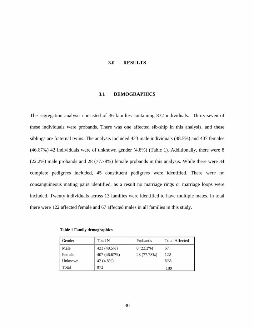
The segregation analysis consisted of 36 families containing 872 individuals. Thirty-seven of
these individuals were probands. There was one affected sib-ship in this analysis, and these
siblings are fraternal twins. The analysis included 423 male individuals (48.5%) and 407 females
(46.67%) 42 individuals were of unknown gender (4.8%) (Table 1). Additionally, there were 8
(22.2%) male probands and 28 (77.78%) female probands in this analysis. While there were 34
complete pedigrees included, 45 constituent pedigrees were identified. There were no
consanguineous mating pairs identified, as a result no marriage rings or marriage loops were
included. Twenty individuals across 13 families were identified to have multiple mates. In total
there were 122 affected female and 67 affected males in all families in this study.
Table 1 Family demographics
Gender Total N Probands Total Affected
Male 423 (48.5%) 8 (22.2%) 67 Female 407 (46.67%) 28 (77.78%) 122 Unknown 42 (4.8%) N/A Total 872 189
31
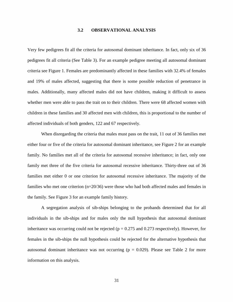
3.2 OBSERVATIONAL ANALYSIS
Very few pedigrees fit all the criteria for autosomal dominant inheritance. In fact, only six of 36
pedigrees fit all criteria (See Table 3). For an example pedigree meeting all autosomal dominant
criteria see Figure 1. Females are predominantly affected in these families with 32.4% of females
and 19% of males affected, suggesting that there is some possible reduction of penetrance in
males. Additionally, many affected males did not have children, making it difficult to assess
whether men were able to pass the trait on to their children. There were 68 affected women with
children in these families and 30 affected men with children, this is proportional to the number of
affected individuals of both genders, 122 and 67 respectively.
When disregarding the criteria that males must pass on the trait, 11 out of 36 families met
either four or five of the criteria for autosomal dominant inheritance, see Figure 2 for an example
family. No families met all of the criteria for autosomal recessive inheritance; in fact, only one
family met three of the five criteria for autosomal recessive inheritance. Thirty-three out of 36
families met either 0 or one criterion for autosomal recessive inheritance. The majority of the
families who met one criterion (n=20/36) were those who had both affected males and females in
the family. See Figure 3 for an example family history.
A segregation analysis of sib-ships belonging to the probands determined that for all
individuals in the sib-ships and for males only the null hypothesis that autosomal dominant
inheritance was occurring could not be rejected (p = 0.275 and 0.273 respectively). However, for
females in the sib-ships the null hypothesis could be rejected for the alternative hypothesis that
autosomal dominant inheritance was not occurring (p = 0.029). Please see Table 2 for more
information on this analysis.
32
Table 2: Simple Segregation Analysis with Gender
Observed Expected (O-E)2/E χ2 p All Individuals Affected 47 42 0.595 1.19 0.275
Unaffected 37 42 0.595 Females Affected 35 27 2.37 4.741 0.029
Unaffected 19 27 2.37 Males Affected 12 15 0.6 1.2 0.273
Unaffected 18 15 0.6
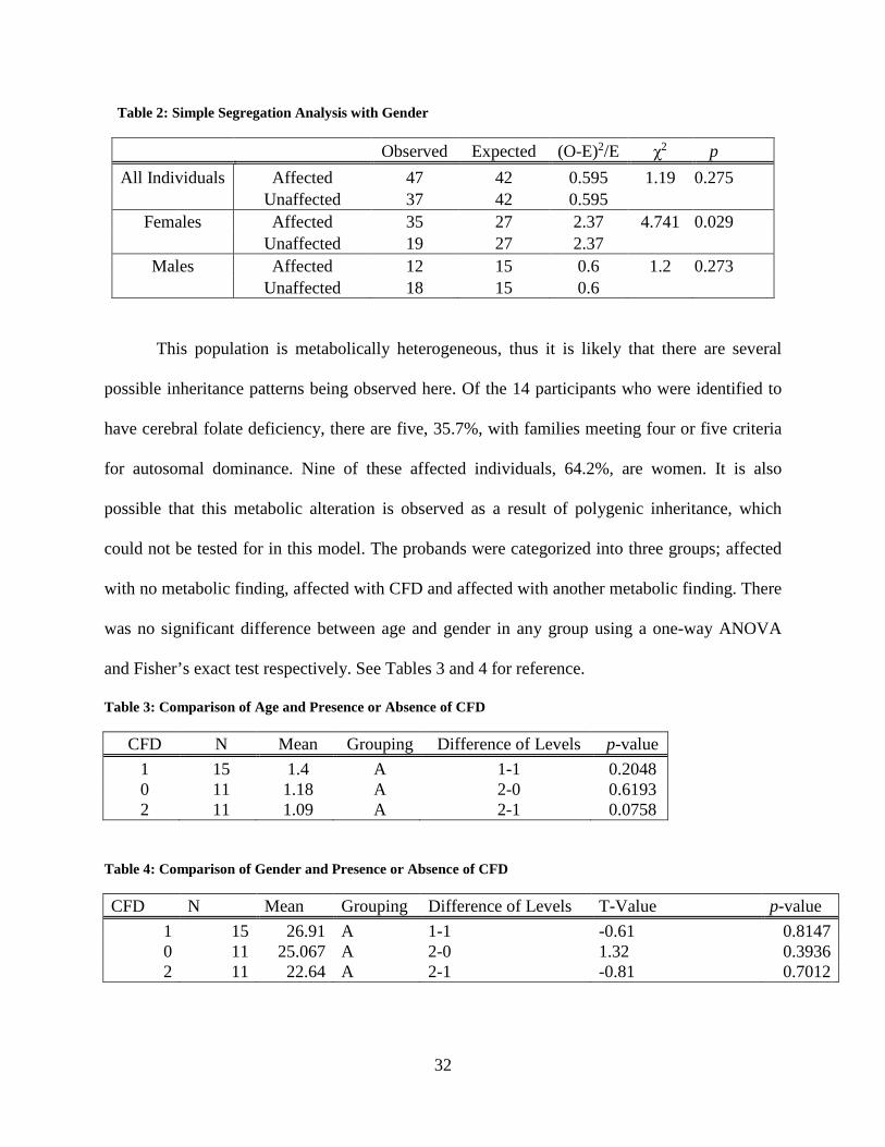
This population is metabolically heterogeneous, thus it is likely that there are several
possible inheritance patterns being observed here. Of the 14 participants who were identified to
have cerebral folate deficiency, there are five, 35.7%, with families meeting four or five criteria
for autosomal dominance. Nine of these affected individuals, 64.2%, are women. It is also
possible that this metabolic alteration is observed as a result of polygenic inheritance, which
could not be tested for in this model. The probands were categorized into three groups; affected
with no metabolic finding, affected with CFD and affected with another metabolic finding. There
was no significant difference between age and gender in any group using a one-way ANOVA
and Fisher’s exact test respectively. See Tables 3 and 4 for reference.
Table 3: Comparison of Age and Presence or Absence of CFD
CFD N Mean Grouping Difference of Levels p-value 1 15 1.4 A 1-1 0.2048 0 11 1.18 A 2-0 0.6193 2 11 1.09 A 2-1 0.0758
Table 4: Comparison of Gender and Presence or Absence of CFD
CFD N Mean Grouping Difference of Levels T-Value p-value 1 15 26.91 A 1-1 -0.61 0.8147 0 11 25.067 A 2-0 1.32 0.3936 2 11 22.64 A 2-1 -0.81 0.7012
33
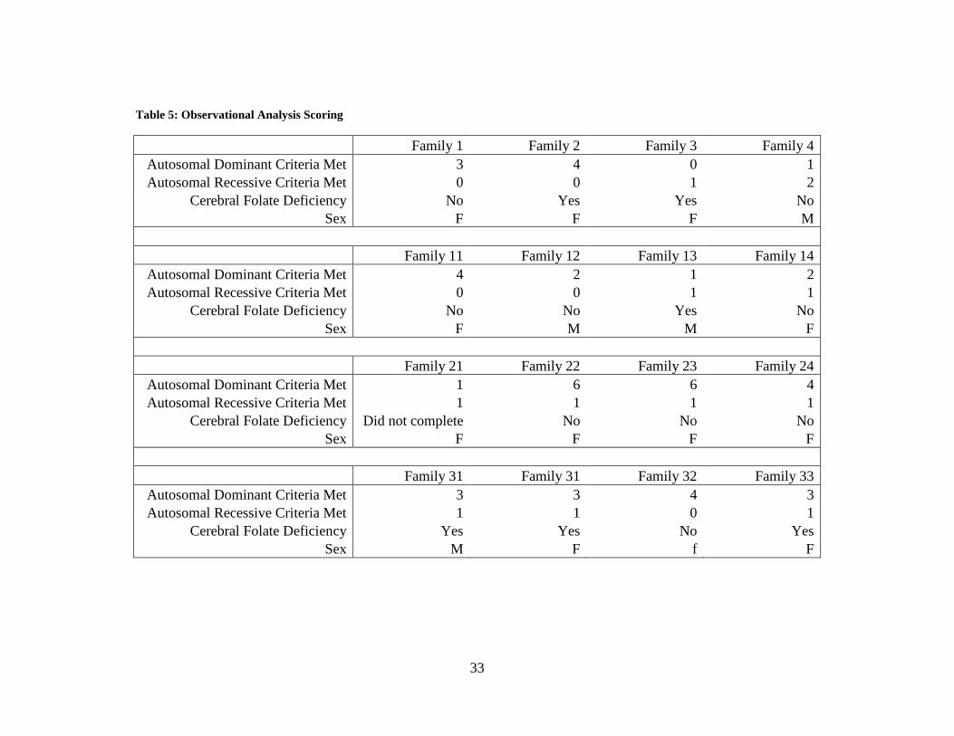
Table 5: Observational Analysis Scoring
Family 1 Family 2 Family 3 Family 4 Autosomal Dominant Criteria Met 3 4 0 1 Autosomal Recessive Criteria Met 0 0 1 2
Cerebral Folate Deficiency No Yes Yes No Sex F F F M
Family 11 Family 12 Family 13 Family 14 Autosomal Dominant Criteria Met 4 2 1 2 Autosomal Recessive Criteria Met 0 0 1 1
Cerebral Folate Deficiency No No Yes No Sex F M M F
Family 21 Family 22 Family 23 Family 24 Autosomal Dominant Criteria Met 1 6 6 4 Autosomal Recessive Criteria Met 1 1 1 1
Cerebral Folate Deficiency Did not complete No No No Sex F F F F
Family 31 Family 31 Family 32 Family 33 Autosomal Dominant Criteria Met 3 3 4 3 Autosomal Recessive Criteria Met 1 1 0 1
Cerebral Folate Deficiency Yes Yes No Yes Sex M F f F
34
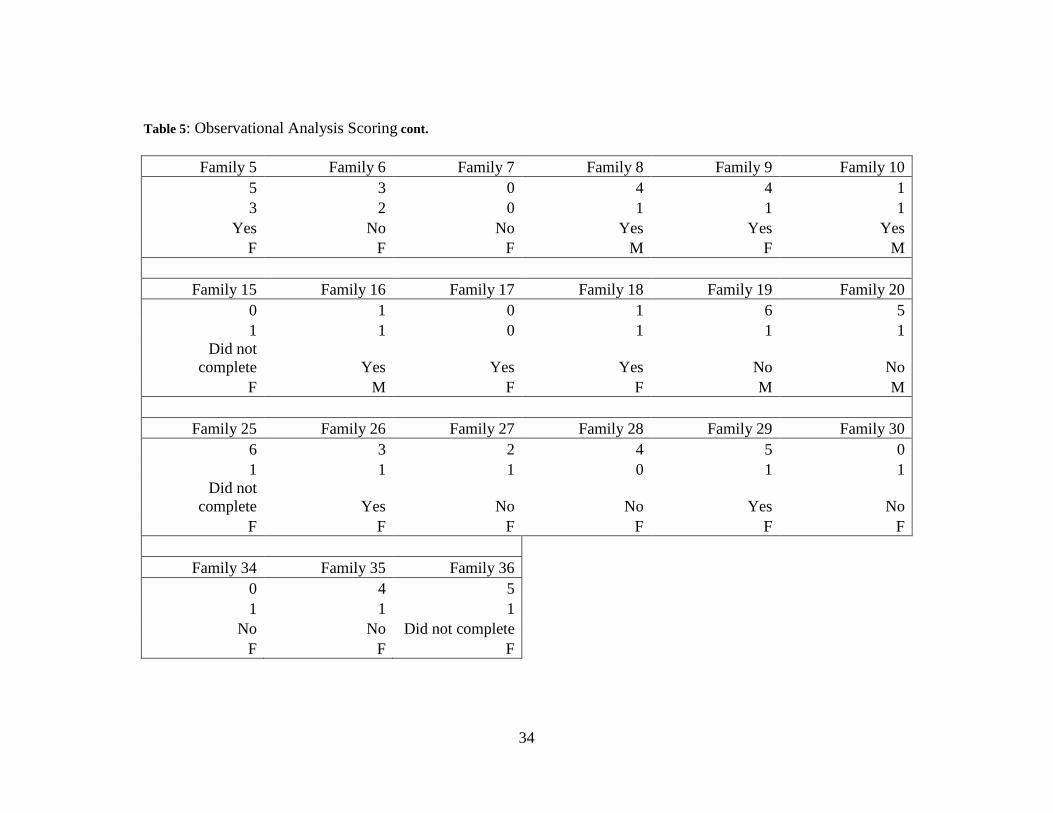
Table 5: Observational Analysis Scoring cont.
Family 5 Family 6 Family 7 Family 8 Family 9 Family 10 5 3 0 4 4 1 3 2 0 1 1 1
Yes No No Yes Yes Yes F F F M F M
Family 15 Family 16 Family 17 Family 18 Family 19 Family 20 0 1 0 1 6 5 1 1 0 1 1 1
Did not complete Yes Yes Yes No No
F M F F M M
Family 25 Family 26 Family 27 Family 28 Family 29 Family 30 6 3 2 4 5 0 1 1 1 0 1 1
Did not complete Yes No No Yes No
F F F F F F
Family 34 Family 35 Family 36 0 4 5 1 1 1 No No Did not complete F F F
35
3.3 SEGREGATION ANALYSIS
The program S.A.G.E.’s sub-program SEGREG was used to assess the segregation of depression
and suicide in these families53. The parameters for the analysis directed the program to run a
likelihood ratio test to determine if the families fit an autosomal dominant mode of inheritance,
compared to polygenic inheritance of disease. The parameters also specified that the program
evaluate trait for this mode of inheritance based on the metabolic findings in our probands. As a
result of the small sample size of the metabolic phenotype, this was ultimately not used in the
analysis.
For this analysis the following is assumed:
H0 = Transmission is occurring in an autosomal dominant manner
HA = Transmission is not occurring in an autosomal dominant manner
This assumption deviates from the original hypothesis as a result of the nature of the segregation
analysis. Because autosomal dominance is a known pattern, the segregation analysis can
determine whether the families assessed fit or do not fit the pattern; however it cannot do the
reverse.
The probability that transmission occurs when a parent has the AA genotype is
0.00000508, for the AB genotype probability is 0.70935667 and for the BB genotype probability
is 0.00000444. The likelihood ratio calculated the difference between what was observed and
what was expected for dominant inheritance. This calculation assumes that expected is 1.0 for
the AA genotype, 0.50 for the AB genotype and 0.00 for the BB genotype. A test statistical D
was calculated as two times the difference in the log-likelihoods. The log-likelihood of the null
36
hypothesis is -361.897, and the log likelihood of the alternative hypothesis is -347.015, thus
D=29.725. D has a chi-squared distribution with three degrees of freedom because there are three
fixed differences between the two models; τ AA, τ AB and τ BB where τ is the transmission
probability of A given the parental genotypes AA, AB or BB, respectively. The result of the
likelihood ratio test is P(χ²(3) ≥ 29.725) = 1.58e-06. At α = 0.05 the null hypothesis that these
families are exhibiting autosomal dominant inheritance was rejected in favor of the alterative
hypothesis, that inheritance is not autosomal dominant.
Simple segregation analysis of the segregation ratio of the sibships in the study showed a
segregation ratio of 1.19 for all sibships (P = 0.275 for test of deviance from the expected ratio of
0.5,), 4.741 for female sibships (P = 0.029), and 1.2 for male sibships (P = 0.273). The null
hypothesis of autosomal dominance was rejected in favor of the alternative hypothesis for female
sibships and not rejected for males or all sibships.

37
4.0 DISCUSSION
In spite of the many associated biologic factors with depression, there is no known cause for
depression, no diagnostic test and few indications for who may develop chronic, treatment
resistant depression21. It is often recurrent and accompanied by significant morbidity and
mortality14. Suicide is highly correlated with depression but there is no reliable method to
determine who is at risk14,99. This study represents a small, metabolically heterogeneous group of
participants with treatment resistant major depression and suicidality. It attempts to describe the
patterns of inheritance in this unique cohort and ultimately add to the growing knowledge
surrounding this newly described group of neuropsychiatric IEM’s72,93.
The majority of individuals in this group are affected by cerebral folate deficiency,
traditionally thought of as an autosomal recessive disorder90,91. It is important to note that these
individuals are not phenotypically identical to those that are homozygous for FOLR1 mutations,
which results in severe neurological disease presenting with ataxia, seizures, and developmental
delay among other symptoms84,90,91. However, individuals in this study are not affected with
biallelic FOLR1 mutations and thus results in a conundrum for discerning the cause for CFD in
this population.
Current understanding of the etiology of depression describes it as a multifactorial
disorder in which both genetic and environmental factors contribute to the development of
disease2,44,47. The segregation analysis performed by S.A.G.E. ultimately supports this
38
conclusion since no Mendelian inheritance pattern was identified through the analysis. Upon
observation, many of the families also did not meet all of the criteria for dominant inheritance,
further ruling out the possibility of a single gene cause for the depression and the underlying
metabolic alteration. Previous GWA studies of depression have been unable to identify anything
more substantial than candidate genes in very large cohorts of individuals30,49.
In simple segregation analysis, the null hypothesis, that autosomal dominance was
occurring, could not be rejected for two of the three groups of the simple segregation analysis
performed on the sib-ships of probands; males and all individuals even though it was rejected for
females. It is important to consider that the alternative hypothesis, that autosomal dominance was
not occurring, was not rejected for males and all individuals because the small sample sizes of
those groups reduce the power to detect such a deviance from the expected. Because this
segregation study assumes that an affected parent is present and passing on the trait it is
important to consider adoption studies. One of which found that maternal depression, but not
paternal depression, increases risk of depression in adoptees100. Additionally, the T-tests
performed on this group of individuals determined that males and females who were affected
were significantly different from one another. As a result it may be important for future studies
to be more particular about the number of individuals in each gender for a more cohesive group
and for interpretation.
Evidence gained from this study allows for the consideration of a more complex pattern
of inheritance within this unique subgroup of individuals. In cases of other types of metabolic
diseases with unknown genetic origin, it has been suggested that many subtle genomic changes
together may lead to the manifestation of disease92. These genomic changes are largely not
understood at this juncture; they may be epigenetic changes, or multiple heterozygous mutations
39
within a pathway or related pathways. It is likely that a similar mechanism is resulting in this
specific group of patients with TR-MDD with metabolic alterations93.
Many studies have determined that depression is difficult to study because groups of
participants are typically highly heterogeneous41. While the cohort for this study is also
heterogeneous, we have taken advantage of a unique group of individuals identified by recent
studies of depression that use metabolomics to determine if there is an underlying metabolic
alteration that may be contributing to the development of depression to discern whether
inheritance in this population aligns with the findings of previous studies of depression. This
metabolic difference has identified a quantitative measure that can be utilized not only for
treatment options but can optimize the ability to study depression93. Studies of this sub-group
may translate into a quantitative test for depression, biomarkers for treatment-resistance or
remain a unique, but alternative method for identification of personalized treatment options for
those who are treatment-resistant with major depressive disorder. Future application of studies of
neuropsychiatric IEM’s may eventually help to create testing options for affected individuals and
their families.
4.1 PUBLIC HEALTH SIGNIFICANCE
The World Health Organization (WHO) predicts MDD will be the second leading cause for
disability by the year 2020, thus placing significant burden on the public101. Approximately 10-
15% of US adults have depression; women are 1.5-3 times more likely to be affected than men,
with this difference beginning in adolescence65,101. The prevailing theory regarding why women
experience depression at higher rates than men involves the complex interaction between

40
environmental experiences and biologic vulnerabilities which differ between men and women102.
Depression can occur at any age, but the most affected age group is within the 25-34 year range,
with 30.1% of affected individuals falling in this age range103. It is estimated that 14 million
people suffer from depression and roughly half of them will seek some form of treatment3,104,.
Some proposed reasons for many individuals with depression not seeking treatment involve
financial and situational circumstances as well as perception and stigma surrounding mental
illness. The majority of people with major depression feel that they could handle or treat the
depressive episode themselves104. Some people do not perceive their condition as an illness;
others feel that it is a normal response to events in their lives104. Additionally, some experience
financial barriers to seeking treatment104.
By identifying the cause of depression within this group it may not only affect the
individuals in this cohort by determining a treatment in addition to a cause, but could also
decrease the cost of treatment and the length of time individuals are non-responsive to treatment
in the larger population of depressed people. In addition, learning who may be at risk for
developing depression in this population would increase the quality of life and general health for
these individuals and their family members. Further, by using a personalized treatment method
the cost of treating depression would decrease. The ability to identify biologic causes for
depression and suicidality may also affect the stigma associated with mental health issues and
depression. Therefore this study and subsequent studies of this group of individuals may also
increase the amount of individuals who seek treatment or who identify their depression as worthy
of seeking treatment. Further, identification of inheritance and depressive etiology in this
population may decrease the cost of treatment overall reducing the public health burden of
depression.
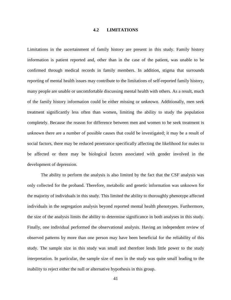
41
4.2 LIMITATIONS
Limitations in the ascertainment of family history are present in this study. Family history
information is patient reported and, other than in the case of the patient, was unable to be
confirmed through medical records in family members. In addition, stigma that surrounds
reporting of mental health issues may contribute to the limitations of self-reported family history,
many people are unable or uncomfortable discussing mental health with others. As a result, much
of the family history information could be either missing or unknown. Additionally, men seek
treatment significantly less often than women, limiting the ability to study the population
completely. Because the reason for difference between men and women to be seek treatment is
unknown there are a number of possible causes that could be investigated; it may be a result of
social factors, there may be reduced penetrance specifically affecting the likelihood for males to
be affected or there may be biological factors associated with gender involved in the
development of depression.
The ability to perform the analysis is also limited by the fact that the CSF analysis was
only collected for the proband. Therefore, metabolic and genetic information was unknown for
the majority of individuals in this study. This limited the ability to thoroughly phenotype affected
individuals in the segregation analysis beyond reported mental health phenotypes. Furthermore,
the size of the analysis limits the ability to determine significance in both analyses in this study.
Finally, one individual performed the observational analysis. Having an independent review of
observed patterns by more than one person may have been beneficial for the reliability of this
study. The sample size in this study was small and therefore lends little power to the study
interpretation. In particular, the sample size of men in the study was quite small leading to the
inability to reject either the null or alternative hypothesis in this group.
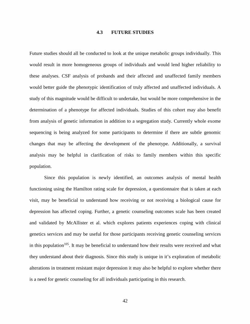
42
4.3 FUTURE STUDIES
Future studies should all be conducted to look at the unique metabolic groups individually. This
would result in more homogeneous groups of individuals and would lend higher reliability to
these analyses. CSF analysis of probands and their affected and unaffected family members
would better guide the phenotypic identification of truly affected and unaffected individuals. A
study of this magnitude would be difficult to undertake, but would be more comprehensive in the
determination of a phenotype for affected individuals. Studies of this cohort may also benefit
from analysis of genetic information in addition to a segregation study. Currently whole exome
sequencing is being analyzed for some participants to determine if there are subtle genomic
changes that may be affecting the development of the phenotype. Additionally, a survival
analysis may be helpful in clarification of risks to family members within this specific
population.
Since this population is newly identified, an outcomes analysis of mental health
functioning using the Hamilton rating scale for depression, a questionnaire that is taken at each
visit, may be beneficial to understand how receiving or not receiving a biological cause for
depression has affected coping. Further, a genetic counseling outcomes scale has been created
and validated by McAllister et al. which explores patients experiences coping with clinical
genetics services and may be useful for those participants receiving genetic counseling services
in this population105. It may be beneficial to understand how their results were received and what
they understand about their diagnosis. Since this study is unique in it’s exploration of metabolic
alterations in treatment resistant major depression it may also be helpful to explore whether there
is a need for genetic counseling for all individuals participating in this research.
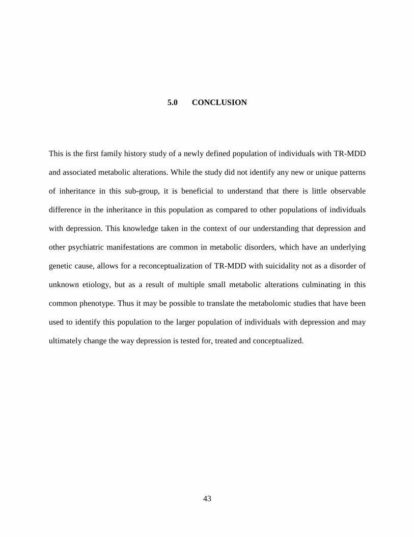
43
5.0 CONCLUSION
This is the first family history study of a newly defined population of individuals with TR-MDD
and associated metabolic alterations. While the study did not identify any new or unique patterns
of inheritance in this sub-group, it is beneficial to understand that there is little observable
difference in the inheritance in this population as compared to other populations of individuals
with depression. This knowledge taken in the context of our understanding that depression and
other psychiatric manifestations are common in metabolic disorders, which have an underlying
genetic cause, allows for a reconceptualization of TR-MDD with suicidality not as a disorder of
unknown etiology, but as a result of multiple small metabolic alterations culminating in this
common phenotype. Thus it may be possible to translate the metabolomic studies that have been
used to identify this population to the larger population of individuals with depression and may
ultimately change the way depression is tested for, treated and conceptualized.

44
APPENDIX A: FIGURES
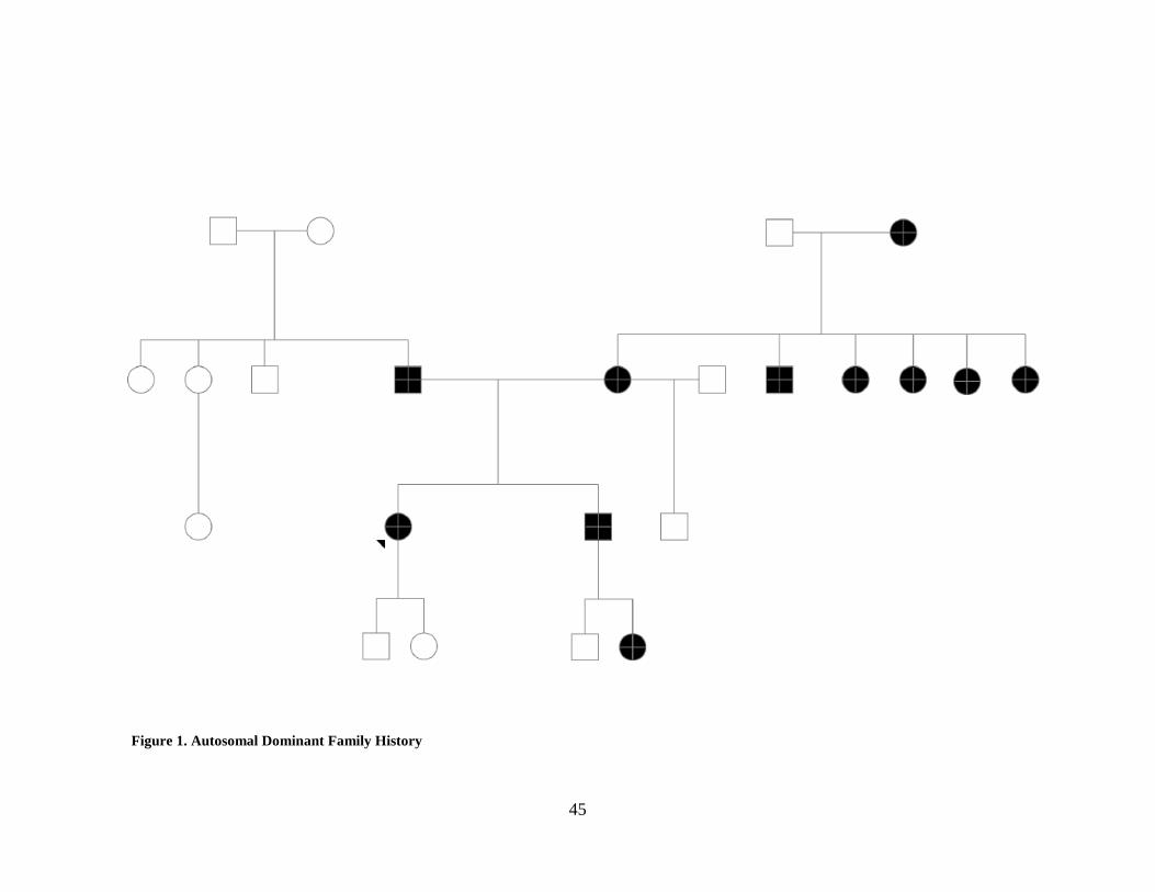
45
Figure 1. Autosomal Dominant Family History
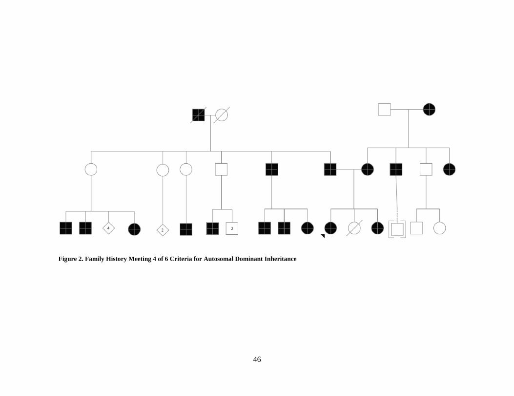
46
Figure 2. Family History Meeting 4 of 6 Criteria for Autosomal Dominant Inheritance
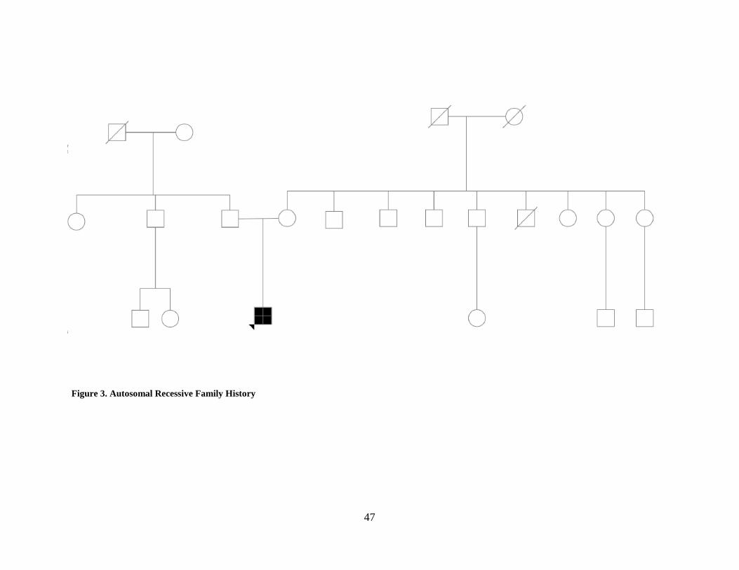
47
Figure 3. Autosomal Recessive Family History
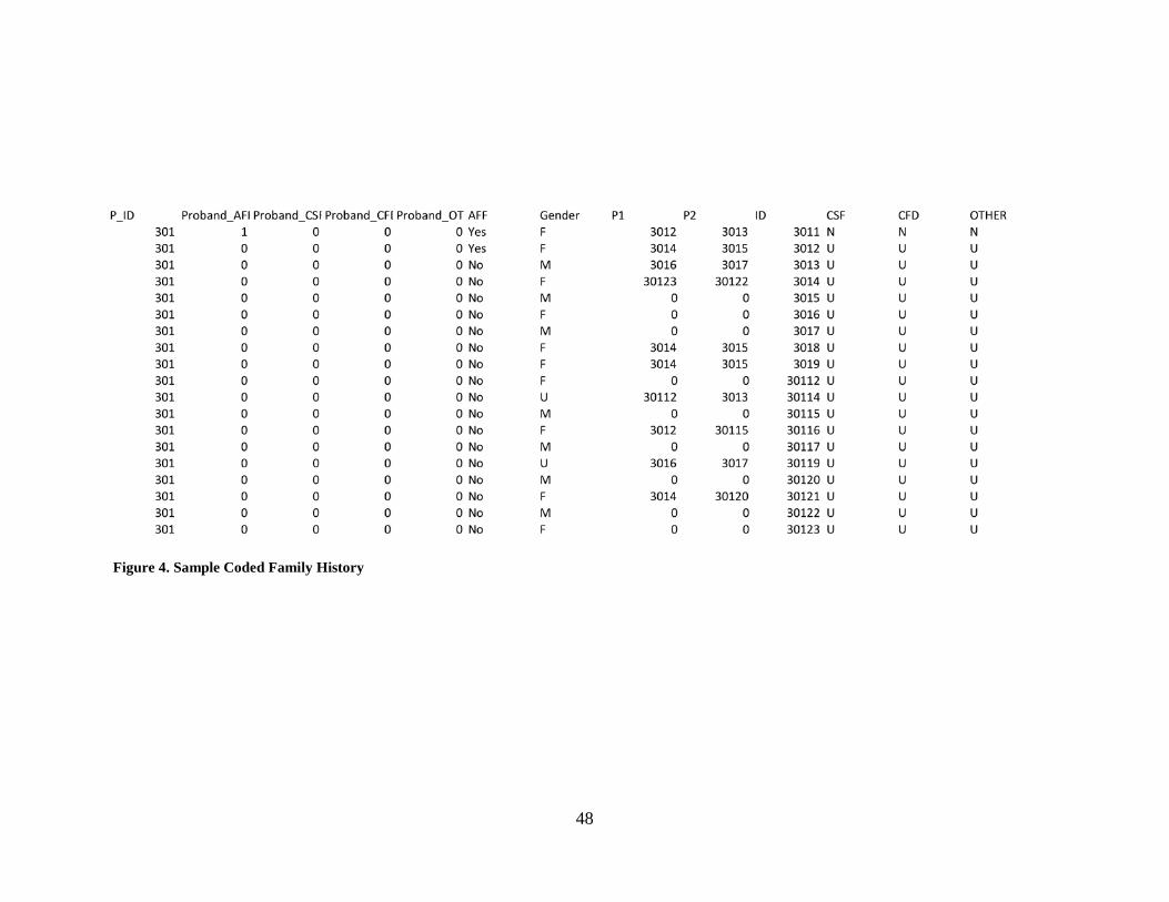
48
Figure 4. Sample Coded Family History
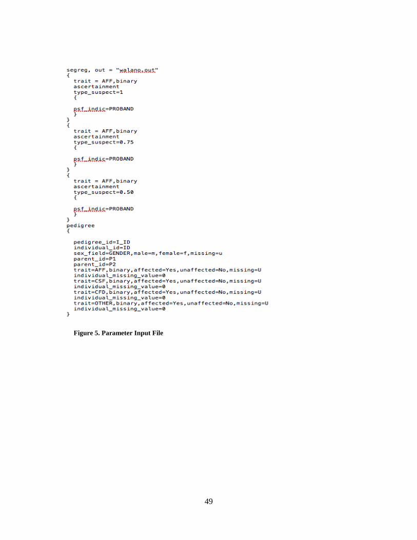
49
Figure 5. Parameter Input File
50
APPENDIX B: IRB APPROVAL
University of Pittsburgh Institutional Review Board
3500 Fifth Avenue Ground Level Pittsburgh, PA 15213 (412) 383-1480 (412) 383-1508 (fax) http://www.irb.pitt.edu
Memorandum To: Lisa Pan, MD From: IRB Office Date: 1/29/2016 IRB#: MOD14060600-04 / PRO14060600 Subject: Metabolomics of Early Suicide Attempt (MESA).
The University of Pittsburgh Institutional Review Board reviewed and approved the requested modifications by the expedited review procedure authorized under 45 CFR 46.110 and 21 CFR 56.110.
Modification Approval Date: 1/29/2016 Expiration Date: 8/17/2016
The following documents were approved by the IRB: We would like to add the following individuals to our study team: Dr Michael
51
Morowitz, and Shilpa Argade. They will be assisting us with analyzing the blood, urine, and CSF samples that we have collected.
Please note that it is the investigator’s responsibility to report to the IRB any unanticipated problems involving risks to subjects or others [see 45 CFR 46.103(b)(5) and 21 CFR 56.108(b)]. Refer to the IRB Policy and Procedure Manual regarding the reporting requirements for unanticipated problems which include, but are not limited to, adverse events. If you have any questions about this process, please contact the Adverse Events Coordinator at 412-383-1480.
The protocol and consent forms, along with a brief progress report must be resubmitted at least one month prior to the renewal date noted above as required by FWA00006790 (University of Pittsburgh), FWA00006735 (University of Pittsburgh Medical Center), FWA00000600 (Children’s Hospital of Pittsburgh), FWA00003567 (Magee-Womens Health Corporation), FWA00003338 (University of Pittsburgh Medical Center Cancer Institute).
Please be advised that your research study may be audited periodically by the University of Pittsburgh Research Conduct and Compliance Office.
University of Pittsburgh Institutional Review Board
3500 Fifth Avenue Pittsburgh, PA 15213 (412) 383-1480 (412) 383-1508 (fax) http://www.irb.pitt.edu
Memorandum To: Lisa Pan, MD From: Aviva Katz, MD, Vice Chair Date: 9/15/2015 IRB#: REN15070250 / PRO11120375 Subject: Metabolomics of treatment refractory depression and early onset suicide attempt
At its full board meeting on 8/19/2015, the University of Pittsburgh Institutional Review Board, Committee G, reviewed the Renewal for the above referenced research study and approved it pending minor modifications. Your responses to these
52
comments have been reviewed and the research submission, in its currently modified form, adequately addresses the concerns of the IRB and is therefore approved.
The risk level designation is Greater Than Minimal Risk. Please note the following information: Approval Date: 9/14/2015 Expiration Date: 8/18/2016 Please note that it is the investigator’s responsibility to report to the IRB any unanticipated problems involving risks to subjects or others [see 45 CFR 46.103(b)(5) and 21 CFR 56.108(b)]. Refer to the IRB Policy and Procedure Manual regarding the reporting requirements for unanticipated problems which include, but are not limited to, adverse events. If you have any questions about this process, please contact the Adverse Events Coordinator at 412-383-1480.
The protocol and consent forms, along with a brief progress report must be resubmitted at least one month prior to the renewal date noted above as required by FWA00006790 (University of Pittsburgh), FWA00006735 (University of Pittsburgh Medical Center), FWA00000600 (Children’s Hospital of Pittsburgh), FWA00003567 (Magee-Womens Health Corporation), FWA00003338 (University of Pittsburgh Medical Center Cancer Institute).
Please be advised that your research study may be audited periodically by the University of Pittsburgh Research Conduct and Compliance Office.
53
APPENDIX C: MTRD ADULT CONSENT FORM
54
55
56
57
58
59
BIBLIOGRAPHY
1. Pratt L, Brody D. Depression in the U.S. Household Population. 2009-2012. NCHS Data Brief No 172. Hyattsville, MD; 2014.
2. Kessler RC, Berglund P, Demler O, et al. The Epidemiology of Major. 2003;289(23):3095-3105.
3. American Psychiatric Association. Diagnostic and Statstical Manual of Mental Disorders. 5th ed.; 2013.
4. Keller MB. Issues in treatment-resistant depression. J Clin Psychiatry. 2005;66(SUPPL. 8):5-12.
5. Hamilton M. A rating scale for depression. J Neurol Neurosurg Psychiatry. 1960;23:56-62. doi:10.1136/jnnp.23.1.56.
6. Al-Harbi KS. Treatment-resistant depression: Therapeutic trends, challenges, and future directions. Patient Prefer Adherence. 2012;6:369-388. doi:10.2147/PPA.S29716.
7. Souery D, Papakostas GI, Trivedi MH. Treatment-resistant depression. J Clin Psychiatry. 2006;67(suppl 6):16-22.
8. Palm U, Hasan A, Strube W, Padberg F. tDCS for the treatment of depression: a comprehensive review. Eur Arch Psychiatry Clin Neurosci. 2016. doi:10.1007/s00406-016-0674-9.
9. Abou-Saleh MT, Coppen A. the Biology Psychiatric Progress of Folate in Depression : Hypotheses Implications for Nutritional of the Psychoses. J Psychiatry. 1986;20(2):91-101.
10. Fava M, Mischoulon D. Folate in depression: Efficacy, safety, differences in formulations, and clinical issues. J Clin Psychiatry. 2009;70(SUPPL. 5):12-17. doi:10.4088/JCP.8157su1c.03.
11. Rush AJ, Trivedi MH, Wisniewski SR, et al. Acute and longer-term outcomes in depressed outpatients requiring one or several treatment steps: A STAR*D report. Am J Psychiatry. 2006;163(11):1905-1917. doi:10.1176/appi.ajp.163.11.1905.
60
12. de Sousa RT, Zanetti M V, Brunoni AR, Machado-Vieira R. Challenging Treatment-Resistant Major Depressive Disorder: A Roadmap for Improved Therapeutics. Curr Neuropharmacol. 2015;13(5):616-635. doi:10.2174/1570159X13666150630173522.
13. Trivedi MH, Rush AJ, Crismon ML, et al. Clinical results for patients with major depressive disorder in the Texas Medication Algorithm Project. Arch Gen Psychiatry. 2004;61(7):669-680. doi:10.1001/archpsyc.61.7.669.
14. Pjevac M, Pregelj P. Neurobiology of suicidal behaviour. Psychiatr Danub. 2012;24(SUPPL.3):336-341. doi:10.1038/nrn1220.
15. Centers for Disease Control and Prevention. Web-based Injury Statistics Query and Reporting System (WISQARS). 2013. www.cdc.gov/injury/wisqars/index.html.
16. Blair-West GW, Cantor CH, Mellsop GW, Eyeson-Annan ML. Lifetime suicide risk in major depression: sex and age determinants. J Affect Disord. 1999;55(2-3):171-178.
17. Peay HL, Veach PM, Palmer CGS, Rosen-Sheidley B, Gettig E, Austin JC. Psychiatric disorders in clinical genetics I: Addressing family histories of psychiatric illness. J Genet Couns. 2008;17(1):6-17. doi:10.1007/s10897-007-9120-5.
18. Austin JC, Palmer CGS, Rosen-Sheidley B, Veach PM, Gettig E, Peay HL. Psychiatric disorders in clinical genetics II: Individualizing recurrence risks. J Genet Couns. 2008;17(1):18-29. doi:10.1007/s10897-007-9121-4.
19. Keitner GI, Miller IW. Family functioning and major depression: An overview. Am J Psychiatry. 1990;147(9):1128-1137.
20. Fava M. Diagnosis and definition of treatment-resistant depression. Biol Psychiatry. 2003;53(8):649-659. doi:10.1016/S0006-3223(03)00231-2.
21. Papakostas GI. Surrogate markers of treatment outcome in major depressive disorder. Int J Neuropsychopharmacol. 2012;15(6):841-854. doi:10.1017/S1461145711001246.
22. Souery D, Oswald P, Massat I, et al. Clinical factors associated with treatment resistance in major depressive disorder: results from a European multicenter study. J Clin Psychiatry. 2007;68(7):1062-1070.
23. Rush a J, Kraemer HC, Sackeim H a, et al. Report by the ACNP Task Force on response and remission in major depressive disorder. Neuropsychopharmacology. 2006;31(9):1841-1853. doi:10.1038/sj.npp.1301131.
24. Stewart JW, Deliyannides D, Taylor BP, Ph D, Davies CA, Klein DF. Effectiveness for Depressed Outpatients. 2005;(June):671-677.
25. Paykel ES. Partial remission, residual symptoms, and relapse in depression. Dialogues Clin Neurosci. 2008;10(4):431-437.
61
26. Riso LP, Klein DN, Ferro T, et al. Understanding the comorbidity between early-onset dysthymia and cluster B personality disorders: A family study. Am J Psychiatry. 1996;153(SUPPL.):900-906.
27. Berman R. Research Articles. 1997;164(June):351-359.
28. Yu YW-Y, Tsai S-J, Chen T-J, Lin C-H, Hong C-J. Association study of the serotonin transporter promoter polymorphism and symptomatology and antidepressant response in major depressive disorders. Mol Psychiatry. 2002;7(10):1115-1119. doi:10.1038/sj.mp.4001141.
29. El-Hage W, Leman S, Camus V, Belzung C. Mechanisms of antidepressant resistance. Front Pharmacol. 2013;4 NOV(November):1-23. doi:10.3389/fphar.2013.00146.
30. Fabbri C, Porcelli S, Serretti A. From pharmacogenetics to pharmacogenomics: The way toward the personalization of antidepressant treatment. Can J Psychiatry. 2014;59(2):62-75.
31. Savitz J, Lucki I, Drevets WC. 5-HT1A receptor function in major depressive disorder. Prog Neurobiol. 2009;88(1):17-31. doi:10.1016/j.pneurobio.2009.01.009.
32. Ke L, Qi ZY, Ping Y, Ren CY. Effect of SNP at position 40237 in exon 7 of the TPH2 gene on susceptibility to suicide. Brain Res. 2006;1122(1):24-26. doi:10.1016/j.brainres.2006.09.007.
33. Tadic a, Rujescu D, Szegedi a, et al. Association of a MAOA gene variant with generalized anxiety disorder, but not with panic disorder or major depression. Am J Med Genet B Neuropsychiatr Genet. 2003;117B(1):1-6. doi:10.1002/ajmg.b.10013.
34. Baune BT, Hohoff C, Berger K, et al. Association of the COMT val158met variant with antidepressant treatment response in major depression. Neuropsychopharmacology. 2008;33(4):924-932. doi:10.1038/sj.npp.1301462.
35. Wray NR, James MR, Dumenil T, et al. Association study of candidate variants of COMT with neuroticism, anxiety and depression. Am J Med Genet Part B Neuropsychiatr Genet. 2008;147(7):1314-1318. doi:10.1002/ajmg.b.30744.
36. Yu YW-Y, Tsai S-J, Hong C-J, Chen T-J, Chen M-C, Yang C-W. Association study of a monoamine oxidase a gene promoter polymorphism with major depressive disorder and antidepressant response. Neuropsychopharmacology. 2005;30(9):1719-1723. doi:10.1038/sj.npp.1300785.
37. Lin E, Chen PS, Huang L-C, Hsu S-Y. Association study of a brain-derived neurotrophic-factor polymorphism and short-term antidepressant response in major depressive disorders. Pharmgenomics Pers Med. 2008;1:1-6. http://www.pubmedcentral.nih.gov/articlerender.fcgi?artid=3513194&tool=pmcentrez&rendertype=abstract.
62
38. Lin JH, Lu AY. Inhibition and induction of cytochrome P450 and the clinical implications. Clin Pharmacokinet. 1998;35(5):361-390. doi:10.2165/00003088-199835050-00003.
39. Serretti A, Drago A, Liebman MN. Pharmacogenetics of antidepressant response. Biomarkers Psychiatr Disord. 2009;36(2):315-353. doi:10.1007/978-0-387-79251-4_14.
40. consortium C. Sparse whole-genome sequencing identifies two loci for major depressive disorder. Nature. 2015;523(7562):588. doi:10.1038/nature14659.
41. Flint J, Chen Y, Shi S, Kendler KS. Epilogue: Lessons from the CONVERGE study of major depressive disorder in China. J Affect Disord. 2012;140(1):1-5. doi:10.1016/j.jad.2011.09.006.
42. Klein DN, Riso LP, Donaldson SK, et al. Family Study of Early-Onset Dysthymia. 1995.
43. Mullins N, Power RA, Fisher HL, et al. Polygenic interactions with environmental adversity in the aetiology of major depressive disorder. Psychol Med. 2015:1-12. doi:10.1017/S0033291715002172.
44. Sullivan PF, Neale MC, Kendler KS. Genetic epidemiology of major depression: Review and meta-analysis. Am J Psychiatry. 2000;157(10):1552-1562. doi:10.1176/appi.ajp.157.10.1552.
45. Kendler KS, Gatz M, Gardner CO, Pedersen NL. A Swedish national twin study of lifetime major depression. Am J Psychiatry. 2006;163(1):109-114. doi:10.1176/appi.ajp.163.1.109.
46. Byrne EM, Carrillo-Roa T, Henders a K, et al. Monozygotic twins affected with major depressive disorder have greater variance in methylation than their unaffected co-twin. Transl Psychiatry. 2013;3(6):e269. doi:10.1038/tp.2013.45.
47. Fernandez-Pujals AM, Adams MJ, Thomson P, et al. Epidemiology and Heritability of Major Depressive Disorder, Stratified by Age of Onset, Sex, and Illness Course in Generation Scotland: Scottish Family Health Study (GS:SFHS). PLoS One. 2015;10(11):e0142197. doi:10.1371/journal.pone.0142197.
48. Parker G. Beyond major depression. Psychol Med. 2005;35(4):467-474. doi:10.1017/S0033291704004210.
49. McGuffin P, Perroud N, Uher R, et al. The genetics of affective disorder and suicide. Eur Psychiatry. 2010;25(5):275-277. doi:10.1016/j.eurpsy.2009.12.012.
50. Li CC. a tale of two thermos bottles : properties of a genetic model for human intelligence. 1971.
51. Lander ES. Initial impact of the sequencing of the human genome. Nature. 2011;470(7333):187-197. doi:10.1038/nature09792.
63
52. Reich DE, Lander ES. On the allelic spectrum of human disease. Trends Genet. 2001;17(9):502-510. doi:10.1016/S0168-9525(01)02410-6.
53. Sekar A, Bialas AR, de Rivera H, et al. Schizophrenia risk from complex variation of complement component 4. Nature. 2016;530(7589):177-183. doi:10.1038/nature16549.
54. Harrison PJ, Weinberger DR. Schizophrenia genes, gene expression, and neuropathology: on the matter of their convergence. Mol Psychiatry. 2005;10(1):40-68; image 5. doi:10.1038/sj.mp.4001558.
55. Levinson DF. The Genetics of Depression: A Review. Biol Psychiatry. 2006;60(2):84-92. doi:10.1016/j.biopsych.2005.08.024.
56. Córdova-Palomera a, Fatjó-Vilas M, Gastó C, Navarro V, Krebs M-O, Fañanás L. Genome-wide methylation study on depression: differential methylation and variable methylation in monozygotic twins. Transl Psychiatry. 2015;5(March):e557. doi:10.1038/tp.2015.49.
57. Johnson B a, Brent D a, Bridge J, Connolly J. The familial aggregation of adolescent suicide attempts. Acta Psychiatr Scand. 1998;97(1):18-24.
58. Lieb R, Bronisch T, Höfler M, Schreier A, Wittchen HU. Maternal suicidality and risk of suicidality in offspring: Findings from a community study. Am J Psychiatry. 2005;162(9):1665-1671. doi:10.1176/appi.ajp.162.9.1665.
59. Adam KS, Bouckoms A, Streiner D. PArental loss and family stability in attempted suicide. Arch Gen Psychiatry. 1982;39(9):1081-1085. doi:10.1001/archpsyc.1982.04290090065013.
60. Runeson B, Åsberg M. Family history of suicide among suicide victims. Am J Psychiatry. 2003;160(8):1525-1526. doi:10.1176/appi.ajp.160.8.1525.
61. Phillips MR, Yang G, Zhang Y, Wang L, Ji H, Zhou M. Risk factors for suicide in China : A national case-control psychology autopsy study psychological autopsy study. Lancet. 2002;360(9347):1728-1736. doi:10.1016/S0140-6736(02)11681-3.
62. Cho H, Guo G, Iritani BJ, Hallfors DD. Genetic contribution to suicidal behaviors and associated risk factors among adolescents in the U.S. Prev Sci. 2006;7(3):303-311. doi:10.1007/s11121-006-0042-5.
63. Mcguffin P, Katz R, Watkins S, Rutherford J. Hospital-Based Heritability Unipolar. Arch Gen Psychiatry. 1996;53(2):129. doi:10.1001/archpsyc.1996.01830020047006.
64. Daly J, Ripke S, Lewis CM, et al. A mega-analysis of genome-wide association studies for major depressive disorder. Mol Psychiatry. 2013;18(4):497-511. doi:10.1038/mp.2012.21.A.

64
65. Xia L, Yao S. The Involvement of Genes in Adolescent Depression : A Systematic Review. 2015;9(December):1-9. doi:10.3389/fnbeh.2015.00329.
66. Devlin AM, Brain U, Austin J, Oberlander TF. Prenatal exposure to maternal depressed mood and the MTHFR C677T variant affect SLC6A4 methylation in infants at birth. PLoS One. 2010;5(8):2-9. doi:10.1371/journal.pone.0012201.
67. Kelly CB, McDonnell AP, Johnston TG, et al. The MTHFR C677T polymorphism is associated with depressive episodes in patients from Northern Ireland. J Psychopharmacol. 2004;18(4):567-571. http://www.ncbi.nlm.nih.gov/entrez/query.fcgi?cmd=Retrieve&db=PubMed&dopt=Citation&list_uids=15582924.
68. Peerbooms OLJ, van Os J, Drukker M, et al. Meta-analysis of MTHFR gene variants in schizophrenia, bipolar disorder and unipolar depressive disorder: Evidence for a common genetic vulnerability? Brain Behav Immun. 2011;25(8):1530-1543. doi:10.1016/j.bbi.2010.12.006.
69. Hickey SE, Curry CJ, Toriello H V. ACMG Practice Guideline: lack of evidence for MTHFR polymorphism testing. Genet Med. 2013;15(2):153-156. doi:10.1038/gim.2012.165.
70. Vol NV, Belousova LV, Kulikov AV. Association between the 5-HTTPLR Polymorphism of Serotonin Transporter Gene and EEG in Young and Postmenopausal Women. Zhurnal Vyss Nervn Deiatelnosti Im I P Pavlov. 2015;65(3).
71. Sedel F, Baumann N, Turpin JC, Lyon-Caen O, Saudubray JM, Cohen D. Psychiatric manifestations revealing inborn errors of metabolism in adolescents and adults. J Inherit Metab Dis. 2007;30(5):631-641. doi:10.1007/s10545-007-0661-4.
72. Pan L, Vockley J. Neuropsychiatric Symptoms in Inborn Errors of Metabolism: Incorporation of Genomic and Metabolomic Analysis into Therapeutics and Prevention. Curr Genet Med Rep. 2013;1(1):65-70. doi:10.1007/s40142-012-0004-0.
73. Cole AL, Lee PJ, Hughes DA, Deegan PB, Waldek S, Lachmann RH. Depression in adults with Fabry disease: A common and under-diagnosed problem. J Inherit Metab Dis. 2007;30(6):943-951. doi:10.1007/s10545-007-0708-6.
74. Sharman R, Sullivan K, Young RM, McGill J. Depressive symptoms in adolescents with early and continuously treated phenylketonuria: Associations with phenylalanine and tyrosine levels. Gene. 2012;504(2):288-291. doi:10.1016/j.gene.2012.05.007.
75. Muelly ER, Moore GJ, Bunce SC, et al. Biochemical correlates of neuropsychiatric illness in maple syrup urine disease. J Clin Invest. 2013;123(4):1809-1820. doi:10.1172/JCI67217.
65
76. Blau N, Hennermann JB, Langenbeck U, Lichter-Konecki U. Diagnosis, classification, and genetics of phenylketonuria and tetrahydrobiopterin (BH4) deficiencies. Mol Genet Metab. 2011;104:2-9. doi:10.1016/j.ymgme.2011.08.017.
77. Vockley J, Andersson HC, Antshel KM, et al. Phenylalanine hydroxylase deficiency: diagnosis and management guideline. Genet Med. 2014;16(2):188-200. doi:10.1038/gim.2013.157.
78. Smith I, Knowles J. Behaviour in early treated phenylketonuria: a systematic review. Eur J Pediatr. 2000;159 Suppl :S89-S93. doi:Doi 10.1007/Pl00014392.
79. González MJ, Gutiérrez AP, Gassió R, Fusté ME, Vilaseca M a, Campistol J. Neurological complications and behavioral problems in patients with phenylketonuria in a follow-up unit. Mol Genet Metab. 2011;104 Suppl:S73-S79. doi:10.1016/j.ymgme.2011.07.015.
80. Koch R, Burton B, Hoganson G, et al. Phenylketonuria in adulthood: a collaborative study. J Inherit Metab Dis. 2002;25(5):333-346. doi:10.1023/A:1020158631102.
81. Möller HE, Weglage J, Wiedermann D, Ullrich K. Blood-brain barrier phenylalanine transport and individual vulnerability in phenylketonuria. J Cereb Blood Flow Metab. 1998;18(11):1184-1191. doi:10.1097/00004647-199811000-00004.
82. Ramaekers V, Sequeira JM, Quadros E V. Clinical recognition and aspects of the cerebral folate deficiency syndromes. Clin Chem Lab Med. 2013;51(3):497-511. doi:10.1515/cclm-2012-0543.
83. Wevers RA, Hansen SI, Hubar JLMVH, Holm J, Hier-madsen M, Jongen PJH. Folate deficiency in cerebrospinal fluid associated with a defect in folate binding protein in the central nervous system. 1994:223-226.
84. Steinfeld R, Grapp M, Kraetzner R, et al. Folate Receptor Alpha Defect Causes Cerebral Folate Transport Deficiency: A Treatable Neurodegenerative Disorder Associated with Disturbed Myelin Metabolism. Am J Hum Genet. 2009;85(3):354-363. doi:10.1016/j.ajhg.2009.08.005.
85. Biology C. Behavioral and Neurochemical Changes in Folate-Deficient Mice. 1995;58(5):935-941.
86. Mangold S, Blau N, Opladen T, et al. Cerebral folate deficiency: A neurometabolic syndrome? Mol Genet Metab. 2011;104(3):369-372. doi:10.1016/j.ymgme.2011.06.004.
87. Ramaekers VT, Blau N. Cerebral folate deficiency. 2004;46(12):843-851. doi:10.1111/j.1469-8749.2008.03185.x.
88. Quijada-Fraile P, O Callaghan Mm, Martín-Hernández E, et al. Follow-up of folinic acid supplementation for patients with cerebral folate deficiency and Kearns-Sayre syndrome. Orphanet J Rare Dis. 2014;9(1):3. doi:10.1186/s13023-014-0217-2.
66
89. Emerich DF, Vasconcellos A V, Elliott RB, Skinner SJM, Borlongan C V. The choroid plexus: function, pathology and therapeutic potential of its transplantation. Expert Opin Biol Ther. 2004;4(8):1191-1201. doi:10.1517/14712598.4.8.1191.
90. Hyland K, Shoffner J, Heales SJ. Cerebral folate deficiency. J Inherit Metab Dis. 2010;33(5):563-570. doi:10.1007/s10545-010-9159-6.
91. Serrano M, Pérez-Dueñas B, Montoya J, Ormazabal A, Artuch R. Genetic causes of cerebral folate deficiency: Clinical, biochemical and therapeutic aspects. Drug Discov Today. 2012;17(23-24):1299-1306. doi:10.1016/j.drudis.2012.07.008.
92. Vockley J, Rinaldo P, Bennett MJ, Matern D, Vladutiu GD. Synergistic heterozygosity: disease resulting from multiple partial defects in one or more metabolic pathways. Mol Genet Metab. 2000;71(1-2):10-18. doi:10.1006/mgme.2000.3066.
93. Pan L, Zimmer T, Martin P, et al. Original research. :1-6.
94. National Society of Genetic Counselors. 2015. NSGC.org.
95. Austin J, Semaka A, Hadjipavlou G. Conceptualizing Genetic Counseling as Psychotherapy in the Era of Genomic Medicine. J Genet Couns. 2014;23(6):903-909. doi:10.1007/s10897-014-9728-1.
96. Austin JC, Honer WG. The genomic era and serious mental illness: a potential application for psychiatric genetic counseling. Psychiatr Serv. 2007;58(2):254-261. doi:10.1176/appi.ps.58.2.254.
97. Hippman C, Lohn Z, Ringrose A, Inglis A, Cheek J, Austin JC. Nothing is absolute in life: Understanding uncertainty in the context of psychiatric genetic counseling from the perspective of those with serious mental Illness. J Genet Couns. 2013;22(5):625-632. doi:10.1007/s10897-013-9594-2.
98. Gibson-Smith D, Bot M, Milaneschi Y, et al. It is illegal to post this copyrighted PDF on any website. J Clin Psychiatry. 2015;(September). doi:dx.doi.org/10.4088/JCP.14m09658.
99. Keogh B, Doyle L, Morrissey J. Suicidal behavior. Emerg Nurse. 2007;15(3):30-35. http://libproxy.dundee.ac.uk/login?url=http://search.ebscohost.com/login.aspx?direct=true&db=jlh&AN=2009608640&site=ehost-live&scope=site.
100. Tully EC, Iacono WG, McGue M. An adoption study of parental depression as an environmental liability for adolescent depression and childhood disruptive disorders. Am J Psychiatry. 2008;165(9):1148-1154. doi:10.1176/appi.ajp.2008.07091438.
101. Reddy MS. Depression: The Disorder and the Burden. Indian J Psychol Med. 2010;32(1):1-2. doi:http://doi.org/10.4103/0253-7176.70510.
102. Kessler RC. Epidemiology of women and depression. J Affect Disord. 2003;74(1):5-13. doi:10.1016/S0165-0327(02)00426-3.
67
103. Blazer DG, Kessler RC, McGonagle K a, Swartz MS. The prevalence and distribution of major depression in a national community sample: the National Comorbidity Survey. Am J Psychiatry. 1994;151(July):979-986.
104. Blumenthal R, Endicott J. Barriers to seeking treatment for major depression. Depress Anxiety. 1996;4(6):273-278. doi:10.1002/(SICI)1520-6394(1996)4:6<273::AID-DA3>3.0.CO;2-D.
105. Mcallister M, Wood A, Dunn G, Shiloh S, Todd C. The Genetic Counseling Outcome Scale: A new patient-reported outcome measure for clinical genetics services. Clin Genet. 2011;79(5):413-424. doi:10.1111/j.1399-0004.2011.01636.x.