Embed Size (px)
Citation preview
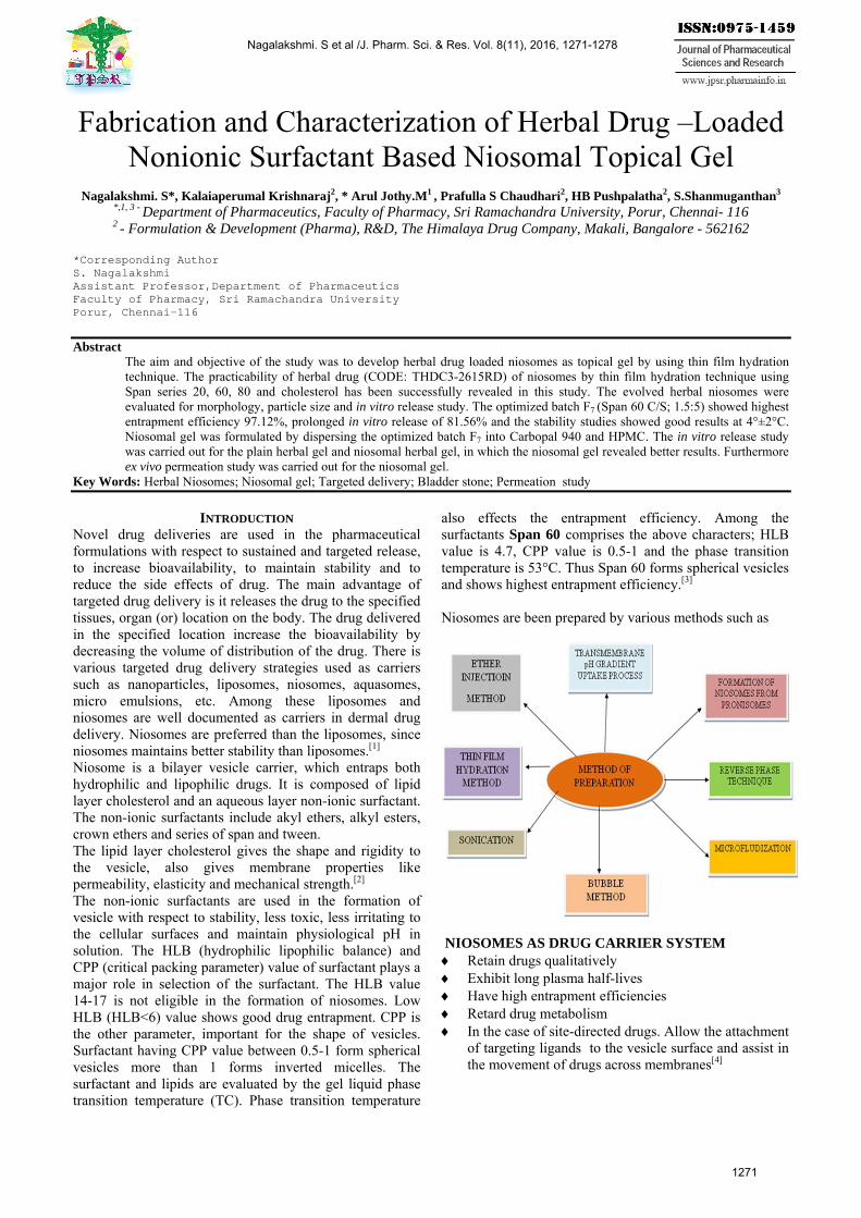
Fabrication and Characterization of Herbal Drug –Loaded Nonionic Surfactant Based Niosomal Topical Gel
Nagalakshmi. S*, Kalaiaperumal Krishnaraj2, * Arul Jothy.M1 , Prafulla S Chaudhari2, HB Pushpalatha2, S.Shanmuganthan3 *,1, 3 - Department of Pharmaceutics, Faculty of Pharmacy, Sri Ramachandra University, Porur, Chennai- 116 2 - Formulation & Development (Pharma), R&D, The Himalaya Drug Company, Makali, Bangalore - 562162
*Corresponding AuthorS. NagalakshmiAssistant Professor,Department of PharmaceuticsFaculty of Pharmacy, Sri Ramachandra UniversityPorur, Chennai-116
Abstract The aim and objective of the study was to develop herbal drug loaded niosomes as topical gel by using thin film hydration technique. The practicability of herbal drug (CODE: THDC3-2615RD) of niosomes by thin film hydration technique using Span series 20, 60, 80 and cholesterol has been successfully revealed in this study. The evolved herbal niosomes were evaluated for morphology, particle size and in vitro release study. The optimized batch F7 (Span 60 C/S; 1.5:5) showed highest entrapment efficiency 97.12%, prolonged in vitro release of 81.56% and the stability studies showed good results at 4°±2°C. Niosomal gel was formulated by dispersing the optimized batch F7 into Carbopal 940 and HPMC. The in vitro release study was carried out for the plain herbal gel and niosomal herbal gel, in which the niosomal gel revealed better results. Furthermore ex vivo permeation study was carried out for the niosomal gel.
Key Words: Herbal Niosomes; Niosomal gel; Targeted delivery; Bladder stone; Permeation study
INTRODUCTION Novel drug deliveries are used in the pharmaceutical formulations with respect to sustained and targeted release, to increase bioavailability, to maintain stability and to reduce the side effects of drug. The main advantage of targeted drug delivery is it releases the drug to the specified tissues, organ (or) location on the body. The drug delivered in the specified location increase the bioavailability by decreasing the volume of distribution of the drug. There is various targeted drug delivery strategies used as carriers such as nanoparticles, liposomes, niosomes, aquasomes, micro emulsions, etc. Among these liposomes and niosomes are well documented as carriers in dermal drug delivery. Niosomes are preferred than the liposomes, since niosomes maintains better stability than liposomes.[1]
Niosome is a bilayer vesicle carrier, which entraps both hydrophilic and lipophilic drugs. It is composed of lipid layer cholesterol and an aqueous layer non-ionic surfactant. The non-ionic surfactants include akyl ethers, alkyl esters, crown ethers and series of span and tween. The lipid layer cholesterol gives the shape and rigidity to the vesicle, also gives membrane properties like permeability, elasticity and mechanical strength.[2]
The non-ionic surfactants are used in the formation of vesicle with respect to stability, less toxic, less irritating to the cellular surfaces and maintain physiological pH in solution. The HLB (hydrophilic lipophilic balance) and CPP (critical packing parameter) value of surfactant plays a major role in selection of the surfactant. The HLB value 14-17 is not eligible in the formation of niosomes. LowHLB (HLB<6) value shows good drug entrapment. CPP isthe other parameter, important for the shape of vesicles.Surfactant having CPP value between 0.5-1 form sphericalvesicles more than 1 forms inverted micelles. Thesurfactant and lipids are evaluated by the gel liquid phasetransition temperature (TC). Phase transition temperature
also effects the entrapment efficiency. Among the surfactants Span 60 comprises the above characters; HLB value is 4.7, CPP value is 0.5-1 and the phase transition temperature is 53°C. Thus Span 60 forms spherical vesicles and shows highest entrapment efficiency.[3]
Niosomes are been prepared by various methods such as
NIOSOMES AS DRUG CARRIER SYSTEM Retain drugs qualitatively Exhibit long plasma half-lives Have high entrapment efficiencies Retard drug metabolism In the case of site-directed drugs. Allow the attachment
of targeting ligands to the vesicle surface and assist in the movement of drugs across membranes[4]
Nagalakshmi. S et al /J. Pharm. Sci. & Res. Vol. 8(11), 2016, 1271-1278
1271
In this study, herbal topical niosomal gel has been formulated for the treatment of bladder stones. Drugs when administrated orally have various disadvantages such as absorption, first pass metabolism which reduces the bioavailability of the drug, drug- food interactions and degradation of the drug due to gastric enzymes. Topical administration the drug gives a localized drug delivery through the skin. Stratum corneum is the major barrier of the skin which is the top layer of the epidermis. In novel drug delivery niosomes are mostly used to enhance the permeation of the drug through the skin.[5]
BLADDER STONE Urinary bladder is an elastic organ which is located in the pelvic cavity. The function of the bladder is, it acts as a tank which stores the body urine. Urine is produced by the kidney as it flows through the ureters to the urinary bladder. The bladder plays salient role retarding and controlling the urination. The capacity of bladder is around 300-350ml of urine. One of the disorders related to the urinary bladder is bladder stone.[6]
THDC3-2615RD is a herbal drug used in the treatment of bladder stone traditionally. Bladder stone is a calculus formed in the urinary bladder. The calculus is hard masses of minerals such as calcium, magnesium ammonium and uric acid salts. It also various in size, shape and texture as small, rigid and spike. The stones are formed when the urine becomes more concentrated (or) due to often dehydration in the body, inflammation in bladder and use of urinary catheters (causes irritation the bladder) and leads to formation of stones. The symptoms are pain in the lower abdomen, burn (or) paining sensation in the urethra at the time of urination. Urine may contain blood and appears cloudy. Initially, bladder stones can be treated by uptake of large amount of fluids, later stage by oral medications. When the size of the stone are found to be large they are broke down using cystolitholapaxy or surgery is an alternative method for the removal of stones.[7]
In this study, a novel approach has been carried out to cure the bladder stones. Niosomal topical gel as been formulated as an alternate to conventional formulation to enhance the bioavailability when compared with oral route of administration. It is having challenges that deliver the drug or reach the herbal drug at bladder site.
MATERIALS AND METHODS
Materials DRUG CODE: THDC3-2615RD was a gift sample from The Himalaya Drug Company, surfactants (Span 20, 60, 80), cholesterol. All the solvents used were in analytical grade. Methods
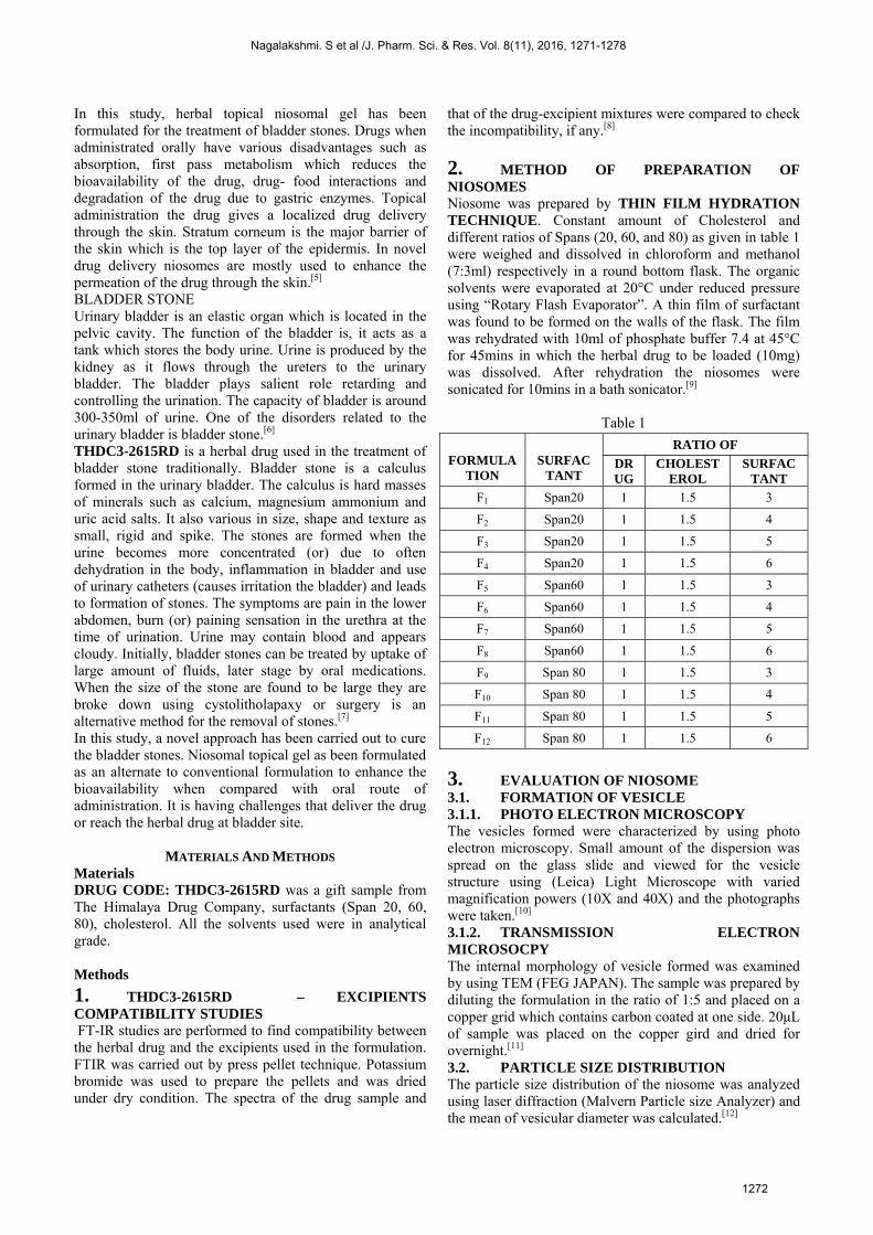
1. THDC3-2615RD – EXCIPIENTS COMPATIBILITY STUDIES FT-IR studies are performed to find compatibility between the herbal drug and the excipients used in the formulation. FTIR was carried out by press pellet technique. Potassium bromide was used to prepare the pellets and was dried under dry condition. The spectra of the drug sample and
that of the drug-excipient mixtures were compared to check the incompatibility, if any.[8]
2. METHOD OF PREPARATION OF NIOSOMES Niosome was prepared by THIN FILM HYDRATION TECHNIQUE. Constant amount of Cholesterol and different ratios of Spans (20, 60, and 80) as given in table 1 were weighed and dissolved in chloroform and methanol (7:3ml) respectively in a round bottom flask. The organic solvents were evaporated at 20°C under reduced pressure using “Rotary Flash Evaporator”. A thin film of surfactant was found to be formed on the walls of the flask. The film was rehydrated with 10ml of phosphate buffer 7.4 at 45°C for 45mins in which the herbal drug to be loaded (10mg) was dissolved. After rehydration the niosomes were sonicated for 10mins in a bath sonicator.[9]
Table 1
3. EVALUATION OF NIOSOME 3.1. FORMATION OF VESICLE 3.1.1. PHOTO ELECTRON MICROSCOPY The vesicles formed were characterized by using photo electron microscopy. Small amount of the dispersion was spread on the glass slide and viewed for the vesicle structure using (Leica) Light Microscope with varied magnification powers (10X and 40X) and the photographs were taken.[10] 3.1.2. TRANSMISSION ELECTRON MICROSOCPY The internal morphology of vesicle formed was examined by using TEM (FEG JAPAN). The sample was prepared by diluting the formulation in the ratio of 1:5 and placed on a copper grid which contains carbon coated at one side. 20μL of sample was placed on the copper gird and dried for overnight.[11]
3.2. PARTICLE SIZE DISTRIBUTION The particle size distribution of the niosome was analyzed using laser diffraction (Malvern Particle size Analyzer) and the mean of vesicular diameter was calculated.[12]
FORMULA
TION
SURFAC
TANT
RATIO OF
DRUG
CHOLESTEROL
SURFACTANT
F1 Span20 1 1.5 3
F2 Span20 1 1.5 4
F3 Span20 1 1.5 5
F4 Span20 1 1.5 6
F5 Span60 1 1.5 3
F6 Span60 1 1.5 4
F7 Span60 1 1.5 5
F8 Span60 1 1.5 6
F9 Span 80 1 1.5 3
F10 Span 80 1 1.5 4
F11 Span 80 1 1.5 5
F12 Span 80 1 1.5 6
Nagalakshmi. S et al /J. Pharm. Sci. & Res. Vol. 8(11), 2016, 1271-1278
1272
3.3. ENTRAPMENT EFFICEINCY The entrapment efficiency of niosomes was determined by centrifugation method. 2 ml of the suspension was centrifuged at 14,000 rpm for 60 minutes maintaining a temperature of 4ºC, in order to separate niosomes from unentrapped drug. The free drug concentration in the supernatant liquid was determined at 265nm using UV-Visible Spectrophotometer The percentage of drug entrapment in niosomes was calculated by using the following formula [13]
% Drug entrapment = (Total drug – unentrapped drug) × 100
Total drug 3.4. DRUG CONTENT The drug content of niosomes was determined by taking niosomal dispersion equivalent to 10mg inn a standard flask and making the volume up to required volume using phosphate buffer pH 7.4. After that 1ml of the solution was withdrawn and diluted up to 10ml using phosphate buffer pH7.4, the amount of drug present was measured at 265nm using UV spectrophotometer.[14]
3.5. IN VITRO RELEASE STUDIES In vitro release of niosomes was carried out using dialysis bag. Prepared niosomes were placed in a dialysis bag length of 5 cm which acts as a donor compartment. Dialysis bag was placed at one end of the open end cylinder and placed in a beaker containing 100 ml of phosphate buffer 7.4, which acts as a receptor compartment. The temperature of the receptor medium was maintained at 37±1ºC and the medium was agitated at a speed of 50 rpm using a magnetic stirrer. 5 ml of the sample was collected at regular time intervals and replaced immediately with the same volume of phosphate buffer pH 7.4. The sink condition was maintained throughout the experiment. The collected samples were analyzed spectrophotometrically at 265 nm using UV-Visible Spectrophotometer.[15]
3.6. STABILITY STUDIES Leaching of drug from niosomes was investigated by carrying out stability studies. Niosomal dispersion was stored in two different conditions freeze condition (4±2°C) and room temperature (25±2°C) for 10 weeks in sealed transparent glass vial. Drug content of the niosomal dispersion was checked periodically.[16]
4. FORMULATION OF GEL Placebo gel (G1) and Herbal drug loaded gel (G2) were prepared by “Cold Mechanism Method” and physical characterization such as appearance, pH and viscosity studies have been performed for the prepared for the gels.[17]
5. EVALUATION OF GEL 5.1 IN VITRO DIFFUSION STUDY The release study for the gel was carried out by diffusion method using Franz-diffusion cell through a cellophane membrane. 1g of a plain herbal gel and herbal drug loaded niosomal gel were weighed and placed on the cellophane membrane. The diffusion study was carried out separately maintaining the temperature at 37±1°C using 100ml of phosphate buffer 7.4 as medium. 5ml of sample was carried
out at regular time intervals for 8hrs. Sink condition was maintained throughout the study. The samples were analyzed spectrophotometrically at 265nm using UV –visible spectrophotometer.[18]
5.2 DRUG RELEASE KINETICS The results obtained from in vitro drug release studies were attempted to fit into various mathematical models such as Zero order release kinetics, First order release kinetics, Higuchi classical diffusion equation, Koresmeyer-Peppas exponential equation and Hixson-Crowell erosion equation to know the mechanism of drug release. The equation with high regression coefficient (r2) for formulation will be the best fit of release data. For Koresmeyer-Peppas equation, if n = 0.5 indicating pure fickian diffusion, n = 0.5-1 indicating anamolous non-fickian diffusion and n=1 indicates zero order release.[19]
5.3. EX VIVO STUDIES Permeation capacity of the gel was evaluated by carrying out ex-vivo permeation study using Goat skin purchased from the slaughter house, Chennai. Skin is stored at -20°C. Skin hairs were trimmed and cleaned triplicate using scissors and the skin contents were removed. Franz diffusion cell was used to carry out the study. The goat dorsal skin was placed between the compartments of the diffusion cell, stratum corneum facing the donor compartment. 12ml of phosphate buffer saline (PBS) pH 7.4 acts as the receiver phase. It was stirred at 500 rpm on a magnetic stirrer, maintaining the temperature at 37±0.5°C. The study was carried out for 8hrs and the amount of drug permeated was found by removing sample (2ml) at regular time intervals. Sink condition was maintained throughout the study. The absorbance was measured at 265nm spectrophotometrically.[20]
RESULTS AND DISCUSSION Herbal drug loaded niosomes were prepared using various non-ionic surfactants (Span 20,60,80) along with cholesterol in different proportions as shown in Table.1 by thin film hydration technique. The prepared herbal drug loaded niosomes were examined for various parameters like vesicle size, shape, particle size, entrapment efficiency and in vitro release study. The prepared optimized niosomal formulation was incorporated in to the gel to formulate the Topical Niosomal Gel. FTIR
Fig:1 FTIR spectra of drug(code: THDC3-2615RD)
Nagalakshmi. S et al /J. Pharm. Sci. & Res. Vol. 8(11), 2016, 1271-1278
1273
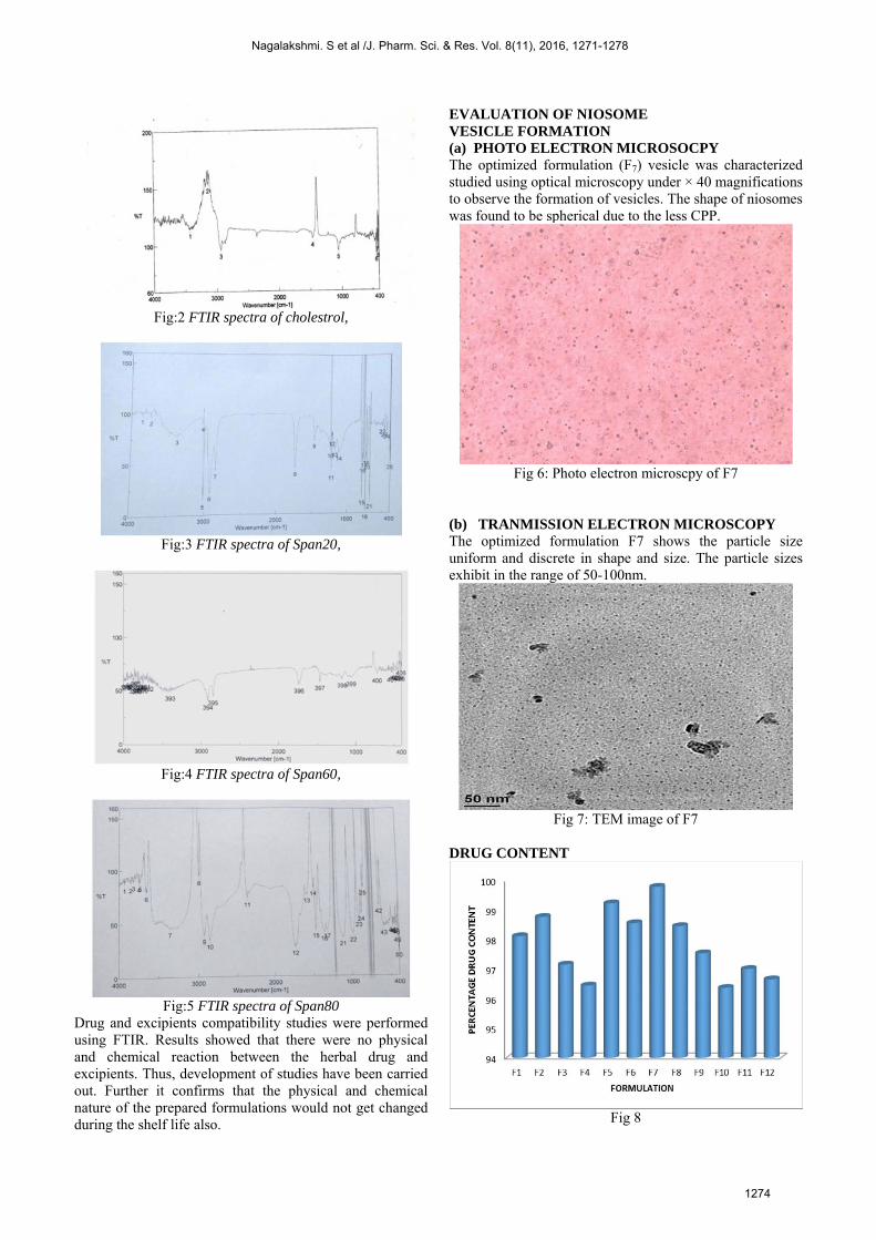
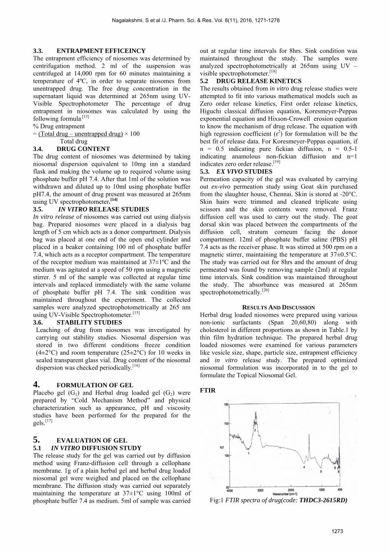
Fig:2 FTIR spectra of cholestrol,
Fig:3 FTIR spectra of Span20,
Fig:4 FTIR spectra of Span60,
Fig:5 FTIR spectra of Span80
Drug and excipients compatibility studies were performed using FTIR. Results showed that there were no physical and chemical reaction between the herbal drug and excipients. Thus, development of studies have been carried out. Further it confirms that the physical and chemical nature of the prepared formulations would not get changed during the shelf life also.
EVALUATION OF NIOSOME VESICLE FORMATION (a) PHOTO ELECTRON MICROSOCPY The optimized formulation (F7) vesicle was characterized studied using optical microscopy under × 40 magnifications to observe the formation of vesicles. The shape of niosomes was found to be spherical due to the less CPP.
Fig 6: Photo electron microscpy of F7
(b) TRANMISSION ELECTRON MICROSCOPY The optimized formulation F7 shows the particle size uniform and discrete in shape and size. The particle sizes exhibit in the range of 50-100nm.
Fig 7: TEM image of F7
DRUG CONTENT
Fig 8
Nagalakshmi. S et al /J. Pharm. Sci. & Res. Vol. 8(11), 2016, 1271-1278
1274
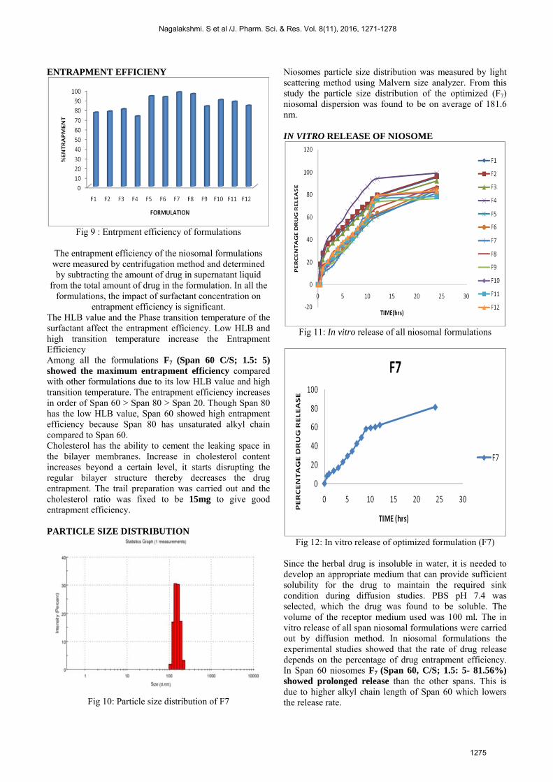
ENTRAPMENT EFFICIENY
Fig 9 : Entrpment efficiency of formulations
The entrapment efficiency of the niosomal formulations were measured by centrifugation method and determined by subtracting the amount of drug in supernatant liquid
from the total amount of drug in the formulation. In all the formulations, the impact of surfactant concentration on
entrapment efficiency is significant. The HLB value and the Phase transition temperature of the surfactant affect the entrapment efficiency. Low HLB and high transition temperature increase the Entrapment Efficiency
Among all the formulations F7 (Span 60 C/S; 1.5: 5) showed the maximum entrapment efficiency compared with other formulations due to its low HLB value and high transition temperature. The entrapment efficiency increases in order of Span 60 > Span 80 > Span 20. Though Span 80 has the low HLB value, Span 60 showed high entrapment efficiency because Span 80 has unsaturated alkyl chain compared to Span 60. Cholesterol has the ability to cement the leaking space in the bilayer membranes. Increase in cholesterol content increases beyond a certain level, it starts disrupting the regular bilayer structure thereby decreases the drug entrapment. The trail preparation was carried out and the cholesterol ratio was fixed to be 15mg to give good entrapment efficiency. PARTICLE SIZE DISTRIBUTION
Fig 10: Particle size distribution of F7
Niosomes particle size distribution was measured by light scattering method using Malvern size analyzer. From this study the particle size distribution of the optimized (F7) niosomal dispersion was found to be on average of 181.6 nm. IN VITRO RELEASE OF NIOSOME
Fig 11: In vitro release of all niosomal formulations
Fig 12: In vitro release of optimized formulation (F7)
Since the herbal drug is insoluble in water, it is needed to develop an appropriate medium that can provide sufficient solubility for the drug to maintain the required sink condition during diffusion studies. PBS pH 7.4 was selected, which the drug was found to be soluble. The volume of the receptor medium used was 100 ml. The in vitro release of all span niosomal formulations were carried out by diffusion method. In niosomal formulations the experimental studies showed that the rate of drug release depends on the percentage of drug entrapment efficiency. In Span 60 niosomes F7 (Span 60, C/S; 1.5: 5- 81.56%) showed prolonged release than the other spans. This is due to higher alkyl chain length of Span 60 which lowers the release rate.
Nagalakshmi. S et al /J. Pharm. Sci. & Res. Vol. 8(11), 2016, 1271-1278
1275
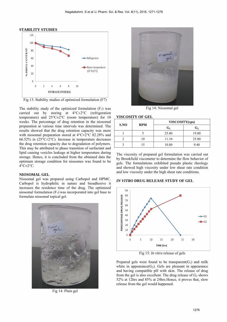
STABILITY STUDIES
Fig 13: Stability studies of optimized formulation (F7)
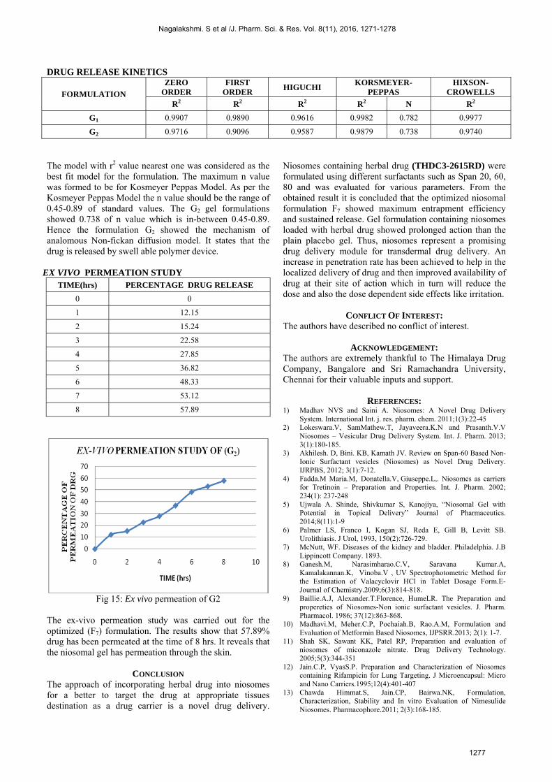
The stability study of the optimized formulation (F7) was carried out by storing at 4°C±2°C (refrigeration temperature) and 25°C±2°C (room temperature) for 10 weeks. The percentage of drug retention in the niosomal preparation at various time intervals was determined. The results showed that the drug retention capacity was more with niosomal preparation stored at 4°C±2°C 82.28% and 66.52% in (25°C±2°C). Increase in temperature decreases the drug retention capacity due to degradation of polymers. This may be attributed to phase transition of surfactant and lipid causing vesicles leakage at higher temperature during storage. Hence, it is concluded from the obtained data the optimum storage condition for niosomes was found to be 4°C±2°C. NIOSOMAL GEL Niosomal gel was prepared using Carbopol and HPMC. Carbopol is hydrophilic in nature and bioadhesive it increases the residence time of the drug. The optimized niosomal formulation (F7) was incorporated into gel base to formulate niosomal topical gel.
Fig 14: Plain gel
Fig 14: Niosomal gel
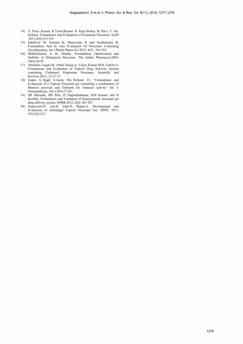
VISCOSITY OF GEL
S.NO RPM VISCOSITY(cps)
G1 G2
1 5 25.80 19.80
2 10 11.10 25.80
3 15 10.80 9.40
The viscosity of prepared gel formulation was carried out by Brookfield viscometer to determine the flow behavior of gels. The formulations exhibited pseudo plastic rheology and showed high viscosity under low shear rate condition and low viscosity under the high shear rate conditions. IN VITRO DRUG RELEASE STUDY OF GEL
Fig 15: In vitro release of gels
Prepared gels were found to be transparent(G1) and milk white in appearance(G2). Gels are pleasant in appearance and having compatible pH with skin. The release of drug from the gel is also excellent. The drug release of G2 shows 52% at 12hrs and 85% at 24hrs.Hence, it proves that, slow release from the gel would happened.
Nagalakshmi. S et al /J. Pharm. Sci. & Res. Vol. 8(11), 2016, 1271-1278
1276
DRUG RELEASE KINETICS
FORMULATION
ZERO ORDER
FIRST ORDER
HIGUCHI KORSMEYER-
PEPPAS HIXSON-
CROWELLS
R2 R2 R2 R2 N R2
G1 0.9907 0.9890 0.9616 0.9982 0.782 0.9977
G2 0.9716 0.9096 0.9587 0.9879 0.738 0.9740
The model with r2 value nearest one was considered as the best fit model for the formulation. The maximum n value was formed to be for Kosmeyer Peppas Model. As per the Kosmeyer Peppas Model the n value should be the range of 0.45-0.89 of standard values. The G2 gel formulations showed 0.738 of n value which is in-between 0.45-0.89. Hence the formulation G2 showed the mechanism of analomous Non-fickan diffusion model. It states that the drug is released by swell able polymer device.
EX VIVO PERMEATION STUDY TIME(hrs) PERCENTAGE DRUG RELEASE
0 0
1 12.15
2 15.24
3 22.58
4 27.85
5 36.82
6 48.33
7 53.12
8 57.89
Fig 15: Ex vivo permeation of G2
The ex-vivo permeation study was carried out for the optimized (F7) formulation. The results show that 57.89% drug has been permeated at the time of 8 hrs. It reveals that the niosomal gel has permeation through the skin.
CONCLUSION The approach of incorporating herbal drug into niosomes for a better to target the drug at appropriate tissues destination as a drug carrier is a novel drug delivery.
Niosomes containing herbal drug (THDC3-2615RD) were formulated using different surfactants such as Span 20, 60, 80 and was evaluated for various parameters. From the obtained result it is concluded that the optimized niosomal formulation F7 showed maximum entrapment efficiency and sustained release. Gel formulation containing niosomes loaded with herbal drug showed prolonged action than the plain placebo gel. Thus, niosomes represent a promising drug delivery module for transdermal drug delivery. An increase in penetration rate has been achieved to help in the localized delivery of drug and then improved availability of drug at their site of action which in turn will reduce the dose and also the dose dependent side effects like irritation.
CONFLICT OF INTEREST: The authors have described no conflict of interest.
ACKNOWLEDGEMENT: The authors are extremely thankful to The Himalaya Drug Company, Bangalore and Sri Ramachandra University, Chennai for their valuable inputs and support.
REFERENCES: 1) Madhav NVS and Saini A. Niosomes: A Novel Drug Delivery
System. International Int. j. res. pharm. chem. 2011;1(3):22-45 2) Lokeswara.V, SamMathew.T, Jayaveera.K.N and Prasanth.V.V
Niosomes – Vesicular Drug Delivery System. Int. J. Pharm. 2013; 3(1):180-185.
3) Akhilesh. D, Bini. KB, Kamath JV. Review on Span-60 Based Non-Ionic Surfactant vesicles (Niosomes) as Novel Drug Delivery. IJRPBS, 2012; 3(1):7-12.
4) Fadda.M Maria.M, Donatella.V, Giuseppe.L,. Niosomes as carriers for Tretinoin – Preparation and Properties. Int. J. Pharm. 2002; 234(1): 237-248
5) Ujwala A. Shinde, Shivkumar S, Kanojiya, “Niosomal Gel with Potential in Topical Delivery” Journal of Pharmaceutics. 2014;8(11):1-9
6) Palmer LS, Franco I, Kogan SJ, Reda E, Gill B, Levitt SB. Urolithiasis. J Urol, 1993, 150(2):726-729.
7) McNutt, WF. Diseases of the kidney and bladder. Philadelphia. J.B Lippincott Company. 1893.
8) Ganesh.M, Narasimharao.C.V, Saravana Kumar.A, Kamalakannan.K, Vinoba.V , UV Spectrophotometric Method for the Estimation of Valacyclovir HCl in Tablet Dosage Form.E-Journal of Chemistry.2009;6(3):814-818.
9) Baillie.A.J, Alexander.T.Florence, HumeLR. The Preparation and propereties of Niosomes-Non ionic surfactant vesicles. J. Pharm. Pharmacol. 1986; 37(12):863-868.
10) Madhavi.M, Meher.C.P, Pochaiah.B, Rao.A.M, Formulation and Evaluation of Metformin Based Niosomes, IJPSRR.2013; 2(1): 1-7.
11) Shah SK, Sawant KK, Patel RP, Preparation and evaluation of niosomes of miconazole nitrate. Drug Delivery Technology. 2005;5(3):344-351
12) Jain.C.P, VyasS.P. Preparation and Characterization of Niosomes containing Rifampicin for Lung Targeting. J Microencapsul: Micro and Nano Carriers.1995;12(4):401-407
13) Chawda Himmat.S, Jain.CP, Bairwa.NK, Formulation, Characterization, Stability and In vitro Evaluation of Nimesulide Niosomes. Pharmacophore.2011; 2(3):168-185.
Nagalakshmi. S et al /J. Pharm. Sci. & Res. Vol. 8(11), 2016, 1271-1278
1277
14) Y. Prem. Kumar, K.Vinod.Kumar, R. Raja Shekar, M. Ravi, V. Sai.Kishore. Formulation and Evaluation of Econazole Niosomes. SAJP.2013;2(4):315-318
15) Sakthivel M, Kannan K, Manavalan R and Senthamarai R,Formulation And In vitro Evaluation Of Niosomes ContainingOxcarbazepine, Int J Pharm Pharm Sci.2012; 4(3) : 563-567.
16) Mullaicharam, A. R., Murthy. Formulation, Optimization andStability of Rifampicin Niosomes. The Indian Pharmacist.2004;34(4):54-58
17) Abraham Lingan.M, Abdul Hasan.A, Vijaya Kumar.M.R, Gokila.A.Formulation and Evaluation of Topical Drug Delivery Systemcontaining Clobetasol Propionate Niosomes. Scientific andReviews.2011; 1(1):7-17.
18) Gupta A, Singh S, Kotla NG, Webster TJ, “Formulation andEvaluation of a Topical Niosomal gel containing a combination ofBenzoyl peroxide and Tretinoin for Antiacne activity” Int. J.Nanomedicine, 10(1) 2014:71-82.
19) SB Shirsand, MS Para, D Nagendrakumar, KM Kanani and DKeerthy, Formulation and evaluation of Ketoconazole niosomal geldrug delivery system. IJPRR.2012, 2[4]: 201-207.
20) Sanjeevani.D, Ajit.D, John.D, Rajani.A. Development andEvaluation of Antifungal Topical Niosomal Gel. IJPPS, 2011;3(5):224-231.
Nagalakshmi. S et al /J. Pharm. Sci. & Res. Vol. 8(11), 2016, 1271-1278
1278


![Fabrication and Characterization of Herbal Drug –Loaded ...niosomes maintains better stability than liposomes.[1] Niosome is a bilayer vesicle carrier, which entraps both hydrophilic](https://img.dokumen.tips/doc/110x75/5f9e794fb1067e646f269ebd/fabrication-and-characterization-of-herbal-drug-aloaded-niosomes-maintains.jpg)