Embed Size (px)
Citation preview
EXTRACTIONS IN ORTHODONTICS21/01/2015
Dr.Gyan P.Singh
Associate professor
Department of Orthodontics&Dentofacial Orthopaedics,FODS,KGMU
CONTENTS
• INTRODUCTION
• EVALUATION OF DIAGNOSTIC ELEMENTS
• CHOICE OF INDIVIDUAL TEETH
INTRODUCTION
• Generally there are three reasons to extract the teeth;
1.To provide space for the alignment of crowded teeth.
2.For the retraction of protruded teeth.3.For camouflaging the skeletal class II and
class III malocclusions.
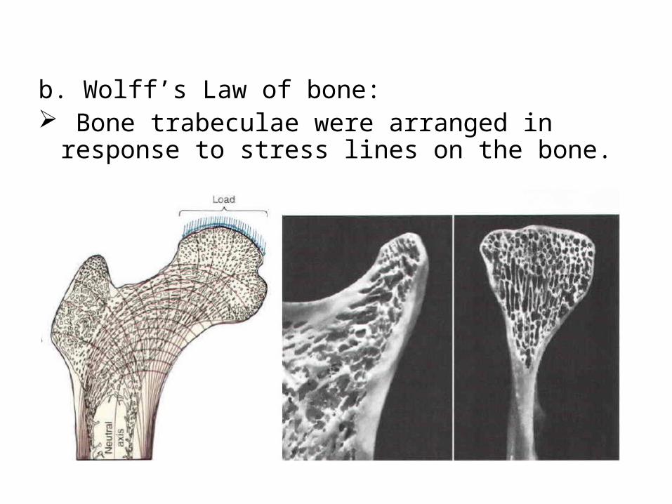
b. Wolff’s Law of bone: Bone trabeculae were arranged in response to
stress lines on the bone.
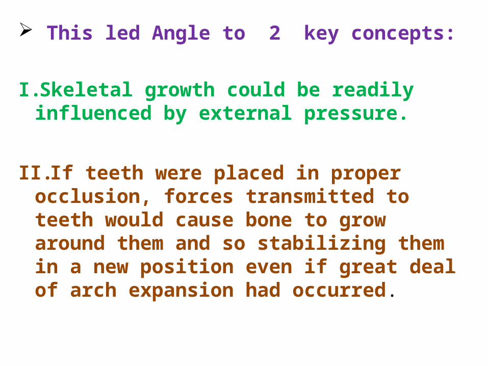
This led Angle to 2 key concepts:
I. Skeletal growth could be readily influenced by external pressure.
II.If teeth were placed in proper occlusion, forces transmitted to teeth would cause bone to grow around them and so stabilizing them in a new position even if great deal of arch expansion had occurred.
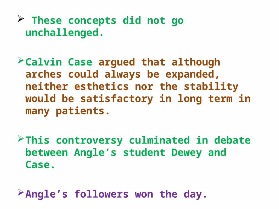
These concepts did not go unchallenged.
Calvin Case argued that although arches could always be expanded, neither esthetics nor the stability would be satisfactory in long term in many patients.
This controversy culminated in debate between Angle’s student Dewey and Case.
Angle’s followers won the day.
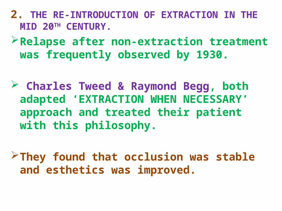
2. THE RE-INTRODUCTION OF EXTRACTION IN THE MID 20TH CENTURY.
Relapse after non-extraction treatment was frequently observed by 1930.
Charles Tweed & Raymond Begg, both adapted ‘EXTRACTION WHEN NECESSARY’ approach and treated their patient with this philosophy.
They found that occlusion was stable and esthetics was improved.
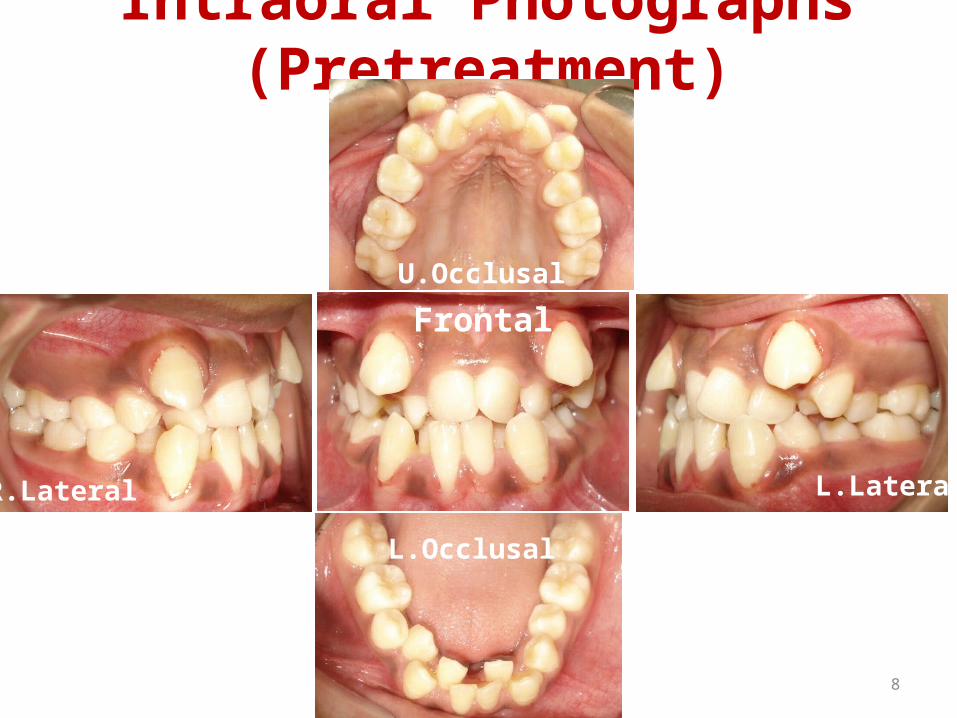
Intraoral Photographs (Pretreatment)
R.Lateral L.Lateral
Frontal
U.Occlusal
L.Occlusal
8
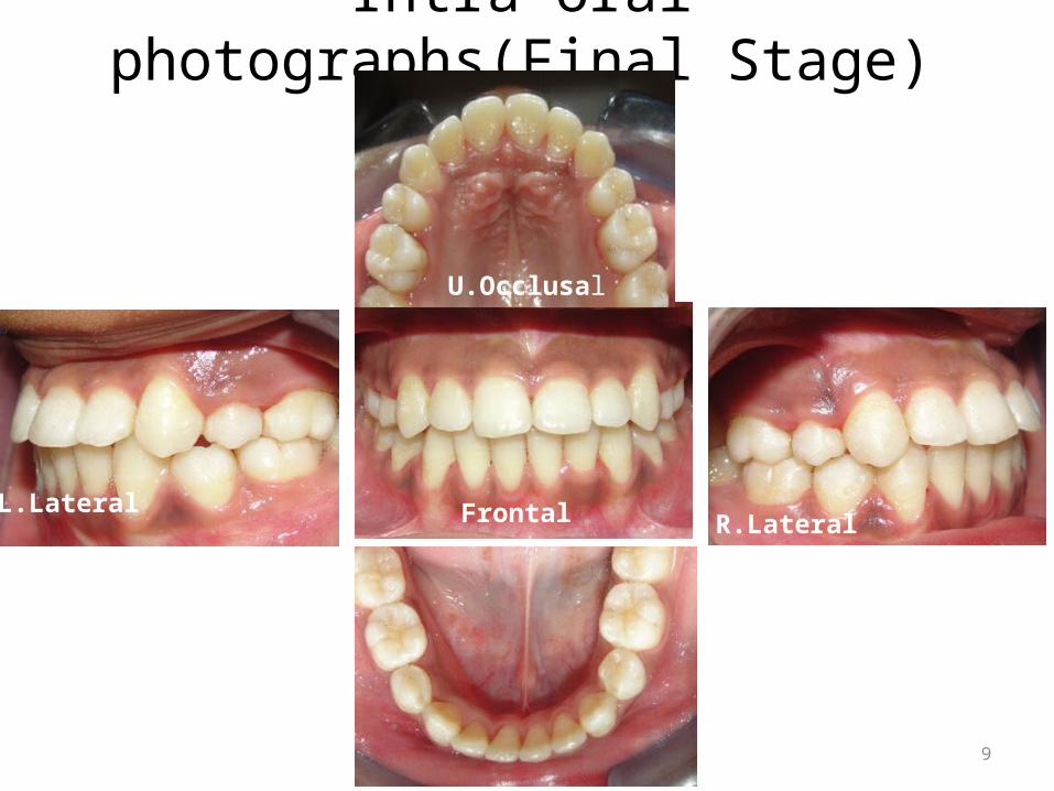
Intra oral photographs(Final Stage)
U.Occlusal
Frontal R.LateralL.Lateral
9
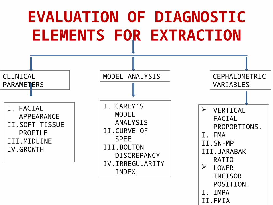
EVALUATION OF DIAGNOSTIC ELEMENTS FOR EXTRACTION
CLINICAL PARAMETERS
I. FACIAL APPEARANCE
II. SOFT TISSUE PROFILE
III. MIDLINEIV. GROWTH
MODEL ANALYSIS
I. CAREY’S MODEL ANALYSIS
II. CURVE OF SPEEIII. BOLTON
DISCREPANCYIV. IRREGULARITY
INDEX
CEPHALOMETRIC VARIABLES
VERTICAL FACIAL PROPORTIONS.
I. FMAII. SN-MPIII. JARABAK RATIO LOWER INCISOR
POSITION.I. IMPAII. FMIAIII. LOWER INCISOR
TO A-Pog DISTANCE
CLINICAL PARAMETERSI. FACIAL APPEARANCE:• Facial appearance -consideration -planning
orthodontic treatment. • Genetic makeup,• Environmental influences,• and cultural background.
• How extractions vs arch expansion affects facial appearance is a major concern for orthodontists.
II. SOFT TISSUE PROFILE.
• How extraction vs non-extraction therapy affects the profile also is a concern.
• Extraction therapy is sometimes believed to be detrimental to the profile.
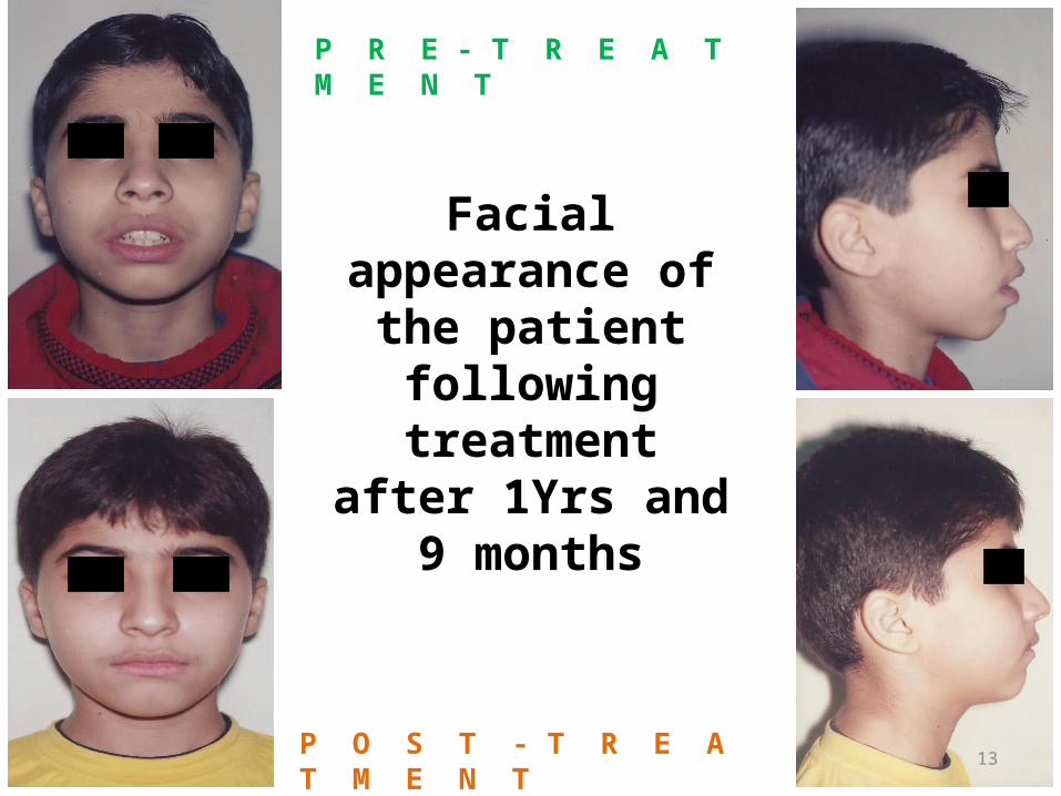
Facial appearance of the patient
following treatment after 1Yrs and 9
months
P R E - T R E A T M E N T
P O S T - T R E A T M E N T13
• This is important because, if a patient has proclined incisors or proclined incisors with crowding.
• it would be virtually impossible to improve the anteroposterior position of the teeth and the patient’s profile without extractions.
III. MIDLINE:
• According to Strang , the harmonic positioning of the midlines relative to each other and to the face is what characterizes normal occlusion.
• Any variation in this combination is indicative of improper relationship between the teeth or dental arches.
• This requires a careful diagnosis because properly assessing the causes behind midline shifts allows professionals to use unique mechanics and asymmetric extractions.
• Patients presenting with severe dental midline deviation relative to the face require tooth extractions.
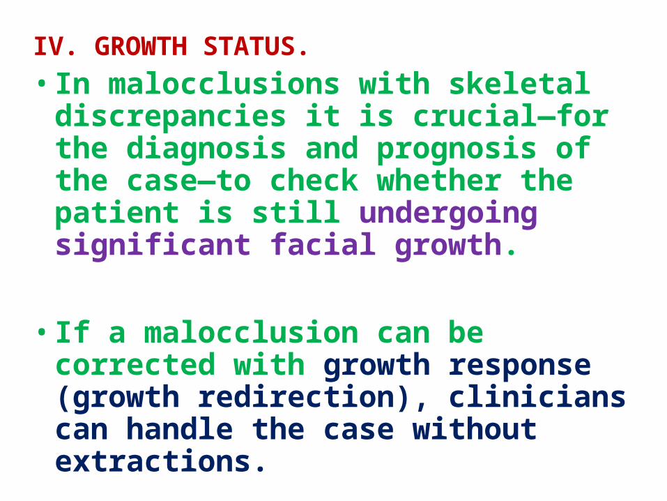
IV. GROWTH STATUS.• In malocclusions with skeletal
discrepancies it is crucial—for the diagnosis and prognosis of the case—to check whether the patient is still undergoing significant facial growth.
• If a malocclusion can be corrected with growth response (growth redirection), clinicians can handle the case without extractions.
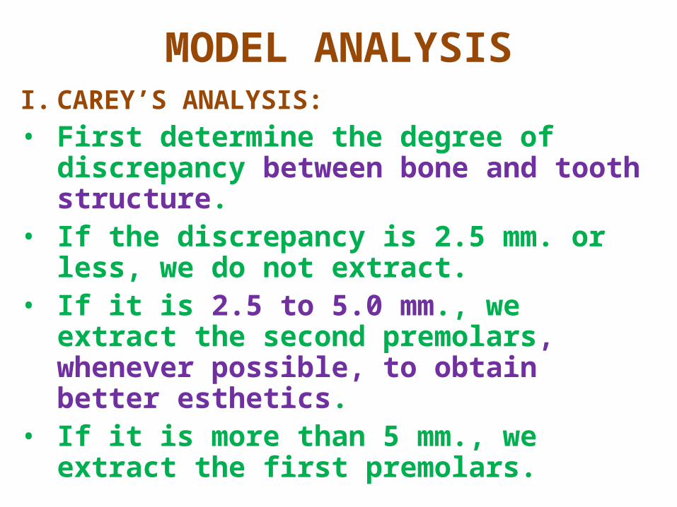
MODEL ANALYSISI. CAREY’S ANALYSIS:• First determine the degree of discrepancy
between bone and tooth structure.• If the discrepancy is 2.5 mm. or less, we do
not extract.• If it is 2.5 to 5.0 mm., we extract the
second premolars, whenever possible, to obtain better esthetics.
• If it is more than 5 mm., we extract the first premolars.
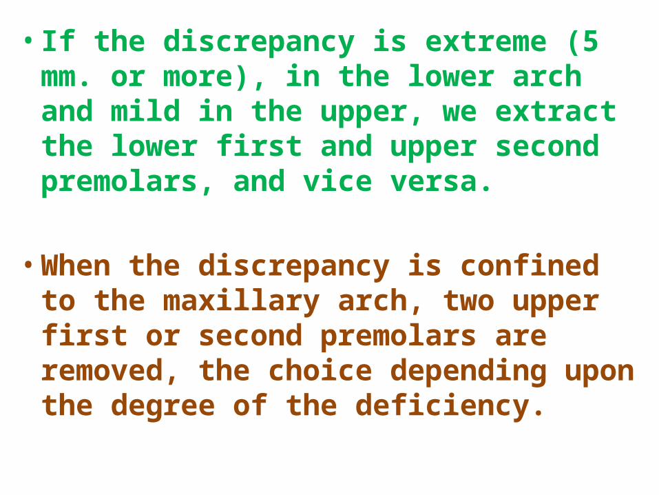
• If the discrepancy is extreme (5 mm. or more), in the lower arch and mild in the upper, we extract the lower first and upper second premolars, and vice versa.
• When the discrepancy is confined to the maxillary arch, two upper first or second premolars are removed, the choice depending upon the degree of the deficiency.
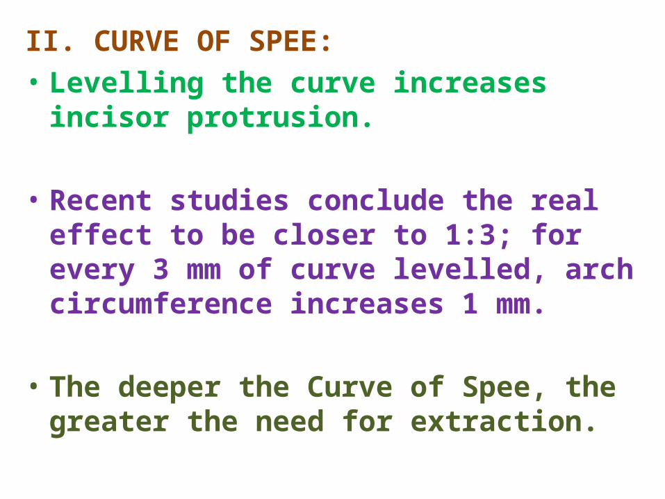
II. CURVE OF SPEE:• Levelling the curve increases incisor protrusion. • Recent studies conclude the real effect to be
closer to 1:3; for every 3 mm of curve levelled, arch circumference increases 1 mm.
• The deeper the Curve of Spee, the greater the
need for extraction.
III. BOLTON DISCREPANCY.
• An interarch tooth-size discrepancy may provide incentive to extract in order to establish a proper occlusion. This diagnostic variable has been popularized as the Bolton discrepancy.
• Clinicians have utilized interproximal reduction to resolve interarch tooth size discrepancies.
• Bolton noted a 4 mm limit to anterior reduction. Thus, extraction may be necessary to resolve a discrepancy greater than this.
CEPHALOMETRIC VARIABLES VERTICAL FACIAL PROPORTIONS:I. SN-MANDIBULAR PLANE ANGLE(SN-MP)• Schudy utilized the angle formed at the
intersection of the sella-nasion and mandibular planes (SN-MP)to aid in his assessments, and found the value of 33 degrees to be average for balanced vertical facial types, with a range of 31 to 34 degrees.
II. FRANKFORT MANDIBULAR PLANE ANGLE(FMA).• The FMA provides an additional vertical
appraisal to the SN-MP measurement. • A normal value for the FMA is in the range of 20
to 30 degrees. • Values above these normal ranges are
associated with skeletal open bite, whereas values below are typically associated with skeletal deep bite.
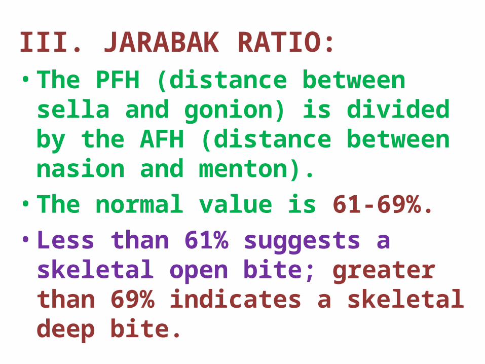
III. JARABAK RATIO:• The PFH (distance between sella and
gonion) is divided by the AFH (distance between nasion and menton). • The normal value is 61-69%. • Less than 61% suggests a skeletal open
bite; greater than 69% indicates a skeletal deep bite.
•Treatment geared toward achieving facial balance is more likely to extract in skeletal open bite and not extract in cases with skeletal deep bite .
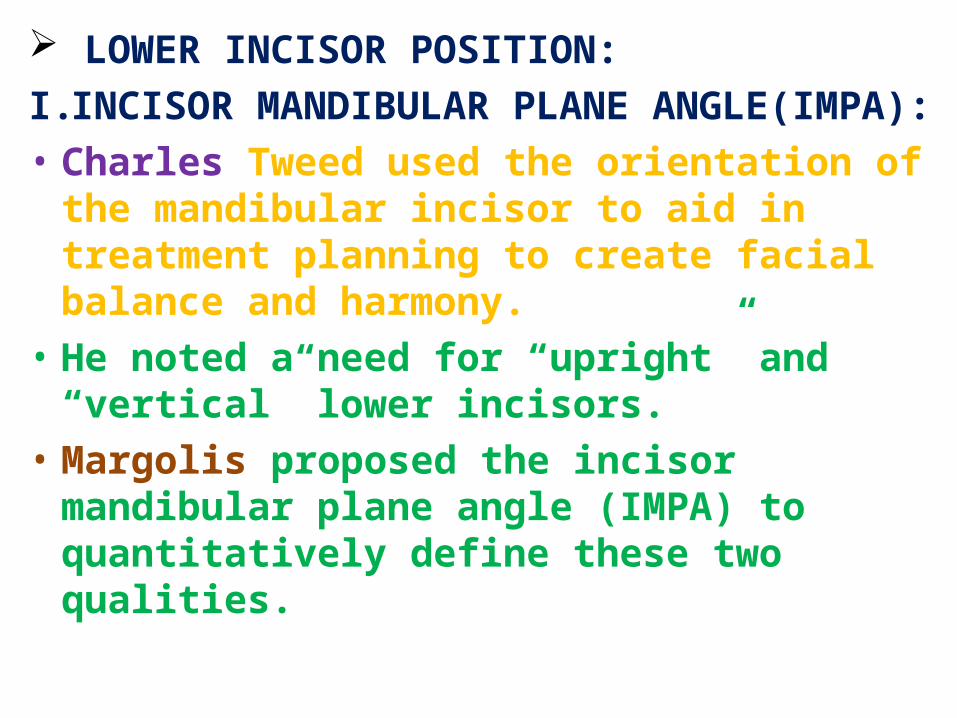
LOWER INCISOR POSITION:I. INCISOR MANDIBULAR PLANE ANGLE(IMPA):• Charles Tweed used the orientation of the
mandibular incisor to aid in treatment planning to create facial balance and harmony.
• He noted a need for “upright” and “vertical” lower incisors.
• Margolis proposed the incisor mandibular plane angle (IMPA) to quantitatively define these two qualities.
• He proposed IMPA to be 90+/-3 degrees in normal, balanced faces. • According to Tweed, this value can range
between 85 and 95 degrees, and vary according to ethnicity.• Values above this range are indicative of
extraction to improve functional and esthetic imbalance.
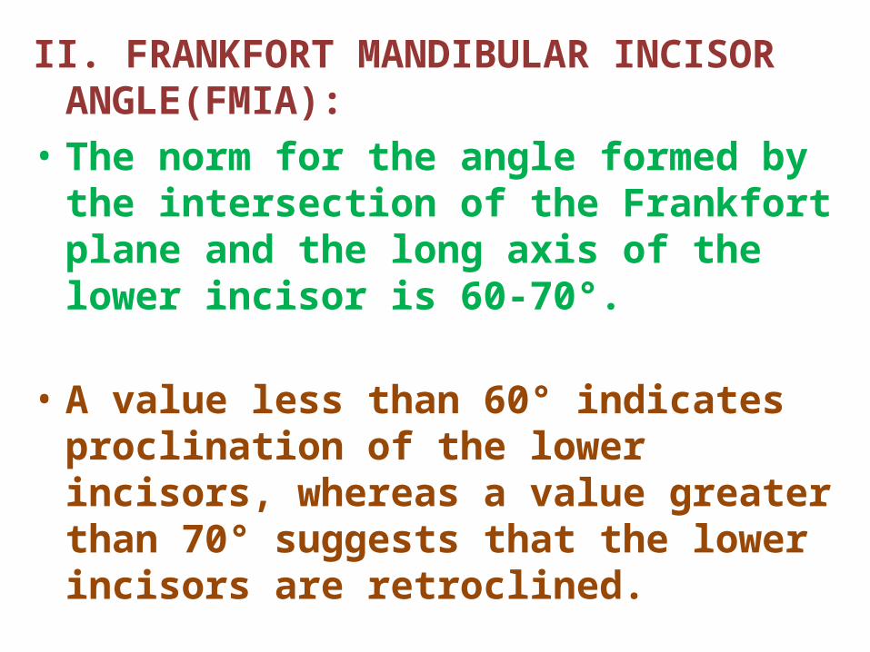
II. FRANKFORT MANDIBULAR INCISOR ANGLE(FMIA):
• The norm for the angle formed by the intersection of the Frankfort plane and the long axis of the lower incisor is 60-70°.
• A value less than 60° indicates proclination of the lower incisors, whereas a value greater than 70° suggests that the lower incisors are retroclined.

III. LOWER INCISOR TO A-Pog DISTANCE:• McNamara found the proper
position of the mandibular incisor to be 1 to 3 millimeters anterior to the line from point A to Pogonion (A-Pog) in a well-balanced face, regardless of age.
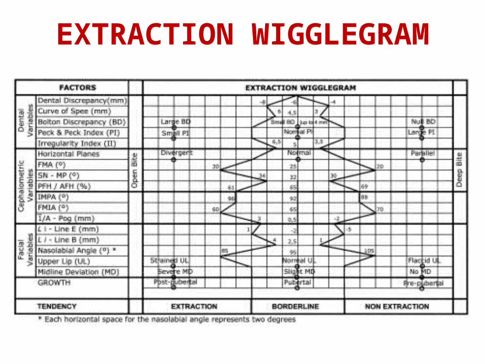
EXTRACTION WIGGLEGRAM

CHOICE OF INDIVIDUAL TEETH
1. UPPER INCISOR EXTRACTION: INDICATIONS:• Unfavorably impacted incsor.• Buccally or lingually blocked out lateral
incisor with good contact between central incisor and canine.
• Congenitally missing one of lateral incisor, opposite incisor may require extraction to maintain arch symmetry.
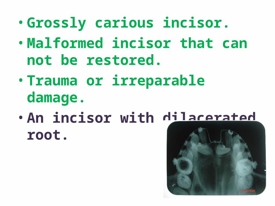
• Grossly carious incisor.• Malformed incisor that can not be
restored.• Trauma or irreparable damage.• An incisor with dilacerated root.
2. LOWER INCISOR EXTRACTION:
• In 1905, Jackson described a case in which two lower incisors were extracted at different times to relieve mandibular crowding.
• Hahn(1942) advocated the removal of a mandibular incisor to close the space and thus reduce the anterior dentition.
INDICATIONS:• Permanent dentition,• Minimal growth potential, • Class I molar relationship, • Harmonious soft-tissue profile,• Minimal-to-moderate overbite, • Little or no crowding in the maxillary arch,• Existing Bolton discrepancy and• Tooth-size-arch-length discrepancy of more
than 5mm in the anterior region
WHICH INCISOR TO BE REMOVED?• Periodontal conditions,
• The presence of gingival recession, and
• The location of any restorations,including endodontic treatment.
• Extraction of a lateral incisor is generally preferred because it is less visible from the front.
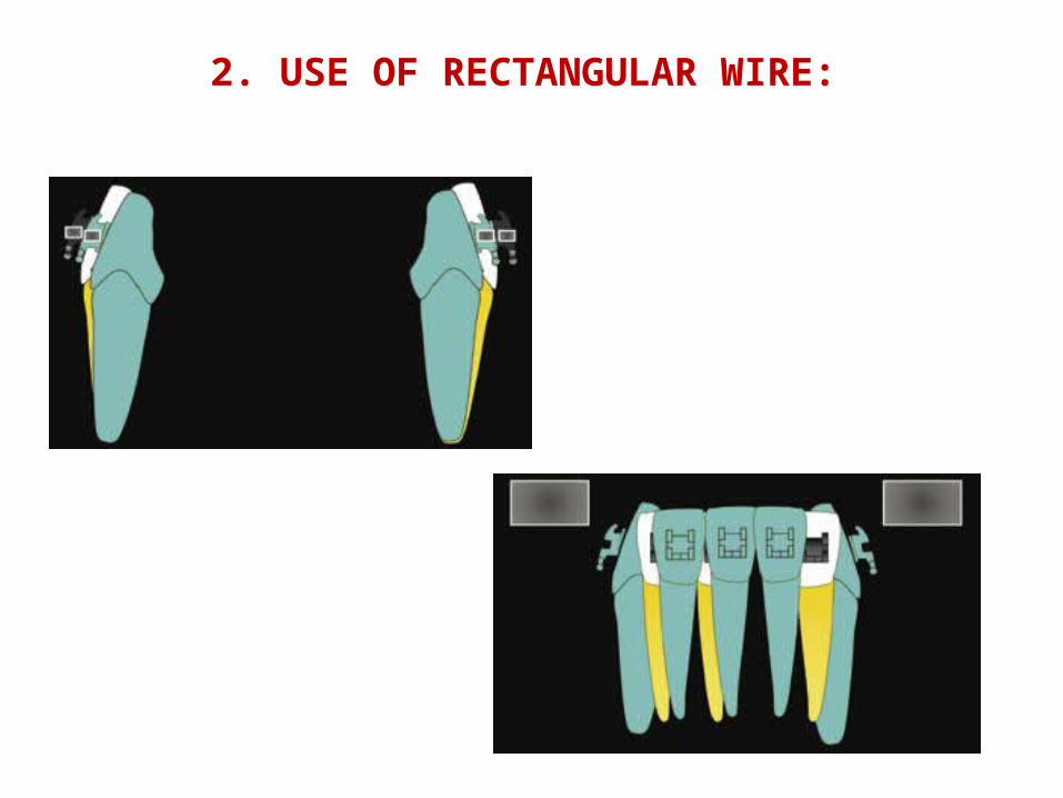
2. USE OF RECTANGULAR WIRE:
3. CANINE EXTRACTION INDICATIONS:•Extremely unfavourable cuspid position.
•Tooth position unfavourable for orthodontic movement.
•Anklosed tooth.
•Internal or external root resorption.
•Severe dilacerataion.
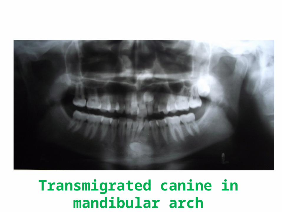
Transmigrated canine in mandibular arch
4. PREMOLAR EXTRACTION:• In 1949, Nance stated that the term extraction
had, at that time, become synonymous with the removal of all four first premolars.
• Augmenting anchorage, maximum lip retraction, better contact between the canines and second premolars , and the fact that first premolars are nearer to anterior crowding are some of the reasons behind favouring their extraction.
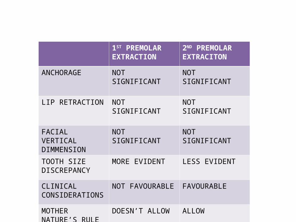
1ST PREMOLAR EXTRACTION
2ND PREMOLAR EXTRACITON
ANCHORAGE NOT SIGNIFICANT NOT SIGNIFICANT
LIP RETRACTION NOT SIGNIFICANT NOT SIGNIFICANT
FACIAL VERTICAL DIMMENSION
NOT SIGNIFICANT NOT SIGNIFICANT
TOOTH SIZE DISCREPANCY
MORE EVIDENT LESS EVIDENT
CLINICAL CONSIDERATIONS
NOT FAVOURABLE FAVOURABLE
MOTHER NATURE’S RULE
DOESN’T ALLOW ALLOW
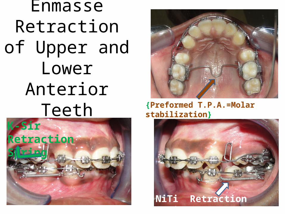
Enmasse Retraction of
Upper and Lower Anterior Teeth
{Preformed T.P.A.=Molar stabilization}
K-Sir Retraction Spring
NiTi Retraction spring42
5. 1ST MOLAR EXTRACTION:• “First permanent molar extractions doubling
the treatment time and halving the prognosis” was the phrase coined by Mills.
• Daugaard-Jensen suggested that first molar cases are no more time consuming than 4 premolar cases.
• Williams and Hosila highlighted the fact that first molar extraction cases are likely to have less effect on the profile than premolar extraction cases.
INDICATIONS:( Sandler et al 2000)• Extensively carious first molars• Hypoplastic first molars• Heavily filled first molars where premolars are
perfectly healthy• Apical pathoses or root canal treated first
molars• Crowding at the distal part of the arches and
wisdom teeth reasonably positioned• High maxillary/mandibular planes angle(Anterior open bite cases)
• TIMING OF EXTRACTIONS:If the upper second molars are unerupted at the
time of extraction of the upper first molars, they will almost completely replace them, thus contributing little space for correction of the malocclusion.
If there is a space requirement in the upper arch therefore, extraction of the first molars must be delayed until the second molars have erupted sufficiently to allow a palatal arch with Nance button or headgear to be placed.
6. 2ND MOLAR EXTRACTION:INDICATIONS(Lehmann 1979)• The second molars are severely carious,
ectopically erupted, or severely rotated.
• Skeletal Class I malocclusions with arch length discrepancy in the distal part of the arch or with mild anterior crowding and
• In Class II "skeletal" cases with only mild crowding of the mandibular arch.
ADVANTAGES:• Disimpaction of third molars
• Faster eruption of third molars
• Prevention of "dished-in" appearance of the face at the end of facial growth
• Prevention of "late" incisor imbrication
• Facilitation of first molar distal movement
TIMINGS OF EXTRACTION: (Kokich 1983)• The third molar crowns should be
completely formed but extractions should be performed before the roots begin to develop;
• The axial inclination of the third molar buds should not be greater than 30 ° relative to the occlusal plane;
• The mandibular third molar should be in close proximity to the second molar roots to ensure adequate mesial drift of the third molar as it erupts.
MCQ:1.Extraction of teeth in conjunction to orthodontic treatment is
necessary in order to
(A)To relieve crowding in the arches especially when jaws are not large enough to accommodate all the teeth
(B)To achieve proper sagittal inter-arch relationship
(C)Just as a procedure of orthodontics
(D)Both A and B
2. The decision of extraction is based on the following factors
(A)Patient’s age
(B)Sex of the patient
(C) The amount of space needed for tooth alignment
(D) All of the above 49
3.The decision to opt for extraction should only be made
(A)After careful clinical evaluation
(B)After model analysis done
(C)After cephalometric tracing done
(D)All of the above
4. Injudicious extraction of teeth can cause
(A)Arch collapse
(B)Deep overbite
(C) Spacing and tissue damage
(D) All of the above
50
5.Who was the major proponent of “ Non Extraction Philosophy”
(A)Edward H Angle
(B)Calvin Case
(C)John Hunter
(D)All of the above
6. Who introduced the concept of extraction as a part of orthodontic treatment.
(A)Calvin Case
(B)Charles Tweed
(C)Angle
(D)Jhon Hunter 51
7.Most commonly extracted teeth for orthodontic purpose are
(A)Maxillary first molars
(B)Maxillary and mandibular premolars
(C)Mandibular incisors
(D)Maxillary incisors
8. The tooth most rarely extracted as a part of orthodontic treatment
(A)Maxillary central incisors
(B)Maxillary third molars
(C)Mandibular third molar
(D)Maxillary and mandibular premolars
52
9. What are the different extraction procedures?
(A)Balanced extraction
(B)Compensatory extraction
(C)Enforced extraction
(D)All of the above
10. Compensatory extraction refers to
(A)Extraction of tooth in the opposite jaw to the same teeth group
(B)The extraction of a tooth in the same jaw to the same teeth group
(C)The extraction of a tooth in the cotralateral side to the same teeth group
(D)None of the above53
REFERENCES
1. Graber TM:Principles and Practicce Orthodontics,WB Saunders,1988
2.Profitt.Contemporary Orthodontics,Elsevier India.3rd ed.,2000.
54

55