Embed Size (px)
Citation preview
Universidade de Lisboa
Faculdade de Ciências
Departamento de Biologia Vegetal
Extracellular matrix and integrins
influence in the regulation of myogenic
precursor cells behaviour
Raquel Rodrigues Vaz
Mestrado em Biologia Molecular Humana
2009
Universidade de Lisboa
Faculdade de Ciências
Departamento de Biologia Vegetal
Extracellular matrix and integrins
influence in the regulation of myogenic
precursor cells behaviour
Raquel Rodrigues Vaz
Dissertação orientada por Professora Doutora Gabriela Rodrigues
Mestrado em Biologia Molecular Humana
2009
Extracellular matrix and integrins influence in the regulation of myogenic precursor cells behaviour
Raquel Rodrigues Vaz 2009 i
Index
Index ....................................................................................................................................... i
Acknowledgments ................................................................................................................. iii
Abstract ................................................................................................................................. iv
Resumo .................................................................................................................................. v
1. Introduction ....................................................................................................................... 1
1.1 Myogenesis ................................................................................................................................................................... 1
1.1.1 Vertebrate myogenesis ....................................................................................................................................... 1
1.1.2 Genetic regulation of myogenesis ..................................................................................................................... 1
1.2 Satellite cells ................................................................................................................................................................. 3
1.3 Notch signaling ............................................................................................................................................................. 3
1.3.1 Notch signaling in myogenesis ........................................................................................................................... 4
1.4 Extracellular matrix and integrins ............................................................................................................................... 4
1.4.1 Extracellular matrix .............................................................................................................................................. 4
1.4.1.1 Laminin .......................................................................................................................................................... 4
1.4.1.2 Fibronectin .................................................................................................................................................... 5
1.4.1.3 Extracellular matrix in myogenesis ............................................................................................................ 5
1.4.2 Integrins ................................................................................................................................................................. 5
1.4.2.1 Integrins in myogenesis .............................................................................................................................. 6
1.5 Aims of this thesis ........................................................................................................................................................ 7
2. Materials and Methods ...................................................................................................... 8
2.1 In vitro studies............................................................................................................................................................... 8
2.1.1 Cell culture ............................................................................................................................................................ 8
2.1.2 Extracellular matrix preparation ......................................................................................................................... 8
2.1.3 Immunofluorescence ........................................................................................................................................... 8
2.2 In vivo studies ............................................................................................................................................................... 9
2.2.1 Mouse embryo collection and culture ................................................................................................................ 9
2.2.2 Immunofluorescence ........................................................................................................................................... 9
2.3 Experimental studies ................................................................................................................................................... 9
2.4 Antibodies .................................................................................................................................................................... 10
2.5 Imaging ........................................................................................................................................................................ 10
2.5.1 Wide-field image acquisition ............................................................................................................................. 10
2.5.2 Time-lapse image acquisition ........................................................................................................................... 10
2.5.3 Confocal image acquisition ............................................................................................................................... 10
2.6 Image analysis and quantifications .......................................................................................................................... 11
2.6.1 In vitro studies .................................................................................................................................................... 11
Extracellular matrix and integrins influence in the regulation of myogenic precursor cells behaviour
Raquel Rodrigues Vaz 2009 ii
2.6.1.1 Quantification of nuclear shape ............................................................................................................... 11
2.6.1.2 Cell tracking from time-lapse movies ...................................................................................................... 11
2.6.2 In vivo studies ..................................................................................................................................................... 12
2.6.2.1 Quantification of dermomyotome fluorescence ..................................................................................... 12
2.7 Statistical analysis ...................................................................................................................................................... 12
3. Results .............................................................................................................................13
3.1 Laminin ........................................................................................................................................................................ 13
3.1.1 Laminin influence in in vitro myogenesis ........................................................................................................ 13
3.1.2 Laminin influence in in vivo myogenesis ......................................................................................................... 13
3.1.2.1 Laminin-α6β1 interaction maintains dermomyotomal cells undifferentiated ...................................... 13
3.1.2.2 Notch signaling is necessary for inhibition of dermomyotomal cells myogenic differentiation ........ 13
3.1.2.3 Notch inhibition and Myf5 expression in the dermomyotome may be inducing an enhancement of
laminin matrix assembly ......................................................................................................................................... 14
3.2 Fibronectin .................................................................................................................................................................. 15
3.2.1 Fibronectin influence in in vitro myogenesis .................................................................................................. 15
3.2.1.1 Fibronectin is important for C2C12 myoblasts alignment ..................................................................... 15
3.2.1.2 Fibronectin-α5β1 integrin interaction appears to be responsible for in vitro cultured myoblasts
alignment .................................................................................................................................................................. 17
3.2.1.3 N-cadherin appears to be necessary for in vitro cultured myoblasts alignment................................ 19
3.2.2 Fibronectin influence in in vivo myogenesis ................................................................................................... 19
3.2.2.1 Fibronectin and α5β1 integrin are present in the myotome ................................................................. 19
3.2.2.2 Fibronectin matrix seems to be important for N-cadherin polarization ............................................... 20
3.2.2.3 Fibronectin matrix-α5β1 integrin interaction might be important in cell alignment ............................ 20
3.2.2.4 Fibronectin-α5β1 interaction maintains dermomyotomal cells undifferentiated ................................ 21
4. Discussion and Concluding Remarks ...............................................................................22
4.1 Laminin ........................................................................................................................................................................ 22
4.2 Fibronectin .................................................................................................................................................................. 24
5. References .......................................................................................................................29
6. Appendix ..........................................................................................................................32
Extracellular matrix and integrins influence in the regulation of myogenic precursor cells behaviour
Raquel Rodrigues Vaz 2009 iii
Acknowledgments
Ao longo deste ano foram muitas as pessoas que se tornaram essenciais para que esta tese pudesse
ser concluída com o maior sucesso possível. Felizmente, neste capítulo tenho a oportunidade de
agradecer a algumas delas.
O meu maior e mais sentido agradecimento vai obviamente para a Gabriela! Começando por
agradecer ter-me aceite no grupo logo que me “auto-propus” para sua mestranda, não posso deixar
de agradecer por tudo o que tem feito por mim. O primeiro contacto com a ciência, a magnifica tese
que me proporcionou e todo o “extra-tese”: o ajudar nas aulas, as oportunidades de mostrar o meu
trabalho por vezes em situações inesperadas, … MUITO OBRIGADA!!! Estes agradecimentos são
igualmente dirigidos à Sólveig, que apesar de não ser minha orientadora (oficialmente), também
sempre me fez sentir muito apoiada. De salientar a constante boa disposição de ambas, a
disponibilidade e o continuarem a exclamar “tão giro!” ao verem as nossas imagens que, com toda a
certeza, já viram parecido dezenas de vezes! É óptimo e super motivador! Com orientadoras assim, é
difícil encontrar melhor!!!
A todo o grupo BD: Luís, Pedro, Ana Lina, Tomás, Ana Martins, Ana Bernardo e à Carla, a “infiltrada”;
aos que resistem, aos que chegaram depois de mim e aos que entretanto saíram, um especial
obrigado! Pelo ambiente, boa disposição, companheirismo, e pela certeza que não poderia ter
escolhido melhor grupo! Em especial agradeço à Ana Gaspar, pela preciosa ajuda a fazer-me uma
“mestre” no criostato; ao Luís, que foi quem me ensinou muito do que sei de cultura de
células/embriões e imunos. À Ana Lina e ao Tomás, os companheiros nesta luta contra o tempo, na
tentativa de conseguir uma super tese e de fazer com que os nossos chefes e companheiros de
grupo se orgulhem de nós!
A todos aqueles que permitiram que a ”vida no laboratório” fosse mais animada: além dos acima
mencionados: à Catarina, à Josefina, …
Ao Gabriel um SUPER OBRIGADO pela infinita ajuda na aquisição e tratamento de imagens, vídeos
e afins. Pela paciência que sempre teve comigo, uma “melga” como poucas, por me ensinar tudo o
que sei de microscopia e associados! Que orgulho aprender com um dos melhores!
Um especial agradecimento a: Raquel Lourenço, Dr. António Cidadão, Dr. Yoshio Wakamatsu e Dr.
Weimin Zhong/Rodney Ritzel que muito atenciosamente me deram Taq e anticorpos for free. Sem
eles não teria sido tão fácil (nem tão divertido)!!!
À Filipa Martins, pela preciosíssima ajuda nas análises estatísticas, que em poucos dias me permitiu
aprender imenso deste tema quase “quebra-cabeças”! À Joana Costa, por ter ajudado na construção
de primers para os RT-PCRs, por nunca se ter recusado aos meus pedidos e “empréstimos”.
Obrigada aos amigos, por se preocuparem e lembrarem que existe vida além do lab, desculpem por
este ano ter sido um pouco “anti-social”…
Um especial agradecimento à minha família, pelo apoio, pelas (infelizmente) constantes viagens
feitas para que eu não chegasse tão tarde a casa, pela paciência quando dizia que o fim-de-semana
iria ser passado no laboratório, pela companhia nas minhas idas ao lab a horas impróprias,…!
Agora em jeito de brincadeira, um agradecimento muito especial aos PCRs e aos Westerns, por não
terem funcionado. Tiraram-me a necessidade de arranjar espaço para os pôr na tese!
A todos, obrigado por este ano super cansativo mas, sem dúvida,
espectacular!
Extracellular matrix and integrins influence in the regulation of myogenic precursor cells behaviour
Raquel Rodrigues Vaz 2009 iv
Abstract
Myogenesis is the process by which undifferentiated dermomyotomal cells are specified for
myogenesis, move towards the myotome where they differentiate into skeletal muscle cells
that fuse into myotubes and later in development form myofibers which will constitute the
skeletal muscles of the adult. The muscle precursor cells arise from the dermomyotome, an
epithelial-like structure that is the source for skeletal muscle and dorsal dermis cells.
Some cells, called satellite cells, go throughout part of this differentiation process but remain
in a quiescent undifferentiated state (although committed to skeletal muscle fate). These
cells are activated in the adult in case of muscle injury or enhanced exercise, for example.
In this work we used a satellite cell-derived cell line, C2C12, and the mouse embryo to study
the extracellular matrix (ECM) and integrins influence in myogenic determination and
differentiation. Integrins are heterodimeric ECM receptors constituted by an α and a β
subunit that can induce, for example, migration or differentiation. The integrin ligand
specificity is acquired by the combination of both subunits.
Our studies have addressed that laminin-α6β1 integrin interaction may be coordinating with
Notch signaling the maintenance of undifferentiated dermomyotomal cells. By inhibiting
Notch signaling, we observed precocious myogenic differentiation of dermomyotomal cells
(by Myf5 expression) and the assembly of a laminin matrix around these cells. This result
suggests that Myf5 induces laminin assembly.
In vitro, fibronectin enhances C2C12 myoblasts alignment and migration. When we observed
the myotubes of cells grown on fibronectin, we believe that the enhanced cell alignment
imposed by fibronectin-α5β1 integrin interaction will facilitate cell fusion. In vivo, we found
that fibronectin is important for dermomyotome epithelial-integrity, especially through the
polarization of N-cadherin, and that α5β1 integrin signaling may also contribute to myogenic
repression in the dermomyotome.
These observations show that the ECM and integrins are of paramount importance in
myoblast cell behaviour.
Key words: myogenesis, laminin, fibronectin, integrins, C2C12

Extracellular matrix and integrins influence in the regulation of myogenic precursor cells behaviour
Raquel Rodrigues Vaz 2009 v
Resumo
A miogénese é o processo através do qual as células precursoras miogénicas são
especificadas, se diferenciam em mioblastos e posteriormente em miócitos que se fundem
formando miotubos que vão constituir as fibras musculares do organismo adulto. Ao longo
deste processo são também especificadas as células satélite, células que se mantêm
indiferenciadas e quiescentes, sendo apenas activadas em caso de lesão muscular ou
exercício intenso (Grounds & McGeachie, 1987; Bischoff & Heintz, 1994).
As células precursoras músculo-esqueléticas do tronco são especificadas ainda no
dermamiótomo, estrutura derivada da porção dorsal do sómito e que se mantém epitelial,
mais concretamente no lábio epaxial do dermamiótomo, correspondente à porção dorso-
medial do dermamiótomo (Christ et al, 1998). No embrião de ratinho, o inicio da
diferenciação destas células é detectado pela expressão do factor de transcrição Myf5,
seguido de outros factores de regulação miogénicos (FRM) como Miogenina, criando uma
espécie de cascata de FRM importantes para a formação correcta dos músculos
esqueléticos (revisto por Cossu et al., 1996). No lábio oposto, denominado hipaxial (porção
mais lateral do dermamiótomo), são definidas as células que vão dar origem aos músculos
dos membros, mas também à musculatura da parede ventral do corpo (Ordahl et al., 1992).
Além de serem a fonte de células precursoras de músculo esquelético, as células do
dermamiótomo também se diferenciam em células da derme dorsal (Brent et al., 2002). A
especificação dos diferentes tipos celulares é possível pois as células do dermamiótomo
recebem diferentes sinais provenientes de diferentes tecidos adjacentes, como tubo neural,
notocorda, ectoderme ou mesoderme lateral, que permitem a “padronização” do
dermamiótomo (Cossu et al., 1996; Cossu et al., 2000; Yusuf et al., 2006). Nesta tese
estivemos particularmente interessados na especificação e diferenciação das células
precursoras de músculo esquelético, células provenientes do lábio epaxial do
dermamiótomo. Mais precisamente, pretendemos entender um pouco mais sobre a
influência da matriz extracelular (MEC) e das integrinas na miogénese, tanto na
diferenciação das células precursoras miogénicas como no seu comportamento.
A MEC corresponde a uma rede altamente dinâmica e complexa constituída por diferentes
moléculas como glicoproteínas, proteoglicanos ou colagénios, que são sintetizadas e
organizadas pelas próprias células. A MEC pode estar organizada na forma de matriz
intersticial, uma estrutura porosa como a do tecido conjuntivo, ou na forma de matriz
pericelular, como a membrana ou lâmina basal, presente por exemplo a rodear o
dermamiótomo (Yurchenco et al., 2004; Schwarzbauer et al., 1999). Estas moléculas
interagem com as células através de receptores específicos, dos quais se destacam as
integrinas, heterodímeros constituídos pela combinação de uma subunidade α (de 18
Extracellular matrix and integrins influence in the regulation of myogenic precursor cells behaviour
Raquel Rodrigues Vaz 2009 vi
existentes) e uma β (de 8). Da combinação das duas subunidades surge a especificidade
para o ligando.
Nesta tese focámo-nos apenas na fibronectina e na laminina, duas glicoproteínas da MEC, e
nas integrinas que interagem com estas moléculas, principalmente α5β1 e α6β1,
respectivamente.
Como modelos de estudo foram usados embriões de ratinho (Mus musculus) e a linha
celular C2C12. Esta linha celular foi estabelecida a partir de células satélite de ratinho adulto
em que a diferenciação destas células em miócitos e miotubos passa pela mesma cascata
de FRM que a miogénese embrionária (Parker et al., 2003) Assim sendo, estas células são
um bom modelo para estudos preliminares de miogénese (Burattini et al., 2004).
Os primeiros estudos realizados nesta tese pretendiam perceber qual o efeito da matriz de
laminina na diferenciação dos mioblastos C2C12. Ao cultivar estas células ao longo de
vários dias em meio de crescimento e de diferenciação sobre laminina e gelatina (controlo),
não observámos qualquer diferença na expressão de diferentes FRM, o que sugere que a
laminina não terá influência na diferenciação destas células.
Estudos anteriores descreveram que, in vivo, a matriz de laminina e a sua interacção com a
integrina α6β1 são importantes para a manutenção do estado não diferenciado das células
do dermamiótomo, e também como barreira que impede a dispersão das células
precursoras musculares (Bajanca et al., 2006). No seguimento deste estudo, tentámos
perceber um pouco mais da regulação do estado indiferenciado das células do
dermamiótomo. Assim, questionámos se a via de sinalização Notch estaria envolvida nesta
regulação.
A importância da via de sinalização Notch, iniciada pela ligação de Notch a Delta e
conduzida intracelularmente através de um domínio de Notch que é clivado, Notch
Intracellular Domain (NICD), tem sido documentada em vários processos, como por exemplo
na diferenciação neuronal ou hematopioese (Wakamatsu, et al, 1999; Weber & Calvi, 2009).
Outros estudos têm relacionado Notch e miogénese. A grande maioria dos estudos tem sido
feita em sistemas in vitro, no entanto alguns estudos têm sido feitos também in vivo. Em
ambos os casos é consensual que a sinalização Notch impede a diferenciação miogénica,
mais especificamente inibindo MyoD (um FRM importante na determinação miogénica;
Braun, et al, 1994), no entanto pouco se sabe sobre quais são os intervenientes celulares
importantes para esta inibição (e.g. Sasai, et al., 1992; Hirsinger et al., 2001; Kopan, Nye, &
Weintraub, 1994; Buas, et al., 2009).
Ao cultivarmos embriões de ratinho com DAPT, um inibidor da γ-secretase, enzima que é
responsável pela clivagem do NICD, observámos a presença de Myf5 nas células do
dermamiótomo. Este resultado confirma que Notch é importante para reprimir a miogénese,
já que o bloqueio da sua sinalização permite que as células do dermamiótomo activem o
Extracellular matrix and integrins influence in the regulation of myogenic precursor cells behaviour
Raquel Rodrigues Vaz 2009 vii
programa de diferenciação miogénica. Este efeito foi semelhante ao observado quando é
inibida a ligação laminina-integrina α6β1 (Bajanca et al., 2006).
Tentando perceber se a inibição da sinalização Notch e consequente diferenciação
miogénica no dermamiótomo teria algum efeito na matriz de laminina, foram feitas
imunofluorescências em embriões cultivados com DAPT (e controlos) e observou-se a
montagem de laminina numa matriz pontilhada onde normalmente não existe, à volta das
células do dermamiótomo, além da lâmina basal. Assim sendo, confirmam-se resultados
anteriores que descrevem que Myf5 será necessário para a montagem da matriz de
laminina, já que embriões mutantes para Myf5 não conseguem fazer a montagem da
laminina numa matriz (Bajanca et al., 2006).
Quando C2C12 foram cultivadas sobre lamelas de fibronectina, cedo notámos que as
células alinhavam precocemente, mas que no entanto esta característica não estava
associada a diferenciação precoce. Ao analisarmos a forma dos núcleos de células
cultivadas sobre fibronectina e sobre gelatina (controlo), confirmámos que as células na
primeira condição estão de facto mais alinhadas, já que os núcleos destas células são mais
elípticos que os núcleos das células cultivadas sobre gelatina.
Para percebermos melhor as diferenças na dinâmica celular nas diferentes matrizes,
colocámos as células numa caixa incubadora associada a uma lupa, na qual eram
adquiridas imagens sequenciais em time-lapse, as quais foram conjugadas num vídeo. Ao
analisar o movimento de várias células em cada vídeo, concluímos que a fibronectina induz
a migração das C2C12, já que sobre a matriz de fibronectina estas células movem-se mais e
a distância entre o ponto inicial e o final no vídeo é significativamente maior. Após verificar
que a integrina α5 é expressa nestas células e ao impedir a ligação destas células á
fibronectina (através de um péptido inibidor da ligação fibronectina-integrina α5β1, o RGD;
Takahashi et al., 2007), deduzimos que as células necessitam das moléculas de fibronectina
solúveis no meio de cultura para conseguirem eficazmente aderir ao substrato e concluímos
que uma matriz de fibronectina induz o alinhamento. Para este alinhamento ser possível, o
citoesqueleto é reorganizado e pensamos que a N-caderina (molécula de adesão célula-
célula) seja também importante para o correcto alinhamento das células. Este alinhamento
parece ser importante quando os miócitos começam a fundir entre si, pois sobre fibronectina
parece haver mais miotubos do que sobre laminina ou gelatina (para o mesmo tempo de
cultura).
Tendo em conta os resultados obtidos com as C2C12, tentámos perceber se, in vivo, a
fibronectina seria necessária tanto para o alinhamento dos precursores musculares após
especificação e nas fases iniciais de diferenciação (embriões mais precoces, com cerca de 9
dias embrionários – E9.0), mas também na altura em que ocorre a fusão dos miócitos
Extracellular matrix and integrins influence in the regulation of myogenic precursor cells behaviour
Raquel Rodrigues Vaz 2009 viii
(E12.5), fase em que apenas estão presentes integrinas que interagem com a fibronectina
(Cachaço et al, 2005). Neste caso não conseguimos detectar diferenças na orientação das
células.
No entanto, ao inibir a montagem da matriz de fibronectina com o fragmento de 70kDa
(McKeown-Longo et al., 1985; Rifes et al., 2007), observámos que a polarização da N-
caderina no dermamiótomo estava perturbada, já que estava presente homogeneamente
nas células, em vez de estar apenas na porção apical das mesmas (revisitando os
resultados obtidos na somitogénese de galinha pelo nosso grupo; Martins et al., in press).
Por outro lado, ao cultivar embriões com RGD durante 12 horas observámos a presença de
algumas células positivas para Myf5 no dermamiótomo. Este resultado sugere que também
a fibronectina, mediada pela interacção com a integrina α5β1, poderá ser importante na
inibição da diferenciação precoce no dermamiótomo.
Nesta tese conseguimos descrever um pouco mais da influência da MEC e das integrinas
na miogénese, nomeadamente na regulação da diferenciação das células do dermamiótomo
em precursores miogénicos. Estabelecemos uma relação entre a laminina e a sinalização
Notch, mas também observámos a influência da fibronectina no alinhamento, migração e na
“eficiência” da fusão de mioblastos in vitro e in vivo e na manutenção das características
epiteliais do dermamiótomo.
Palavras-chave: miogénese, laminina, fibronectina, integrinas, C2C12
Extracellular matrix and integrins influence in the regulation of myogenic precursor cells behaviour
Raquel Rodrigues Vaz 2009 1
1. Introduction
1.1 Myogenesis
1.1.1 Vertebrate myogenesis
Embryonic myogenesis is the process by which cells differentiate into skeletal muscle cells. If
this highly coordinated process does not proceed properly, several defects may decrease
newborn survival and adult health. Skeletal muscle cells begin their differentiation when they
leave the dermomyotome and move to the myotome. The dermomyotome is a transient
structure that arise from somites, blocks of epithelial cells that segment on both sides of the
neural tube from the pre-somitic mesoderm and differentiate in a rostro-caudal gradient
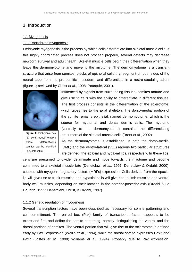
(figure 1; reviewed by Christ et al., 1998; Pourquié, 2001).
Influenced by signals from surrounding tissues, somites mature and
give rise to cells with the ability to differentiate in different tissues.
The first process consists in the differentiation of the sclerotome,
which gives rise to the axial skeleton. The dorso-medial portion of
the somite remains epithelial, named dermomyotome, which is the
source for myotomal and dorsal dermis cells. The myotome
(ventrally to the dermomyotome) contains the differentiating
precursors of the skeletal muscle cells (Brent et al., 2002).
As the dermomyotome is established, in both the dorso-medial
(DML) and the ventro-lateral (VLL) regions two particular structures
are defined: the epaxial and hypaxial lips, respectively. In these lips,
cells are presumed to divide, delaminate and move towards the myotome and become
committed to a skeletal muscle fate (Denetclaw, et al., 1997; Denetclaw & Ordahl, 2000),
coupled with myogenic regulatory factors (MRFs) expression. Cells derived from the epaxial
lip will give rise to trunk muscles and hypaxial cells will give rise to limb muscles and ventral
body wall muscles, depending on their location in the anterior-posterior axis (Ordahl & Le
Douarin, 1992; Denetclaw, Christ, & Ordahl, 1997).
1.1.2 Genetic regulation of myogenesis
Several transcription factors have been described as necessary for somite patterning and
cell commitment. The paired box (Pax) family of transcription factors appears to be
expressed first and define the somite patterning, namely distinguishing the ventral and the
dorsal portions of somites. The ventral portion that will give rise to the sclerotome is defined
early by Pax1 expression (Wallin et al., 1994), while the dorsal somite expresses Pax3 and
Pax7 (Jostes et al., 1990; Williams et al., 1994). Probably due to Pax expression,
Figure 1: Embryonic day
(E) 10.5 mouse embryo
where differentiating
somites can be identified
(e.g. asterisks).
Extracellular matrix and integrins influence in the regulation of myogenic precursor cells behaviour
Raquel Rodrigues Vaz 2009 2
dermomyotomal cells are apoptosis-protected and do not express differentiating factors
(Kassar-Duchossoy et al., 2005; Relaix et al., 2005).
After the establishment of the dermomyotome and the lips, cells that will differentiate in
skeletal muscle are induced to express the beta helix-loop-helix myogenic class of MRFs
Myf5 (first in mouse), MyoD (first in chicken), Mrf4 and Myogenin (Cossu et al., 1996). Later
in myoblast differentiation, Desmin (intermediate filament) and Myosin will be expressed,
defining the myocytes.
The first MRF to be expressed in the mouse embryo is Myf5, whose mRNA is detected on
the dorso-medial quadrant of E8.0 embryos most anterior somites. From E11.5 stages on its
expression decreases being undetectable at E14, suggesting that Myf5 is mostly related to
early muscle determination (Ott et al., 1991).
In vivo studies showed that MyoD is activated in response to dorsal ectoderm and axial
structures signals (Cossu et al., 1996). This MRF, the major responsible for hypaxial
myogenic precursors (Kablar et al., 1997), is expressed in muscle progenitors and mature
myofibers.
It has been described that MRF4 is strongly expressed in embryonic myotome after Myf5
and Myogenin expression and earlier than MyoD, being maintained in adult muscles at high
levels, which suggests that MRF4 might be needed to regulate maturation and maintenance
of adult muscle phenotype (Bober et al., 1991; Hinterberger et al., 1991).
Myogenin is detected in E8.5 embryos in the most anterior somites, when myocytes appear,
being associated with fusion and differentiation (Smith et al., 1994). In Myogenin knock-out
mice, a normal number of myoblasts was observed but myofibers were absent, suggesting
that this MRF is expressed in cells entering the terminal differentiation program (Nabeshima
et al., 1993; Hasty et al., 1993).
Desmin is the muscle specific intermediate filament that is present in muscles (for example in
the Z-disk of striated muscles) (reviewed by Paulin & Li, 2004). This protein appears in
myotomes of E9.0 mouse embryos (Schaart et al., 1989), especially in differentiated
myotubes (Kaufman et al., 1988). Although Desmin-null mutants survive and muscle fibers
maturation does not appear to be affected, these animals suffer from myopathies and have
less tolerance to exercise. This means that muscle formation is not compromised but
myofibers are severely disorganized and their function is compromised (reviewed by Paulin &
Li, 2004).
Similarly to Desmin, Myosin is first detected between E9.0 and E10.0 in the rostralmost
myotomes, being implicated in myoblast fusion and present in myofibers. Myosin is
maintained in adult skeletal muscles (Lyons et al., 1990).
Extracellular matrix and integrins influence in the regulation of myogenic precursor cells behaviour
Raquel Rodrigues Vaz 2009 3
1.2 Satellite cells
As described above during muscle development, some cells, called satellite cells, remain
undifferentiated and quiescent, lying in contact with the basal lamina of muscle fibers
(Schultz et al., 1978; Bischoff & Heintz, 1994). In post-natal life and throughout adulthood, in
case of muscle injury or enhanced exercise, mononucleated satellite cells are activated,
proliferate and are induced to differentiate (Grounds & McGeachie, 1987; Schultz &
McCormick, 1994). The fact that these cells divide asymmetrically, one of which differentiates
and the other remains in the undifferentiated niche connected to the basal lamina (Cossu et
al., 2007), led to the idea that these cells represent a type of stem cells (Cossu & Tajbakhsh,
2007; Zammit et al., 2006). Satellite cells are characterized in vivo by the expression of Pax7
and, in many muscle masses, Pax3 (reviewed by Buckingham, 2007) as well as Myf5,
although some heterogeneity is detected (reviewed by Kuang & Rudnicki, 2008). Once
activated, satellite cells differentiate similarly to embryonic myogenesis (Parker et al., 2003),
namely expressing the characteristics MRFs and then they fuse with pre-existing fibers or
with themselves creating new ones.
In order to study several processes concerning satellite cells, three major approaches are
possible: 1) in vivo manipulation; 2) removal of skeletal-muscles satellite cells and ex vivo
culture or 3) in vitro studies with C2C12 cell line. The third approach is frequently used, as
C2C12 is an immortalized satellite cell line, being easy to culture and maintain, and allows
several useful studies (Burattini et al., 2004).
1.3 Notch signaling
The Notch signaling pathway has been extensively studied in many different models. When
Notch interacts with Delta, Serrate or Lag2 (DSL) ligands, it undergoes a proteolytic cleavage
by γ-secretases or others proteinases, releasing the Notch intracellular domain (NICD) to the
cytoplasm. As the NICD is translocated to the nucleus, it associates with the CBF1, Su(H) or
LAG-1 (CSL) transcription factors, modulating a broad range of signaling pathways. Most
studies concerning Notch signaling in cell differentiation in development have been
performed in Drosophila melanogaster nervous system and muscle development (Corbin et
al., 1991; Ruiz Gómez & Bate, 1997; Roegiers & Jan, 2004), although in vertebrates some
studies have addressed a role of Notch in myogenic regulation, as well as in other systems
such as neuronal differentiation, eritropoiesis and hematopoiesis (Wakamatsu et al., 1999;
Cheng et al., 2008; Weber & Calvi, 2009). This signaling pathway has also been implicated
in systems approaching stem cells maintenance or differentiation. In stem cell niches Notch
signaling inhibits differentiation and, in some cases, inducing proliferation (reviewed by
Lathia et al., 2007).
Extracellular matrix and integrins influence in the regulation of myogenic precursor cells behaviour
Raquel Rodrigues Vaz 2009 4
1.3.1 Notch signaling in myogenesis
Notch signaling pathway has been implicated as being important to prevent myogenic
differentiation, even in committed myoblasts (Kopan et al., 1994) or avian embryos (Hirsinger
et al., 2001). Several transcription factors have been associated with the Notch intracellular
signaling, but all studies describe that Notch signaling antagonizes or represses MyoD (e.g.
(Sasai et al., 1992; Shawber et al., 1996; Kuroda et al., 1999). Knock-out experiments
showed that in Notch1 null-embryos the somitogenesis timing is perturbed, although
epithelial somites form and the muscle differentiation markers are present (Conlon et al.,
1995).
1.4 Extracellular matrix and integrins
1.4.1 Extracellular matrix
Cells within all multicellular organisms are surrounded by a multi-component structure, called
the extracellular matrix (ECM). The ECM is a dynamic network composed of several proteins
(glycoproteins; collagens; proteoglicans like perlecan and others) synthesized and organized
by the cells. This network can exist in different forms: as intersticial matrices (like the one of
the connective tissue, a porous structure that allows cell movements and support) and as
pericellular matrices (like the basement membrane, also known as the basal lamina, a sheet-
like structure that serves as a barrier), providing support to cells and tissues (Schwarzbauer,
1999; Yurchenco et al., 2004). As the ECM interacts with cells directly and serves as a
reservoir for growth factors, it gives the positional and environmental information needed for
cells to coordinate their (own) behaviour. Several studies have been describing the ECM as
a key player in embryogenesis (reviewed by Zagris, 2001). From the large variety of ECM
molecules, laminin and fibronectin glycoproteins are the most studied so far, and those are
the ones studied in this thesis.
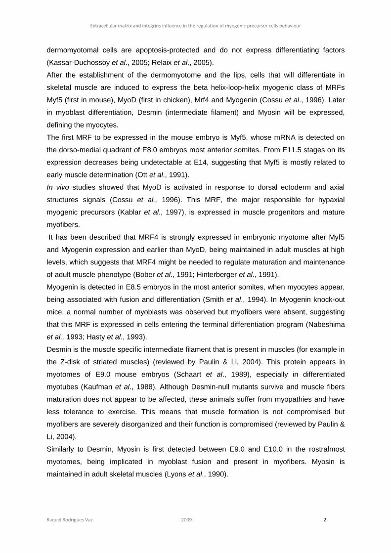
1.4.1.1 Laminin
Functional laminins (LN) are heterotrimers composed of a combination
of one α, one β and one γ chain (figure 2). These glycoproteins,
important components of basement membranes, can form a large
variety of laminins, depending on the combination of the chains
(Colognato and Yurchenco, 2000), although not all possible
combinations exist. Studies of specific laminin-deficient embryos show
that laminin is important in muscle formation (Sunada & Yamadas,
1994; Hynes, 1996).
Figure 2: Schematic
representation of the
laminin molecule and
its receptors, integrins
and dystroglycan. In
Schwarzbauer, 2005.
Extracellular matrix and integrins influence in the regulation of myogenic precursor cells behaviour
Raquel Rodrigues Vaz 2009 5
1.4.1.2 Fibronectin
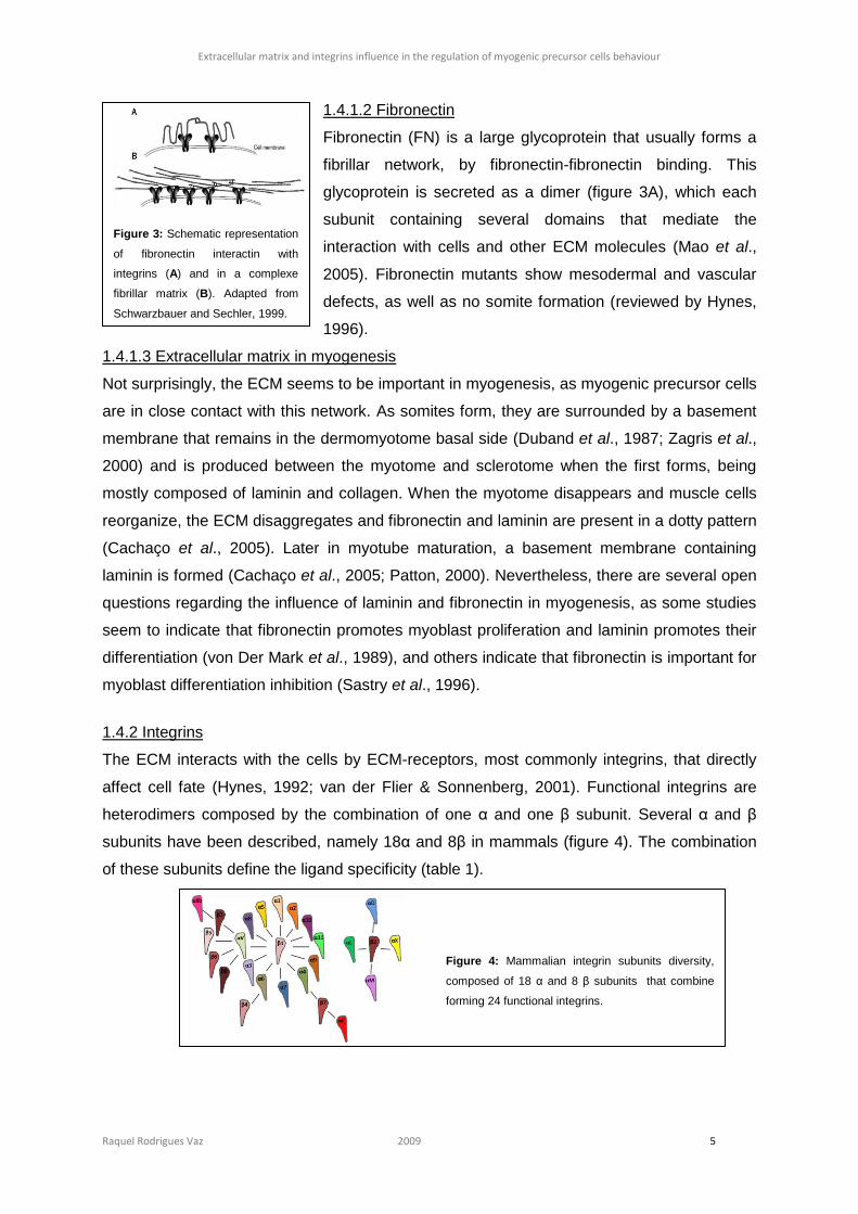
Fibronectin (FN) is a large glycoprotein that usually forms a
fibrillar network, by fibronectin-fibronectin binding. This
glycoprotein is secreted as a dimer (figure 3A), which each
subunit containing several domains that mediate the
interaction with cells and other ECM molecules (Mao et al.,
2005). Fibronectin mutants show mesodermal and vascular
defects, as well as no somite formation (reviewed by Hynes,
1996).
1.4.1.3 Extracellular matrix in myogenesis
Not surprisingly, the ECM seems to be important in myogenesis, as myogenic precursor cells
are in close contact with this network. As somites form, they are surrounded by a basement
membrane that remains in the dermomyotome basal side (Duband et al., 1987; Zagris et al.,
2000) and is produced between the myotome and sclerotome when the first forms, being
mostly composed of laminin and collagen. When the myotome disappears and muscle cells
reorganize, the ECM disaggregates and fibronectin and laminin are present in a dotty pattern
(Cachaço et al., 2005). Later in myotube maturation, a basement membrane containing
laminin is formed (Cachaço et al., 2005; Patton, 2000). Nevertheless, there are several open
questions regarding the influence of laminin and fibronectin in myogenesis, as some studies
seem to indicate that fibronectin promotes myoblast proliferation and laminin promotes their
differentiation (von Der Mark et al., 1989), and others indicate that fibronectin is important for
myoblast differentiation inhibition (Sastry et al., 1996).
1.4.2 Integrins
The ECM interacts with the cells by ECM-receptors, most commonly integrins, that directly
affect cell fate (Hynes, 1992; van der Flier & Sonnenberg, 2001). Functional integrins are
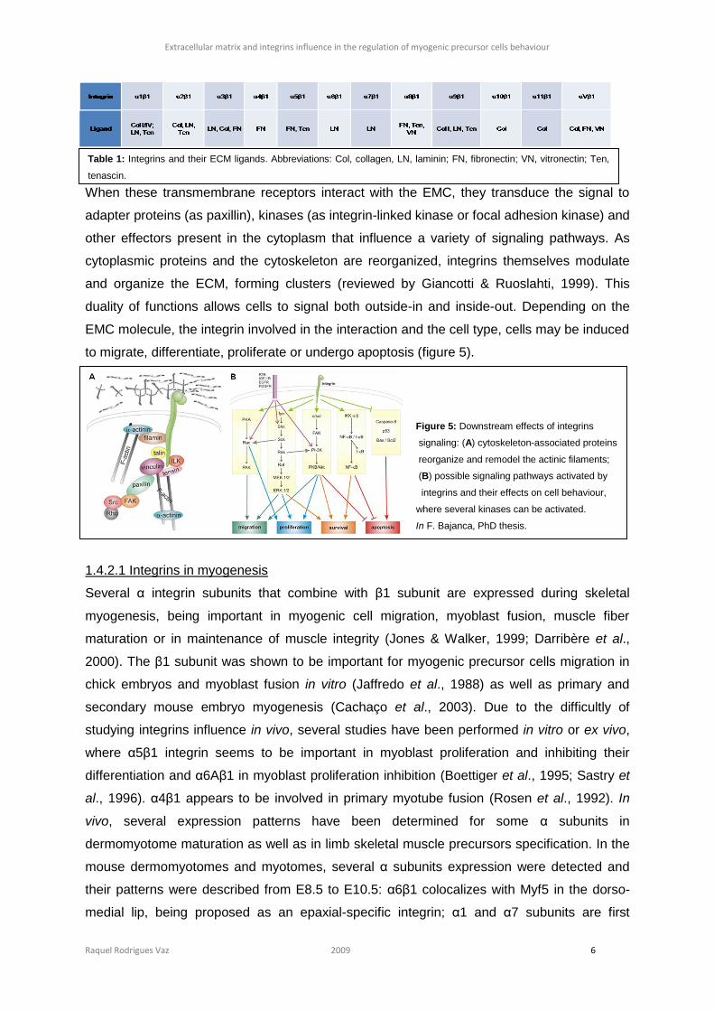
heterodimers composed by the combination of one α and one β subunit. Several α and β
subunits have been described, namely 18α and 8β in mammals (figure 4). The combination
of these subunits define the ligand specificity (table 1).
Figure 4: Mammalian integrin subunits diversity,
composed of 18 α and 8 β subunits that combine
forming 24 functional integrins.
Figure 3: Schematic representation
of fibronectin interactin with
integrins (A) and in a complexe
fibrillar matrix (B). Adapted from
Schwarzbauer and Sechler, 1999.
Extracellular matrix and integrins influence in the regulation of myogenic precursor cells behaviour
Raquel Rodrigues Vaz 2009 6
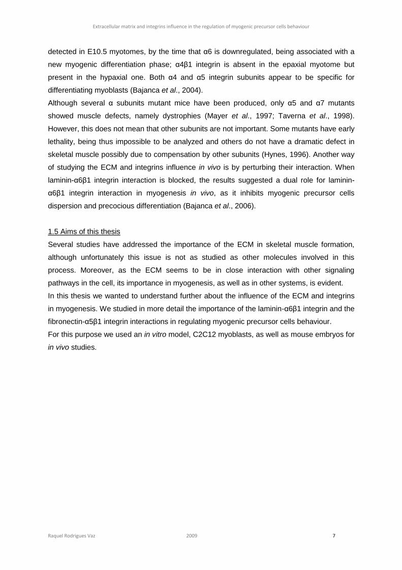
Figure 5: Downstream effects of integrins
signaling: (A) cytoskeleton-associated proteins
reorganize and remodel the actinic filaments;
(B) possible signaling pathways activated by
integrins and their effects on cell behaviour,
where several kinases can be activated.
In F. Bajanca, PhD thesis.
When these transmembrane receptors interact with the EMC, they transduce the signal to
adapter proteins (as paxillin), kinases (as integrin-linked kinase or focal adhesion kinase) and
other effectors present in the cytoplasm that influence a variety of signaling pathways. As
cytoplasmic proteins and the cytoskeleton are reorganized, integrins themselves modulate
and organize the ECM, forming clusters (reviewed by Giancotti & Ruoslahti, 1999). This
duality of functions allows cells to signal both outside-in and inside-out. Depending on the
EMC molecule, the integrin involved in the interaction and the cell type, cells may be induced
to migrate, differentiate, proliferate or undergo apoptosis (figure 5).
1.4.2.1 Integrins in myogenesis
Several α integrin subunits that combine with β1 subunit are expressed during skeletal
myogenesis, being important in myogenic cell migration, myoblast fusion, muscle fiber
maturation or in maintenance of muscle integrity (Jones & Walker, 1999; Darribère et al.,
2000). The β1 subunit was shown to be important for myogenic precursor cells migration in
chick embryos and myoblast fusion in vitro (Jaffredo et al., 1988) as well as primary and
secondary mouse embryo myogenesis (Cachaço et al., 2003). Due to the difficultly of
studying integrins influence in vivo, several studies have been performed in vitro or ex vivo,
where α5β1 integrin seems to be important in myoblast proliferation and inhibiting their
differentiation and α6Aβ1 in myoblast proliferation inhibition (Boettiger et al., 1995; Sastry et
al., 1996). α4β1 appears to be involved in primary myotube fusion (Rosen et al., 1992). In
vivo, several expression patterns have been determined for some α subunits in
dermomyotome maturation as well as in limb skeletal muscle precursors specification. In the
mouse dermomyotomes and myotomes, several α subunits expression were detected and
their patterns were described from E8.5 to E10.5: α6β1 colocalizes with Myf5 in the dorso-
medial lip, being proposed as an epaxial-specific integrin; α1 and α7 subunits are first
Table 1: Integrins and their ECM ligands. Abbreviations: Col, collagen, LN, laminin; FN, fibronectin; VN, vitronectin; Ten,
tenascin.
Extracellular matrix and integrins influence in the regulation of myogenic precursor cells behaviour
Raquel Rodrigues Vaz 2009 7
detected in E10.5 myotomes, by the time that α6 is downregulated, being associated with a
new myogenic differentiation phase; α4β1 integrin is absent in the epaxial myotome but
present in the hypaxial one. Both α4 and α5 integrin subunits appear to be specific for
differentiating myoblasts (Bajanca et al., 2004).
Although several α subunits mutant mice have been produced, only α5 and α7 mutants
showed muscle defects, namely dystrophies (Mayer et al., 1997; Taverna et al., 1998).
However, this does not mean that other subunits are not important. Some mutants have early
lethality, being thus impossible to be analyzed and others do not have a dramatic defect in
skeletal muscle possibly due to compensation by other subunits (Hynes, 1996). Another way
of studying the ECM and integrins influence in vivo is by perturbing their interaction. When
laminin-α6β1 integrin interaction is blocked, the results suggested a dual role for laminin-
α6β1 integrin interaction in myogenesis in vivo, as it inhibits myogenic precursor cells
dispersion and precocious differentiation (Bajanca et al., 2006).
1.5 Aims of this thesis
Several studies have addressed the importance of the ECM in skeletal muscle formation,
although unfortunately this issue is not as studied as other molecules involved in this
process. Moreover, as the ECM seems to be in close interaction with other signaling
pathways in the cell, its importance in myogenesis, as well as in other systems, is evident.
In this thesis we wanted to understand further about the influence of the ECM and integrins
in myogenesis. We studied in more detail the importance of the laminin-α6β1 integrin and the
fibronectin-α5β1 integrin interactions in regulating myogenic precursor cells behaviour.
For this purpose we used an in vitro model, C2C12 myoblasts, as well as mouse embryos for
in vivo studies.
Extracellular matrix and integrins influence in the regulation of myogenic precursor cells behaviour
Raquel Rodrigues Vaz 2009 8
2. Materials and Methods
2.1 In vitro studies
2.1.1 Cell culture
C2C12 myoblasts were maintained in culture in DMEM GlutaMax (ref. 31966, Invitrogen)
supplemented with 10% Fetal Bovine Serum (FBS) (ref. 10500, Invitrogen) and 100U/mL of
streptomycin and penicillin antibiotics (ref. 15140, Invitrogen) in a humidified atmosphere of
5% CO2 and 37ºC. Cells were detached from the culture flasks when strictly subconfluent
(80% confluency) using 0,05% Tripsin-EDTA (ref. 25300, Invitrogen) to maintain their
differentiation potential.
2.1.2 Extracellular matrix preparation
Laminin: Laminin (ref. L2020, Sigma) diluted at 5μg/mL in 1X PBS (137mM NaCl; 2,68mM
KCl; 8,1mM Na2HPO4; 1,47mM KH2PO4; pH 7,3 in deionized water) was placed on sterile
glass coverslips and incubated for 1 hour at 37ºC for coating. Next, the excess was removed,
washed with 1X PBS and the laminin-coated coverslips were ready to use.
Fibronectin: 2D fibronectin pre-coated coverslips were obtained from BD Biocoat Cellware®
(ref. 354088).
Gelatin: Sterile 1% gelatin was placed on sterile glass coverslips, left for one hour at 37ºC for
polymerization coating and the excess removed. This matrix was used as control.
2.1.3 Immunofluorescence
C2C12 cells were passaged onto 6- or 12-well plates with the pre-coated fibronectin
coverslips and sterilized coverslips coated with laminin and gelatin, as described above.
After cells reached the desired confluence, they were washed twice briefly with 1X PBS fixed
with 1% paraformaldehyde (PFA) in 1X PBS for 30 minutes (or 2% PFA in differentiated
cells), washed with 1X PBS and permeabilized with 0,5% Triton X-100 in 1X PBS for 5
minutes. After washing briefly with 1X PBS, blocking was performed with 2% bovine serum
albumin (BSA) in 1X PBS solution for 30 minutes. Coverslips with cells were transferred to
parafilm-coated dishes where they were incubated with primary antibodies in blocking
solution for 2 hours at room temperature (RT). Before and after incubation with secondary
antibodies for 2 hours (with nucleic acid stain ToPro3 and RNase in blocking solution), cells
were washed 3x10min with 1X PBS. In the case nuclei staining was performed with DAPT,
ToPro3 and 1:100 RNase were not used and cells were incubated with 5µg/mL of DAPI for 1
minute and then washed 3x10min with 1X PBS. Finally they were mounted on microscope
slides with propyl gallate-PBS-glycerol and sealed with nail varnish.
Extracellular matrix and integrins influence in the regulation of myogenic precursor cells behaviour
Raquel Rodrigues Vaz 2009 9
2.2 In vivo studies
2.2.1 Mouse embryo collection and culture
Charles-River mice were used to generate embryos. The morning plug was counted as E0.5
and females were sacrificed by cervical dislocation to collect embryos at the desired stage,
which ranged from E9 to E12.5. For culture, embryos were dissected in Dulbecco’s Modified
Eagle Medium/F12 GlutaMax medium (DMEM/F12) (ref. 31331, Invitrogen) supplemented
with 10mM HEPES (organic buffer ideal for maintain physiological pH), 1mM sodium
pyruvate (ref. 11360-039, Invitrogen), penicillin and streptomycin antibiotics (100U/mL) and
cultured on 0,8μm Milipore size filter (ref. ATTPo2500, IsoporeTM) floating on culture medium
(dissection medium without additional HEPES) at 37ºC and 5% CO2 humidified atmosphere
for the desired time.
2.2.2 Immunofluorescence
Mouse embryos were fixed in 0,2% PFA ON at 4ºC, dehydrated in an increasing glucose
series until included in gelatin solution and then frozen in dry ice-chilled liquid isopentane and
stored at -80ºC. Embryos were sectioned in 30 thick slices using a cryostat (Bright Clinicut)
and placed on SuperFrost Ultra Plus miscroscope slides (Menzel-Gläser).
The embryo transversal sections (and coronal sections in E12.0 and E12.5 cultured
embryos) were washed 3x5min with 1X PBS, permeabilized for 40 minutes in 0,2% Triton X-
100 in 1X PBS, washed again 3x5min and blocked for 1 hour in 1% BSA in 1X PBS. Primary
antibodies in blocking solution were added and left ON at 4ºC. Then, embryo sections were
washed 3x10min and incubation with secondary antibodies (in blocking solution, with ToPro3
and 1:100 RNase) was performed for at least 2 hours at RT. Embryo sections were washed
with 4X PBS for 20 minutes and 3x10min with 1X PBS to eliminate non-specific antibody
binding and finally mounted with glass coverslips on propyl gallate-PBS-glycerol, sealed with
nail varnish and kept in the dark at 4ºC.
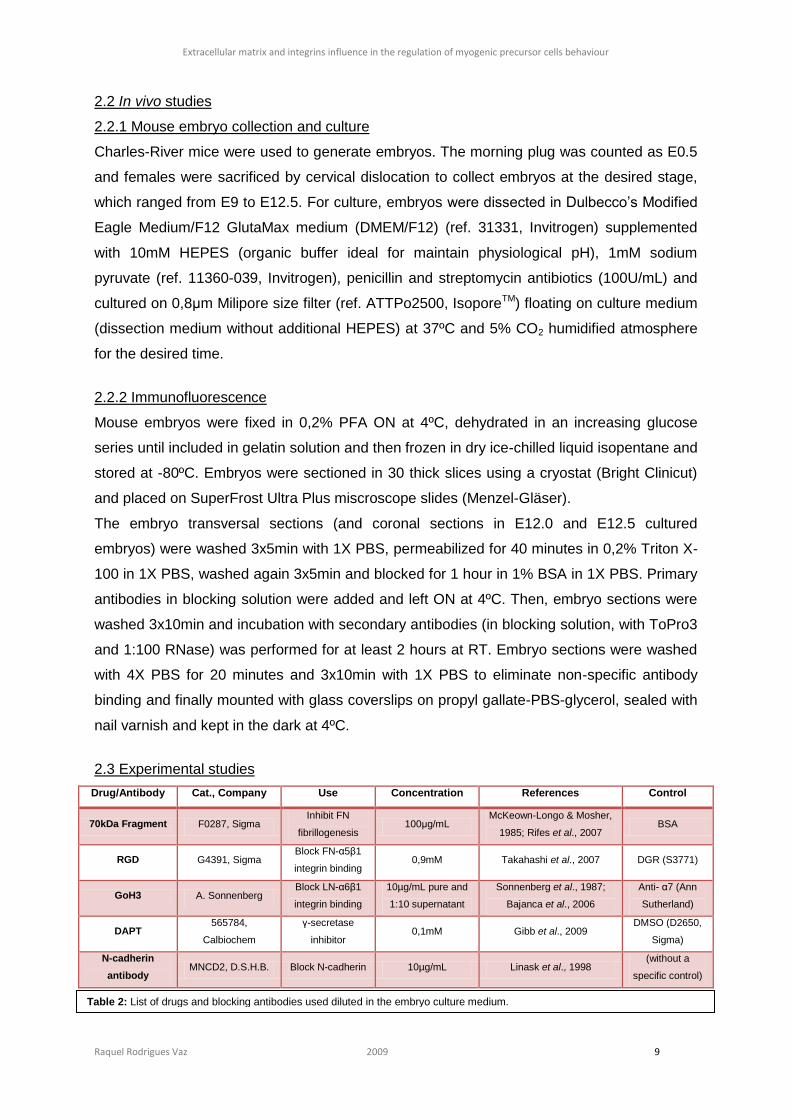
2.3 Experimental studies
Drug/Antibody Cat., Company Use Concentration References Control
70kDa Fragment F0287, Sigma Inhibit FN
fibrillogenesis 100μg/mL
McKeown-Longo & Mosher,
1985; Rifes et al., 2007 BSA
RGD G4391, Sigma Block FN-α5β1
integrin binding 0,9mM Takahashi et al., 2007 DGR (S3771)
GoH3 A. Sonnenberg Block LN-α6β1
integrin binding
10µg/mL pure and
1:10 supernatant
Sonnenberg et al., 1987;
Bajanca et al., 2006
Anti- α7 (Ann
Sutherland)
DAPT 565784,
Calbiochem
γ-secretase
inhibitor 0,1mM Gibb et al., 2009
DMSO (D2650,
Sigma)
N-cadherin
antibody MNCD2, D.S.H.B. Block N-cadherin 10µg/mL Linask et al., 1998
(without a
specific control)
Table 2: List of drugs and blocking antibodies used diluted in the embryo culture medium.
Extracellular matrix and integrins influence in the regulation of myogenic precursor cells behaviour
Raquel Rodrigues Vaz 2009 10
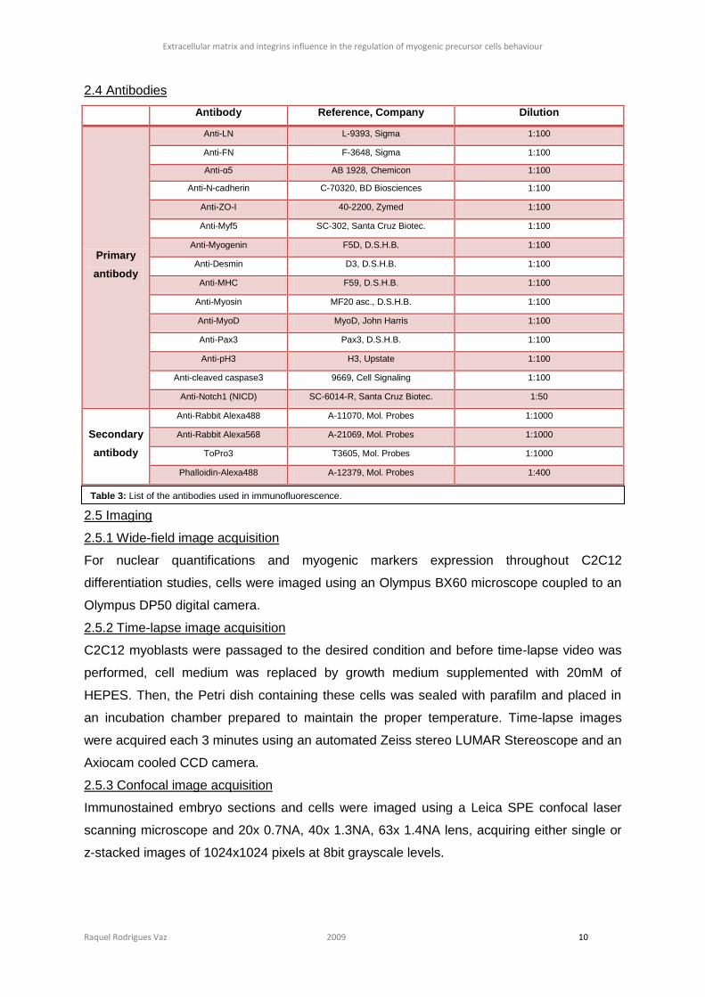
2.4 Antibodies
Antibody Reference, Company Dilution
Primary
antibody
Anti-LN L-9393, Sigma 1:100
Anti-FN F-3648, Sigma 1:100
Anti-α5 AB 1928, Chemicon 1:100
Anti-N-cadherin C-70320, BD Biosciences 1:100
Anti-ZO-I 40-2200, Zymed 1:100
Anti-Myf5 SC-302, Santa Cruz Biotec. 1:100
Anti-Myogenin F5D, D.S.H.B. 1:100
Anti-Desmin D3, D.S.H.B. 1:100
Anti-MHC F59, D.S.H.B. 1:100
Anti-Myosin MF20 asc., D.S.H.B. 1:100
Anti-MyoD MyoD, John Harris 1:100
Anti-Pax3 Pax3, D.S.H.B. 1:100
Anti-pH3 H3, Upstate 1:100
Anti-cleaved caspase3 9669, Cell Signaling 1:100
Anti-Notch1 (NICD) SC-6014-R, Santa Cruz Biotec. 1:50
Secondary
antibody
Anti-Rabbit Alexa488 A-11070, Mol. Probes 1:1000
Anti-Rabbit Alexa568 A-21069, Mol. Probes 1:1000
ToPro3 T3605, Mol. Probes 1:1000
Phalloidin-Alexa488 A-12379, Mol. Probes 1:400
2.5 Imaging
2.5.1 Wide-field image acquisition
For nuclear quantifications and myogenic markers expression throughout C2C12
differentiation studies, cells were imaged using an Olympus BX60 microscope coupled to an
Olympus DP50 digital camera.
2.5.2 Time-lapse image acquisition
C2C12 myoblasts were passaged to the desired condition and before time-lapse video was
performed, cell medium was replaced by growth medium supplemented with 20mM of
HEPES. Then, the Petri dish containing these cells was sealed with parafilm and placed in
an incubation chamber prepared to maintain the proper temperature. Time-lapse images
were acquired each 3 minutes using an automated Zeiss stereo LUMAR Stereoscope and an
Axiocam cooled CCD camera.
2.5.3 Confocal image acquisition
Immunostained embryo sections and cells were imaged using a Leica SPE confocal laser
scanning microscope and 20x 0.7NA, 40x 1.3NA, 63x 1.4NA lens, acquiring either single or
z-stacked images of 1024x1024 pixels at 8bit grayscale levels.
Table 3: List of the antibodies used in immunofluorescence.
Extracellular matrix and integrins influence in the regulation of myogenic precursor cells behaviour
Raquel Rodrigues Vaz 2009 11
Images and videos were then treated and analyzed using ImageJ, Amira v4.2 and Imaris
v5.7.2 software.
2.6 Image analysis and quantifications
2.6.1 In vitro studies
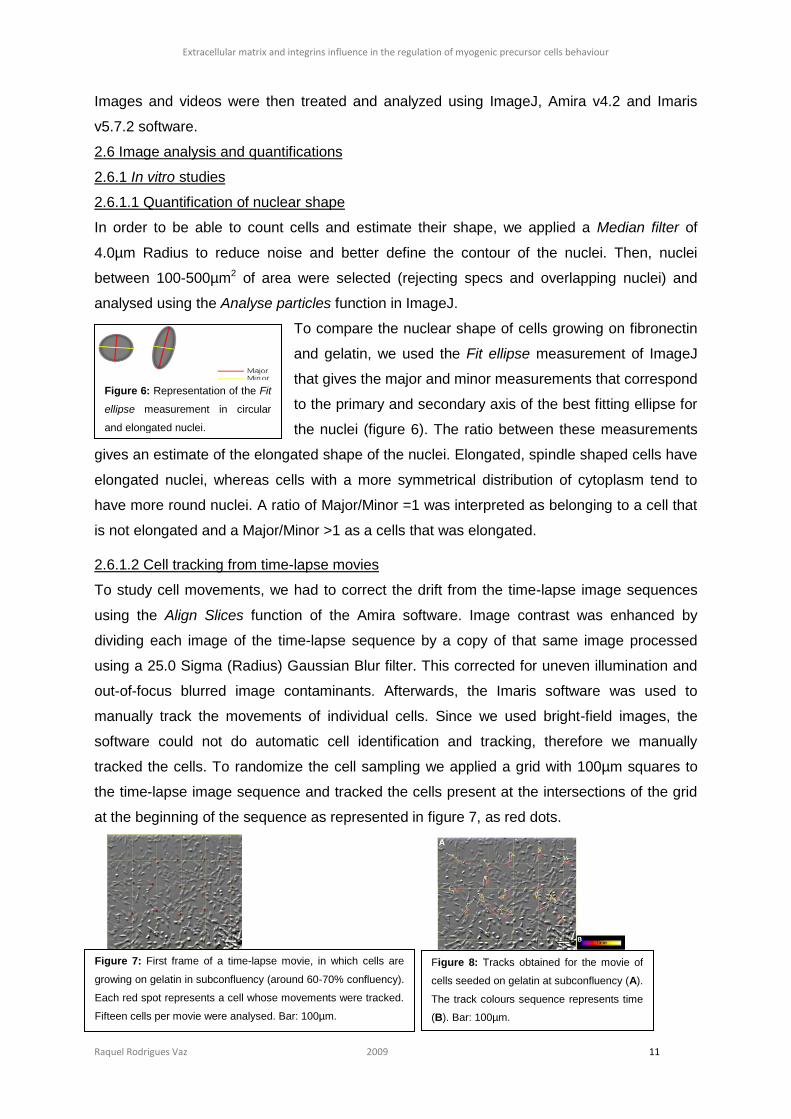
2.6.1.1 Quantification of nuclear shape
In order to be able to count cells and estimate their shape, we applied a Median filter of
4.0µm Radius to reduce noise and better define the contour of the nuclei. Then, nuclei
between 100-500µm2 of area were selected (rejecting specs and overlapping nuclei) and
analysed using the Analyse particles function in ImageJ.
To compare the nuclear shape of cells growing on fibronectin
and gelatin, we used the Fit ellipse measurement of ImageJ
that gives the major and minor measurements that correspond
to the primary and secondary axis of the best fitting ellipse for
the nuclei (figure 6). The ratio between these measurements
gives an estimate of the elongated shape of the nuclei. Elongated, spindle shaped cells have
elongated nuclei, whereas cells with a more symmetrical distribution of cytoplasm tend to
have more round nuclei. A ratio of Major/Minor =1 was interpreted as belonging to a cell that
is not elongated and a Major/Minor >1 as a cells that was elongated.
2.6.1.2 Cell tracking from time-lapse movies
To study cell movements, we had to correct the drift from the time-lapse image sequences
using the Align Slices function of the Amira software. Image contrast was enhanced by
dividing each image of the time-lapse sequence by a copy of that same image processed
using a 25.0 Sigma (Radius) Gaussian Blur filter. This corrected for uneven illumination and
out-of-focus blurred image contaminants. Afterwards, the Imaris software was used to
manually track the movements of individual cells. Since we used bright-field images, the
software could not do automatic cell identification and tracking, therefore we manually
tracked the cells. To randomize the cell sampling we applied a grid with 100µm squares to
the time-lapse image sequence and tracked the cells present at the intersections of the grid
at the beginning of the sequence as represented in figure 7, as red dots.
Figure 6: Representation of the Fit
ellipse measurement in circular
and elongated nuclei.
Figure 7: First frame of a time-lapse movie, in which cells are
growing on gelatin in subconfluency (around 60-70% confluency).
Each red spot represents a cell whose movements were tracked.
Fifteen cells per movie were analysed. Bar: 100µm.
Figure 8: Tracks obtained for the movie of
cells seeded on gelatin at subconfluency (A).
The track colours sequence represents time
(B). Bar: 100µm.
Extracellular matrix and integrins influence in the regulation of myogenic precursor cells behaviour
Raquel Rodrigues Vaz 2009 12
Each chosen cell was followed during the whole time-lapse sequence by placing a spot in the
center of the cell body at each time-point. The tracks were then created, analysed and
compared for some parameters that Imaris calculates, the cell displacement and track length.
The tracks were then calculated by the software (figure 8), which also calculated the
displacement (linear distance between beginning and ending position) and full track length.
2.6.2 In vivo studies
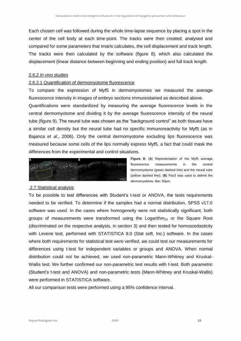
2.6.2.1 Quantification of dermomyotome fluorescence
To compare the expression of Myf5 in dermomyotomes we measured the average
fluorescence intensity in images of embryo sections immunostained as described above.
Quantifications were standardized by measuring the average fluorescence levels in the
central dermomyotome and dividing it by the average fluorescence intensity of the neural
tube (figure 9). The neural tube was chosen as the “background control” as both tissues have
a similar cell density but the neural tube had no specific immunoreactivity for Myf5 (as in
Bajanca et al., 2006). Only the central dermomyotome excluding lips fluorescence was
measured because some cells of the lips normally express Myf5, a fact that could mask the
differences from the experimental and control situations.
Figure 9: (A) Representation of the Myf5 average
fluorescence measurements in the central
dermomyotome (green dashed line) and the neural tube
(yellow dashed line); (B) Pax3 was used to delimit the
dermomyotome. Bar: 50µm.
2.7 Statistical analysis
To be possible to test differences with Student’s t-test or ANOVA, the tests requirements
needed to be verified. To determine if the samples had a normal distribution, SPSS v17.0
software was used. In the cases where homogeneity were not statistically significant, both
groups of measurements were transformed using the Logarithm10 or the Square Root
(discriminated on the respective analysis, in section 3) and then tested for homoscedasticity
with Levene test, performed with STATISTICA 8.0 (Stat soft, Inc.) software. In the cases
where both requirements for statistical test were verified, we could test our measurements for
differences using t-test for independent variables or groups and ANOVA. When normal
distribution could not be achieved, we used non-parametric Mann-Whitney and Kruskal-
Wallis test. We further confirmed our non-parametric test results with t-test. Both parametric
(Student’s t-test and ANOVA) and non-parametric tests (Mann-Whitney and Kruskal-Wallis)
were performed in STATISTICA software.
All our comparison tests were performed using a 95% confidence interval.
Extracellular matrix and integrins influence in the regulation of myogenic precursor cells behaviour
Raquel Rodrigues Vaz 2009 13
3. Results
3.1 Laminin
3.1.1 Laminin influence in in vitro myogenesis
To study the effect of a laminin matrix on myogenic differentiation, we cultured C2C12 cells
on laminin- or gelatin-coated coverslips and compared the dynamics of MRFs expression
throughout the activation and development of the myogenic program performing
immunofluorescence in cells maintained over several days in growth and differentiation
medium. In the first 8 days of C2C12 differentiation, we could not observe any difference in
MRFs, namely Myf5 and Myogenin (figure S1, section 6) nor in Myosin expression and the
correspondent myotubes (figure S2, section 6).
3.1.2 Laminin influence in in vivo myogenesis
It has been previously shown that the laminin matrix is necessary to inhibit myogenic
differentiation in the dermomyotomal cells (Bajanca et al., 2006). One possibility is that it
could be related to the Notch signaling pathway.
3.1.2.1 Laminin-α6β1 interaction maintains dermomyotomal cells undifferentiated
In a first approach, we tried to reproduce the laminin-α6β1 interaction blocking experiments
performed in Bajanca, 2006, but it was very difficult to achieve it fruitfully. When E9.0 mouse
embryos were cultured for 24 hours with GoH3 (blocking antibody) we observed in some
embryos a slight increase of Myf5 protein in the dermomyotome as compared with the
control embryos. However, when the fluorescence measurements were compared with the t-
test, after transformation with Log10, the difference was not statistically significant (t=1.2505,
ncontrol=49, nGoH3=53, p=0.214) (figure S3, section 6).
3.1.2.2 Notch signaling is necessary for inhibition of dermomyotomal cells myogenic
differentiation
To test the Notch signaling pathway involvement in myogenic differentiation, we blocked the
γ-secretase activity with DAPT thus inhibiting the nuclear translocation of the NICD.
Our first approach was to confirm that in embryos cultured with DAPT, the NICD is more
retained in the membrane, in a non-cleaved form, than in control embryos. In the neural tube
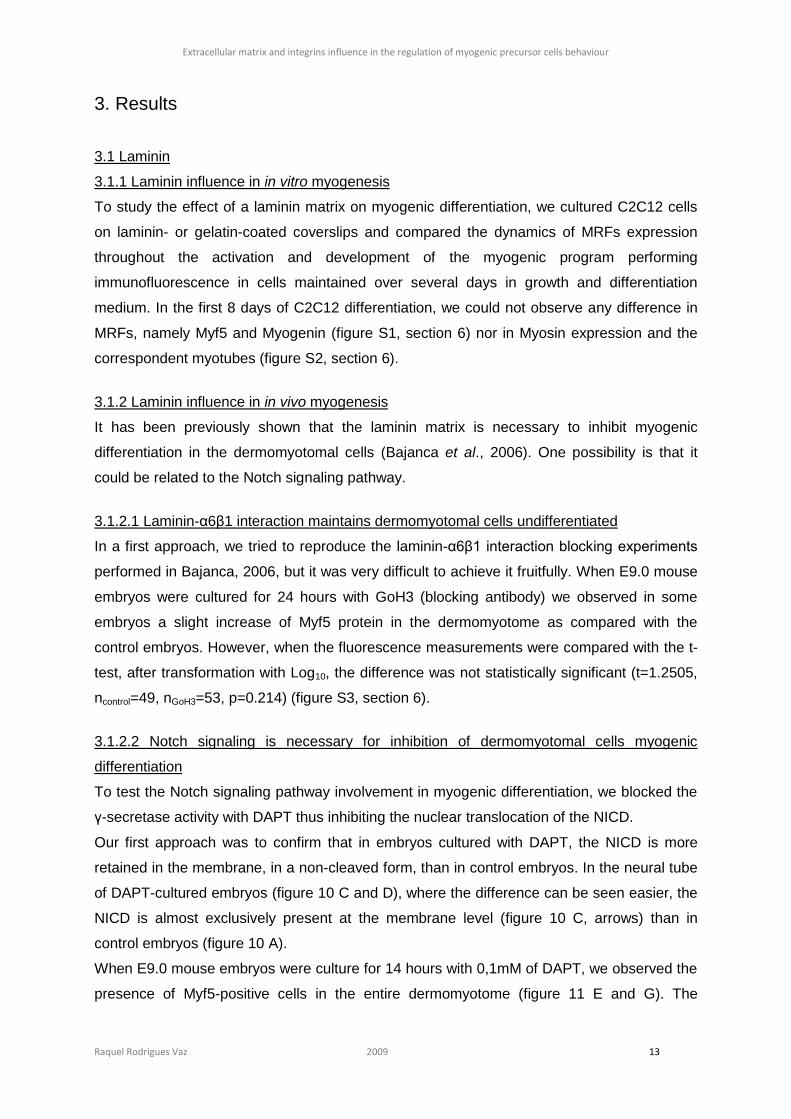
of DAPT-cultured embryos (figure 10 C and D), where the difference can be seen easier, the
NICD is almost exclusively present at the membrane level (figure 10 C, arrows) than in
control embryos (figure 10 A).
When E9.0 mouse embryos were culture for 14 hours with 0,1mM of DAPT, we observed the
presence of Myf5-positive cells in the entire dermomyotome (figure 11 E and G). The
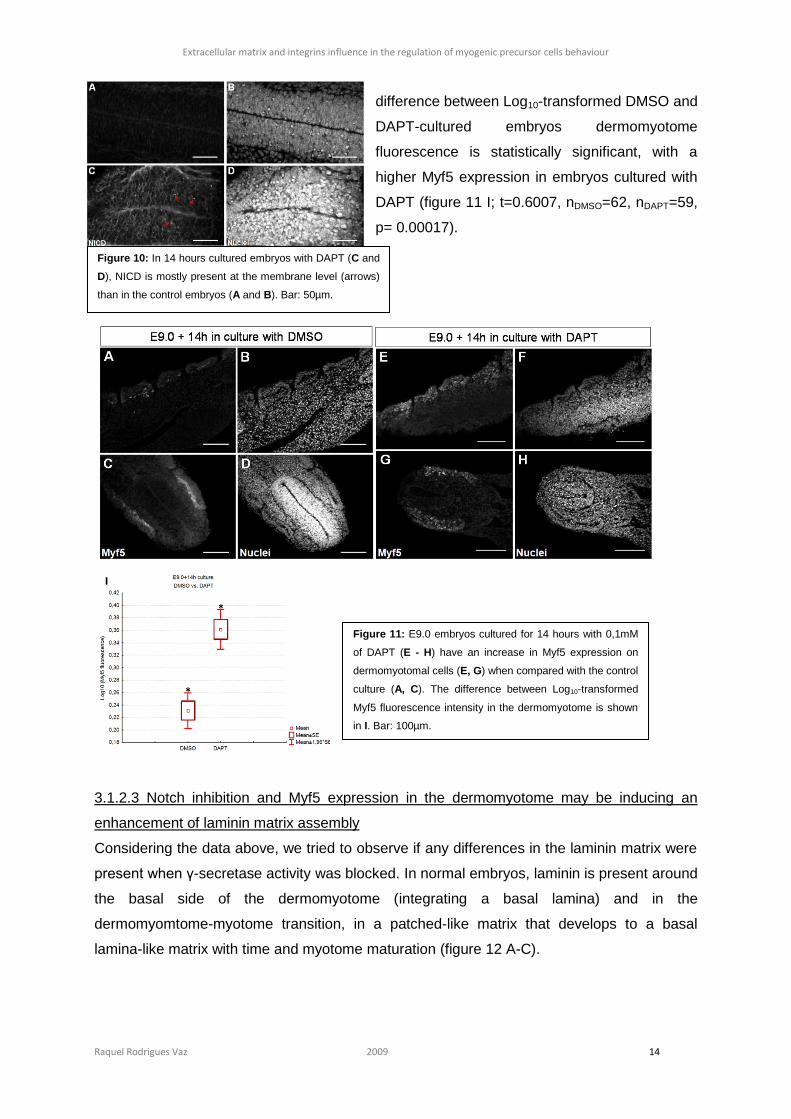
Extracellular matrix and integrins influence in the regulation of myogenic precursor cells behaviour
Raquel Rodrigues Vaz 2009 14
difference between Log10-transformed DMSO and
DAPT-cultured embryos dermomyotome
fluorescence is statistically significant, with a
higher Myf5 expression in embryos cultured with
DAPT (figure 11 I; t=0.6007, nDMSO=62, nDAPT=59,
p= 0.00017).
3.1.2.3 Notch inhibition and Myf5 expression in the dermomyotome may be inducing an
enhancement of laminin matrix assembly
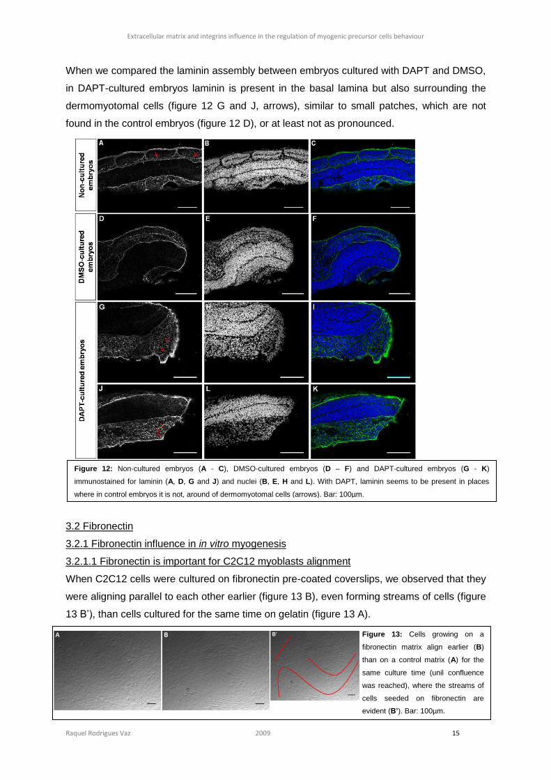
Considering the data above, we tried to observe if any differences in the laminin matrix were
present when γ-secretase activity was blocked. In normal embryos, laminin is present around
the basal side of the dermomyotome (integrating a basal lamina) and in the
dermomyomtome-myotome transition, in a patched-like matrix that develops to a basal
lamina-like matrix with time and myotome maturation (figure 12 A-C).
Figure 10: In 14 hours cultured embryos with DAPT (C and
D), NICD is mostly present at the membrane level (arrows)
than in the control embryos (A and B). Bar: 50µm.
Figure 11: E9.0 embryos cultured for 14 hours with 0,1mM
of DAPT (E - H) have an increase in Myf5 expression on
dermomyotomal cells (E, G) when compared with the control
culture (A, C). The difference between Log10-transformed
Myf5 fluorescence intensity in the dermomyotome is shown
in I. Bar: 100µm.
Extracellular matrix and integrins influence in the regulation of myogenic precursor cells behaviour
Raquel Rodrigues Vaz 2009 15
When we compared the laminin assembly between embryos cultured with DAPT and DMSO,
in DAPT-cultured embryos laminin is present in the basal lamina but also surrounding the
dermomyotomal cells (figure 12 G and J, arrows), similar to small patches, which are not
found in the control embryos (figure 12 D), or at least not as pronounced.
3.2 Fibronectin
3.2.1 Fibronectin influence in in vitro myogenesis
3.2.1.1 Fibronectin is important for C2C12 myoblasts alignment
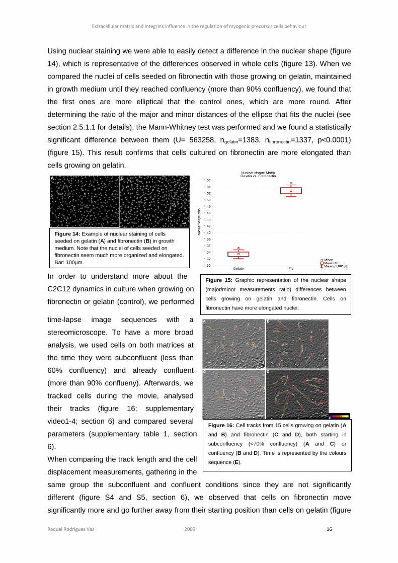
When C2C12 cells were cultured on fibronectin pre-coated coverslips, we observed that they
were aligning parallel to each other earlier (figure 13 B), even forming streams of cells (figure
13 B’), than cells cultured for the same time on gelatin (figure 13 A).
Figure 13: Cells growing on a
fibronectin matrix align earlier (B)
than on a control matrix (A) for the
same culture time (unil confluence
was reached), where the streams of
cells seeded on fibronectin are
evident (B’). Bar: 100µm.
Figure 12: Non-cultured embryos (A - C), DMSO-cultured embryos (D – F) and DAPT-cultured embryos (G - K)
immunostained for laminin (A, D, G and J) and nuclei (B, E, H and L). With DAPT, laminin seems to be present in places
where in control embryos it is not, around of dermomyotomal cells (arrows). Bar: 100µm.
Extracellular matrix and integrins influence in the regulation of myogenic precursor cells behaviour
Raquel Rodrigues Vaz 2009 16
Using nuclear staining we were able to easily detect a difference in the nuclear shape (figure
14), which is representative of the differences observed in whole cells (figure 13). When we
compared the nuclei of cells seeded on fibronectin with those growing on gelatin, maintained
in growth medium until they reached confluency (more than 90% confluency), we found that
the first ones are more elliptical that the control ones, which are more round. After
determining the ratio of the major and minor distances of the ellipse that fits the nuclei (see
section 2.5.1.1 for details), the Mann-Whitney test was performed and we found a statistically
significant difference between them (U= 563258, ngelatin=1383, nfibronectin=1337, p<0.0001)
(figure 15). This result confirms that cells cultured on fibronectin are more elongated than
cells growing on gelatin.
In order to understand more about the
C2C12 dynamics in culture when growing on
fibronectin or gelatin (control), we performed
time-lapse image sequences with a
stereomicroscope. To have a more broad
analysis, we used cells on both matrices at
the time they were subconfluent (less than
60% confluency) and already confluent
(more than 90% conflueny). Afterwards, we
tracked cells during the movie, analysed
their tracks (figure 16; supplementary
video1-4; section 6) and compared several
parameters (supplementary table 1, section
6).
When comparing the track length and the cell
displacement measurements, gathering in the
same group the subconfluent and confluent conditions since they are not significantly
different (figure S4 and S5, section 6), we observed that cells on fibronectin move
significantly more and go further away from their starting position than cells on gelatin (figure
Figure 15: Graphic representation of the nuclear shape
(major/minor measurements ratio) differences between
cells growing on gelatin and fibronectin. Cells on
fibronectin have more elongated nuclei.
Figure 16: Cell tracks from 15 cells growing on gelatin (A
and B) and fibronectin (C and D), both starting in
subconfluency (<70% confluency) (A and C) or
confluency (B and D). Time is represented by the colours
sequence (E).
Bar: 100µm.
Figure 14: Example of nuclear staining of cells
seeded on gelatin (A) and fibronectin (B) in growth
medium. Note that the nuclei of cells seeded on
fibronectin seem much more organized and elongated.
Bar: 100µm.
Extracellular matrix and integrins influence in the regulation of myogenic precursor cells behaviour
Raquel Rodrigues Vaz 2009 17
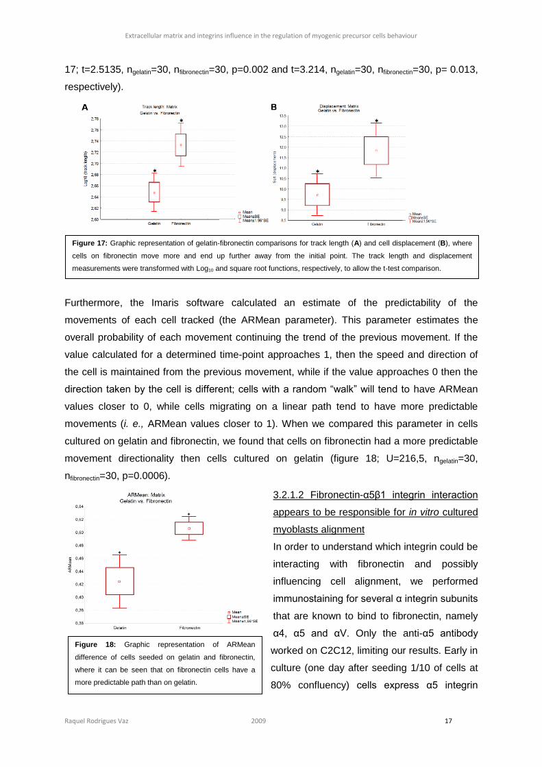
17; t=2.5135, ngelatin=30, nfibronectin=30, p=0.002 and t=3.214, ngelatin=30, nfibronectin=30, p= 0.013,
respectively).
Furthermore, the Imaris software calculated an estimate of the predictability of the
movements of each cell tracked (the ARMean parameter). This parameter estimates the
overall probability of each movement continuing the trend of the previous movement. If the
value calculated for a determined time-point approaches 1, then the speed and direction of
the cell is maintained from the previous movement, while if the value approaches 0 then the
direction taken by the cell is different; cells with a random “walk” will tend to have ARMean
values closer to 0, while cells migrating on a linear path tend to have more predictable
movements (i. e., ARMean values closer to 1). When we compared this parameter in cells
cultured on gelatin and fibronectin, we found that cells on fibronectin had a more predictable
movement directionality then cells cultured on gelatin (figure 18; U=216,5, ngelatin=30,
nfibronectin=30, p=0.0006).
3.2.1.2 Fibronectin-α5β1 integrin interaction
appears to be responsible for in vitro cultured
myoblasts alignment
In order to understand which integrin could be
interacting with fibronectin and possibly
influencing cell alignment, we performed
immunostaining for several α integrin subunits
that are known to bind to fibronectin, namely
α4, α5 and αV. Only the anti-α5 antibody
worked on C2C12, limiting our results. Early in
culture (one day after seeding 1/10 of cells at
80% confluency) cells express α5 integrin
Figure 18: Graphic representation of ARMean
difference of cells seeded on gelatin and fibronectin,
where it can be seen that on fibronectin cells have a
more predictable path than on gelatin.
Figure 17: Graphic representation of gelatin-fibronectin comparisons for track length (A) and cell displacement (B), where
cells on fibronectin move more and end up further away from the initial point. The track length and displacement
measurements were transformed with Log10 and square root functions, respectively, to allow the t-test comparison.
Extracellular matrix and integrins influence in the regulation of myogenic precursor cells behaviour
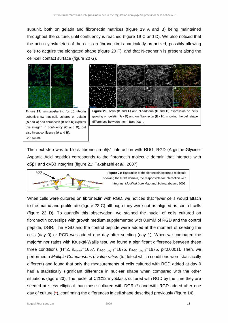
Raquel Rodrigues Vaz 2009 18
subunit, both on gelatin and fibronectin matrices (figure 19 A and B) being maintained
throughout the culture, until confluency is reached (figure 19 C and D). We also noticed that
the actin cytoskeleton of the cells on fibronectin is particularly organized, possibly allowing
cells to acquire the elongated shape (figure 20 F), and that N-cadherin is present along the
cell-cell contact surface (figure 20 G).
The next step was to block fibronectin-α5β1 interaction with RDG. RGD (Arginine-Glycine-
Aspartic Acid peptide) corresponds to the fibronectin molecule domain that interacts with
α5β1 and αVβ3 integrins (figure 21; Takahashi et al., 2007).
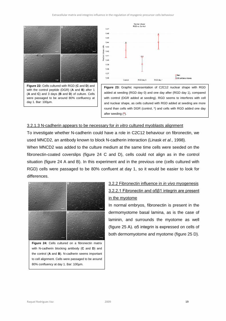
When cells were cultured on fibronectin with RGD, we noticed that fewer cells would attach
to the matrix and proliferate (figure 22 C) although they were not as aligned as control cells
(figure 22 D). To quantify this observation, we stained the nuclei of cells cultured on
fibronectin coverslips with growth medium supplemented with 0,9mM of RGD and the control
peptide, DGR. The RGD and the control peptide were added at the moment of seeding the
cells (day 0) or RGD was added one day after seeding (day 1). When we compared the
major/minor ratios with Kruskal-Wallis test, we found a significant difference between these
three conditions (H=2, ncontrol=1657, nRGD day 2=1675, nRGD day 1=1675, p<0.0001). Then, we
performed a Multiple Comparisons p value ratios (to detect which conditions were statistically
different) and found that only the measurements of cells cultured with RGD added at day 0
had a statistically significant difference in nuclear shape when compared with the other
situations (figure 23). The nuclei of C2C12 myoblasts cultured with RGD by the time they are
seeded are less elliptical than those cultured with DGR (*) and with RGD added after one
day of culture (*), confirming the differences in cell shape described previously (figure 14).
Figure 19: Immunostaining for α5 integrin
subunit show that cells cultured on gelatin
(A and C) and fibronectin (B and D) express
this integrin in confluency (C and D), but
also in subconfluency (A and B).
Bar: 50µm.
Figure 20: Actin (B and F) and N-cadherin (C and G) expression on cells
growing on gelatin (A - D) and on fibronectin (E - H), showing the cell shape
differences between them. Bar: 40µm.
Figure 21: Illustration of the fibronectin secreted molecule
showing the RGD domain, the responsible for interaction with
integrins. Modified from Mao and Schwarzbauer, 2005.
Extracellular matrix and integrins influence in the regulation of myogenic precursor cells behaviour
Raquel Rodrigues Vaz 2009 19
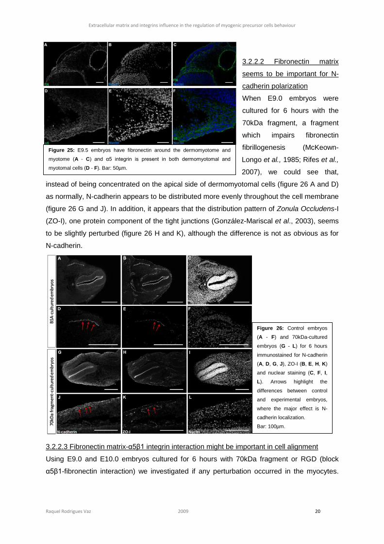
3.2.1.3 N-cadherin appears to be necessary for in vitro cultured myoblasts alignment
To investigate whether N-cadherin could have a role in C2C12 behaviour on fibronectin, we
used MNCD2, an antibody known to block N-cadherin interaction (Linask et al., 1998).
When MNCD2 was added to the culture medium at the same time cells were seeded on the
fibronectin-coated coverslips (figure 24 C and D), cells could not align as in the control
situation (figure 24 A and B). In this experiment and in the previous one (cells cultured with
RGD) cells were passaged to be 80% confluent at day 1, so it would be easier to look for
differences.
3.2.2 Fibronectin influence in in vivo myogenesis
3.2.2.1 Fibronectin and α5β1 integrin are present
in the myotome
In normal embryos, fibronectin is present in the
dermomyotome basal lamina, as is the case of
laminin, and surrounds the myotome as well
(figure 25 A). α5 integrin is expressed on cells of
both dermomyotome and myotome (figure 25 D).
Figure 22: Cells cultured with RGD (C and D) and
with the control peptide (DGR) (A and B) after 1
(A and C) and 3 days (B and D) of culture. Cells
were passaged to be around 80% confluency at
day 1. Bar: 100µm.
Figure 23: Graphic representation of C2C12 nuclear shape with RGD
added at seeding (RGD day 0) and one day after (RGD day 1), compared
with control (DGR added at seeding). RGD seems to interferes with cell
and nuclear shape, as cells cultured with RGD added at seeding are more
round than cells with DGR (control, *) and cells with RGD added one day
after seeding (*).
Figure 24: Cells cultured on a fibronectin matrix
with N-cadherin blocking antibody (C and D) and
the control (A and B). N-cadherin seems important
to cell alignment. Cells were passaged to be around
80% confluency at day 1. Bar: 100µm.
Extracellular matrix and integrins influence in the regulation of myogenic precursor cells behaviour
Raquel Rodrigues Vaz 2009 20
3.2.2.2 Fibronectin matrix
seems to be important for N-
cadherin polarization
When E9.0 embryos were
cultured for 6 hours with the
70kDa fragment, a fragment
which impairs fibronectin
fibrillogenesis (McKeown-
Longo et al., 1985; Rifes et al.,
2007), we could see that,
instead of being concentrated on the apical side of dermomyotomal cells (figure 26 A and D)
as normally, N-cadherin appears to be distributed more evenly throughout the cell membrane
(figure 26 G and J). In addition, it appears that the distribution pattern of Zonula Occludens-I
(ZO-I), one protein component of the tight junctions (González-Mariscal et al., 2003), seems
to be slightly perturbed (figure 26 H and K), although the difference is not as obvious as for
N-cadherin.
3.2.2.3 Fibronectin matrix-α5β1 integrin interaction might be important in cell alignment
Using E9.0 and E10.0 embryos cultured for 6 hours with 70kDa fragment or RGD (block
α5β1-fibronectin interaction) we investigated if any perturbation occurred in the myocytes.
Figure 25: E9.5 embryos have fibronectin around the dermomyotome and
myotome (A - C) and α5 integrin is present in both dermomyotomal and
myotomal cells (D - F). Bar: 50µm.
Figure 26: Control embryos
(A - F) and 70kDa-cultured
embryos (G - L) for 6 hours
immunostained for N-cadherin
(A, D, G, J), ZO-I (B, E, H, K)
and nuclear staining (C, F, I,
L). Arrows highlight the
differences between control
and experimental embryos,
where the major effect is N-
cadherin localization.
Bar: 100µm.

Extracellular matrix and integrins influence in the regulation of myogenic precursor cells behaviour
Raquel Rodrigues Vaz 2009 21
We could not see any obvious change in myoblasts alignment and orientation (figure S6,
section 6).
It has been previously shown that in E12.5 embryos only fibronectin and fibronectin-binding
integrins are present in primary myocytes as they begin to fuse into myofibers (Cachaço et
al., 2005). Therefore, we questioned if fibronectin is important for proper myocytes alignment
and fusion in E12.5 staged embryos. E12.0 mouse embryos were cultured for 6 hours with
the 70kDa fragment or RGD and we did not find any difference in myotome derived myocytes
of both cultures (figure S7, section 6). We cultured E12.5 embryos for 14 hours and could not
see a difference in cell alignment as well (data not shown).
3.2.2.4 Fibronectin-α5β1 interaction maintains dermomyotomal cells undifferentiated
During these experiments we observed that, in some sections of the E9.0 embryos cultured
for 12 hours with RGD, Myf5 protein was present in the dermomyotome, similar to what we
saw when embryos were cultured with DAPT (see section 3.1.2.2) (figure 27 D).
Figure 27: E9.0 embryos cultured with RGD for 12 hours (D - F), where in some sections we could detect Myf5
expression in the dermomyotome (D), never detected on control cultured-embryos (A - C). Bar: 100µm.
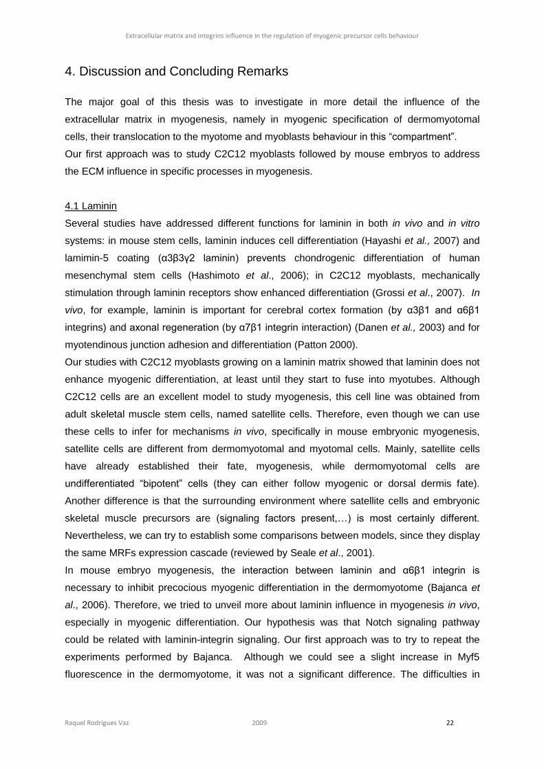
Extracellular matrix and integrins influence in the regulation of myogenic precursor cells behaviour
Raquel Rodrigues Vaz 2009 22
4. Discussion and Concluding Remarks
The major goal of this thesis was to investigate in more detail the influence of the
extracellular matrix in myogenesis, namely in myogenic specification of dermomyotomal
cells, their translocation to the myotome and myoblasts behaviour in this “compartment”.
Our first approach was to study C2C12 myoblasts followed by mouse embryos to address
the ECM influence in specific processes in myogenesis.
4.1 Laminin
Several studies have addressed different functions for laminin in both in vivo and in vitro
systems: in mouse stem cells, laminin induces cell differentiation (Hayashi et al., 2007) and
lamimin-5 coating (α3β3γ2 laminin) prevents chondrogenic differentiation of human
mesenchymal stem cells (Hashimoto et al., 2006); in C2C12 myoblasts, mechanically
stimulation through laminin receptors show enhanced differentiation (Grossi et al., 2007). In
vivo, for example, laminin is important for cerebral cortex formation (by α3β1 and α6β1
integrins) and axonal regeneration (by α7β1 integrin interaction) (Danen et al., 2003) and for
myotendinous junction adhesion and differentiation (Patton 2000).
Our studies with C2C12 myoblasts growing on a laminin matrix showed that laminin does not
enhance myogenic differentiation, at least until they start to fuse into myotubes. Although
C2C12 cells are an excellent model to study myogenesis, this cell line was obtained from
adult skeletal muscle stem cells, named satellite cells. Therefore, even though we can use
these cells to infer for mechanisms in vivo, specifically in mouse embryonic myogenesis,
satellite cells are different from dermomyotomal and myotomal cells. Mainly, satellite cells
have already established their fate, myogenesis, while dermomyotomal cells are
undifferentiated “bipotent” cells (they can either follow myogenic or dorsal dermis fate).
Another difference is that the surrounding environment where satellite cells and embryonic
skeletal muscle precursors are (signaling factors present,…) is most certainly different.
Nevertheless, we can try to establish some comparisons between models, since they display
the same MRFs expression cascade (reviewed by Seale et al., 2001).
In mouse embryo myogenesis, the interaction between laminin and α6β1 integrin is
necessary to inhibit precocious myogenic differentiation in the dermomyotome (Bajanca et
al., 2006). Therefore, we tried to unveil more about laminin influence in myogenesis in vivo,
especially in myogenic differentiation. Our hypothesis was that Notch signaling pathway
could be related with laminin-integrin signaling. Our first approach was to try to repeat the
experiments performed by Bajanca. Although we could see a slight increase in Myf5
fluorescence in the dermomyotome, it was not a significant difference. The difficulties in

Extracellular matrix and integrins influence in the regulation of myogenic precursor cells behaviour
Raquel Rodrigues Vaz 2009 23
reproducing Bajanca’s results may be related to deficient conditions of the blocking
antibodies.
In vitro studies showed that when Delta binds Notch, the Notch intracellular domain (NICD) is
cleaved and translocated into the nucleus where it represses myogenesis by inhibiting MyoD
activity (Kopan et al., 1996; Kuroda et al., 1999). In these studies several transcription factors
downstream of Notch have been addressed as important for this process, although the exact
inhibition pathway is not known (Shawber et al., 1996; Nofziger et al., 1999; Buas et al.,
2009). In vivo, most studies have been performed in chicken embryos by overexpression
assays of several components of the Notch pathway, where it is clear that Notch controls
MyoD activation (Delfini et al., 2000; Hirsinger et al., 2001). When we cultured E9.0 mouse
embryos for 14 hours with DAPT, a γ-secretase inhibitor, we detected Myf5+ cells in the
dermomyotome. This result is in accordance with our predictions: if Notch is repressing
myogenesis in the dermomyotome, the inhibition of Notch signaling would promote
precocious differentiation.
Furthermore, we looked for possible differences in laminin matrix in DAPT-cultured embryos.
Myf5-null embryos do not have a laminin matrix in the dermomyotome: although laminin is
produced and secreted, the assembly does not occur (Bajanca et al., 2006). We, therefore,
hypothesized that if Notch signaling inhibition “forced” dermomyotomal cells to follow a
myogenic fate by Myf5 expression, maybe we could be enhancing laminin assembly in
places it normally does not assemble. In mouse embryos cultured with DAPT, we observed
the maintenance of the laminin basal lamina and, as expected, laminin was present between
dermomyotomal cells, in a patched-like matrix. We can now conclude that Myf5 is important
for laminin assembly, namely enhancing the process.
We therefore propose a preliminary model in which laminin-α6β1 integrin interaction would
somehow influence Notch signaling in dermomyotomal cells, namely maintaining the
translocation of the NICD into the nuclei. At the epaxial lip, dermomyotomal cells would
detach from the laminin matrix, move towards the myotome, activate Myf5 expression and
assemble a new laminin matrix (this has been the model proposed for this process in our
group). But the question concerning the relation between laminin, mediated by α6β1 integrin
interaction, and Notch remains. As Notch expression appears in both avian dermomyotome
and myotome (Hirsinger et al., 2001), it seems necessary that a repressor exists to allow cell
differentiation. Our hypothesis is that a previously described Notch repressor, Numb, may be
involved in this process.
Numb has been described as a blocker of the NICD nuclear translocation (Frise et al., 1996;
Guo et al., 1996; Wakamatsu et al., 1999) and an inducer of its endocytosis for degradation
(Santolini et al., 2000; Berdnik et al., 2002). Furthermore, it has been shown that Numb
overexpression promotes differentiation of skeletal muscle progenitors (Conboy et al., 2002).
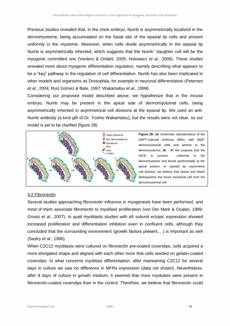
Extracellular matrix and integrins influence in the regulation of myogenic precursor cells behaviour
Raquel Rodrigues Vaz 2009 24
Previous studies revealed that, in the chick embryo, Numb is asymmetrically localized in the
dermomyotome, being accumulated on the basal site of the epaxial lip cells and present
uniformly in the myotome. Moreover, when cells divide asymmetrically in the epaxial lip
Numb is asymmetrically inherited, which suggests that the Numb+ daughter cell will be the
myogenic committed one (Venters & Ordahl, 2005; Holowacz et al., 2006). These studies
revealed more about myogenic differentiation regulation, namely describing what appears to
be a “key” pathway in the regulation of cell differentiation. Numb has also been implicated in
other models and organisms as Drosophila, for example in neuronal differentiation (Petersen
et al., 2004; Ruiz Gómez & Bate, 1997; Wakamatsu et al., 1999).
Considering our proposed model described above, we hypothesize that in the mouse
embryo, Numb may be present in the apical side of dermomyotomal cells, being
asymmetrically inherited in asymmetrical cell divisions at the epaxial lip. We used an anti-
Numb antibody (a kind gift of Dr. Yoshio Wakamatsu), but the results were not clear, so our
model is yet to be clarified (figure 28).
4.2 Fibronectin
Several studies approaching fibronectin influence in myogenesis have been performed, and
most of them associate fibronectin to myoblast proliferation (von Der Mark & Ocalan, 1989;
Grossi et al., 2007). In quail myoblasts studies with α5 subunit ectopic expression showed
increased proliferation and differentiation inhibition even in confluent cells, although they
concluded that the surrounding environment (growth factors present,…) is important as well
(Sastry et al., 1996).
When C2C12 myoblasts were cultured on fibronectin pre-coated coverslips, cells acquired a
more elongated shape and aligned with each other more that cells seeded on gelatin-coated
coverslips. In what concerns myoblast differentiation, after maintaining C2C12 for several
days in culture we saw no difference in MFRs expression (data not shown). Nevertheless,
after 8 days of culture in growth medium, it seemed that more myotubes were present in
fibronectin-coated coverslips than in the control. Therefore, we believe that fibronectin could
Figure 28: (A) Schematic representation of the
DAPT-cultured embryos effect, with Myf5+
dermomyotomal cells and laminin in the
dermomyotome; (B – F) We propose that the
NICD is present uniformly in the
dermomyotome and Numb preferentially at the
apical portion. In epaxial lip asymmetric
cell division, we believe that Numb and Notch
distinguishes the future myotomal cell from the
dermomyotomal cell.
Extracellular matrix and integrins influence in the regulation of myogenic precursor cells behaviour
Raquel Rodrigues Vaz 2009 25
enhance cell alignment to prepare them for fusion, although no enhanced differentiation is
observed.
When the nuclear shape was compared, we found that the nuclei of cells seeded on
fibronectin were more elliptical than those of cells seeded on gelatin. Using the time-lapse
image acquisition we found that fibronectin promotes cell migration and that cells on
fibronectin have a more predictable path than cells seeded on gelatine. All these results
made us hypothesize that fibronectin is responsible for early myoblasts alignment but also for
cell migration.
We then investigated which integrins, from those which interact with fibronectin, were present
in cells growing on fibronectin and could be responsible for this effect. Several studies have
been performed to understand if fibronectin is important for the migratory behaviour.
Depending on the cells and the experimental methodology used, in some cases α5 integrin
subunit is not important for cell migration or invasiveness, in other cells the addition of anti-α5
or anti-β1 blocking antibodies do indeed affect cell migration (reviewed by Akiyama et al.,
1995; Truong & Danen, 2009). Our results revealed that early in culture α5 integrin subunit is
present in cells growing on both a fibronectin and a gelatin matrix and that it is maintained
throughout the culture time. Our hypothesis so far was that fibronectin, mediated by α5β1
integrin interaction, would make cells elongate and align with each other. This interaction
would possibly remodel the actin cytoskeleton and increasing the N-cadherin in the
membrane (Huttenlocher et al., 1998). To test if α5β1 integrin was indeed important to the
alignment, we used the RGD fragment known to block fibronectin-α5β1 integrin interaction
(Takahashi et al., 2007) on cells seeded on fibronectin pre-coated coverslips. Although RGD
blocks fibronectin interaction with α5β1 integrin and αV integrin subunit, it is known that
fibronectin has another binding site for αV (Takahashi et al., 2007). The FN-α4β1 integrin
interaction is not affected because this integrin binds to fibronectin in another domain of the
ECM molecule (IIICS domain) (Sechler et al., 2000). Therefore, by adding RGD to the culture
medium, we are affecting only FN-α5β1 interaction. When we cultured cells on fibronectin
with RGD at the moment of seeding, we noticed that only a few cells would effectively attach
to the coverslip and those those that could attach were not elongated as the control cells.
After 2 days of culture, we observed that those cells managed to proliferate but still they were
not as aligned as those seeded with the control peptide. We believe that this could be due to
a change in integrins that were expressed, so they could attach to the surface (fibronectin
matrix) by other receptors. When we compared the nuclear Major/Minor ratios between them,
we found a statistically significant difference between nuclei cultured with RGD and the
control, with the first ones being more rounded that the control nuclei. In this experiment we
also added RGD one day after culture, when cells were around 80% confluent, but in this
case we did not perturb cell elongation. In fact, the nuclei of these cells were not different
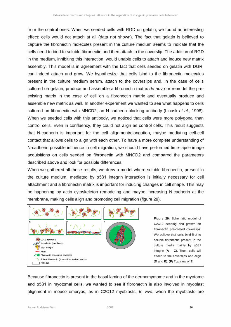
Extracellular matrix and integrins influence in the regulation of myogenic precursor cells behaviour
Raquel Rodrigues Vaz 2009 26
from the control ones. When we seeded cells with RGD on gelatin, we found an interesting
effect: cells would not attach at all (data not shown). The fact that gelatin is believed to
capture the fibronectin molecules present in the culture medium seems to indicate that the
cells need to bind to soluble fibronectin and then attach to the coverslip. The addition of RGD
in the medium, inhibiting this interaction, would unable cells to attach and induce new matrix
assembly. This model is in agreement with the fact that cells seeded on gelatin with DGR,
can indeed attach and grow. We hypothesize that cells bind to the fibronectin molecules
present in the culture medium serum, attach to the coverslips and, in the case of cells
cultured on gelatin, produce and assemble a fibronectin matrix de novo or remodel the pre-
existing matrix in the case of cell on a fibronectin matrix and eventually produce and
assemble new matrix as well. In another experiment we wanted to see what happens to cells
cultured on fibronectin with MNCD2, an N-cadherin blocking antibody (Linask et al., 1998).
When we seeded cells with this antibody, we noticed that cells were more polygonal than
control cells. Even in confluency, they could not align as control cells. This result suggests
that N-cadherin is important for the cell alignment/elongation, maybe mediating cell-cell
contact that allows cells to align with each other. To have a more complete understanding of
N-cadherin possible influence in cell migration, we should have performed time-lapse image
acquisitions on cells seeded on fibronectin with MNCD2 and compared the parameters
described above and look for possible differences.
When we gathered all these results, we drew a model where soluble fibronectin, present in
the culture medium, mediated by α5β1 integrin interaction is initially necessary for cell
attachment and a fibronectin matrix is important for inducing changes in cell shape. This may
be happening by actin cytoskeleton remodeling and maybe increasing N-cadherin at the
membrane, making cells align and promoting cell migration (figure 29).
Because fibronectin is present in the basal lamina of the dermomyotome and in the myotome
and α5β1 in myotomal cells, we wanted to see if fibronectin is also involved in myoblast
alignment in mouse embryos, as in C2C12 myoblasts. In vivo, when the myoblasts are
Figure 29: Schematic model of
C2C12 seeding and growth on
fibronectin pre-coated coverslips.
We believe that cells bind first to
soluble fibronectin present in the
culture media mainly by α5β1
integrin (A – C). Then, cells will
attach to the coverslips and align
(D and E). (F) Top view of E.
Extracellular matrix and integrins influence in the regulation of myogenic precursor cells behaviour
Raquel Rodrigues Vaz 2009 27
specified and move towards the medial portion of the myotome, they align along the antero-
posterior axis. When we cultured E9.0 and E10.0 mouse embryos for 6 hours with both the
70kDa fragment and RGD, we could not see any difference in myoblasts orientation. This
result is, in fact, not as surprising as it looked initially because myoblasts at this stage are
surrounded by a complex matrix (like laminin, fibronectin, collagen and perlecan; Adams &
Watt, 1993). We also cultured embryos in these developmental stages for 12 hours but
70kDa fragment-cultured embryos showed higher apoptosis levels so they were not used for
analysis. Nevertheless, in embryos cultured 6 hours with the 70kDa fragment we observed
that N-cadherin had lost its polarized location, i.e., it would be present evenly in
dermomyotomal cells instead of being restricted to the apical domain. Furthermore,
immunostaining for ZO-1 showed that the localization of this tight junction protein was slightly
perturbed although not as much as N-cadherin, because it is still present in the apical portion
of the dermomyotomal but forming a more discontinuous line as compared to control
embryos. This result is similar to the effect of the 70kDa fragment on chick somitogenesis,
where the fragment inhibits new somites formation and has an effect on the distribution of N-
cadherin (Martins et al, in press). The loss of N-cadherin polarization in the 70kDa cultured
embryos may indicate that fibronectin is important for epithelia maintenance.
We then used mouse embryos in later stages of development, E12 and E12.5, cultured for 6
and 14 hours respectively, since it was previously reported that, at E12.5, trunk myocytes
downregulate laminin-receptors and only express α1, α4, α5 and αV integrin subunits. It was
suggested that fibronectin could be important for myocytes correct alignment for fusion, the
following process in myogenesis (Cachaço et al., 2005). As before, we could not find an
effect in myocytes alignment. We analysed myotome-derived pre-muscle masses, those
derived from dermomyotomal cells that differentiated into skeletal muscle precursors and
occupied the myotome. In these cultures we could not find a visible difference in myocytes
alignment and orientation as well. This could be due to the fact that we used the same
concentration of the 70kDa fragment and RGD as in younger embryos which may by
insufficient because E12.0 mouse embryos have a much more complex structure and cell
density. In future experiments will definitely include the increase of the fragment and peptide
concentration in E12.0 or E12.5 cultured embryos.
Nevertheless, when we cultured E9.0 for 12 hours with RGD, we detected in a few sections
of each RGD-cultured embryo Myf5 protein in the dermomyotome. We hypothesize that
fibronectin, mediated by α5β1 integrin interaction, may be important in the maintenance of
undifferentiated dermomyotomal cells.
In summary, in this work we were able to unveil a little more about the extracellular matrix
influence in myogenesis, namely in myogenic precursor cells determination or specification
Extracellular matrix and integrins influence in the regulation of myogenic precursor cells behaviour
Raquel Rodrigues Vaz 2009 28
and behaviour. Using C2C12 myoblasts we found that laminin does not enhance cell
differentiation and in vivo we descibed that Notch signaling pathway inhibition leads to
myogenic differentiation of dermomyotomal cells, seen by Myf5 expression. Now we
hypothesize that Notch signaling may be interacting with laminin-α6β1 signaling, probably
mediated by Numb. Furthermore, we found that α5β1 integrin, mediated by fibronectin
interaction, may be important in myogenic differentiation, namely in the maintenance of
undifferentiated dermomyotomal cells.
Fibronectin seems to be essential for C2C12 myoblasts alignment, mediated by α5β1
integrin interaction. Furthermore, this ECM molecule is important for myoblast migration.
Although we did not detect any differences in MRFs expression, it seems that fibronectin, by
making cells align, may be conferring cells the physical properties necessary to fuse, as we
saw more myotubes on cells seeded on fibronectin than in cells seeded on laminin or gelatin,
after 8 days of culture with growth medium. We also tried to see if fibronectin is important in
similar process in vivo, in myoblasts and myocytes alignment and consequently fusion.
Perturbing both fibronectin fibrillogenesis and fibronectin-α5β1 integrin interaction, we could
not find an effect in cell alignment but we described that fibronectin is important for N-
cadherin polarization and general epithelial maintenance, namely in the dermomyotome.
Extracellular matrix and integrins influence in the regulation of myogenic precursor cells behaviour
Raquel Rodrigues Vaz 2009 29
5. References
Adams, J. C., and Watt, F. M. (1993). Regulation of development and differentiation by the extracellular matrix. Development (Cambridge, England) 117, 1183-98.
Akiyama, S. K., Olden, K., and Yamada, K. M. (1995). Fibronectin and integrins in invasion and metastasis. Cancer metastasis reviews 14, 173-89.
Bajanca, F., Luz, M., Duxson, M. J., and Thorsteinsdóttir, S. (2004). Integrins in the mouse myotome: developmental changes and differences between the epaxial and hypaxial lineage. Developmental dynamics : an official publication of the American Association of Anatomists 231, 402-15.
Bajanca, F., Luz, M., Raymond, K., Martins, G. G., Sonnenberg, A., Tajbakhsh, S., Buckingham, M., and Thorsteinsdóttir, S. (2006). Integrin alpha6beta1-laminin interactions regulate early myotome formation in the mouse embryo. Development (Cambridge, England) 133, 1635-44.
Berdnik, D., Török, T., González-Gaitán, M., and Knoblich, J. A. (2002). The endocytic protein alpha-Adaptin is required for numb-mediated asymmetric cell division in Drosophila. Developmental cell 3, 221-31.
Bischoff, R., and Heintz, C. (1994). Enhancement of skeletal muscle regeneration. Developmental dynamics : an official publication of the American Association of Anatomists 201, 41-54.
Bober, E., Lyons, G. E., Braun, T., Cossu, G., Buckingham, M., and Arnold, H. H. (1991). The muscle regulatory gene, Myf-6, has a biphasic pattern of expression during early mouse development. The Journal of cell biology 113, 1255-65.
Boettiger, D., Enomoto-Iwamoto, M., Yoon, H. Y., Hofer, U., Menko, A. S., and Chiquet-Ehrismann, R. (1995). Regulation of integrin alpha 5 beta 1 affinity during myogenic differentiation. Developmental biology 169, 261-72.
Braun, T., Bober, E., Rudnicki, M. A., Jaenisch, R., and Arnold, H. H. (1994). MyoD expression marks the onset of skeletal myogenesis in Myf-5 mutant mice. Development (Cambridge, England) 120, 3083-92.
Brent, A. E., and Tabin, C. J. (2002). Developmental regulation of somite derivatives: muscle, cartilage and tendon. Current opinion in genetics & development 12, 548-57.
Buas, M. F., Kabak, S., and Kadesch, T. (2009). Inhibition of myogenesis by Notch: evidence for multiple pathways. Journal of cellular physiology 218, 84-93.
Buckingham, M. (2007). Skeletal muscle progenitor cells and the role of Pax genes. Comptes rendus biologies 330, 530-3. Burattini, S., Ferri, P., Battistelli, M., Curci, R., Luchetti, F., and Falcieri, E. (2004). C2C12 murine myoblasts as a model of
skeletal muscle development: morpho-functional characterization. European journal of histochemistry : EJH 48, 223-33.
Cachaço, A. S., Chuva De Sousa Lopes, S. M., Kuikman, I., Bajanca, F., Abe, K., Baudoin, C., Sonnenberg, A., Mummery, C. L., and Thorsteinsdóttir, S. (2003). Knock-in of integrin beta 1D affects primary but not secondary myogenesis in mice. Development (Cambridge, England) 130, 1659-71.
Cachaço, A. S., Pereira, C. S., Pardal, R. G., Bajanca, F., and Thorsteinsdóttir, S. (2005). Integrin repertoire on myogenic cells changes during the course of primary myogenesis in the mouse. Developmental dynamics : an official publication of the American Association of Anatomists 232, 1069-78.
Cheng, X., Huber, T. L., Chen, V. C., Gadue, P., and Keller, G. M. (2008). Numb mediates the interaction between Wnt and Notch to modulate primitive erythropoietic specification from the hemangioblast. Development (Cambridge, England) 135, 3447-58.
Christ, B., Schmidt, C., Huang, R., Wilting, J., and Brand-Saberi, B. (1998). Segmentation of the vertebrate body. Anatomy and embryology 197, 1-8.
Conboy, I. M., and Rando, T. A. (2002). The regulation of Notch signaling controls satellite cell activation and cell fate determination in postnatal myogenesis. Developmental cell 3, 397-409.
Conlon, R. a., Reaume, a. G., and Rossant, J. (1995). Notch1 is required for the coordinate segmentation of somites. Development (Cambridge, England) 121, 1533-45.
Corbin, V., Michelson, A. M., Abmayr, S. M., Neel, V., Alcamo, E., Maniatis, T., and Young, M. W. (1991). A role for the Drosophila neurogenic genes in mesoderm differentiation. Cell 67, 311-23.
Cossu, G. et al. (2000). Determination, diversification and multipotency of mammalian myogenic cells. The International journal of developmental biology 44, 699-706.
Cossu, G., Kelly, R., Tajbakhsh, S., Di Donna, S., Vivarelli, E., and Buckingham, M. (1996). Activation of different myogenic pathways: myf-5 is induced by the neural tube and MyoD by the dorsal ectoderm in mouse paraxial mesoderm. Development (Cambridge, England) 122, 429-37.
Cossu, G., Tajbakhsh, S., and Buckingham, M. (1996). How is myogenesis initiated in the embryo? Trends in genetics : TIG 12, 218-23.
Cossu, G., and Tajbakhsh, S. (2007). Oriented cell divisions and muscle satellite cell heterogeneity. Cell 129, 859-61. Danen, E. H., and Sonnenberg, A. (2003). Integrins in regulation of tissue development and function. The Journal of pathology
201, 632-41. Darribère, T., Skalski, M., Cousin, H. L., Gaultier, A., Montmory, C., and Alfandari, D. (2000). Integrins: regulators of
embryogenesis. Biology of the cell / under the auspices of the European Cell Biology Organization 92, 5-25. Delfini, M. C., Hirsinger, E., Pourquié, O., and Duprez, D. (2000). Delta 1-activated notch inhibits muscle differentiation without
affecting Myf5 and Pax3 expression in chick limb myogenesis. Development (Cambridge, England) 127, 5213-24. Denetclaw, W. F., Christ, B., and Ordahl, C. P. (1997). Location and growth of epaxial myotome precursor cells. Development
(Cambridge, England) 124, 1601-10. Denetclaw, W. F., and Ordahl, C. P. (2000). The growth of the dermomyotome and formation of early myotome lineages in
thoracolumbar somites of chicken embryos. Development (Cambridge, England) 127, 893-905. Duband, J. L., Dufour, S., Hatta, K., Takeichi, M., Edelman, G. M., and Thiery, J. P. (1987). Adhesion molecules during
somitogenesis in the avian embryo. The Journal of cell biology 104, 1361-74. Frise, E., Knoblich, J. a., Younger-Shepherd, S., Jan, L. Y., and Jan, Y. N. (1996). The Drosophila Numb protein inhibits
signaling of the Notch receptor during cell-cell interaction in sensory organ lineage. Proceedings of the National Academy of Sciences of the United States of America 93, 11925-32.
Giancotti, F. G., and Ruoslahti, E. (1999). Integrin signaling. Science (New York, N.Y.) 285, 1028-32. Gibb, S., Zagorska, A., Melton, K., Tenin, G., Vacca, I., Trainor, P., Maroto, M., and Dale, J. K. (2009). Interfering with Wnt
signalling alters the periodicity of the segmentation clock. Developmental biology 330, 21-31. González-Mariscal, L., Betanzos, a., Nava, P., and Jaramillo, B. E. (2003). Tight junction proteins. Progress in biophysics and
molecular biology 81, 1-44.
Extracellular matrix and integrins influence in the regulation of myogenic precursor cells behaviour
Raquel Rodrigues Vaz 2009 30
Grossi, A., Yadav, K., and Lawson, M. a. (2007). Mechanical stimulation increases proliferation, differentiation and protein expression in culture: stimulation effects are substrate dependent. Journal of biomechanics 40, 3354-62.
Grounds, M. D., and McGeachie, J. K. (1987). A model of myogenesis in vivo, derived from detailed autoradiographic studies of regenerating skeletal muscle, challenges the concept of quantal mitosis. Cell and tissue research 250, 563-9.
Guo, M., Jan, L. Y., and Jan, Y. N. (1996). Control of daughter cell fates during asymmetric division: interaction of Numb and Notch. Neuron 17, 27-41.
Hashimoto, J., Kariya, Y., and Miyazaki, K. (2006). Regulation of proliferation and chondrogenic differentiation of human mesenchymal stem cells by laminin-5 (laminin-332). Stem cells (Dayton, Ohio) 24, 2346-54.
Hasty, P., Bradley, A., Morris, J. H., Edmondson, D. G., Venuti, J. M., Olson, E. N., and Klein, W. H. (1993). Muscle deficiency and neonatal death in mice with a targeted mutation in the myogenin gene. Nature 364, 501-6.
Hayashi, Y. et al. (2007). Integrins regulate mouse embryonic stem cell self-renewal. Stem cells (Dayton, Ohio) 25, 3005-15. Hinterberger, T. J., Sassoon, D. A., Rhodes, S. J., and Konieczny, S. F. (1991). Expression of the muscle regulatory factor
MRF4 during somite and skeletal myofiber development. Developmental biology 147, 144-56. Hirsinger, E., Malapert, P., Dubrulle, J., Delfini, M. C., Duprez, D., Henrique, D., Ish-Horowicz, D., and Pourquié, O. (2001).
Notch signalling acts in postmitotic avian myogenic cells to control MyoD activation. Development (Cambridge, England) 128, 107-16.
Holowacz, T., Zeng, L., and Lassar, A. B. (2006). Asymmetric localization of numb in the chick somite and the influence of myogenic signals. Developmental dynamics : an official publication of the American Association of Anatomists 235, 633-45.
Huttenlocher, A., Lakonishok, M., Kinder, M., Wu, S., Truong, T., Knudsen, K. A., and Horwitz, A. F. (1998). Integrin and cadherin synergy regulates contact inhibition of migration and motile activity. The Journal of cell biology 141, 515-26.
Hynes, R. O. (1992). Integrins: versatility, modulation, and signaling in cell adhesion. Cell 69, 11-25. Hynes, R. O. (1996). Targeted mutations in cell adhesion genes: what have we learned from them? Developmental biology 180,
402-12. Jaffredo, T., Horwitz, a. F., Buck, C. a., Rong, P. M., and Dieterlen-Lievre, F. (1988). Myoblast migration specifically inhibited in
the chick embryo by grafted CSAT hybridoma cells secreting an anti-integrin antibody. Development (Cambridge, England) 103, 431-46.
Jones, J. L., and Walker, R. a. (1999). Integrins: a role as cell signalling molecules. Molecular pathology : MP 52, 208-13. Jostes, B., Walther, C., and Gruss, P. (1990). The murine paired box gene, Pax7, is expressed specifically during the
development of the nervous and muscular system. Mechanisms of development 33, 27-37. Kablar, B., Krastel, K., Ying, C., Asakura, A., Tapscott, S. J., and Rudnicki, M. A. (1997). MyoD and Myf-5 differentially regulate
the development of limb versus trunk skeletal muscle. Development (Cambridge, England) 124, 4729-38. Kassar-Duchossoy, L., Giacone, E., Gayraud-Morel, B., Jory, A., Gomès, D., and Tajbakhsh, S. (2005). Pax3/Pax7 mark a
novel population of primitive myogenic cells during development. Genes & development 19, 1426-31. Kaufman, S. J., and Foster, R. F. (1988). Replicating myoblasts express a muscle-specific phenotype. Proceedings of the
National Academy of Sciences of the United States of America 85, 9606-10. Kopan, R., Nye, J. S., and Weintraub, H. (1994). The intracellular domain of mouse Notch: a constitutively activated repressor of
myogenesis directed at the basic helix-loop-helix region of MyoD. Development (Cambridge, England) 120, 2385-96. Kopan, R., Schroeter, E. H., Weintraub, H., and Nye, J. S. (1996). Signal transduction by activated mNotch: importance of
proteolytic processing and its regulation by the extracellular domain. Proceedings of the National Academy of Sciences of the United States of America 93, 1683-8.
Kuang, S., and Rudnicki, M. A. (2008). The emerging biology of satellite cells and their therapeutic potential. Trends in molecular medicine 14, 82-91.
Kuroda, K., Tani, S., Tamura, K., Minoguchi, S., Kurooka, H., and Honjo, T. (1999). Delta-induced Notch signaling mediated by RBP-J inhibits MyoD expression and myogenesis. The Journal of biological chemistry 274, 7238-44.
Lathia, J. D., Rao, M. S., Mattson, M. P., and Ffrench-Constant, C. (2007). The microenvironment of the embryonic neural stem cell: lessons from adult niches? Developmental dynamics : an official publication of the American Association of Anatomists 236, 3267-82.
Linask, K. K., Ludwig, C., Han, M. D., Liu, X., Radice, G. L., and Knudsen, K. A. (1998). N-cadherin/catenin-mediated morphoregulation of somite formation. Developmental biology 202, 85-102.
Linask, K. K., Ludwig, C., Han, M. D., Liu, X., Radice, G. L., and Knudsen, K. a. (1998). N-cadherin/catenin-mediated morphoregulation of somite formation. Developmental biology 202, 85-102.
Lyons, G. E., Ontell, M., Cox, R., Sassoon, D., and Buckingham, M. (1990). The expression of myosin genes in developing skeletal muscle in the mouse embryo. The Journal of cell biology 111, 1465-76.
Mao, Y., and Schwarzbauer, J. E. (2005). Fibronectin fibrillogenesis, a cell-mediated matrix assembly process. Matrix biology : journal of the International Society for Matrix Biology 24, 389-99.
Mayer, U., Saher, G., Fässler, R., Bornemann, A., Echtermeyer, F., von Der Mark, H., Miosge, N., Pöschl, E., and von Der Mark, K. (1997). Absence of integrin alpha 7 causes a novel form of muscular dystrophy. Nature genetics 17, 318-23.
McKeown-Longo, P. J., and Mosher, D. F. (1985). Interaction of the 70,000-mol-wt amino-terminal fragment of fibronectin with the matrix-assembly receptor of fibroblasts. The Journal of cell biology 100, 364-74.
Nabeshima, Y., Hanaoka, K., Hayasaka, M., Esumi, E., Li, S., and Nonaka, I. (1993). Myogenin gene disruption results in perinatal lethality because of severe muscle defect. Nature 364, 532-5.
Nofziger, D., Miyamoto, A., Lyons, K. M., and Weinmaster, G. (1999). Notch signaling imposes two distinct blocks in the differentiation of C2C12 myoblasts. Development (Cambridge, England) 126, 1689-702.
Ordahl, C. P., and Le Douarin, N. M. (1992). Two myogenic lineages within the developing somite. Development (Cambridge, England) 114, 339-53.
Ott, M. O., Bober, E., Lyons, G., Arnold, H., and Buckingham, M. (1991). Early expression of the myogenic regulatory gene, myf-5, in precursor cells of skeletal muscle in the mouse embryo. Development (Cambridge, England) 111, 1097-107.
Parker, M. H., Seale, P., and Rudnicki, M. A. (2003). Looking back to the embryo: defining transcriptional networks in adult myogenesis. Nature reviews. Genetics 4, 497-507.
Patton, B. L. (2000). Laminins of the neuromuscular system. Microscopy research and technique 51, 247-61. Paulin, D., and Li, Z. (2004). Desmin: a major intermediate filament protein essential for the structural integrity and function of
muscle. Experimental cell research 301, 1-7. Petersen, P. H., Zou, K., Krauss, S., and Zhong, W. (2004). Continuing role for mouse Numb and Numbl in maintaining
progenitor cells during cortical neurogenesis. Nature neuroscience 7, 803-11. Pourquié, O. (2001). Vertebrate somitogenesis. Annual review of cell and developmental biology 17, 311-50.
Extracellular matrix and integrins influence in the regulation of myogenic precursor cells behaviour
Raquel Rodrigues Vaz 2009 31
Relaix, F., Rocancourt, D., Mansouri, A., and Buckingham, M. (2005). A Pax3/Pax7-dependent population of skeletal muscle progenitor cells. Nature 435, 948-53.
Rifes, P., Carvalho, L., Lopes, C., Andrade, R. P., Rodrigues, G., Palmeirim, I., and Thorsteinsdóttir, S. (2007). Redefining the role of ectoderm in somitogenesis: a player in the formation of the fibronectin matrix of presomitic mesoderm. Development (Cambridge, England) 134, 3155-65.
Roegiers, F., and Jan, Y. N. (2004). Asymmetric cell division. Current opinion in cell biology 16, 195-205. Rosen, G. D., Sanes, J. R., LaChance, R., Cunningham, J. M., Roman, J., and Dean, D. C. (1992). Roles for the integrin VLA-4
and its counter receptor VCAM-1 in myogenesis. Cell 69, 1107-19. Ruiz Gómez, M., and Bate, M. (1997). Segregation of myogenic lineages in Drosophila requires numb. Development
(Cambridge, England) 124, 4857-66. Santolini, E., Puri, C., Salcini, A. E., Gagliani, M. C., Pelicci, P. G., Tacchetti, C., and Di Fiore, P. P. (2000). Numb is an
endocytic protein. The Journal of cell biology 151, 1345-52. Sasai, Y., Kageyama, R., Tagawa, Y., Shigemoto, R., and Nakanishi, S. (1992). Two mammalian helix-loop-helix factors
structurally related to Drosophila hairy and Enhancer of split. Genes & Development 6, 2620-2634. Sastry, S. K., Lakonishok, M., Thomas, D. a., Muschler, J., and Horwitz, a. F. (1996). Integrin alpha subunit ratios, cytoplasmic
domains, and growth factor synergy regulate muscle proliferation and differentiation. The Journal of cell biology 133, 169-84.
Schaart, G., Viebahn, C., Langmann, W., and Ramaekers, F. (1989). Desmin and titin expression in early postimplantation mouse embryos. Development (Cambridge, England) 107, 585-96.
Schultz, E. (1996). Satellite cell proliferative compartments in growing skeletal muscles. Developmental biology 175, 84-94. Schultz, E., Gibson, M. C., and Champion, T. (1978). Satellite cells are mitotically quiescent in mature mouse muscle: an EM
and radioautographic study. The Journal of experimental zoology 206, 451-6. Schultz, E., and McCormick, K. M. (1994). Skeletal muscle satellite cells. Reviews of physiology, biochemistry and
pharmacology 123, 213-57. Schwarzbauer, J. (1999). Basement membranes: Putting up the barriers. Current biology : CB 9, R242-4. Schwarzbauer, J. E., and Sechler, J. L. (1999). Fibronectin fibrillogenesis: a paradigm for extracellular matrix assembly. Current
opinion in cell biology 11, 622-7. Seale, P., Asakura, A., and Rudnicki, M. A. (2001). The potential of muscle stem cells. Developmental cell 1, 333-42. Sechler, J. L., Cumiskey, a. M., Gazzola, D. M., and Schwarzbauer, J. E. (2000). A novel RGD-independent fibronectin
assembly pathway initiated by alpha4beta1 integrin binding to the alternatively spliced V region. Journal of cell science 113 ( Pt 8, 1491-8.
Shawber, C., Nofziger, D., Hsieh, J. J., Lindsell, C., Bögler, O., Hayward, D., and Weinmaster, G. (1996). Notch signaling inhibits muscle cell differentiation through a CBF1-independent pathway. Development (Cambridge, England) 122, 3765-73.
Smith, T. H., Kachinsky, A. M., and Miller, J. B. (1994). Somite subdomains, muscle cell origins, and the four muscle regulatory factor proteins. The Journal of cell biology 127, 95-105.
Sonnenberg, A., Janssen, H., Hogervorst, F., Calafat, J., and Hilgers, J. (1987). A complex of platelet glycoproteins Ic and IIa identified by a rat monoclonal antibody. The Journal of biological chemistry 262, 10376-83.
Sunada, Y., and Yamadas, Y. (1994). Deficiency of Merosin in Dystrophic. Control. Takahashi, S. et al. (2007). The RGD motif in fibronectin is essential for development but dispensable for fibril assembly. The
Journal of cell biology 178, 167-78. Taverna, D., Disatnik, M. H., Rayburn, H., Bronson, R. T., Yang, J., Rando, T. a., and Hynes, R. O. (1998). Dystrophic muscle in
mice chimeric for expression of alpha5 integrin. The Journal of cell biology 143, 849-59. Truong, H., and Danen, E. H. (2009). Integrin switching modulates adhesion dynamics and cell migration. Cell adhesion &
migration 3, 179-81. Venters, S. J., and Ordahl, C. P. (2005). Asymmetric cell divisions are concentrated in the dermomyotome dorsomedial lip
during epaxial primary myotome morphogenesis. Anatomy and embryology 209, 449-60. Wakamatsu, Y., Maynard, T. M., Jones, S. U., and Weston, J. A. (1999). NUMB localizes in the basal cortex of mitotic avian
neuroepithelial cells and modulates neuronal differentiation by binding to NOTCH-1. Neuron 23, 71-81. Wallin, J., Wilting, J., Koseki, H., Fritsch, R., Christ, B., and Balling, R. (1994). The role of Pax-1 in axial skeleton development.
Development (Cambridge, England) 120, 1109-21. Weber, J. M., and Calvi, L. M. (2009). Notch signaling and the bone marrow hematopoietic stem cell niche. Bone. Williams, B. A., and Ordahl, C. P. (1994). Pax-3 expression in segmental mesoderm marks early stages in myogenic cell
specification. Development (Cambridge, England) 120, 785-96. Yurchenco, P. D., Amenta, P. S., and Patton, B. L. (2004). Basement membrane assembly, stability and activities observed
through a developmental lens. Matrix biology : journal of the International Society for Matrix Biology 22, 521-38. Yurchenco, P. D., and Wadsworth, W. G. (2004). Assembly and tissue functions of early embryonic laminins and netrins.
Current opinion in cell biology 16, 572-9. Yusuf, F., and Brand-Saberi, B. (2006). The eventful somite: patterning, fate determination and cell division in the somite.
Anatomy and embryology 211 Suppl, 21-30. Zagris, N. (2001). Extracellular matrix in development of the early embryo. Micron (Oxford, England : 1993) 32, 427-38. Zagris, N., Chung, a. E., and Stavridis, V. (2000). Differential expression of laminin genes in early chick embryo. The
International journal of developmental biology 44, 815-8. Zammit, P. S., Partridge, T. a., and Yablonka-Reuveni, Z. (2006). The skeletal muscle satellite cell: the stem cell that came in
from the cold. The journal of histochemistry and cytochemistry : official journal of the Histochemistry Society 54, 1177-91.
van der Flier, A., and Sonnenberg, A. (2001). Function and interactions of integrins. Cell and Tissue Research 305, 285-298. von Der Mark, K., and Ocalan, M. (1989). Antagonistic effects of laminin and fibronectin on the expression of the myogenic
phenotype. Differentiation; research in biological diversity 40, 150-7.
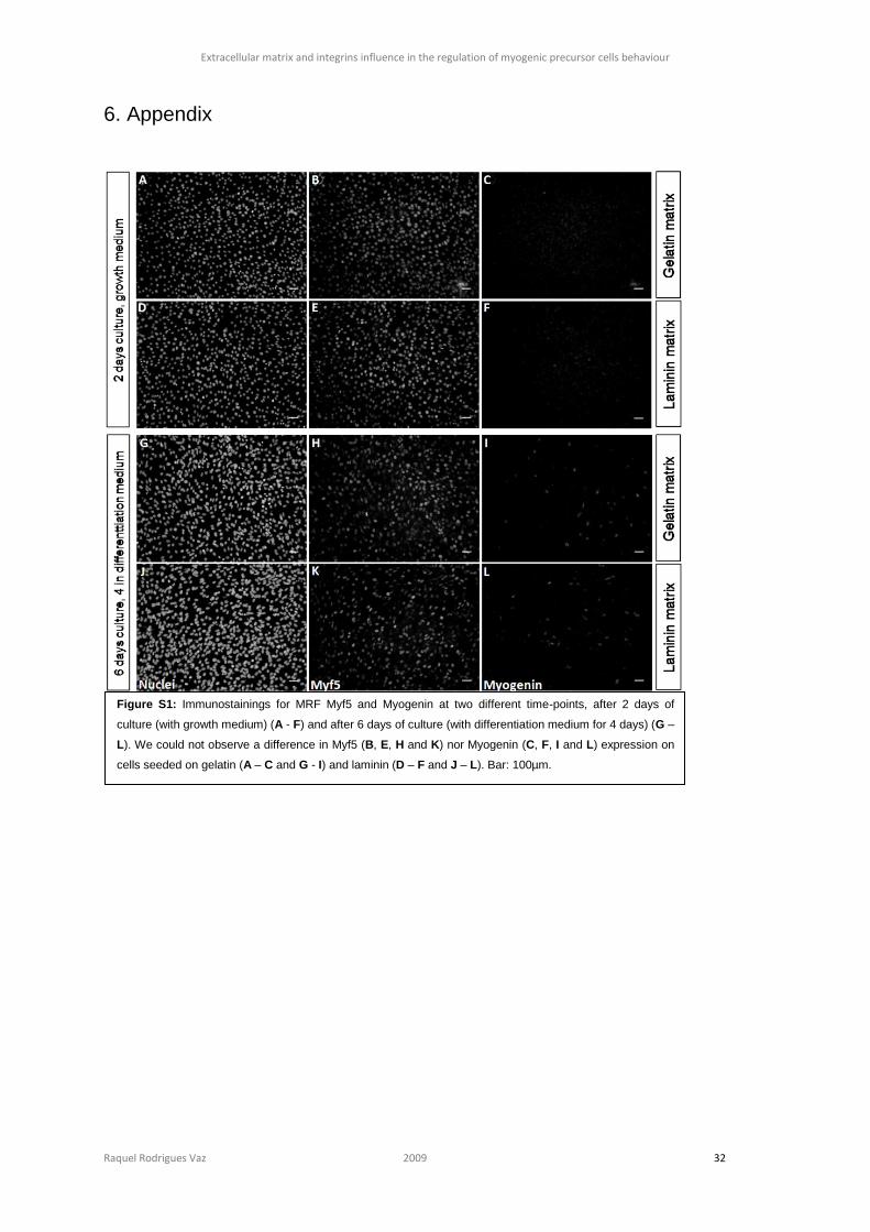
Extracellular matrix and integrins influence in the regulation of myogenic precursor cells behaviour
Raquel Rodrigues Vaz 2009 32
6. Appendix
Figure S1: Immunostainings for MRF Myf5 and Myogenin at two different time-points, after 2 days of
culture (with growth medium) (A - F) and after 6 days of culture (with differentiation medium for 4 days) (G –
L). We could not observe a difference in Myf5 (B, E, H and K) nor Myogenin (C, F, I and L) expression on
cells seeded on gelatin (A – C and G - I) and laminin (D – F and J – L). Bar: 100µm.
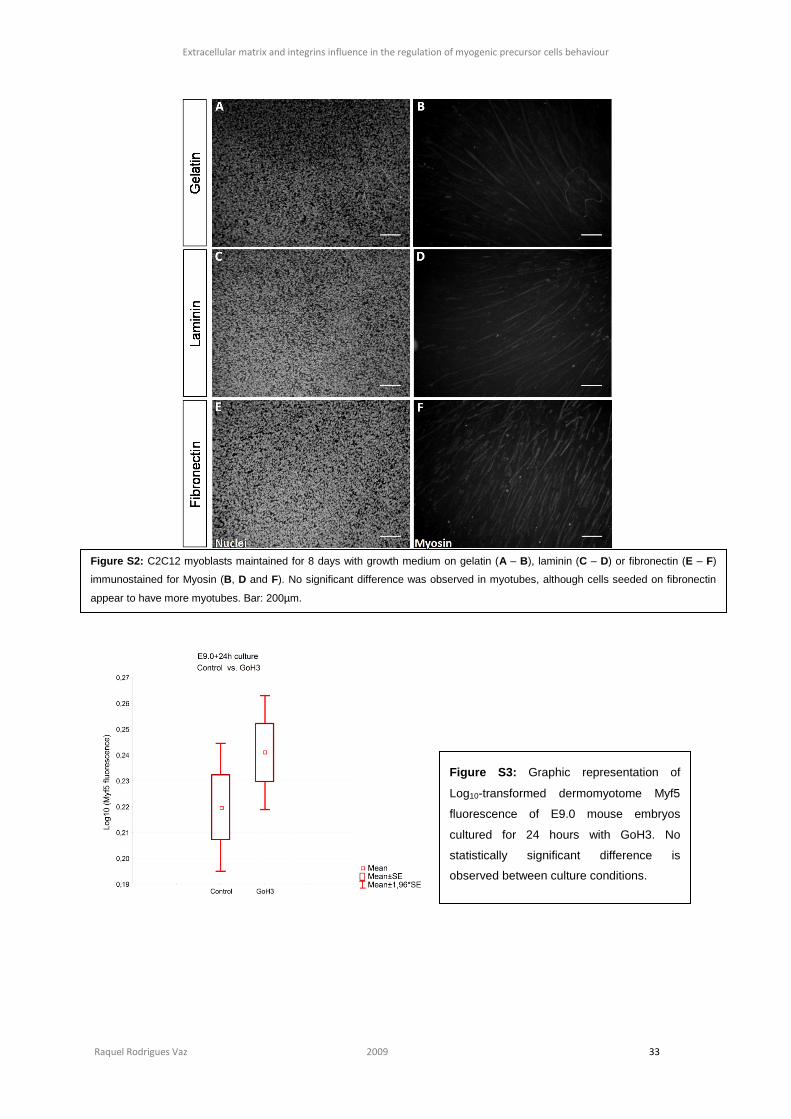
Extracellular matrix and integrins influence in the regulation of myogenic precursor cells behaviour
Raquel Rodrigues Vaz 2009 33
Figure S2: C2C12 myoblasts maintained for 8 days with growth medium on gelatin (A – B), laminin (C – D) or fibronectin (E – F)
immunostained for Myosin (B, D and F). No significant difference was observed in myotubes, although cells seeded on fibronectin
appear to have more myotubes. Bar: 200µm.
Figure S3: Graphic representation of
Log10-transformed dermomyotome Myf5
fluorescence of E9.0 mouse embryos
cultured for 24 hours with GoH3. No
statistically significant difference is
observed between culture conditions.
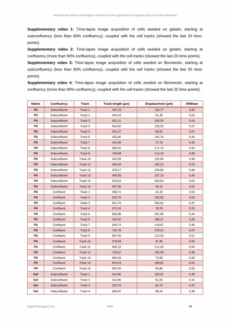
Extracellular matrix and integrins influence in the regulation of myogenic precursor cells behaviour
Raquel Rodrigues Vaz 2009 34
Supplementary video 1: Time-lapse image acquisition of cells seeded on gelatin, starting at
subconfluency (less than 60% confluency), coupled with the cell tracks (showed the last 20 time-
points).
Supplementary video 2: Time-lapse image acquisition of cells seeded on gelatin, starting at
confluency (more than 90% confluency), coupled with the cell tracks (showed the last 20 time-points).
Supplementary video 3: Time-lapse image acquisition of cells seeded on fibronectin, starting at
subconfluency (less than 60% confluency), coupled with the cell tracks (showed the last 20 time-
points).
Supplementary video 4: Time-lapse image acquisition of cells seeded on fibronectin, starting at
confluency (more than 90% confluency), coupled with the cell tracks (showed the last 20 time-points).
Matrix Confluency Track Track length (µm) Displacement (µm) ARMean
FN Subconfluent Track 1 425,78 136,77 0,50
FN Subconfluent Track 2 454,43 51,36 0,53
FN Subconfluent Track 3 401,21 105,35 0,54
FN Subconfluent Track 4 562,92 156,24 0,57
FN Subconfluent Track 5 551,27 88,91 0,57
FN Subconfluent Track 6 432,66 142,76 0,46
FN Subconfluent Track 7 444,38 37,78 0,45
FN Subconfluent Track 8 589,52 171,70 0,52
FN Subconfluent Track 9 799,68 113,18 0,55
FN Subconfluent Track 10 432,59 152,94 0,49
FN Subconfluent Track 11 440,29 182,22 0,53
FN Subconfluent Track 12 478,17 154,96 0,48
FN Subconfluent Track 13 465,58 237,13 0,49
FN Subconfluent Track 14 519,53 200,64 0,52
FN Subconfluent Track 15 547,36 46,12 0,52
FN Confluent Track 1 408,71 31,20 0,52
FN Confluent Track 2 549,70 103,83 0,53
FN Confluent Track 3 641,74 363,62 0,47
FN Confluent Track 4 672,24 78,78 0,55
FN Confluent Track 5 626,88 261,66 0,44
FN Confluent Track 6 442,92 186,37 0,36
FN Confluent Track 7 509,74 118,97 0,49
FN Confluent Track 8 742,78 279,21 0,57
FN Confluent Track 9 687,59 110,26 0,51
FN Confluent Track 10 278,83 97,46 0,50
FN Confluent Track 11 565,19 111,45 0,54
FN Confluent Track 12 733,67 265,08 0,38
FN Confluent Track 13 665,93 73,58 0,56
FN Confluent Track 14 819,43 438,62 0,53
FN Confluent Track 15 802,49 90,86 0,55
Gel Subconfluent Track 1 410,96 165,01 0,48
Gel Subconfluent Track 2 314,85 61,04 0,41
Gel Subconfluent Track 3 323,79 82,74 0,37
Gel Subconfluent Track 4 360,87 89,19 0,39
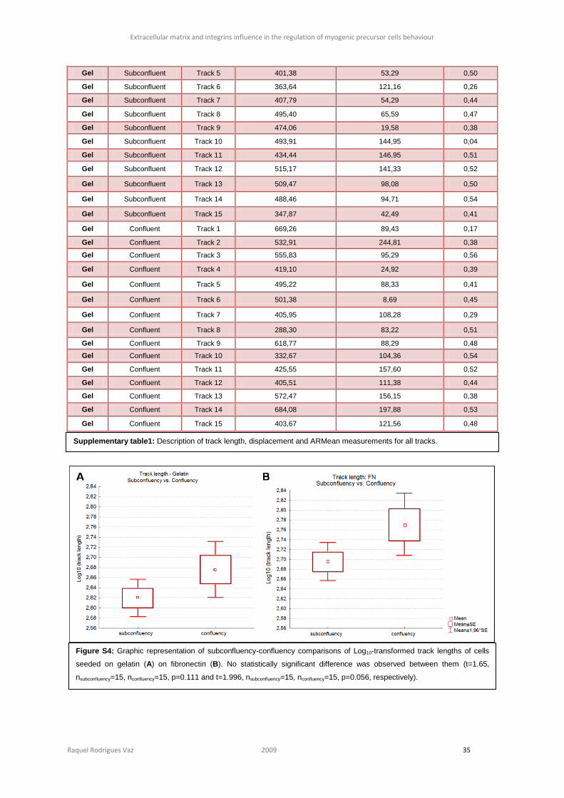
Extracellular matrix and integrins influence in the regulation of myogenic precursor cells behaviour
Raquel Rodrigues Vaz 2009 35
Gel Subconfluent Track 5 401,38 53,29 0,50
Gel Subconfluent Track 6 363,64 121,16 0,26
Gel Subconfluent Track 7 407,79 54,29 0,44
Gel Subconfluent Track 8 495,40 65,59 0,47
Gel Subconfluent Track 9 474,06 19,58 0,38
Gel Subconfluent Track 10 493,91 144,95 0,04
Gel Subconfluent Track 11 434,44 146,95 0,51
Gel Subconfluent Track 12 515,17 141,33 0,52
Gel Subconfluent Track 13 509,47 98,08 0,50
Gel Subconfluent Track 14 488,46 94,71 0,54
Gel Subconfluent Track 15 347,87 42,49 0,41
Gel Confluent Track 1 669,26 89,43 0,17
Gel Confluent Track 2 532,91 244,81 0,38
Gel Confluent Track 3 555,83 95,29 0,56
Gel Confluent Track 4 419,10 24,92 0,39
Gel Confluent Track 5 495,22 88,33 0,41
Gel Confluent Track 6 501,38 8,69 0,45
Gel Confluent Track 7 405,95 108,28 0,29
Gel Confluent Track 8 288,30 83,22 0,51
Gel Confluent Track 9 618,77 88,29 0,48
Gel Confluent Track 10 332,67 104,36 0,54
Gel Confluent Track 11 425,55 157,60 0,52
Gel Confluent Track 12 405,51 111,38 0,44
Gel Confluent Track 13 572,47 156,15 0,38
Gel Confluent Track 14 684,08 197,88 0,53
Gel Confluent Track 15 403,67 121,56 0,48
Supplementary table1: Description of track length, displacement and ARMean measurements for all tracks.
Figure S4: Graphic representation of subconfluency-confluency comparisons of Log10-transformed track lengths of cells
seeded on gelatin (A) on fibronectin (B). No statistically significant difference was observed between them (t=1.65,
nsubconfluency=15, nconfluency=15, p=0.111 and t=1.996, nsubconfluency=15, nconfluency=15, p=0.056, respectively).
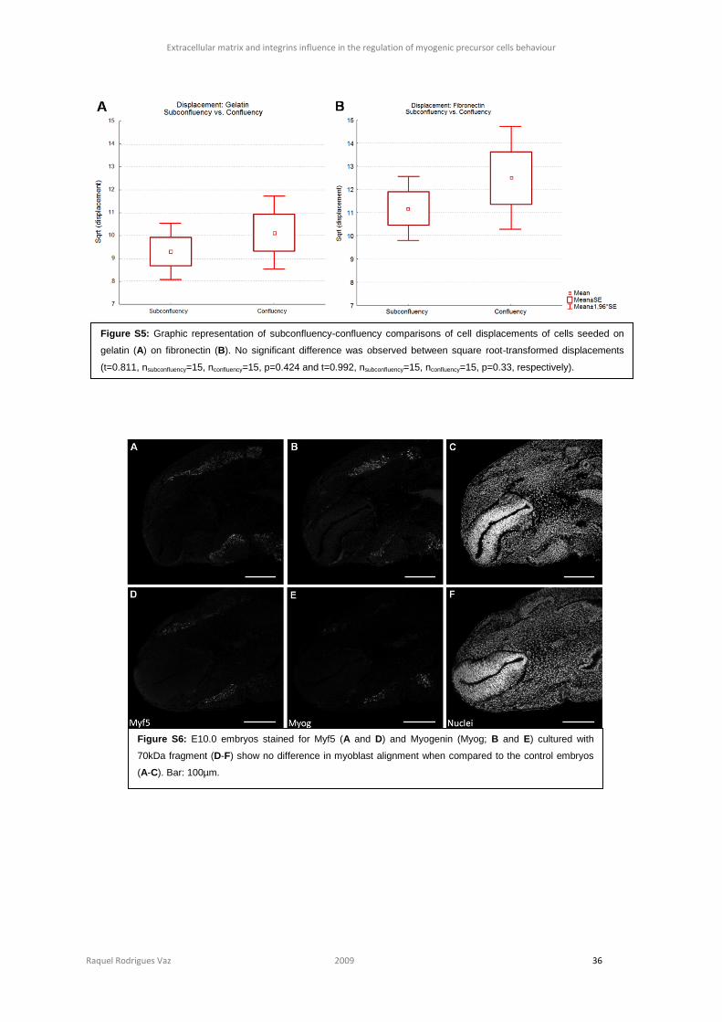
Extracellular matrix and integrins influence in the regulation of myogenic precursor cells behaviour
Raquel Rodrigues Vaz 2009 36
Figure S5: Graphic representation of subconfluency-confluency comparisons of cell displacements of cells seeded on
gelatin (A) on fibronectin (B). No significant difference was observed between square root-transformed displacements
(t=0.811, nsubconfluency=15, nconfluency=15, p=0.424 and t=0.992, nsubconfluency=15, nconfluency=15, p=0.33, respectively).
Figure S6: E10.0 embryos stained for Myf5 (A and D) and Myogenin (Myog; B and E) cultured with
70kDa fragment (D-F) show no difference in myoblast alignment when compared to the control embryos
(A-C). Bar: 100µm.
Extracellular matrix and integrins influence in the regulation of myogenic precursor cells behaviour
Raquel Rodrigues Vaz 2009 37
Figure S7: E12.0 embryos cultured with BSA (control; A-C) or the 70kDa fragment (D-I). When compared inter-
vertebrae myocytes location and alignment by Myf5 (A, D and G) and MHC (B, E and H) immnunostaining, we did
not detected any difference. Bar: 100µm.


![[CANCER RESEARCH 60, 712–721, February 1, 2000] Integrins ...cancerres.aacrjournals.org/content/canres/60/3/712.full.pdf · [CANCER RESEARCH 60, 712–721, February 1, 2000] Integrins](https://img.dokumen.tips/doc/110x75/5d288c9488c99392328c0bac/cancer-research-60-712721-february-1-2000-integrins-cancer-research.jpg)