Embed Size (px)
Citation preview
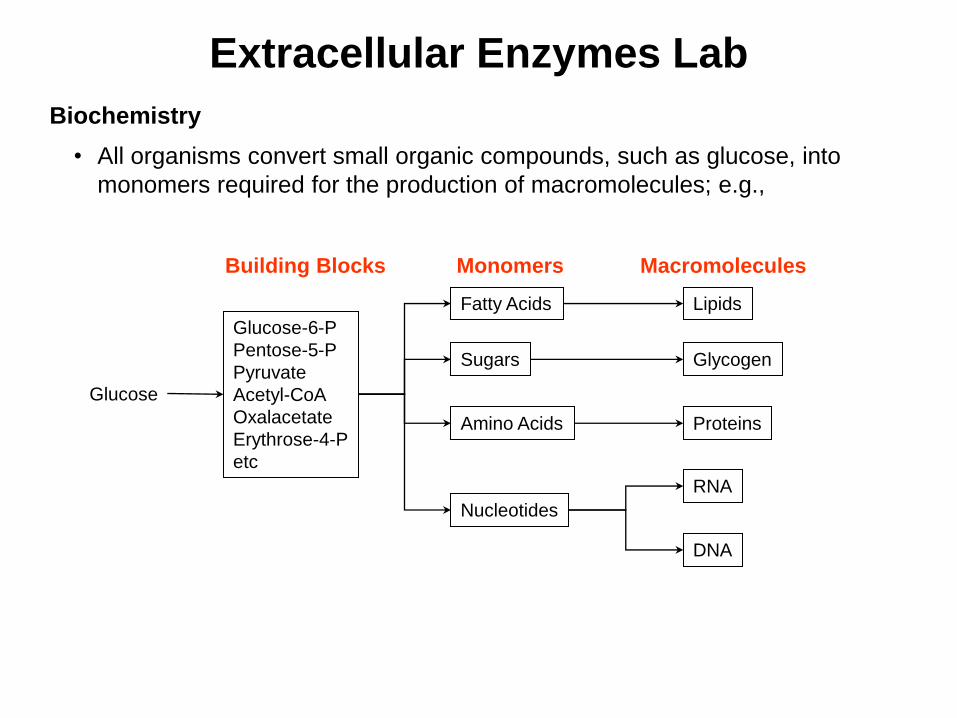
Extracellular Enzymes Lab
Biochemistry
• All organisms convert small organic compounds, such as glucose, into
monomers required for the production of macromolecules; e.g.,
Glucose
Glucose-6-P
Pentose-5-P
Pyruvate
Acetyl-CoA
Oxalacetate
Erythrose-4-P
etc
Fatty Acids
Sugars
Amino Acids
Nucleotides
Lipids
Glycogen
Proteins
RNA
DNA
Building Blocks Monomers Macromolecules
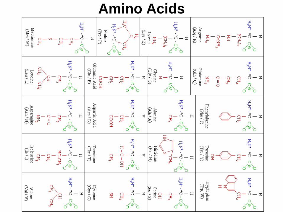
Amino Acids
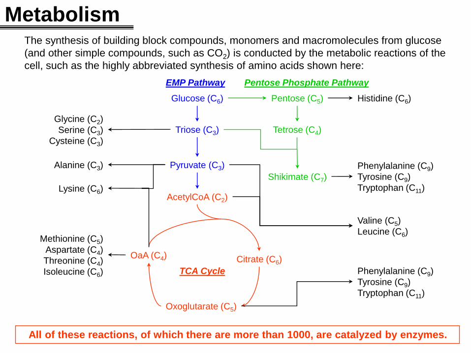
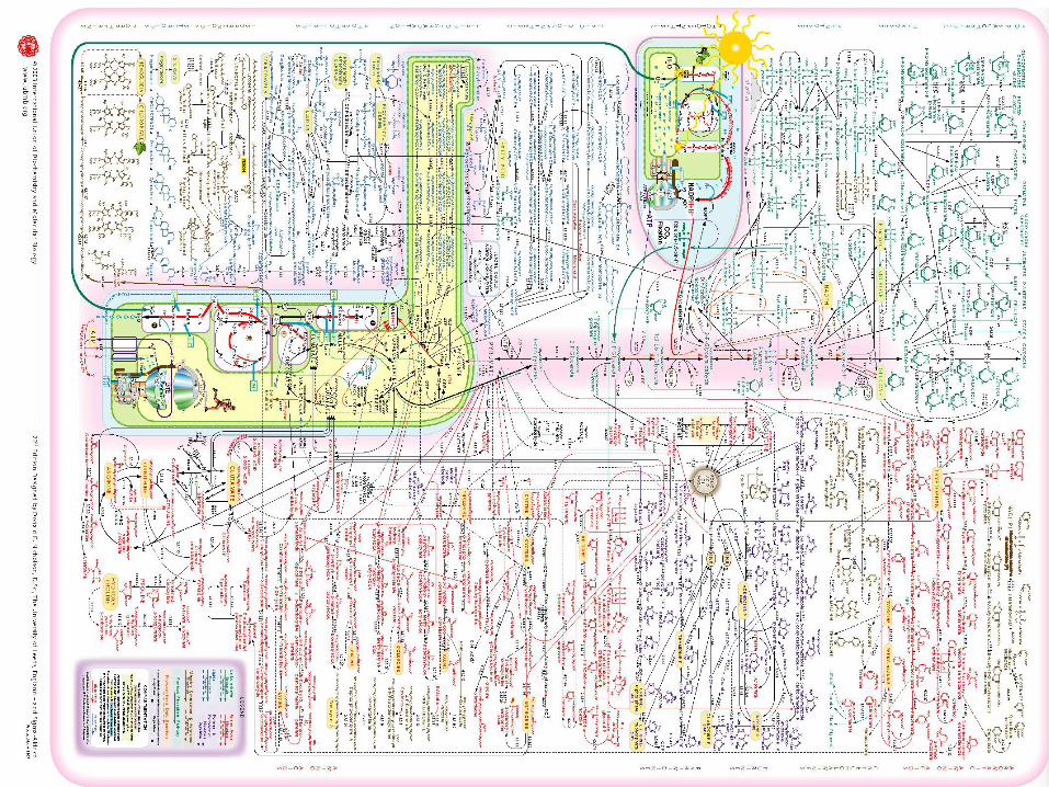
Metabolism The synthesis of building block compounds, monomers and macromolecules from glucose
(and other simple compounds, such as CO2) is conducted by the metabolic reactions of the
cell, such as the highly abbreviated synthesis of amino acids shown here:
All of these reactions, of which there are more than 1000, are catalyzed by enzymes.
Glucose (C6) Pentose (C5)
Triose (C3)
Pyruvate (C3)
AcetylCoA (C2)
Citrate (C6)
Oxoglutarate (C5)
OaA (C4)
Tetrose (C4)
Shikimate (C7)
Glycine (C2)
Serine (C3)
Cysteine (C3)
Phenylalanine (C9)
Tyrosine (C9)
Tryptophan (C11) Lysine (C6)
Alanine (C3)
Methionine (C5)
Aspartate (C4)
Threonine (C4)
Isoleucine (C6)
Histidine (C6)
Valine (C5)
Leucine (C6)
Phenylalanine (C9)
Tyrosine (C9)
Tryptophan (C11)
TCA Cycle
Pentose Phosphate Pathway EMP Pathway
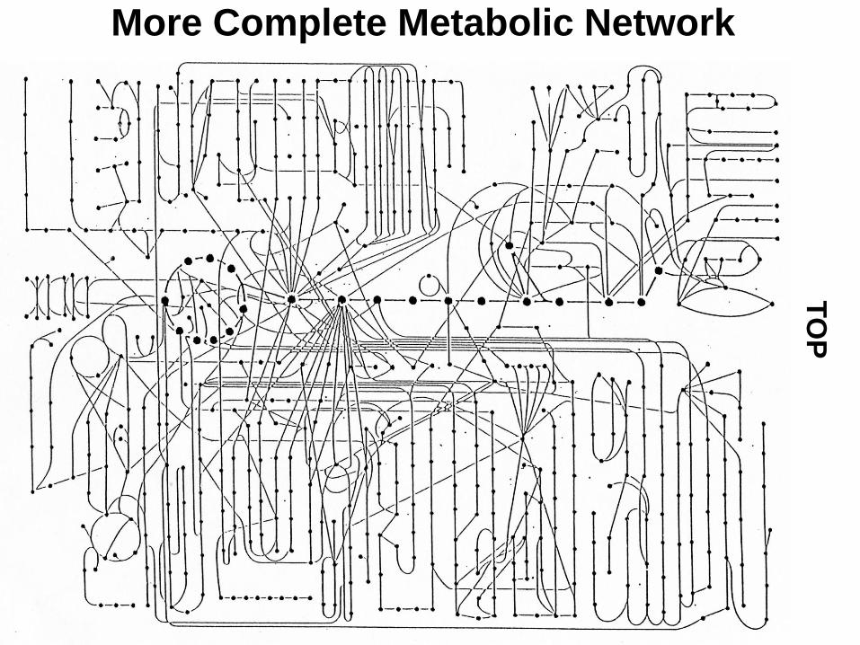
More Complete Metabolic Network T
OP
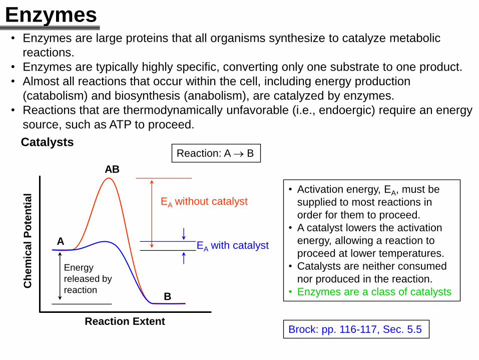
Enzymes • Enzymes are large proteins that all organisms synthesize to catalyze metabolic
reactions.
• Enzymes are typically highly specific, converting only one substrate to one product.
• Almost all reactions that occur within the cell, including energy production
(catabolism) and biosynthesis (anabolism), are catalyzed by enzymes.
• Reactions that are thermodynamically unfavorable (i.e., endoergic) require an energy
source, such as ATP to proceed.
Ch
em
ical
Po
ten
tial
Reaction Extent
A
B
AB
Catalysts
EA without catalyst
Reaction: A B
EA with catalyst
• Activation energy, EA, must be
supplied to most reactions in
order for them to proceed.
• A catalyst lowers the activation
energy, allowing a reaction to
proceed at lower temperatures.
• Catalysts are neither consumed
nor produced in the reaction.
• Enzymes are a class of catalysts
Energy
released by
reaction
Brock: pp. 116-117, Sec. 5.5
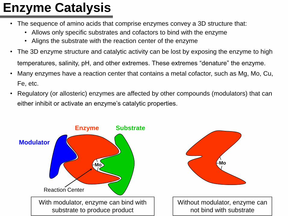
Enzyme Catalysis • The sequence of amino acids that comprise enzymes convey a 3D structure that:
• Allows only specific substrates and cofactors to bind with the enzyme
• Aligns the substrate with the reaction center of the enzyme
• The 3D enzyme structure and catalytic activity can be lost by exposing the enzyme to high
temperatures, salinity, pH, and other extremes. These extremes “denature” the enzyme.
• Many enzymes have a reaction center that contains a metal cofactor, such as Mg, Mo, Cu,
Fe, etc.
• Regulatory (or allosteric) enzymes are affected by other compounds (modulators) that can
either inhibit or activate an enzyme’s catalytic properties.
Mo Mo
Modulator
Enzyme Substrate
Reaction Center
With modulator, enzyme can bind with
substrate to produce product
Without modulator, enzyme can
not bind with substrate
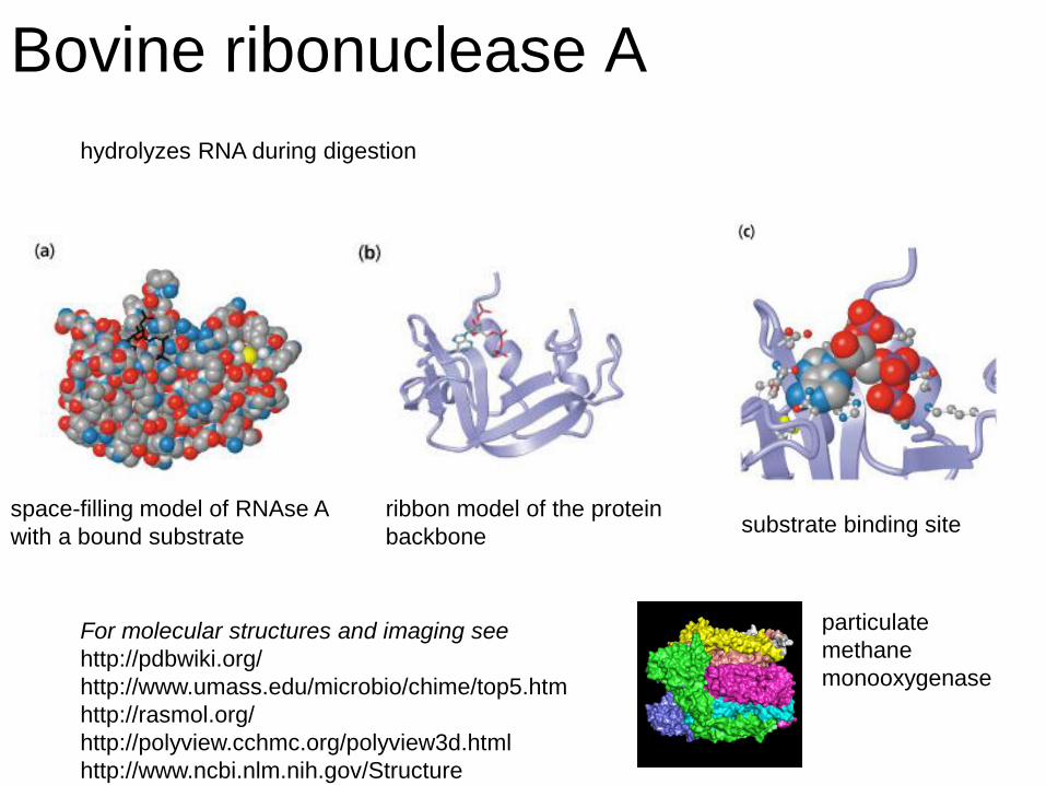
Bovine ribonuclease A
hydrolyzes RNA during digestion
space-filling model of RNAse A
with a bound substrate
ribbon model of the protein
backbone
substrate binding site
For molecular structures and imaging see
http://pdbwiki.org/
http://www.umass.edu/microbio/chime/top5.htm
http://rasmol.org/
http://polyview.cchmc.org/polyview3d.html
http://www.ncbi.nlm.nih.gov/Structure
particulate
methane
monooxygenase
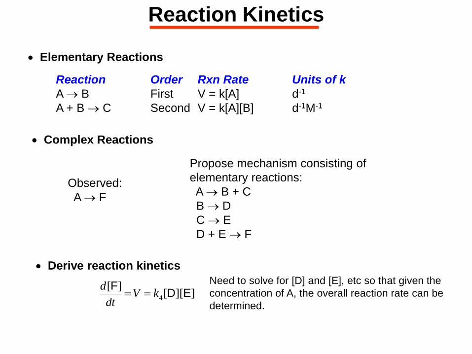
Reaction Kinetics
Elementary Reactions
Reaction Order Rxn Rate Units of k
A B First V = k[A] d-1
A + B C Second V = k[A][B] d-1M-1
Complex Reactions
Observed:
A F
Derive reaction kinetics
]][[][
4 EDF
kVdt
d
Need to solve for [D] and [E], etc so that given the
concentration of A, the overall reaction rate can be
determined.
Propose mechanism consisting of
elementary reactions:
A B + C
B D
C E
D + E F
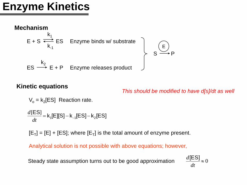
Enzyme Kinetics
Mechanism
E + S ES Enzyme binds w/ substrate
ES E + P Enzyme releases product
k1
k-1
k2
S P
E
Kinetic equations
Vo = k2[ES] Reaction rate.
[ET] = [E] + [ES]; where [ET] is the total amount of enzyme present.
Analytical solution is not possible with above equations; however,
[ES]k[ES]k[E][S]kES
211 dt
d ][
0dt
d [ES]Steady state assumption turns out to be good approximation
This should be modified to have d[s]/dt as well
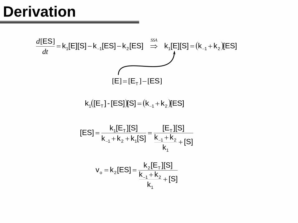
Derivation
[ES]kk[E][S]k[ES]k[ES]k[E][S]kES
211211
SSA
dt
d ][
][][][ ESEE T
[ES]kk[S][ES]-][Ek 21T1
[S]k
kk
][S][E
[S]kkk
][S][Ek[ES]
1
21
T
121
T1
[S]k
kk
][S][Ek[ES]kv
1
21
T22o
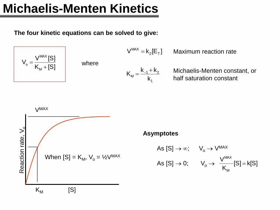
Michaelis-Menten Kinetics
The four kinetic equations can be solved to give:
[S]K
[S]VV
M
MAX
o
where
1
21M
T2
MAX
k
kkK
][EkV
Maximum reaction rate
Michaelis-Menten constant, or
half saturation constant
[S]
Reaction r
ate
, V
o
VMAX
KM
When [S] = KM, Vo = ½VMAX
Asymptotes
As [S] ; Vo VMAX
As [S] 0; Vo k[S][S]K
V
M
MAX
0 5 10 15 20
Time
0
2
4
6
8
10
a,
b
0 5 10 15 20
Time
0
2
4
6
8
10
a,
b
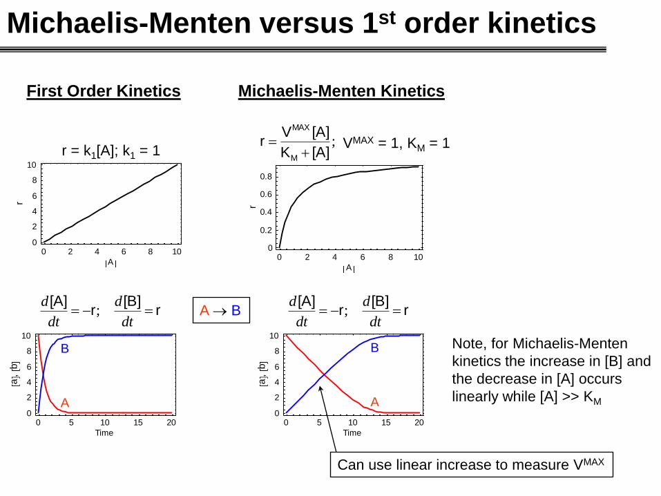
Michaelis-Menten versus 1st order kinetics
First Order Kinetics
r[B]
r[A]
dt
d
dt
d;
r = k1[A]; k1 = 1
Michaelis-Menten Kinetics
;[A]K
[A]Vr
M
MAX
VMAX = 1, KM = 1
r[B]
r[A]
dt
d
dt
d;A B
A
B
A
B Note, for Michaelis-Menten
kinetics the increase in [B] and
the decrease in [A] occurs
linearly while [A] >> KM
Can use linear increase to measure VMAX
0 2 4 6 8 10
A
0
2
4
6
8
10
r
0 2 4 6 8 10
A
0
0.2
0.4
0.6
0.8
r
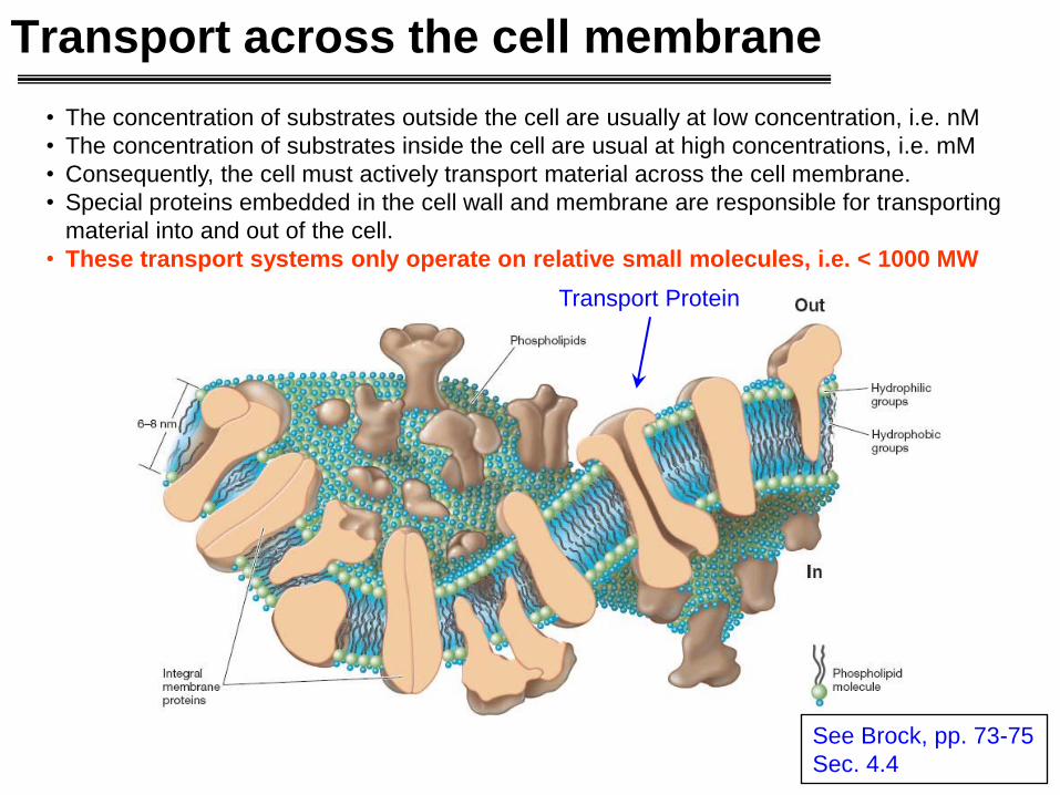
Transport across the cell membrane
• The concentration of substrates outside the cell are usually at low concentration, i.e. nM
• The concentration of substrates inside the cell are usual at high concentrations, i.e. mM
• Consequently, the cell must actively transport material across the cell membrane.
• Special proteins embedded in the cell wall and membrane are responsible for transporting
material into and out of the cell.
• These transport systems only operate on relative small molecules, i.e. < 1000 MW
See Brock, pp. 73-75
Sec. 4.4
Transport Protein
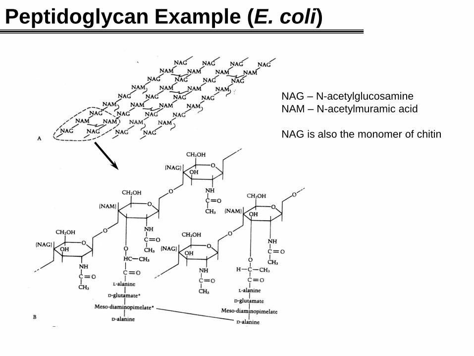
Peptidoglycan Example (E. coli)
NAG – N-acetylglucosamine
NAM – N-acetylmuramic acid
NAG is also the monomer of chitin
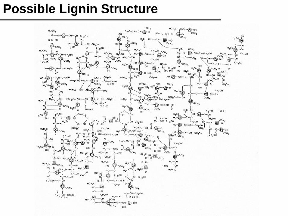
Possible Lignin Structure
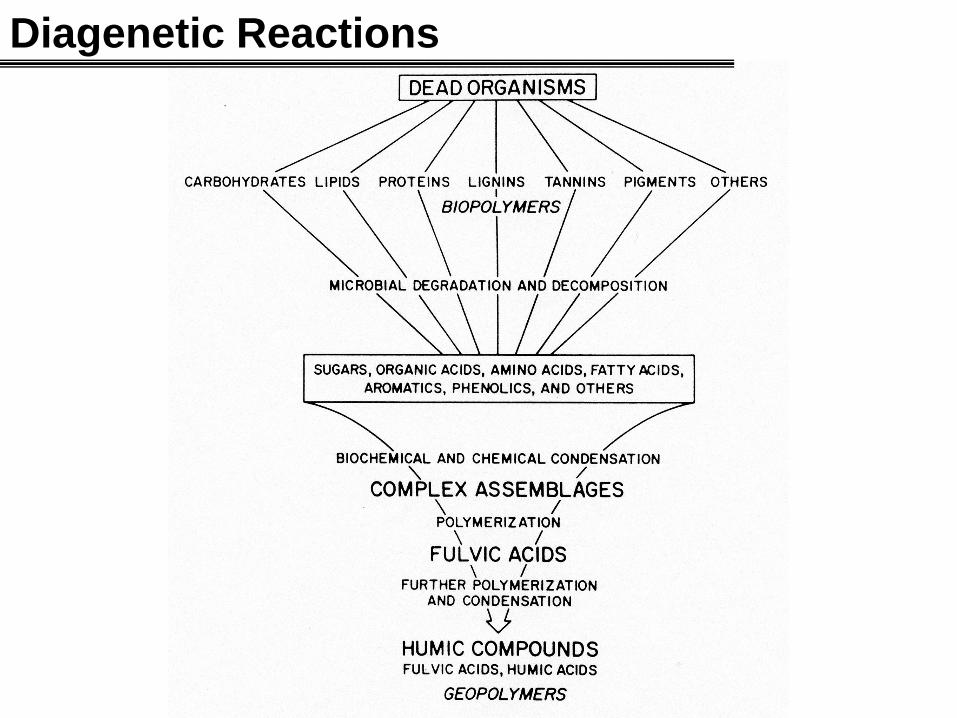
Diagenetic Reactions
Bacterial Substrates
Problem: monomers exist at low concentration and make up only a
small percent of the extracellular POM+DOM pool.
How do bacteria breakdown and consume the large polymeric material?
• All organisms are comprise of mostly polymeric material: protein, cellulose, starch, lipids,
peptidoglycans, lignin, RNA, DNA, etc.
• Consequently, dead organic material available for bacterial consumption is mostly large
polymeric material with high molecular weights.
• Large polymeric compounds can not be transported across the cell wall.
• As organic material is exposed to environmental factors, such as ultraviolet light,
absorption onto minerals (clay, etc) and bacterial degradation, the organic material
becomes even more amorphous.
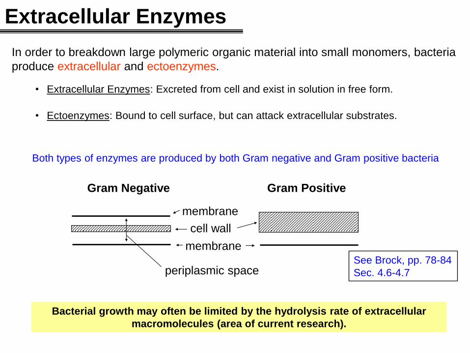
Extracellular Enzymes
Gram Negative Gram Positive
cell wall
membrane
membrane
periplasmic space
In order to breakdown large polymeric organic material into small monomers, bacteria
produce extracellular and ectoenzymes.
• Extracellular Enzymes: Excreted from cell and exist in solution in free form.
• Ectoenzymes: Bound to cell surface, but can attack extracellular substrates.
Both types of enzymes are produced by both Gram negative and Gram positive bacteria
See Brock, pp. 78-84
Sec. 4.6-4.7
Bacterial growth may often be limited by the hydrolysis rate of extracellular
macromolecules (area of current research).
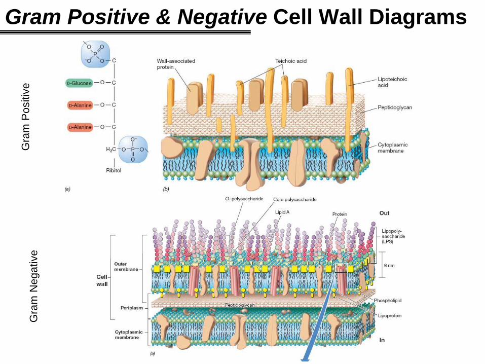
Gram Positive & Negative Cell Wall Diagrams G
ram
Positiv
e
Gra
m N
egative
Enzyme Assay
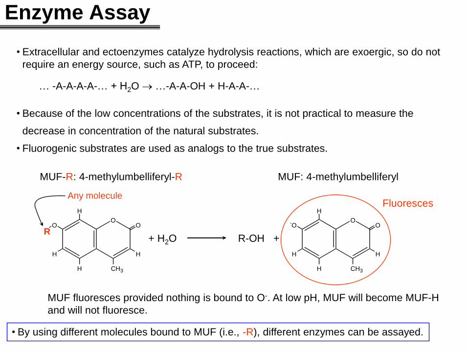
• Extracellular and ectoenzymes catalyze hydrolysis reactions, which are exoergic, so do not
require an energy source, such as ATP, to proceed:
… -A-A-A-A-… + H2O …-A-A-OH + H-A-A-…
• Because of the low concentrations of the substrates, it is not practical to measure the
decrease in concentration of the natural substrates.
• Fluorogenic substrates are used as analogs to the true substrates.
O
CH 3 H
H
O
H
O
H
R
MUF-R: 4-methylumbelliferyl-R
O
CH 3 H
H
- O
H
O
H
R-OH + + H2O
MUF fluoresces provided nothing is bound to O-. At low pH, MUF will become MUF-H
and will not fluoresce.
MUF: 4-methylumbelliferyl
Any molecule
• By using different molecules bound to MUF (i.e., -R), different enzymes can be assayed.
Fluoresces
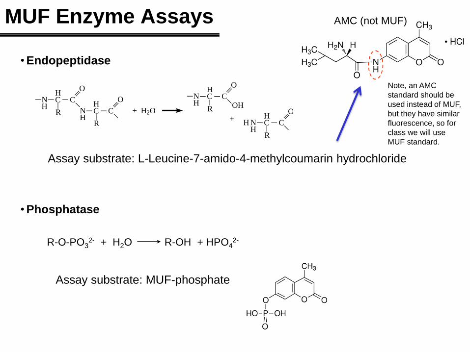
MUF Enzyme Assays
N C C
O
HR
H
N C C
O
HR
H+ H2O
N C C
O
HR
H
+N C C
O
HR
H
OH
H
Assay substrate: L-Leucine-7-amido-4-methylcoumarin hydrochloride
• Endopeptidase
R-O-PO32- + H2O R-OH + HPO4
2-
• Phosphatase
Assay substrate: MUF-phosphate
AMC (not MUF)
Note, an AMC
standard should be
used instead of MUF,
but they have similar
fluorescence, so for
class we will use
MUF standard.
O
H
HOH
CH2OH
H
H
HO
O
H
HOH
CH2OH
H
H
O
HHO
OH
HOH
+ H2O 2 Glucose
•-1,4-glucosidase (cellobiase)
Assay substrate: MUF-β-D-glucopyranoside
• N--D-acetyl-glucosaminidase (Chitobiase)
O
H
HOH
CH2OH
H
H
HO
O
H
HOH
CH2OH
H
H
O
HHN
OH
HNH
+ H2O 2 N-acetyl-glucosaminide
C=O
CH3
C=O
CH3
Assay substrate: MUF-N--D-acetyl-glucosaminide
More MUF Enzyme Assays
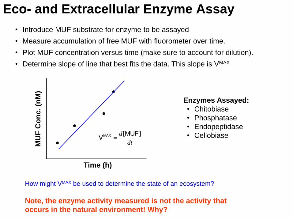
Eco- and Extracellular Enzyme Assay
• Introduce MUF substrate for enzyme to be assayed
• Measure accumulation of free MUF with fluorometer over time.
• Plot MUF concentration versus time (make sure to account for dilution).
• Determine slope of line that best fits the data. This slope is VMAX
Time (h)
MU
F C
on
c. (n
M)
dt
d ][MUFVMAX
Enzymes Assayed:
• Chitobiase
• Phosphatase
• Endopeptidase
• Cellobiase
Note, the enzyme activity measured is not the activity that
occurs in the natural environment! Why?
How might VMAX be used to determine the state of an ecosystem?
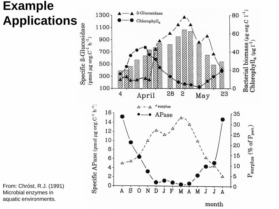
Example
Applications
From: Chróst, R.J. (1991)
Microbial enzymes in
aquatic environments.