Embed Size (px)
Citation preview
Exposure to radio frequency radiation emitted by cell
phone and mortality in chick embryos (Gallus
domesticus)
Author(s): I. V. Ingole, S. K. Ghosh
Vol. 17, No. 3 (2006-09 - 2006-12)
Biomedical Research 2006; 17 (3): 205-210
I. V. Ingole, S. K. Ghosh
Department of Anatomy, Mahatma Gandhi Institute of Medical Sciences, Sevagram,
India
Key words: Cell phone, EMR, radio frequency, radiation, mortality, chick embryo
Accepted September 24, 2006
Abstract
The rapidly increasing use of cell phones since late 1990s has caused a general concern
on the possible hazardous health effects of exposure to radio frequency electro-magnetic
radiation (EMR) emitted by them. While considering the bio-logical effects of EMR on
the human body, its intensity, frequency of radiation and duration of exposure are
important determinants. Many researchers have reported several harmful effects
associated with exposure to radio frequency EMR while some of the studies could not
show any effect at all. The present study addresses the effect of cell phone emanating
EMR in 900 MHz radio fre-quency (widely used) range in determining the health of
developing chick em-bryos. A standard cell phone hand set working at a radio frequency
band of 900 MHz, power of 2 W and Specific Absorption Ratio (SAR) 0.37W/Kg was
used. Sets of fertile hen eggs (Gallus domesticus) in batches were incubated in a spe-
cially designed incubator (radio transparent) at a constant temperature, humidity. During
incubation one set of eggs was exposed for a short period and the other set was exposed
for a longer duration to radiation of the cell phone while the remaining set of eggs was
not exposed to any radiation. It was observed that exposure to radiation increased the
mortality in chick embryos. It was further observed that the rate of mortality is the
function of EMR energy comprising power density at the egg location and duration of
exposure.
Introduction
With the proliferation of personal wireless telecommunication instruments like cordless
phones the cell phone has taken over the society by storm. Its compact size, low weight
and ease of use have found millions of users. Though introduced only in 1990 the user
base of cell phone has crossed 40 million marks within a decade espe-cially in a country
like India. Unlike other electronic equipments the transmitter in the cell phones which
radi-ates EMR happens to be close to human body hence it calls for an attention for its
effects on human. Scientific researches have highlighted some extremely hazardous
effects of exposure to radiation emitted by the cell phones on the human body like
disruption of learned behavior, headache, fatigue, dysaesthesia etc [1,2,3]. Significant
increases in single and double strand DNA breakages in rat’s brain cells have been
reported [4] after exposure to radio frequency radiation. These changes at molecular level
are potentially dangerous to the animal and may prove lethal. Radio frequency radiation
exposure has been reported to induce changes in the prenatal development [5]. Effect of
role of frequency modulation of microwaves on ornithine decarboxylase activity has been
reported [6]. Use of cell phones has been incriminated as a risk for brain tumours [7].
Lethal effects leading to high rate of mortality were reported in chick embryos following
exposure of eggs to cell phone radiation in the radio frequency bandwidth and 2W power
[8]. Six times the mortality in the exposed chick embryos than that of the control group
was reported by the above investigators. In another study, during the embryonic
development chick embryo mortality was reported to have increased to 75% as a result of
exposure to EMF from GSM mobile phone as compared to 16% in the control group [9].
Conversely some of the investigations have reported no adverse biological effects of
exposure to radio frequency radiation emitted by the cell phone. The studies aimed to
investigate the effects of microwaves emitted by cell phones on peripheral blood
parameters of rats did not reveal any difference in the experimental group from sham-
exposed group. But the birth weight of offspring in the experimental group was signifi-
cantly lower than in the sham-exposed group [10]. Cyto-genetic investigations on
microwaves emitted by a 455.7MHz car phone have failed to show any significant
difference in chromosomal aberration or sister chromatid exchanges between microwave
exposed human lymphocytes and unexposed control samples[11].
In considering the biological effects of exposure to radiation, the frequency, its power
density and duration of ex-posure are the important determinants [12]. Regulatory
organizations in different countries have established norms for human radio wave
exposure and Specific Ab-sorption Ratio (SAR). Considering the controversial reports of
the effects of exposure to RF radiation and the varying situations of exposure duration,
frequency and intensity of radiation, the present article addresses the concern regarding
the safety of exposure to radio frequency radiation emitted by handheld communication
devices commonly known as cell phone handset on grow-ing embryos particularly of
chick as a first pointer.
Materials and Methods
Fertile hen eggs were acquired from State Government Poultry Farm, Camp, Amravati
444602, India.
A popular brand cell phone hand set and service provider were selected with a frequency
in the bandwidth of 900 MHz, power of 2 Watt and Specific Absorption Rate (SAR) of
0.37 W/Kg.
An egg incubator cubicle (W-35 cm, H-30 cm, and D-30 cm) made from specific heat
resistant non-metallic material to avoid any internal reflection of the cell phone radiation
within the enclosure was used. It had temperature and humidity controller with forced air
ventilation. Fur-thermore it had an arrangement for automatic rotation of eggs twice a
day. It had an arrangement for mounting the cell phone at on the top of the eggs. A
radiation exposure duration meter (EDM) was used to measure the cumula tive time of
cell phone radiation having an inbuilt control-ler to initiate the cumulative recording of
time whenever the cell phone started radiating outgoing signal.
Positioning the eggs and cell phone
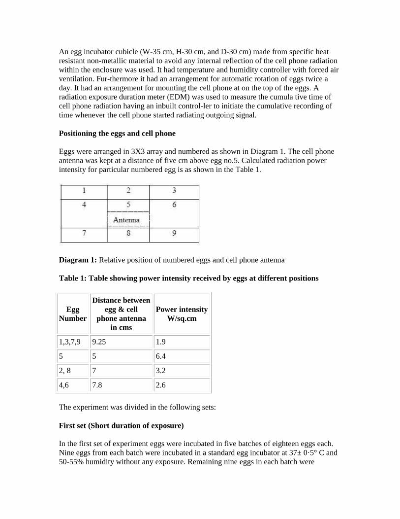
Eggs were arranged in 3X3 array and numbered as shown in Diagram 1. The cell phone
antenna was kept at a distance of five cm above egg no.5. Calculated radiation power
intensity for particular numbered egg is as shown in the Table 1.
Diagram 1: Relative position of numbered eggs and cell phone antenna
Table 1: Table showing power intensity received by eggs at different positions
Egg
Number
Distance between
egg & cell
phone antenna
in cms
Power intensity
W/sq.cm
1,3,7,9 9.25 1.9
5 5 6.4
2, 8 7 3.2
4,6 7.8 2.6
The experiment was divided in the following sets:
First set (Short duration of exposure)
In the first set of experiment eggs were incubated in five batches of eighteen eggs each.
Nine eggs from each batch were incubated in a standard egg incubator at 37± 0·5° C and
50-55% humidity without any exposure. Remaining nine eggs in each batch were
incubated giving radiation in sessions of half an hour each and sessions spaced evenly at
twelve hours interval. For exposure, the cell phone was placed in the incubator after
dialing a particular number. The cell phone started emitting signal and the duration was
recorded by the exposure duration meter (EDM). This was followed by a silent interval
during which there was an automatic redialing but there was no emission of signal. After
the redialing was complete, cell phone again started emitting signal and it’s duration was
detected and recorded by EDM. Thus the total duration of exposure of half an hour in
each session was not continuous but interrupted by short exposure-free periods. The
exposed groups of all the five batches were exposed for four sessions, thus giving two
hours of total exposure in the first forty eight hours of incubation. The eggs from these
five batches were sacrificed at the completion of 4th, 5th, 10th, 12th and 14th day
respectively (Table 2)..
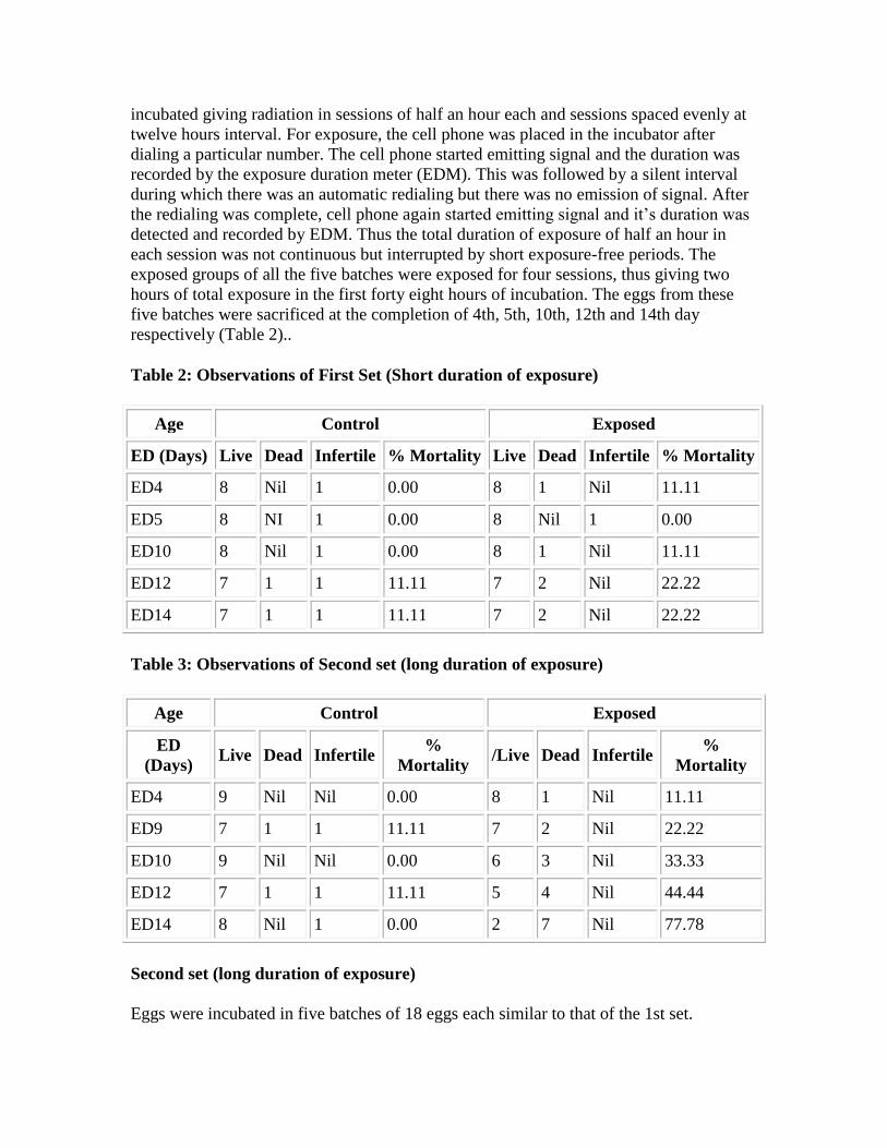
Table 2: Observations of First Set (Short duration of exposure)
Age Control Exposed
ED (Days) Live Dead Infertile % Mortality Live Dead Infertile % Mortality
ED4 8 Nil 1 0.00 8 1 Nil 11.11
ED5 8 NI 1 0.00 8 Nil 1 0.00
ED10 8 Nil 1 0.00 8 1 Nil 11.11
ED12 7 1 1 11.11 7 2 Nil 22.22
ED14 7 1 1 11.11 7 2 Nil 22.22
Table 3: Observations of Second set (long duration of exposure)
Age Control Exposed
ED
(Days) Live Dead Infertile
%
Mortality /Live Dead Infertile
%
Mortality
ED4 9 Nil Nil 0.00 8 1 Nil 11.11
ED9 7 1 1 11.11 7 2 Nil 22.22
ED10 9 Nil Nil 0.00 6 3 Nil 33.33
ED12 7 1 1 11.11 5 4 Nil 44.44
ED14 8 Nil 1 0.00 2 7 Nil 77.78
Second set (long duration of exposure)
Eggs were incubated in five batches of 18 eggs each similar to that of the 1st set.
Nine eggs in each batch were incubated as control in an incubator at 37±0.5°C and 50-
55% humidity. Remaining nine eggs in each batch were incubated in the special in-
cubator (as mentioned for the first set). Eggs were placed on the numbered slots and were
incubated at 37±0.5 ° C and 50-55% humidity (as in the first set). The cell phone was
placed in the same position as for exposed groups of first set. Exposures were given in
sessions of half an hour each, spaced at 12 hrs intervals. Duration of exposure was
recorded by EDM. The five batches were exposed to radiation exposure schedules of four
h, four h, six h, nine h and tens h respectively. These five batches were sacrificed at the
completion of 4th, 9th, 10th, 12th and 14th day of incubation respectively (Table 3). The
embryos were taken out and observed for viability and gross morphology.
Observations and Discussion:
First Set: (short duration of exposure)
At the completion of 4th, 5th, 10th, 12th and 14th day of in-cubation, the condition of the
embryos in the control group receiving no radiation and the condition of the embryos in
the exposed group receiving exposure to radia-tion during the first 48 h of incubation is
tabulated in Ta-ble 2.
Second Set: (long duration of exposure):
In the control groups where no exposure was given, at the completion of 4th, 9th, 10th ,
12th and 14th day of incubation, the condition of the eggs was as shown in Table 3.
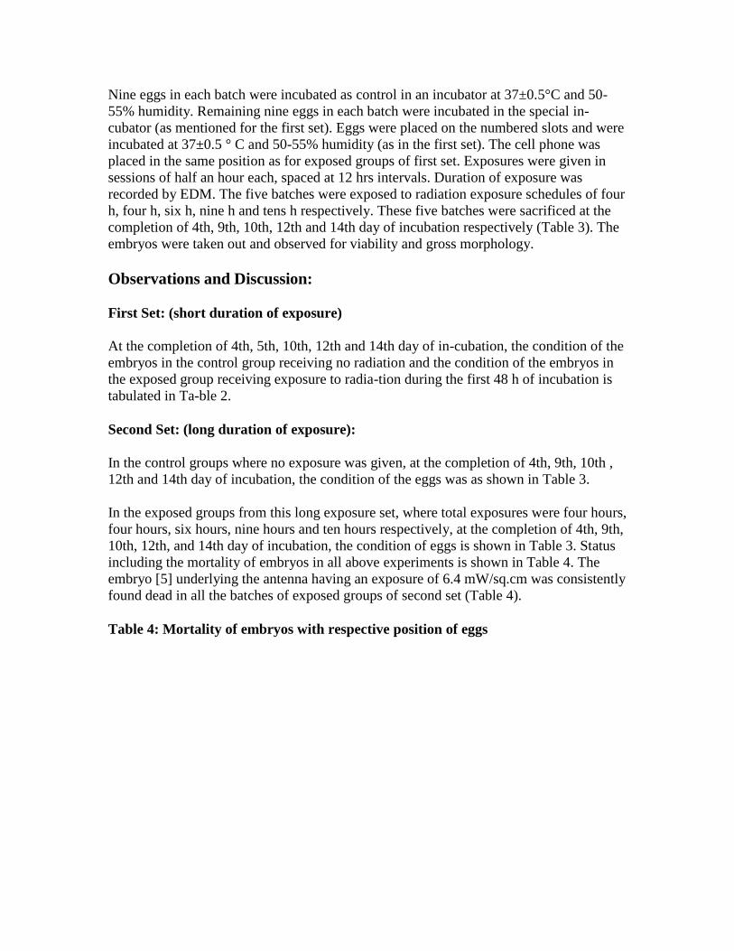
In the exposed groups from this long exposure set, where total exposures were four hours,
four hours, six hours, nine hours and ten hours respectively, at the completion of 4th, 9th,
10th, 12th, and 14th day of incubation, the condition of eggs is shown in Table 3. Status
including the mortality of embryos in all above experiments is shown in Table 4. The
embryo [5] underlying the antenna having an exposure of 6.4 mW/sq.cm was consistently
found dead in all the batches of exposed groups of second set (Table 4).
Table 4: Mortality of embryos with respective position of eggs
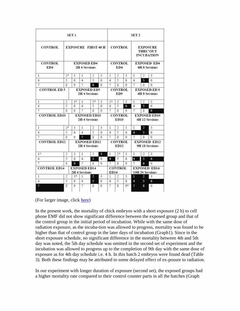
(For larger image, click here)
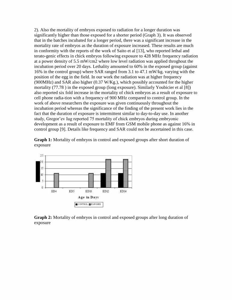
In the present work, the mortality of chick embryos with a short exposure (2 h) to cell
phone EMF did not show significant difference between the exposed group and that of
the control group in the initial period of incubation. While with the same dose of
radiation exposure, as the incuba-tion was allowed to progress, mortality was found to be
higher than that of control group in the later days of incubation (Graph1). Since in the
short exposure schedule, no significant difference in the mortality between 4th and 5th
day was noted, the 5th day schedule was omitted in the second set of experiment and the
incubation was allowed to progress up to the completion of 9th day with the same dose of
exposure as for 4th day schedule i.e. 4 h. In this batch 2 embryos were found dead (Table
3). Both these findings may be attributed to some delayed effect of ex-posure to radiation.
In our experiment with longer duration of exposure (second set), the exposed groups had
a higher mortality rate compared to their control counter parts in all the batches (Graph
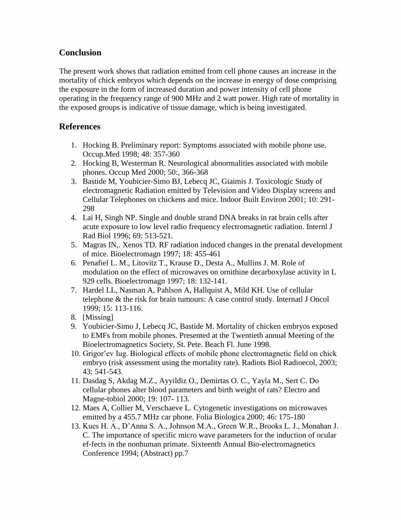
2). Also the mortality of embryos exposed to radiation for a longer duration was
significantly higher than those exposed for a shorter period (Graph 3). It was observed
that in the batches incubated for a longer period, there was a significant increase in the
mortality rate of embryos as the duration of exposure increased. These results are much
in conformity with the reports of the work of Saito et al [13], who reported lethal and
terato-genic effects in chick embryos following exposure to 428 MHz frequency radiation
at a power density of 5.5 mW/cm2 where low level radiation was applied throghout the
incubation period over 20 days. Lethality amounted to 60% in the exposed group (against
16% in the control group) where SAR ranged from 3.1 to 47.1 mW/kg. varying with the
position of the egg in the field. In our work the radiation was at higher frequency
(900MHz) and SAR also higher (0.37 W/Kg.), which possibly accounted for the higher
mortality (77.78 ) in the exposed group (long exposure). Similarly Youbicier et al [8])
also reported six fold increase in the mortality of chick embryos as a result of exposure to
cell phone radia-tion with a frequency of 900 MHz compared to control group. In the
work of above researchers the exposure was given continuously throughout the
incubation period whereas the significance of the finding of the present work lies in the
fact that the duration of exposure is intermittent similar to day-to-day use. In another
study, Gregor’ev Iug reported 75 mortality of chick embryos during embryonic
development as a result of exposure to EMF from GSM mobile phone as against 16% in
control group [9]. Details like frequency and SAR could not be ascertained in this case.
Graph 1: Mortality of embryos in control and exposed groups after short duration of
exposure
Graph 2: Mortality of embryos in control and exposed groups after long duration of
exposure
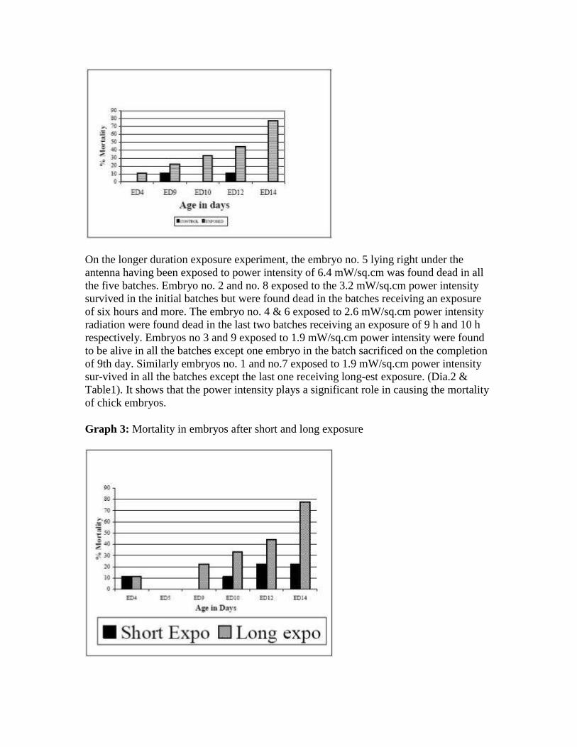
On the longer duration exposure experiment, the embryo no. 5 lying right under the
antenna having been exposed to power intensity of 6.4 mW/sq.cm was found dead in all
the five batches. Embryo no. 2 and no. 8 exposed to the 3.2 mW/sq.cm power intensity
survived in the initial batches but were found dead in the batches receiving an exposure
of six hours and more. The embryo no. 4 & 6 exposed to 2.6 mW/sq.cm power intensity
radiation were found dead in the last two batches receiving an exposure of 9 h and 10 h
respectively. Embryos no 3 and 9 exposed to 1.9 mW/sq.cm power intensity were found
to be alive in all the batches except one embryo in the batch sacrificed on the completion
of 9th day. Similarly embryos no. 1 and no.7 exposed to 1.9 mW/sq.cm power intensity
sur-vived in all the batches except the last one receiving long-est exposure. (Dia.2 &
Table1). It shows that the power intensity plays a significant role in causing the mortality
of chick embryos.
Graph 3: Mortality in embryos after short and long exposure
Conclusion
The present work shows that radiation emitted from cell phone causes an increase in the
mortality of chick embryos which depends on the increase in energy of dose comprising
the exposure in the form of increased duration and power intensity of cell phone
operating in the frequency range of 900 MHz and 2 watt power. High rate of mortality in
the exposed groups is indicative of tissue damage, which is being investigated.
References
1. Hocking B. Preliminary report: Symptoms associated with mobile phone use.
Occup.Med 1998; 48: 357-360
2. Hocking B, Westerman R. Neurological abnormalities associated with mobile
phones. Occup Med 2000; 50:, 366-368
3. Bastide M, Youbicier-Simo BJ, Lebecq JC, Giaimis J. Toxicologic Study of
electromagnetic Radiation emitted by Television and Video Display screens and
Cellular Telephones on chickens and mice. Indoor Built Environ 2001; 10: 291-
298
4. Lai H, Singh NP. Single and double strand DNA breaks in rat brain cells after
acute exposure to low level radio frequency electromagnetic radiation. Internl J
Rad Biol 1996; 69: 513-521.
5. Magras IN,. Xenos TD. RF radiation induced changes in the prenatal development
of mice. Bioelectromagn 1997; 18: 455-461
6. Penafiel L. M., Litovitz T., Krause D., Desta A., Mullins J. M. Role of
modulation on the effect of microwaves on ornithine decarboxylase activity in L
929 cells. Bioelectromagn 1997; 18: 132-141.
7. Hardel LL, Nasman A, Pahlson A, Hallquist A, Mild KH. Use of cellular
telephone & the risk for brain tumours: A case control study. Internatl J Oncol
1999; 15: 113-116.
8. [Missing]
9. Youbicier-Simo J, Lebecq JC, Bastide M. Mortality of chicken embryos exposed
to EMFs from mobile phones. Presented at the Twentieth annual Meeting of the
Bioelectromagnetics Society, St. Pete. Beach Fl. June 1998.
10. Grigor’ev Iug. Biological effects of mobile phone electromagnetic field on chick
embryo (risk assessment using the mortality rate). Radiots Biol Radioecol, 2003;
43: 541-543.
11. Dasdag S, Akdag M.Z., Ayyildiz O., Demirtas O. C., Yayla M., Sert C. Do
cellular phones alter blood parameters and birth weight of rats? Electro and
Magne-tobiol 2000; 19: 107- 113.
12. Maes A, Collier M, Verschaeve L. Cytogenetic investigations on microwaves
emitted by a 455.7 MHz car phone. Folia Biologica 2000; 46: 175-180
13. Kues H. A., D’Anna S. A., Johnson M.A., Green W.R., Brooks L. J., Monahan J.
C. The importance of specific micro wave parameters for the induction of ocular
ef-fects in the nonhuman primate. Sixteenth Annual Bio-electromagnetics
Conference 1994; (Abstract) pp.7
14. Saito K., Suzuki K., Motoyoshi S. Lethal & teratogenic effects of long term low-
intensity Radio Frequency radiation at 428 MHz on developing chick embryo.
Teratology 1991;43: 609-614
Correspondence:
I. V. Ingole Department of Anatomy, Mahatma Gandhi Institute of Medical Sciences
Sevagram (Maharashtra) 442 102, India
e-mail: vijayingole ( at ) hotmail.com