Embed Size (px)
Citation preview
Exploring Visuo-Haptic Augmented Reality UserInterfaces for Stereo-Tactic Neurosurgery Planning
Ulrich Eck1, Philipp Stefan1, Hamid Laga2, Christian Sandor3, Pascal Fallavollita1,and Nassir Navab1
1 Technische Universitat Munchen, Germany, Chair of Computer Aided Medical Procedures,[email protected]
2 Murdoch University, Australia, School of Engineering & Information Technology3 Nara Institute of Science and Technology, Japan, Interactive Media Design Lab
Abstract. Stereo-tactic neurosurgery planning is a time-consuming and com-plex task that requires detailed understanding of the patient anatomy and the af-fected regions in the brain to precisely deliver the treatment and to avoid prox-imity to any known risk structures. Traditional user interfaces for neurosurgeryplanning use keyboard and mouse for interaction and visualize the medical dataon a screen. Previous research, however, has shown that 3D user interfaces aremore intuitive for navigating volumetric data and enable users to understand spa-tial relations more quickly. Furthermore, new imaging modalities and automatedsegmentation of relevant structures provide important information to medical ex-perts. However, displaying such information requires frequent context switchesor occludes otherwise important information.In collaboration with medical experts, we analyzed the planning workflow forstereo-tactic neurosurgery interventions and identified two tasks in the processthat can be improved: volume exploration and trajectory refinement. In this pa-per, we present a novel 3D user interface for neurosurgery planning that is imple-mented using a head-mounted display and a haptic device. The proposed systemimproves volume exploration with bi-manual interaction to control oblique slic-ing of volumetric data and reduces visual clutter with the help of haptic guidesthat enable users to precisely target regions of interest and to avoid proximity toknown risk structures.
Keywords: augmented reality, haptics, neurosurgery, planning
1 IntroductionStereo-tactic neurosurgery is a form of minimally invasive surgery that uses a stereo-tactic frame to locate targets inside the brain. This method enables interventions thatrequire highly precise targeting, like biopsy, ablation, lesion, injection, stimulation, im-plantation, and radio-surgery. The two most important criteria when planning such in-terventions are: to precisely deliver the treatment at the desired target, and to avoidproximity to any risk structures along the trajectory, such as blood vessels or criticalbrain regions.
The planning of trajectories for a stereo-tactic neurosurgery procedure is a highlycomplex task, which requires good understanding of the location and structure of the

(a) Oblique volume slicing. (b) Segmented structures. (c) Trajectory verification.
Fig. 1: The Visuo-Haptic Augmented Reality (VHAR) user interface of the neuro-surgery application enables users to naturally interact with medical volume datasets:(a) Users explore volumetric data using oblique slicing and bi-manual interaction. (b)Segmented structures and planned trajectories (yellow lines) are displayed. (c) Users in-spect and refine trajectories to avoid proximity to risk structures using an oblique sliceplane that is perpendicular to the trajectory.
targeted brain regions, as well as all regions affected by any planned trajectory. Neu-rologists and neurosurgeons explore the original image datasets and segmented vol-umetric structures to gain understanding about the patient’s anatomy. They performtrajectory planning before a surgery to identify target regions and possible paths, andduring surgery, where the pre-planned trajectories are refined and verified to avoid anyproximity to risk structures. The intra-operative planning procedure is performed whilethe patient is under full anesthesia, after the stereo-tactic frame was mounted, and af-ter the pre-operative images are registered with an intra-operative CT scan so that theplanned trajectories can be computed in frame coordinates. The intra-operative plan-ning can take up to two hours while the patient is in the operating theatre. In this paperwe propose a novel Visuo-Haptic Augmented Reality (VHAR) user interface for neu-rosurgery planning, which reduces the required time for planning and therefore reducesthe negative impact on the patient and makes the procedure more cost-effective.
Volumetric image data exploration and trajectory planning in medical practice istypically done using desktop-based workstations with mouse and keyboard. Specializedplanning software provides 2D/3D views onto the available patient image data. Opera-tors define trajectories in slice views onto the medical volumes. During planning, theyiterate between volume exploration using mouse and keyboard, relocating trajectorypoints with the mouse to avoid proximity with identified risk structures, and verifyingthat the recently changed path does not interfere with previously identified risk areas.They also switch often between different image modalities, since not all structures arevisible in all images. The type and amount of information available to neurosurgeonsand neurologists has vastly increased. This includes new imaging modalities [6], im-proved computer assisted segmentation of volumetric data [4], computer assisted riskassessment [15], and automated trajectory planning [4, 5, 3]. Selecting the appropriatedata and visualizing it still remains a challenge especially since additional visual over-lays often interfere with the requirement to see the original image dataset with all thedetails.

The VHAR user interface of our neurosurgery planning application integrates aforce feedback haptic device into an Augmented Reality (AR) system. Operators seepatient data visualization using a Head-Mounted Display (HMD) and can intuitivelyexplore it using bi-manual 3D interaction (see Figure 1). The haptic device providesforce feedback that enables precise 3D targeting and informs operators about nearbyrisk structures. By providing haptic guides, we can reduce visual clutter while still pro-viding sufficient information to efficiently plan the required trajectories.
2 Related WorkNeurosurgical intervention planning requires precise understanding of spatial relationsbetween target areas, access paths, and critical regions. Current planning systems oftenuse slice visualization for volumetric data and user interfaces with keyboard and mousefor navigation and planning, but previous research has shown that 3D user interfacescan greatly enhance the spatial understanding for such tasks. Goble et al. [9] presenteda novel user interface for neurosurgical visualization. Their system tracks passive in-terface props like a solid sphere, cutting plane, and pointer for user interaction. Theyfound that medical experts prefer the intuitive bi-manual 3D interaction over traditionalslice viewers. Our planning system improves on their work so that the medical visu-alization appears co-located with the interface props using an AR display. Eaglesonet al. [7] presented an interactive neurosurgery planning system where multiple userscan explore and annotate patient datasets using a tabletop display with a touch surface.They use a haptic device that is placed onto the display to provide 3D input for volumeexploration, but their system does not provide any haptic feedback.
Researchers have also studied the benefits of AR for surgery planning. Abhari etal. [1] evaluated user performance during the planning of a brain tumor resection.Such tasks require a good understanding of the spatial relationships between relevantanatomy like tumors and risk-structures. They compared task performance of medi-cal experts when using different visualization techniques. In a preliminary study theyshowed that users performed as good or better in AR than in all other modes. The ben-efits of AR are however larger for novices than for experts. Shamir et al. [14] presentedan augmented reality user interface for improved risk assessment in image guided key-hole surgery. They augment the risk of all possible trajectories onto the surface of a headphantom. Based on an expert review they conclude that the proposed AR user interfaceis beneficial for planning of difficult operations and for education. Our system buildsupon and enhances their interaction techniques, and adds haptic feedback as anothermodality to display important information.
Recent advances in image registration and segmentation algorithms enable researchersto build complete image processing pipelines. D’Albis et al. [4] presented a fully in-tegrated and automated planning solution for deep-brain stimulation (PyDBS). Theirworkflow automates pre-operative, intra-operative, and post-operative imaging tasks,such as registration and segmentation. Our neurosurgery planning system integrates thedata generated by PyDBS.
Researchers are also working on automated trajectory planning systems for neuro-surgery interventions [5, 15, 2], which autonomously suggest candidate trajectories tooperators. These systems can reduce planning times and help users to better estimate

the risk of the planned trajectory. However, such automated planning methods require abroad body of medical knowledge in digital form and accurately segmented and labeledpatient data in order to produce acceptable results.
Information generated by automated trajectory planning systems can be either usedto suggest trajectories, or to improve guidance of users during the planning procedure.During interviews with medical experts we realized that they often prefer access toadditional information over automated processes, which they are not fully in control of.Operators can for example be guided during trajectory planning using visual and hapticguides that use data extracted from automated pipelines. Such guides inform operatorsabout accessibility and the risk of certain configurations in real-time.
The contributions of our work are improved interaction techniques for neurosurgeryplanning using VHAR, which combine bi-manual volume exploration with haptic vi-sualization and guides. The intuitive positioning and slicing of medical volumes andsegmented structures helps operators to better understand the spatial relations, whichreduces the time required to identify target regions and risk structures. The applicationprovides haptic guides that display context-dependent constraints as forces in additionto the visual rendering of medical image data and segmented structures in AR. Replac-ing some of the visual information with haptic feedback reduces visual clutter whilestill providing the required information. The presented trajectory planning method fur-ther reduces the required time for planning of trajectories by reducing the number ofiterations needed for refining and verifying trajectories.
3 System DesignThis section presents the system design of the proposed user interface for neurosurgeryplanning that simplifies the exploration of medical datasets and the trajectory planningprocedure. When using the planning system, users wear a HMD and hold a haptic styluswith their dominant hand. In the other hand, they hold a tracked handle with buttonsused as secondary input to the application. As shown in Figure 1, a patient datasetconsisting of volumetric images and segmented structures is displayed in the hapticworkspace. The proposed application allows users to pick up, transform, and place thepatient dataset with the tracked handle in order to adjust the viewing angle. They usethe haptic stylus to control slicing operation during exploration and verification, and todefine trajectories.
3.1 Volume ExplorationThe volume exploration component visualizes volumetric and geometric informationinteractively. During volume exploration, users mainly need control over the displayedcontent, effective ways to locate regions of interest inside the volume, and views thatallow them to understand spatial relations between risk structures and planned trajecto-ries.
In the proposed planning tool, users can directly manipulate the pose of the volumeusing the tracked handle, which gives them an intuitive way to place and orient themedical dataset as needed, similar to [9]. At the same time, they operate the hapticstylus with their dominant hand to interactively explore the volume using slice viewson the volumetric data.

OV
EP
TP
nV
p
nT
nS
Volume (V)
Viewpoint (VP)
Haptic Stylus (S)
Sliceplane (SP)
Trajectory (T)
(a) Definition of slice planes.
[0,0,0]
[1,1,1]
[1,0,0]
[0,1,0]
x1
x2 x3
x4
x5
(b) Rendering of oblique sliceplanes.
Fig. 2: Oblique slicing of medical datasets in AR: (a) Slice plane SP is defined usingpoint p and normal vector n. The plane normal can be controlled by the view plane ofthe current camera nV , the haptic stylus nS, or the trajectory nT . (b) Volumetric medicaldataset with oblique slice plane. Vertices xi of the slice plane and their correspondingtexture coordinates are computed for real-time rendering using OpenGL shaders.
The visual augmentations are rendered using a multi-volume raycaster for volumet-ric image datasets (see Figure 1a) and a geometry renderer for segmented structures (seeFigure 1b). Users inspect data using the oblique slicing method [13], which is a genericversion of the typically used axis-aligned slice planes. In contrast to axis-aligned sliceplanes, users can additionally control the orientation of the slice. The combination ofoblique slicing with the two-handed interaction allows operators to quickly change vol-ume pose and slice properties. Preliminary reviews with medical experts showed thatthe proposed method reduces the required time to find regions of interest and help tounderstand spatial relations within the volume.
The concept of the oblique slicing technique is shown in Figure 2a. Users place avolume V at a convenient pose OV and control the slice plane SP using a haptic stylusS. The plane SP(p,n) is defined by a point p, which is controlled by the position ofthe haptic stylus S, and a normal vector n. The normal vector is selected dependingon the current task and the user’s preference. Currently, the oblique slicing componentsupports three viewing modes, which differ only by the data source for the plane normaln:
– View-Aligned: The slice plane is always parallel to the view plane of the currentcamera and therefore provides the most detailed view onto the current slice. Userscan additionaly use head movement to control the slicing. The plane normal n isthe normal of the camera view-plane.
– Stylus-Aligned: The slice plane is controlled by the haptic stylus, which gives theoperator maximum control over the slicing operation. The plane normal n is theunit-vector along the longitudinal axis of the haptic stylus S.
– Trajectory-Aligned: The slice plane is always perpendicular to the current trajec-tory. This mode was suggested by medical experts for trajectory verification. Theplane normal n is the unit-vector along the current trajectory T .
The oblique slice view is automatically activated once the stylus tip S is inside the vol-ume bounding box (V ). The slice plane is computed every frame relative to the volumeorigin OV using the current pose of the haptic stylus and the selected plane normal. Theslice plane SP controls a clipping plane, which hides unwanted geometry and volumet-ric image data. Additionally, the locations of three to six vertices, xi, are computed thatdefine a polygon, which represents the intersection of the slice plane with the volumebounding box. In addition to the vertex locations, we determine their texture coordi-nates in relation to the volume bounding box (see Figure 2b). The polygon is texturedusing a OpenGL shader by interpolating over data from a three-dimensional texture thatrepresents the medical volume.
Once users have found the desired view on the data, they can lock the slice plane. Alocked slice plane enables a haptic plane effect, which attracts the stylus tip to the sliceplane if close enough. The haptic feedback guides users to stay on the selected plane,for example when placing points for trajectory planning.
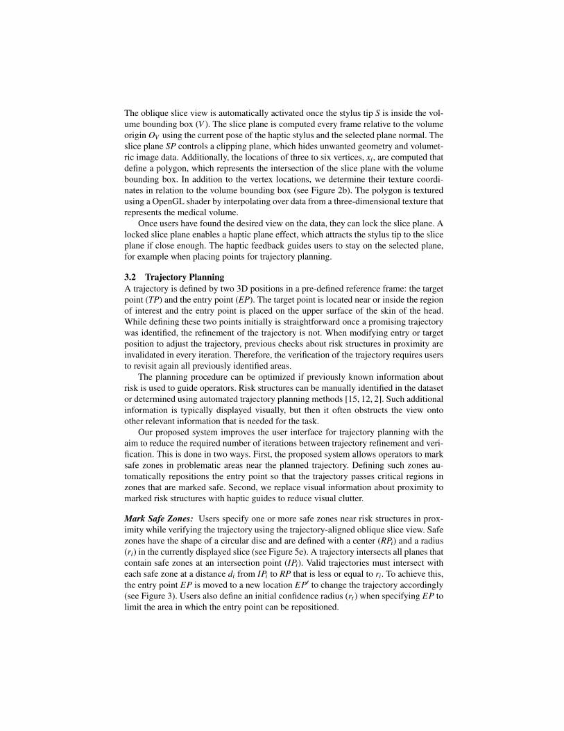
3.2 Trajectory PlanningA trajectory is defined by two 3D positions in a pre-defined reference frame: the targetpoint (TP) and the entry point (EP). The target point is located near or inside the regionof interest and the entry point is placed on the upper surface of the skin of the head.While defining these two points initially is straightforward once a promising trajectorywas identified, the refinement of the trajectory is not. When modifying entry or targetposition to adjust the trajectory, previous checks about risk structures in proximity areinvalidated in every iteration. Therefore, the verification of the trajectory requires usersto revisit again all previously identified areas.
The planning procedure can be optimized if previously known information aboutrisk is used to guide operators. Risk structures can be manually identified in the datasetor determined using automated trajectory planning methods [15, 12, 2]. Such additionalinformation is typically displayed visually, but then it often obstructs the view ontoother relevant information that is needed for the task.
Our proposed system improves the user interface for trajectory planning with theaim to reduce the required number of iterations between trajectory refinement and veri-fication. This is done in two ways. First, the proposed system allows operators to marksafe zones in problematic areas near the planned trajectory. Defining such zones au-tomatically repositions the entry point so that the trajectory passes critical regions inzones that are marked safe. Second, we replace visual information about proximity tomarked risk structures with haptic guides to reduce visual clutter.
Mark Safe Zones: Users specify one or more safe zones near risk structures in prox-imity while verifying the trajectory using the trajectory-aligned oblique slice view. Safezones have the shape of a circular disc and are defined with a center (RPi) and a radius(ri) in the currently displayed slice (see Figure 5e). A trajectory intersects all planes thatcontain safe zones at an intersection point (IPi). Valid trajectories must intersect witheach safe zone at a distance di from IPi to RP that is less or equal to ri. To achieve this,the entry point EP is moved to a new location EP′ to change the trajectory accordingly(see Figure 3). Users also define an initial confidence radius (rt ) when specifying EP tolimit the area in which the entry point can be repositioned.

TP
EP
EP’
RPi’
RP2’
RP1’
RP1
RP2
RPi
IP1IP2
IPi
r
d
y
Fig. 3: The initial (blue) trajectory TP—EP is optimized with three refinement pointsRP1, RP2, RPi. Safe zones are defined as conic volumes around the refinement pointswith an additional radius. A new (red) trajectory TP—EP’ is calculated where the mass-spring system defined by the projected points RP1’, RP2’, RPi’ is at equilibrium.
Reposition the Entry Point: The goal when repositioning the entry point is that a re-fined trajectory should pass only through areas that are marked as safe. This is achievedusing springs that pull the trajectory towards the safe zone centers. The proposed al-gorithm simulates the trajectory as pendulum with the springs attached at equilibriumto calculate the new entry point EP′ using the constraints specified as safe zones. Theapplication invokes the algorithm whenever a trajectory point was modified. Prior to in-vocation, the target point, the entry point, and all centers of safe zones are transformedinto a reference frame (I) with T P as its origin and the negative y-axis pointing towardsthe entry point. From this definition follows that T P = [0,0,0] and EP = [0,−lt ,0],where lt is the length of the trajectory. The position of the new entry point EP′ is de-fined in generalized coordinates α0 and β0.
EP′ =
lt sin(α0)−lt cos(α0)cos(β0)−lt sin(β0)cos(α0)
(1)
To complete the definition of the pendulum, a rigid sphere with mass m1 is attached atthe location of EP′ and a link between T P and EP′ is defined with mass m2. Withoutconsidering any safe zones and assuming normal gravity g = [0,−1,0]× 9.81 m
s2 , thependulum is in a stable state at α = 0 and β = 0.
Safe zones affect the trajectory as follows. First, all centers of safe zones RPi =[xi,yi,zi] and their radii (ri) are projected onto a plane defined by the point EP and thevector [0,−1,0] as its normal.
RP′i = RPi−ltyi
(2)
r′i = ri−ltyi
(3)
Springs between the projected centers RP′i and EP′ pull the trajectory away from nearbyrisk structures by applying forces (Fi) to the pendulum as shown in Figure 3. The springsare parameterized using a stiffness factor (k) and the ratio between the initial confidenceradius (rt ) and r′i.
Fi = krt
r′i(EP′−RP′i ) (4)

TPEP
TIP
RPr
dt
IP
Fg
y
Fig. 4: Haptic guides are computed based on the specified trajectory and the markedsafe zones. This example shows a trajectory with one safe zone that is defined by itscenter RP and its radius r. A conical volume defined by RP, TP, r marks a safe corridor.If the haptic stylus TIP is outside the safe corridor, a spring-damper system is activatedthat pulls the stylus back into the area marked grey. The conic volumes are intersectedif multiple refinement points are active.
The motion of the pendulum is limited using a damping force (Fd) with the factor (c)that is calculated using the current velocity (V ) of EP′.
Fd =−cV (5)
The effects of all forces that are applied to the pendulum are simulated using Kane’smethod [11] to calculate the new location of EP′ where all forces are at equilibrium.The static parameters used in the current system are: m1 = 0.1 kg, m2 = 0.01 kg, c = 5,and k = 25. With these parameters, the duration of the simulation can be limited to 0.3seconds to reach the equilibrium. The new location EP′, however, can be computedfaster than real-time so that the position updates are supplied without noticeable delayin the current implementation. The final entry point for the surgical procedure is locatedat the intersection of the refined trajectory and the patient’s head surface.
Haptic Guides for Safe Zones: Safe zones are also used to visualize the boundariesof safe corridors using force feedback. Such haptic guides help operators to stay withinpreviously defined safe zones while editing the trajectory or to identify impossible con-figurations earlier in the planning procedure. Impossible configurations exist when notrajectory passes safe zones within the defined distance to the center and therefore nosafe path with the current configuration is possible.
Figure 4 shows an example how haptic guides are calculated using a single refine-ment point. The haptic stylus TIP is placed near the trajectory. A safe zone is definedby its center RP and a radius r. In this example the TIP is outside of the conical volume(grey) described by TP, RP, and r. In this case, a haptic guide is activated that pullsthe stylus back into the safe zone by displaying a force. The force is generated using aspring-damper system that pulls the TIP towards the safe volume (blue arrow). Multi-ple safe zones generate multiple overlapping conical volumes. The intersection of thesevolumes is visualized using force feedback.
Feedback forces to display haptic guides are computed as follows. First, for eachsafe zone, we transform the TIP position (T ), the target point (T P), the entry point (EP),

and the center (RP) into a reference frame with T P as its origin and the positive y-axispointing towards the entry point. The trajectory is therefore defined by T P = [0,0,0]and EP = [0, lt ,0], where lt is the length of the trajectory. Then, we define a ray fromthe origin T P through T = [xt ,yt ,zt ] and compute the intersection point (IP) of the raywith the plane that is defined by the center RP and the normal [0,1,0].
IP = Tltyt
(6)
Then, we compute the distance dt from IP to RP. If dt is larger than the radius (r) of thecurrent safe zone, the system activates the haptic guide. Finally, we calculate the guideforce Fg using the unit vector vg =
RP−IP||RP−IP|| , the magnitute mg = dt − r, and a stiffness
factor k.Fg = k(mgvg) (7)
To ensure stability of the system, a damping force is displayed if any haptic guide isactive. The damping force (Fd) is computed using the velocity (V ) of the TIP and adamping factor (c) (see Equation 5). The stiffness factor k and the damping factor cneed to be adapted to the properties of the haptic device and the end-to-end latency ofthe haptic rendering component.
Finally, all forces that are generated by active haptic guides are added and dividedby the number of active guides. Then, we add the damping force, transform the resultingforce vector back into world coordinates, and display the haptic feedback to the user.The feedback forces are disabled if the distance of the TIP to the closest safe corridor isabove an activation distance to avoid unwanted forces when moving the haptic stylus.Furthermore, the haptic effect is disabled if the TIP is too close to T P since the safecorridor becomes too narrow to render stable forces.
4 Implementation
We implemented our system for exploring and evaluating novel interaction techniquesin neurosurgery trajectory planning. The system was developed with H3DAPI for visualand haptic rendering. We integrated the tracking and sensor-fusion library Ubitrack [10]and calibrated the complete system using the calibration method presented by Eck etal. [8]. The remaining registration errors between haptic and visual stimuli are typicallybetween 1−2 mm after calibration. The target environment for running the applicationis a VHAR workspace that consists of a custom HMD, an AR-Tracking DTrack system,a PHANToM Premium haptic device, and a tracked Kensington Wireless Presenter.
As previously discussed in Section 3, the user interface consists of two main com-ponents: volume exploration and trajectory planning. Both components access a sharedmodel consisting of all volumetric images and segmented structures including theirattributes and spatial relations. The implemented planning workflow guides operatorsthrough the planning process and provides sensible defaults for the visibility of informa-tion in the visual and haptic channels. Patient data is loaded from PyDBS [4] datasets.
Figure 5 presents screen shots from the different trajectory planning stages: (a) Theplanning process starts with volume exploration. Users can attach the dataset to the

(a) Oblique slicing. (b) Define target point. (c) Define entry point.
(d) Verify trajectory. (e) Add refinement point. (f) Verify trajectory with re-finement point.
Fig. 5: Screenshots from the application. (b-f) area of interest is magnified for bettervisibility (not part of the visualization).
sub-dominant hand controller to alter its position and orientation. With the haptic stylustip they control the oblique slicing with viewpoint alignment to explore the volumeand to identify the region of interest. (b) A target point is specified on a locked sliceplane near or inside the region of interest. (c) The display switches to rendering riskstructures for identifying possible entry point regions. Once found, the entry point andits confidence region are defined. Users receive haptic feedback when they hit the headsurface. (d) Verification is required after each modification of the trajectory. Operatorsload different image modalities and inspect the regions near the planned trajectory usingthe trajectory-aligned slice view. (e) if they find any risk structures nearby they mark asafe corridor near the identified risk, which alters the planned trajectory. (f) Operator isinspecting a refined trajectory. Note how the trajectory does not directly pass the definedtrajectory points anymore, but shows the result of the physics-based optimization.
Haptic feedback is generated with context-dependent force effects. These effectsreceive the haptic pose in real-time and display guide forces. The following effectsgenerate haptic feedback:
– Plane Constraints are active when oblique slice planes are locked or during inter-action with other planar user interface elements like menus. They are defined by aplane, force calculation parameters (stiffness and damping), and an activation dis-tance that disables the force effect if the distance of the haptic stylus exceeds thelimit. Once the haptic stylus is close enough, the force effect pulls the haptic stylustowards the plane.

– Rigid Surfaces are rendered for selected segmented structures like the head surfaceduring entry point definition. Such guides help during 3D point placement, becauseoperators do not need to verify the placement from different perspectives.
– Safe Corridors are defined by safe zones. Section 3.2 provides details about hapticguides for safe corridors.
During interviews with medical experts, we gathered feedback in various stages dur-ing the development. The volume exploration component received positive feedbackduring reviews with junior and senior medical experts. The experts did not require anytraining to use the system and stated that navigating the medical dataset is very intuitiveand efficient. They suggested to extend the current implementation with capabilities forscaling the dataset and to provide magnification lenses so that operators can interactmore precisely and get a more detailed view on the patient data. The final review witha junior medical expert showed that the current application provides a useful interfacefor trajectory planning that should be further evaluated with a group of more experi-enced neurosurgeons and neurologists to validate the utility of the proposed planningtool. During the review, the junior expert tested the trajectory planning workflow byperforming several planning tasks. He found that the haptic guide that displays the headsurface is very useful to correctly position the initial entry point. The proposed methodto improve the trajectory refinement looks promising, however, the definition of safezones using a disc-like shape could be improved by enabling operators to mark themwith a more flexible approach like free-hand segmentation.
5 Conclusions and Future WorkThe neurosurgery planning application provides a novel user interface for trajectoryplanning in stereo-tactic interventions. The intuitive user interface simplifies volumeexploration. The additional haptic guides reduce visual clutter and help operators toefficiently plan trajectories. The interactive refinement approach reduces the number ofiterations that are required for editing and refining trajectories by optimizing the entrypoint using manually defined safe corridors.
In future work, we plan to evaluate the VHAR user interface for trajectory planningwith a group of medical experts. Such evaluation would include the comparison of userperformance and accuracy for systems with and without haptic risk guides, comparingthe proposed oblique slicing method with traditional cross-section slices, studying thebenefits of the proposed viewing modes for the interactive volume exploration com-ponent, and comparing the proposed system to current state-of-the-art desktop basedplanning systems.
Furthermore, we plan to extend the algorithms that refine the trajectory and pro-vide haptic guides to support more flexible shapes for marking safe zones during theplanning process. A possible approach is to allow operators to draw safe zones in theslice images instead of specifying a center and a radius. With more detailed informationabout risk structures in patient datasets such as blood vessels and critical brain regions,the haptic guides could also be automatically generated once a target point has beendefined, similar to Shamir et al. [15].
Finally, the work presented in this paper enables the evaluation of 3D user interfacesfor trajectory planning. The presented interaction techniques can potentially improve

the outcome of stereo-tactic interventions for patients by reducing the required time forplanning and by increasing the safety of the planned trajectories.
References1. Abhari, K., Baxter, J.S.H., Chen, E.S., Khan, A.R., Wedlake, C., Peters, T., Eagleson, R.,
de Ribaupierre, S.: The Role of Augmented Reality in Training the Planning of Brain Tu-mor Resection. Proceedings of Augmented Reality Environments for Medical Imaging andComputer-Assisted Interventions: International Workshops 8090, 241–248 (2013)
2. Beriault, S., Al Subaie, F., Collins, D.L., Sadikot, A.F., Pike, G.B.: A Multi-Modal Approachto Computer-Assisted Deep Brain Stimulation Trajectory Planning. International Journal ofComputer Assisted Radiology and Surgery 7(5), 687–704 (2012)
3. Beriault, S., Al Subaie, F., Mok, K., Sadikot, A.F., Pike, G.B.: Automatic Trajectory Planningof DBS Neurosurgery from Multi-Modal MRI Datasets. In: Proceedings of the InternationalConference on Medical Image Computing and Computer-Assisted Intervention. pp. 259–266. Springer Berlin Heidelberg, Toronto, Canada (Sep 2011)
4. D Albis, T., Haegelen, C., Essert, C., Fernandez-Vidal, S., Lalys, F., Jannin, P.: PyDBS: AnAutomated Image Processing Workflow for Deep Brain Stimulation Surgery. InternationalJournal of Computer Assisted Radiology and Surgery 10(2), 117–128 (2015)
5. De Momi, E., Caborni, C., Cardinale, F., Castana, L., Casaceli, G., Cossu, M., Antiga,L., Ferrigno, G.: Automatic Trajectory Planner for Stereo-Electro-Encephalo-Graphy Pro-cedures: A Retrospective Study. IEEE Trans. on Biomedical Engineering 60(4), 986–993(Apr 2013)
6. Dormont, D., Seidenwurm, D., Galanaud, D., Cornu, P., Yelnik, J., Bardinet, E.: Neuroimag-ing and Deep Brain Stimulation. American Journal of Neuroradiology 31(1), 15–23 (2010)
7. Eagleson, R., Wucherer, P., Stefan, P., Duschko, Y., de Ribaupierre, S., Vollmar, C., Fallavol-lita, P., Navab, N.: Collaborative Table-Top VR Display for Neurosurgical Planning. Pro-ceedings of the IEEE Virtual Reality Conference pp. 169–170 (2015)
8. Eck, U., Pankratz, F., Sandor, C., Klinker, G., Laga, H.: Precise Haptic Device Co-Locationfor Visuo-Haptic Augmented Reality. IEEE Transactions on Computer Graphics and Visual-ization 21(12), 1427–1441 (2015)
9. Goble, J.C., Hinckley, K., Pausch, R., Snell, J.W., Kassell, N.F.: Two-Handed Spatial Inter-face Tools for Neurosurgical Planning. IEEE Computer 1(7), 20–26 (1995)
10. Huber, M., Pustka, D., Keitler, P., Echtler, F., Klinker, G.: A System Architecture for Ubiq-uitous Tracking Environments. In: Proceedings of the 6th IEEE and ACM InternationalSymposium on Mixed and Augmented Reality. pp. 211–214. IEEE Computer Society, Nara,Japan (2007)
11. Kane, T.R., Levinson, D.A.: Dynamics. Theory and Applications, McGraw-Hill Book Com-pany (1985)
12. Khlebnikov, R., Kainz, B., Muehl, J., Schmalstieg, D.: Crepuscular Rays for Tumor Ac-cessibility Planning. IEEE Transactions on Visualization and Computer Graphics 17(12),2163–2172 (Dec 2011)
13. Preim, B., Bartz, D.: Visualization in Medicine. Morgan Kaufmann (2007)14. Shamir, R.R., Horn, M., Blum, T., Mehrkens, J.H., Shoshan, Y., Joskowicz, L., Navab,
N.: Trajectory Planning with Augmented Reality for Improved Risk Assessment in Image-Guided Keyhole Neurosurgery. Proceedings of the 8th IEEE International Symposium onBiomedical Imaging pp. 1873–1876 (2011)
15. Shamir, R.R., Joskowicz, L., Tamir, I., Dabool, E., Pertman, L., Ben-Ami, A., Shoshan, Y.:Reduced Risk Trajectory Planning in Image-Guided Keyhole Neurosurgery. Medical Physics39(5), 2885–2895 (May 2012)