Embed Size (px)
Citation preview
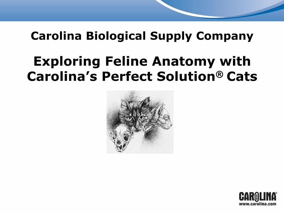
Exploring Feline Anatomy with Carolina’s Perfect Solution® Cats
Carolina Biological Supply Company
Objectives
• Introduce the general structure, anatomy, and physiology of the cat
• Explore the benefits of using the cat as a model organism for your next mammalian dissection
• Experience the quality of Carolina’s Perfect Solution® specimens
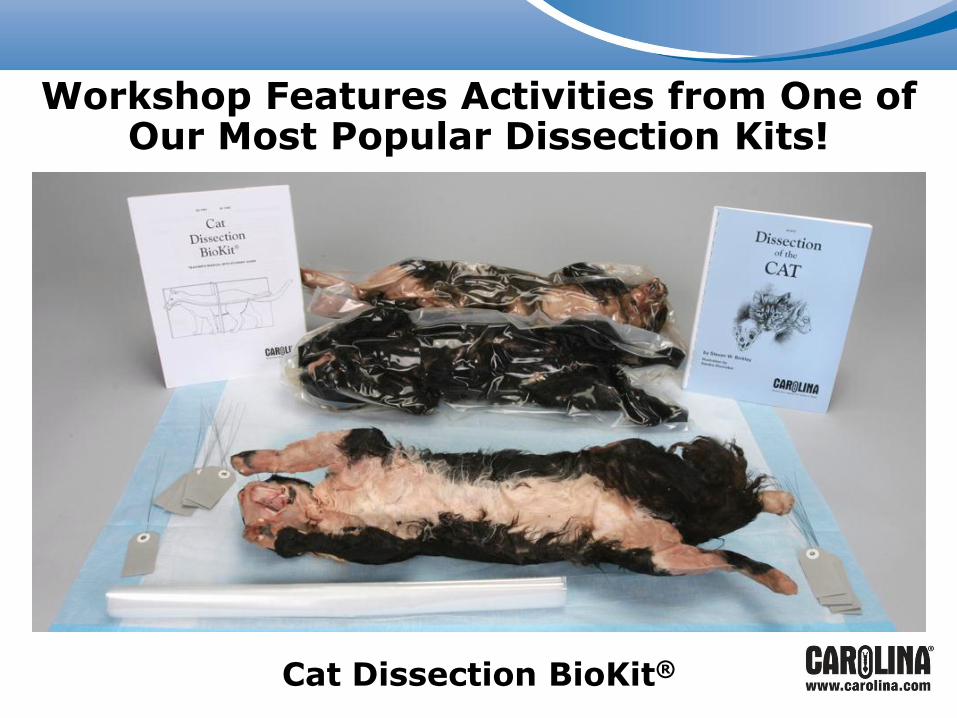
Workshop Features Activities from One of Our Most Popular Dissection Kits!
Cat Dissection BioKit®
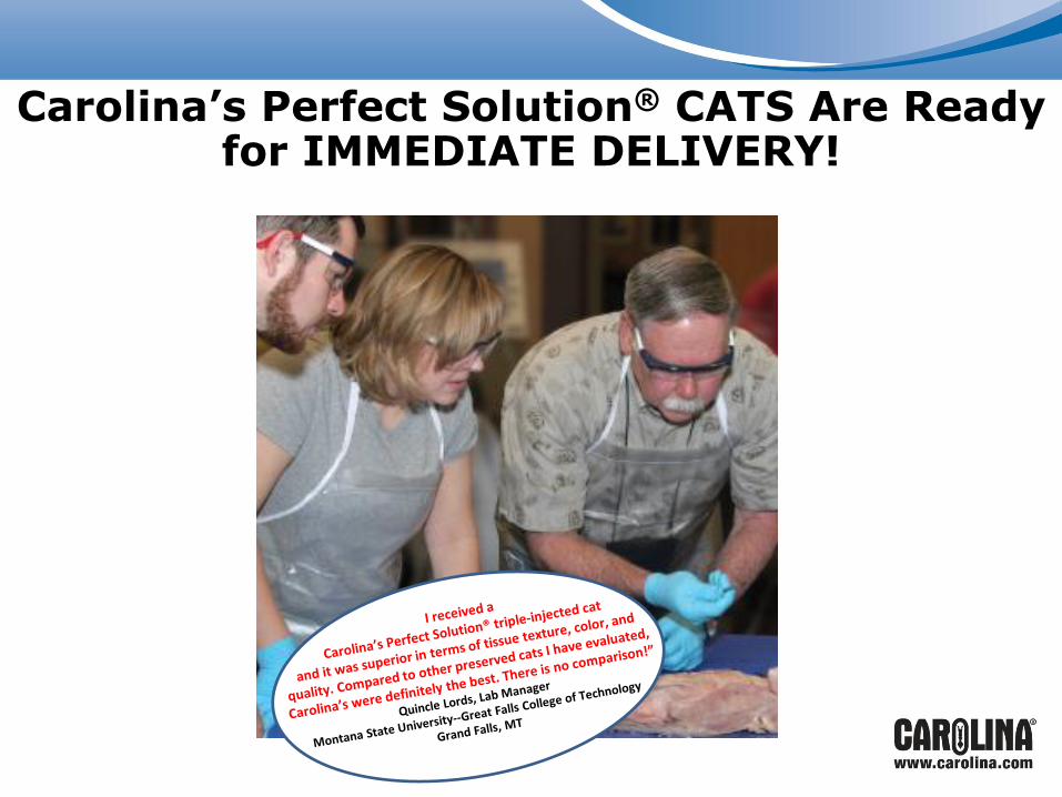
Carolina’s Perfect Solution® CATS Are Ready for IMMEDIATE DELIVERY!

We will work in groups of 4.
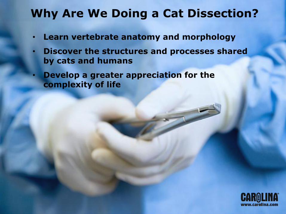
Why Are We Doing a Cat Dissection?
• Learn vertebrate anatomy and morphology
• Discover the structures and processes shared by cats and humans
• Develop a greater appreciation for the complexity of life
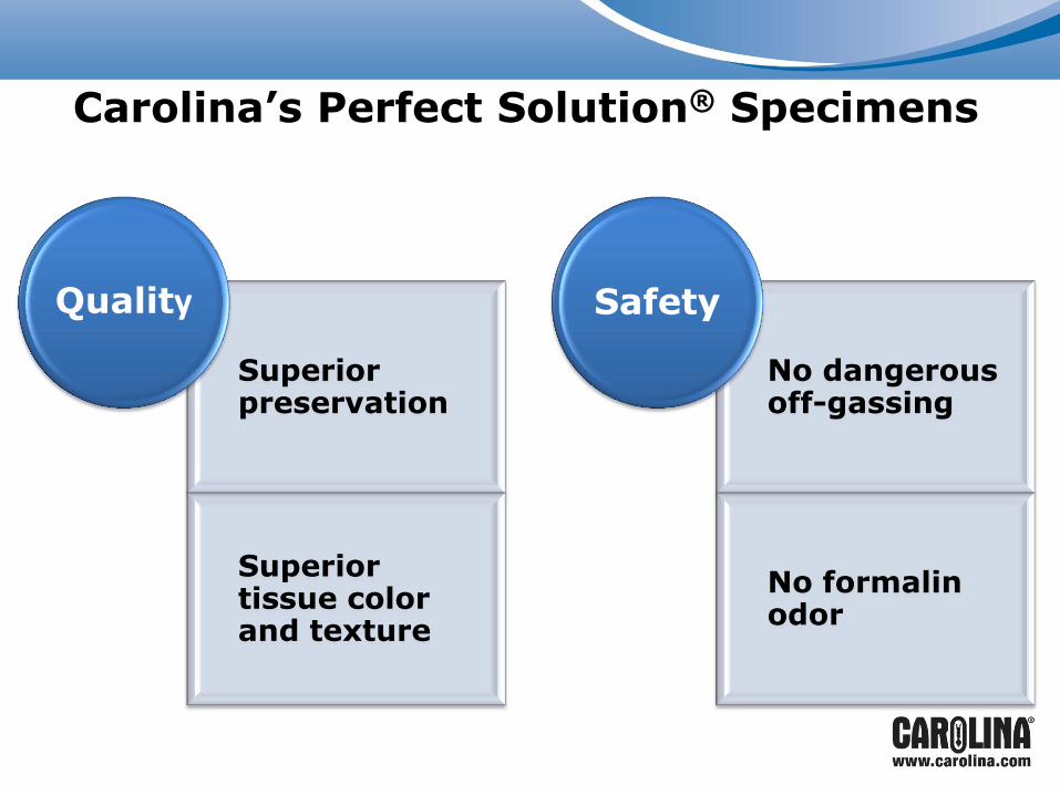
Carolina’s Perfect Solution® Specimens
Superior preservation
Superior tissue color and texture
Quality
No dangerous off-gassing
No formalin odor
Safety
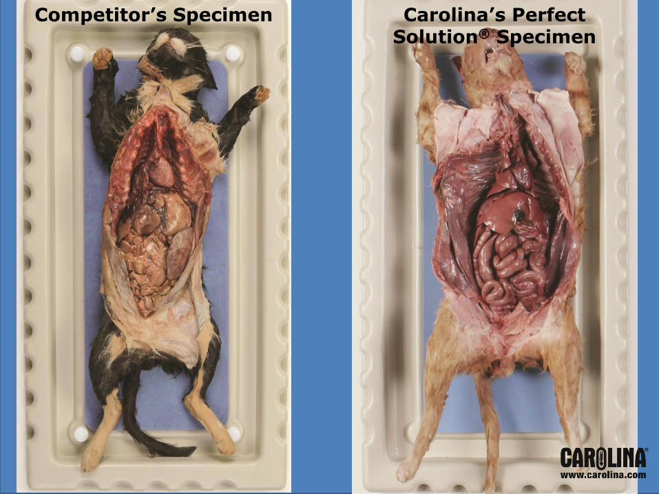
Carolina’s Perfect Solution® Specimen
Competitor’s Specimen
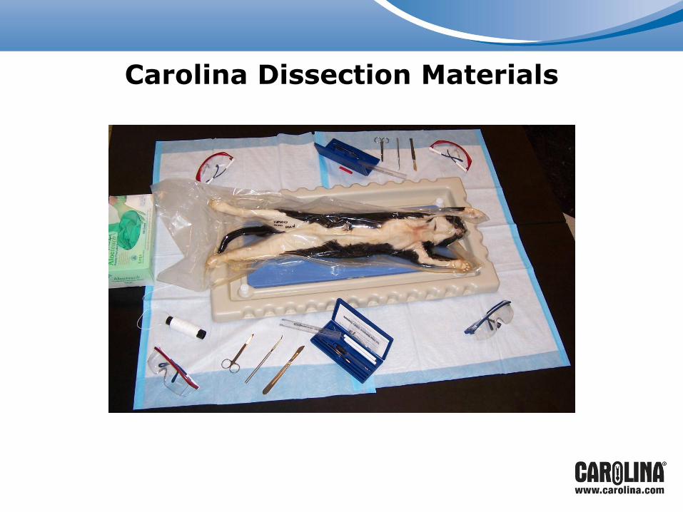
Carolina Dissection Materials

Safety Issues
• Personal Protective Equipment
Gloves, goggles, and lab aprons
• Dissection Tools
New tools = sharp scalpels
• Safety Tip
If you are not using an instrument, set it down
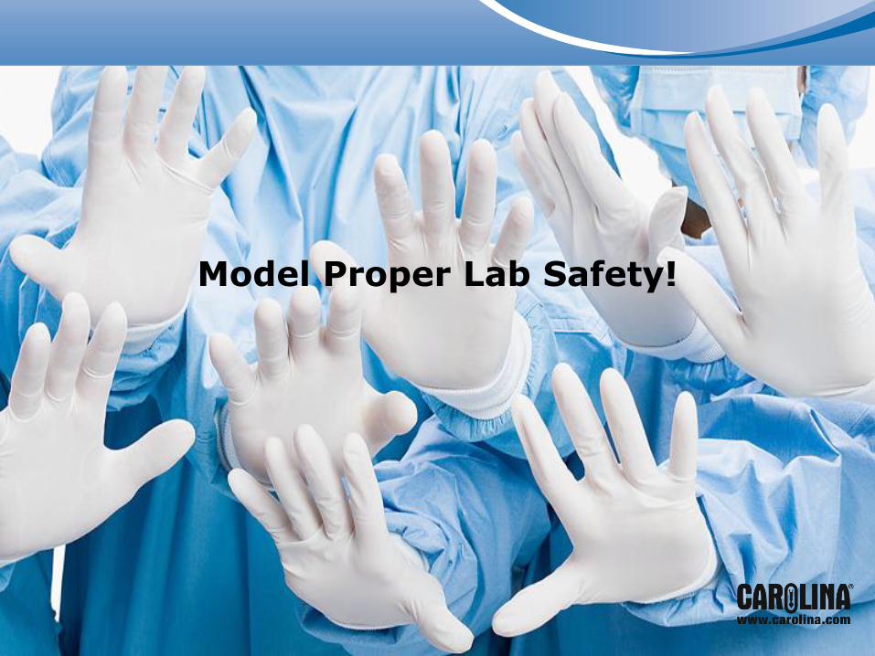
Model Proper Lab Safety!

Ready?
Lay out your instruments so they are easy to
access.
Organize your dissection area.
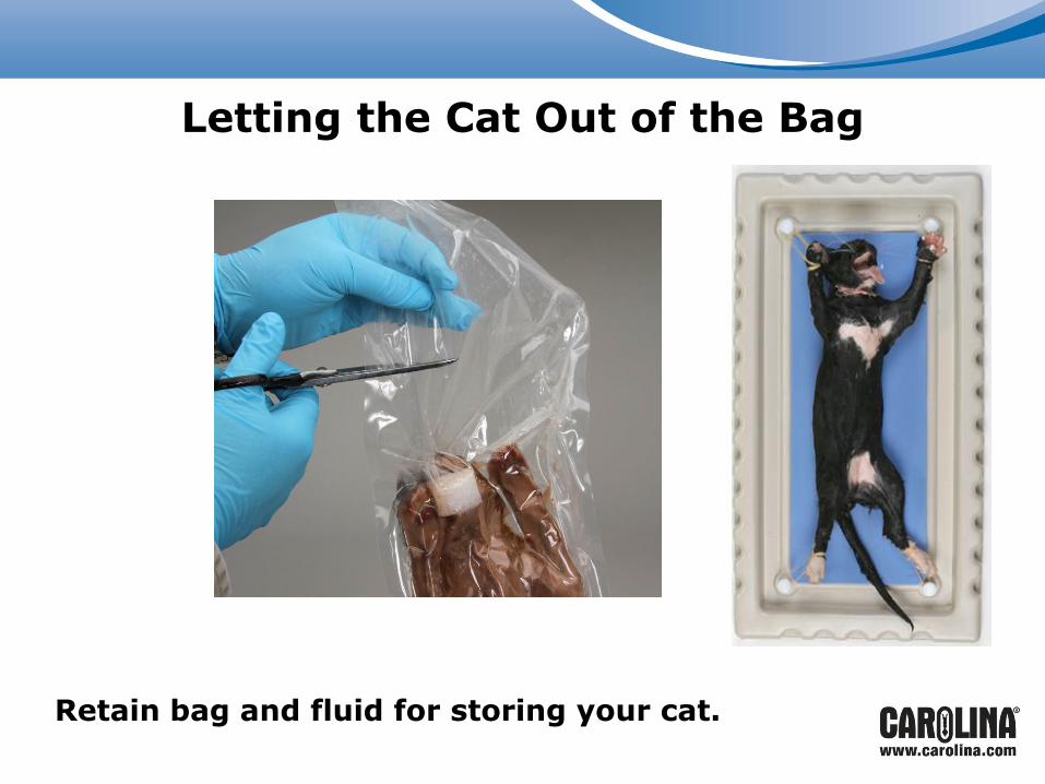
Letting the Cat Out of the Bag
Retain bag and fluid for storing your cat.
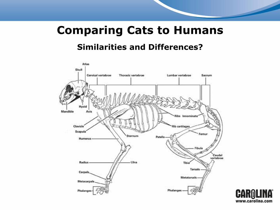
Comparing Cats to Humans
Similarities and Differences?
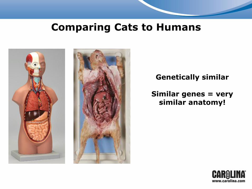
Comparing Cats to Humans
Genetically similar
Similar genes = very similar anatomy!
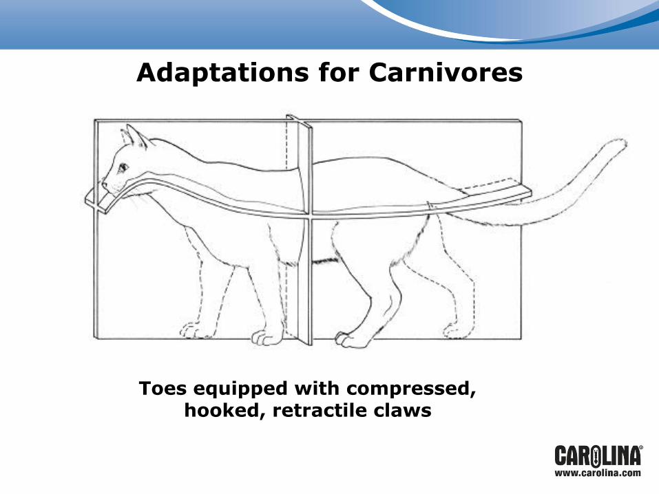
Adaptations for Carnivores
Agility…
The long tail
Toes equipped with compressed, hooked, retractile claws

Configuration of the Head
• Elongated jaw forms a muzzle
• Modified teeth
Carefully observe the teeth and compare them to human teeth.

Modification of Teeth
• Canines
• Incisors
• Molariforms (aka carnassial): designed for tearing tendons and ligaments of prey
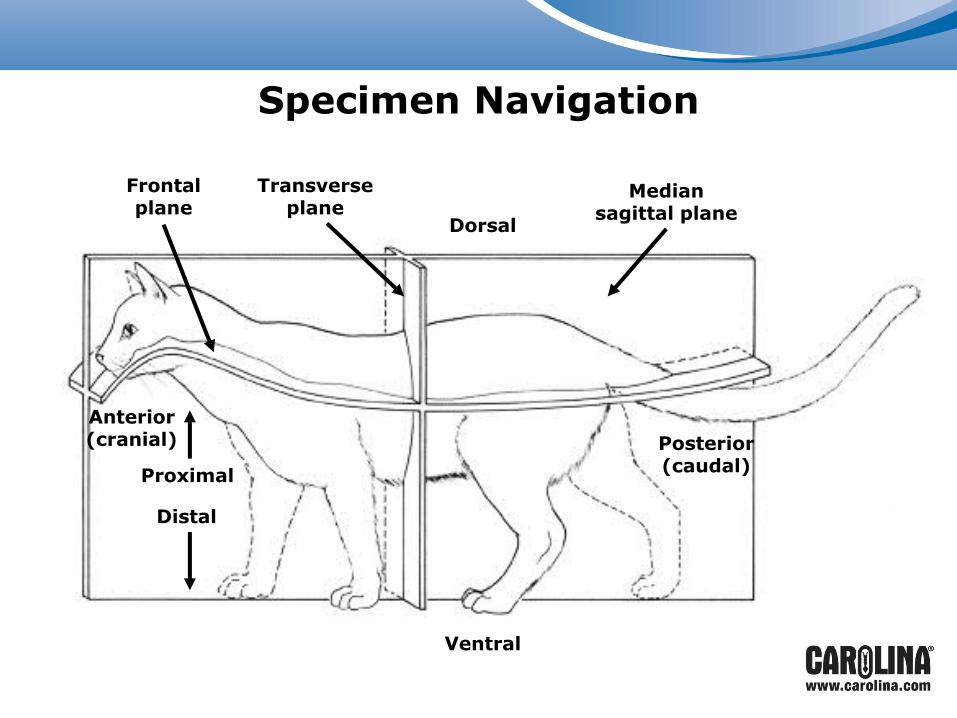
Specimen Navigation
Frontal plane
Transverse plane
Dorsal
Median sagittal plane
Posterior (caudal)
Ventral
Anterior (cranial)
Proximal
Distal
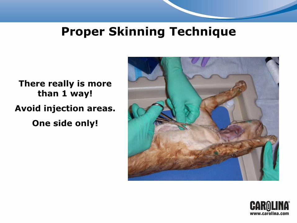
Proper Skinning Technique
There really is more than 1 way!
Avoid injection areas.
One side only!
For dissection by STUDENTS:
Allows for pelt to be placed around cat for storage after
each dissection session
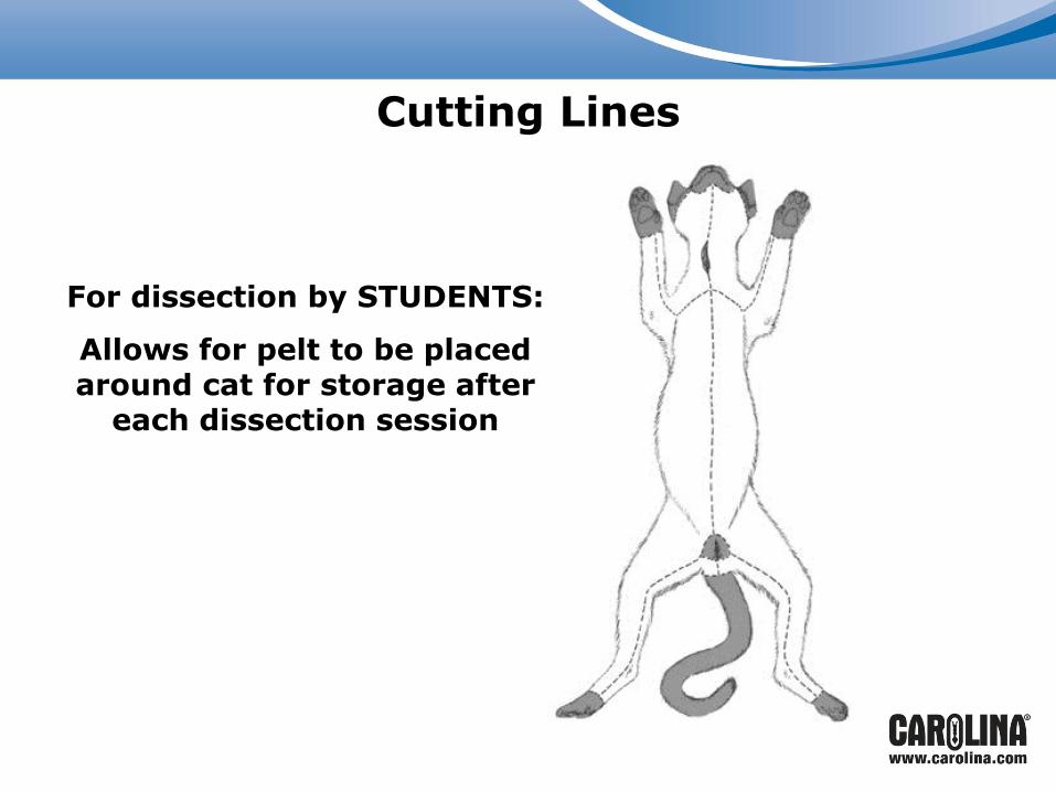
Cutting Lines
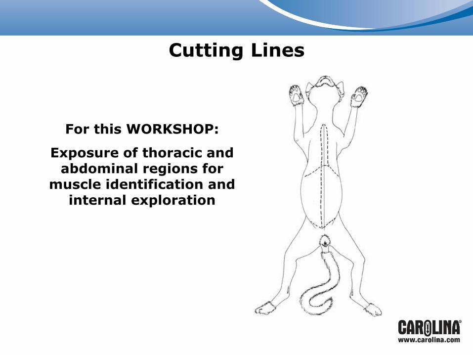
For this WORKSHOP:
Exposure of thoracic and abdominal regions for
muscle identification and internal exploration
Cutting Lines
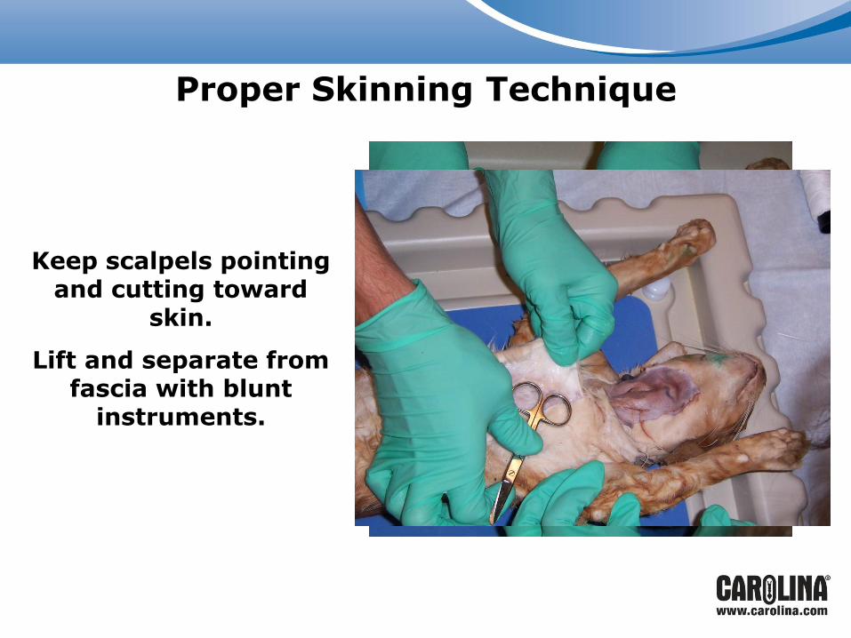
Proper Skinning Technique
Keep scalpels pointing
and cutting toward skin.
Lift and separate from fascia with blunt
instruments.
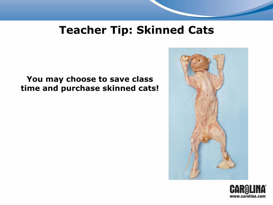
Teacher Tip: Skinned Cats
You may choose to save class time and purchase skinned cats!
Muscle Isolation
Opportunity for extensive study of:
• Morphology
• Kinesiology
• General muscle architecture
Muscle types:
• Smooth or striated
• Shape: convergent, strap, fan, or pinnate
• Points of origin and insertion marked by dense connective tissue
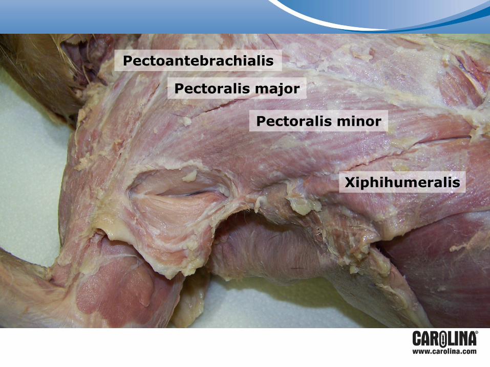
Pectoantebrachialis
Pectoralis major
Pectoralis minor
Xiphihumeralis
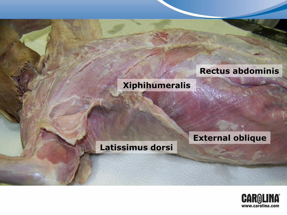
External oblique Latissimus dorsi
Rectus abdominis
Xiphihumeralis
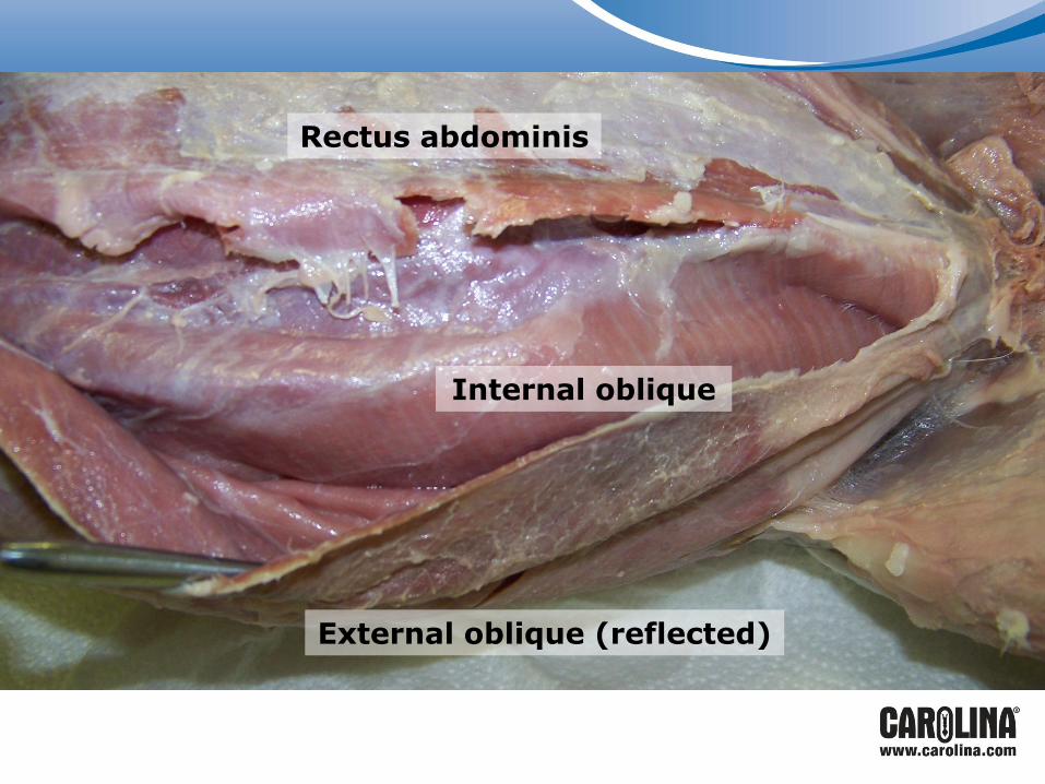
External oblique (reflected)
Internal oblique
Rectus abdominis
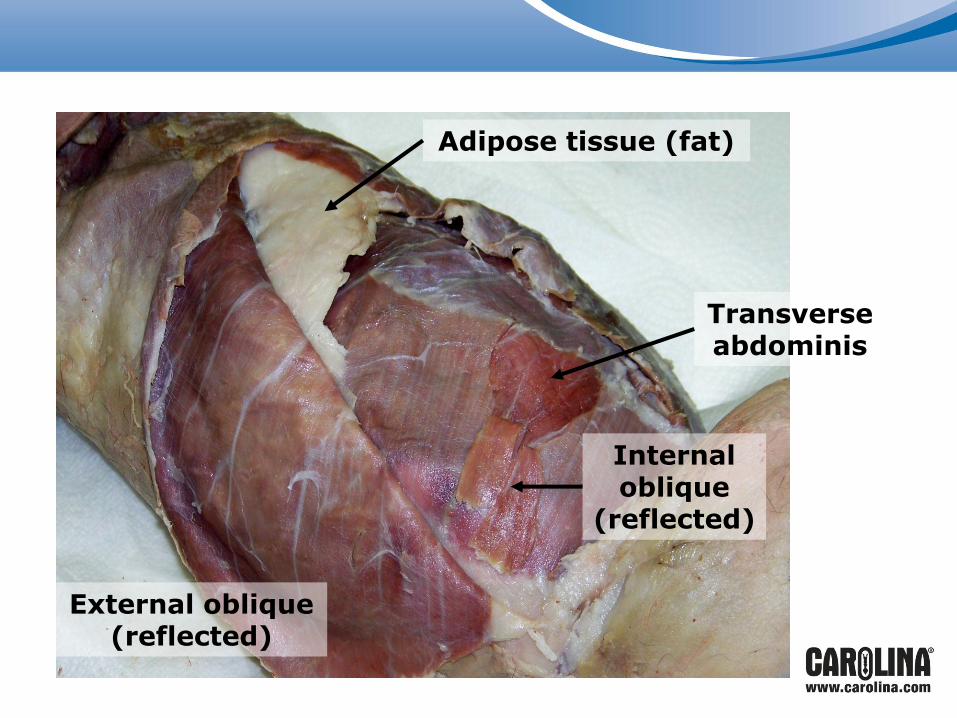
Adipose tissue (fat)
External oblique (reflected)
Internal oblique
(reflected)
Transverse abdominis
Opening the Thoracic Cavity
Use scalpel or scissors to expose throat/neck region and the sternal region.
Be careful to make shallow incisions so that the thyroid and thymus glands remain intact.
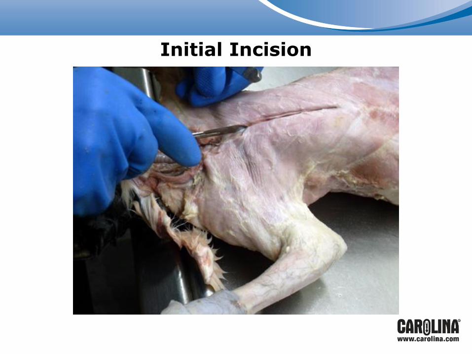
Initial Incision
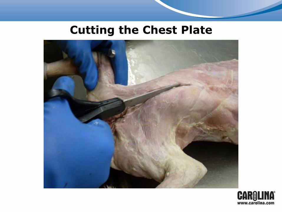
Cutting the Chest Plate

Thyroid gland
Trachea
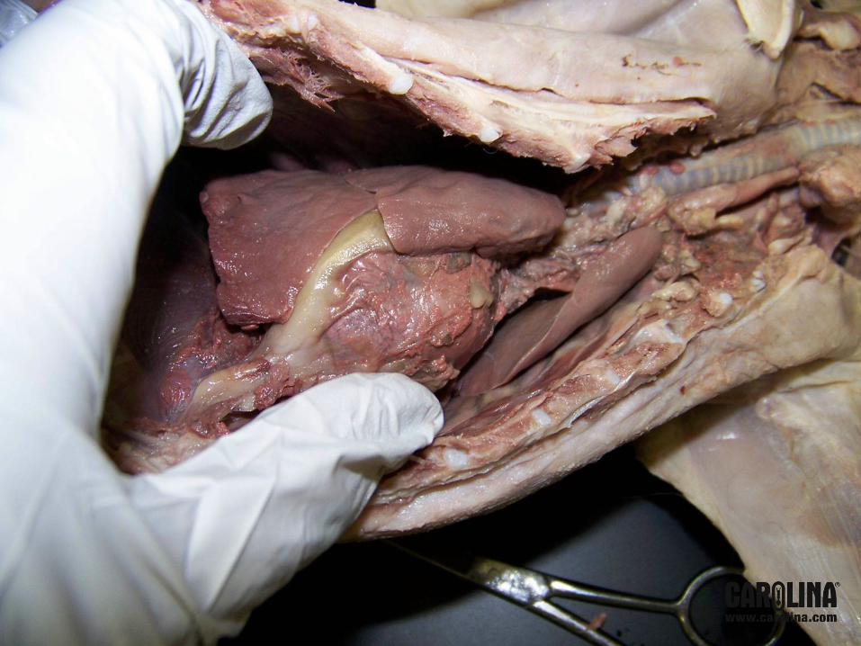
Thoracic Cavity
Cut away ribs to open thoracic region.
Observe thymus gland on the anterior surface of the heart prior to removing pericardial sac.
Remove pericardial sac.
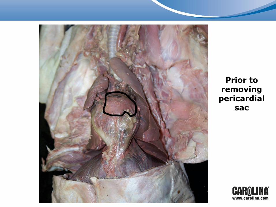
Prior to removing
pericardial sac
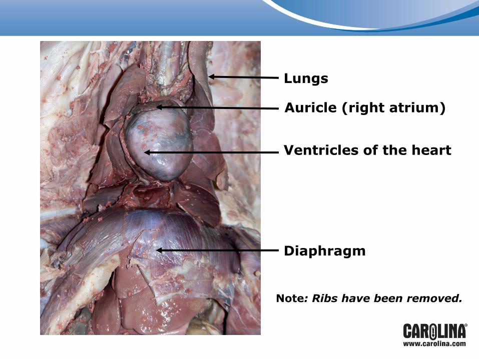
Ventricles of the heart
Diaphragm
Auricle (right atrium)
Lungs
Note: Ribs have been removed.
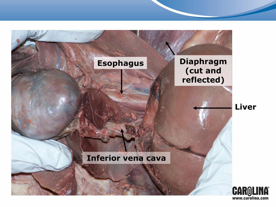
Diaphragm (cut and
reflected)
Liver
Inferior vena cava
Esophagus

Heading to the Abdominal Cavity
Muscle layer is very thin here and structures lie just below the surface.
Using the scissors or scalpel, make a midline incision.
When you reach the genital region, make a lateral incision through the inguinal region on each side.

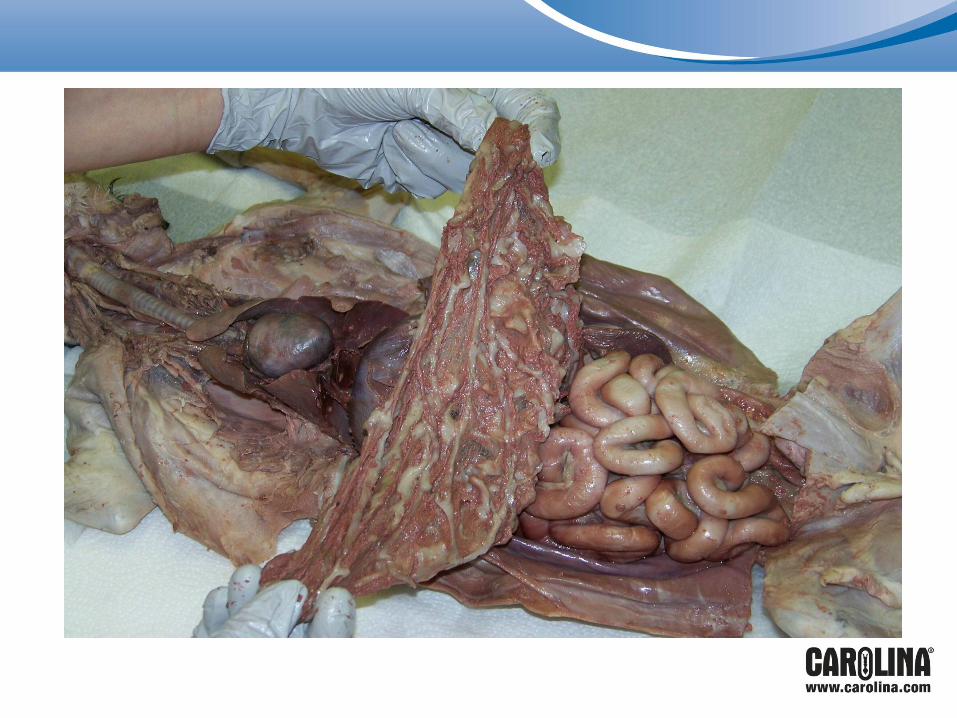
Liver Stomach
Spleen
Greater omentum

The Lesser and Greater Omentums
Peritoneal folds that appear like an apron over the abdominal organs.
Have points of attachment at the stomach, liver, small intestine, spleen, and colon.
Serve as fat storage.
Remove carefully along points of attachment.
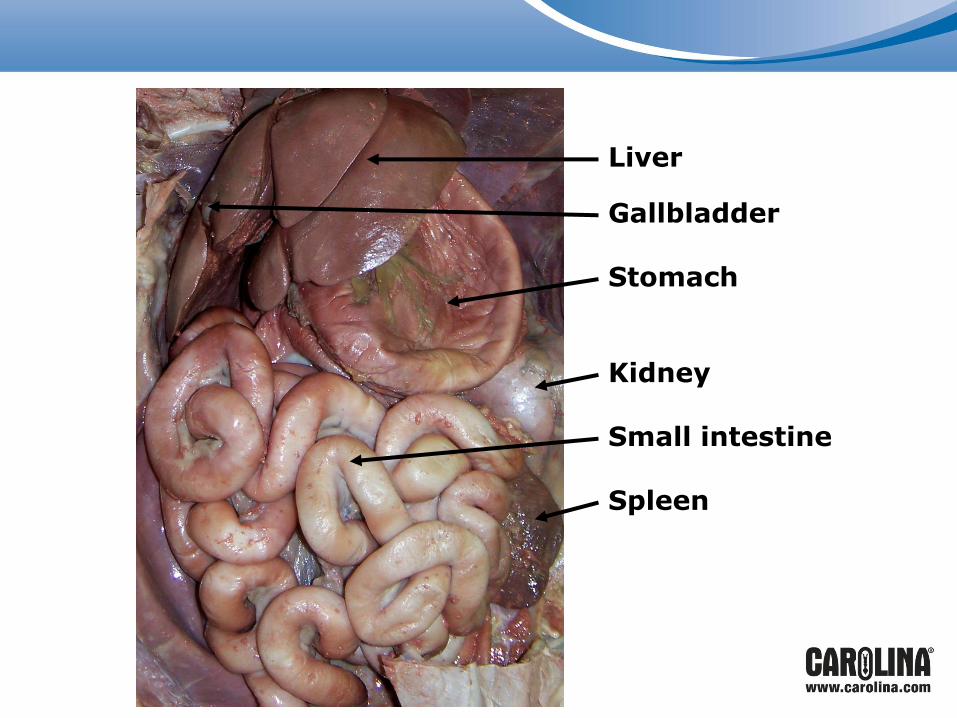
Liver
Gallbladder
Stomach
Kidney
Small intestine
Spleen

Liver
Gallbladder
Stomach
Small intestine
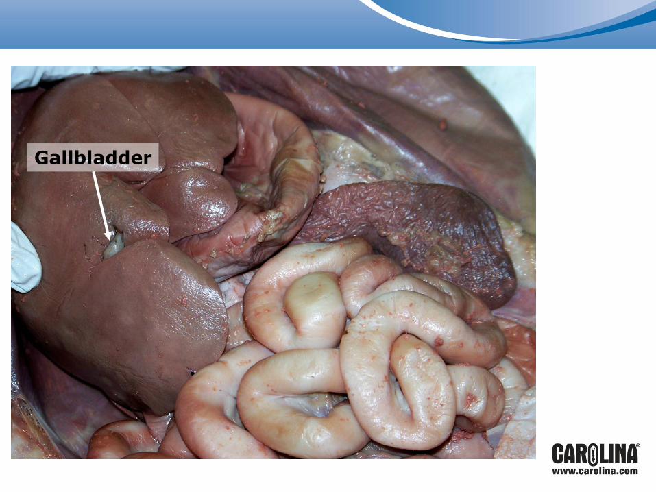
Gallbladder

Lobe of liver is reflected.
The gallbladder
and common bile duct are
exposed.
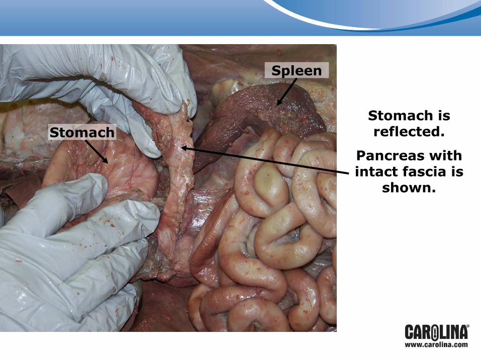
Stomach is reflected.
Pancreas with intact fascia is
shown.
Spleen
Stomach
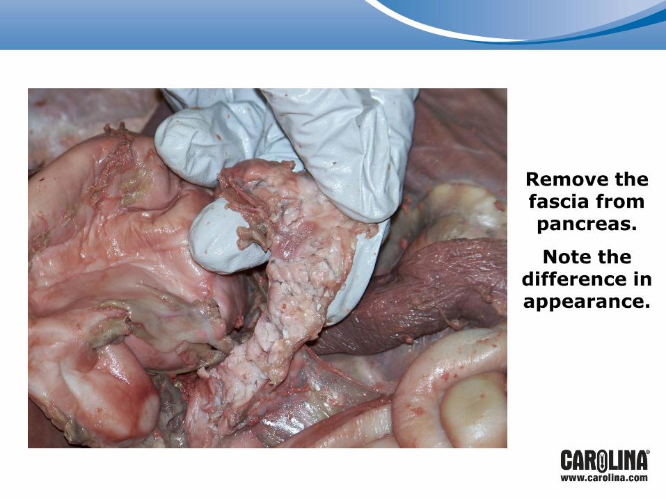
Remove the fascia from pancreas.
Note the difference in appearance.

Small and Large Intestines
Follow the small intestine from the attachment at the stomach.
Note the mesentery and the mesenteric blood vessels.
Note where the small intestine becomes the large intestine.
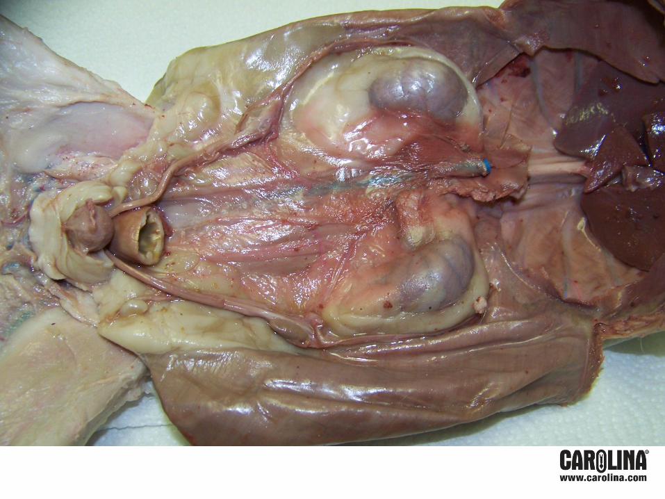
Viewing the Urogenital System
Cut the large intestine at the lowest point possible in the pelvis.
Reflect the intestines and other abdominal organs up and back or remove them at the top of the stomach to
view the urogenital system.
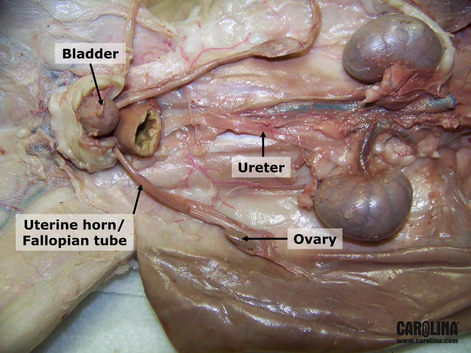
Ureter
Ovary Uterine horn/ Fallopian tube
Bladder

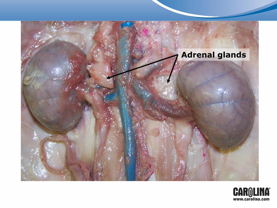
Adrenal glands

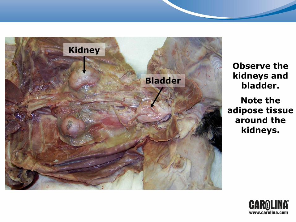
Observe the kidneys and
bladder.
Note the adipose tissue
around the kidneys.
Bladder
Kidney
Notes for Urogenital System
Adrenal glands in cats are more superior and medial than in humans; not directly on the anterior surface of
the kidney.
There is a great convergence of blood vessels and ureters in this area; injection makes this easier to see.
Cleanup Instructions
• KEEP GLOVES ON!
• Place ONLY animal waste in buckets.
• All other trash goes in trash bags.
• Wipe out pans, clean tools, and wipe off tables.
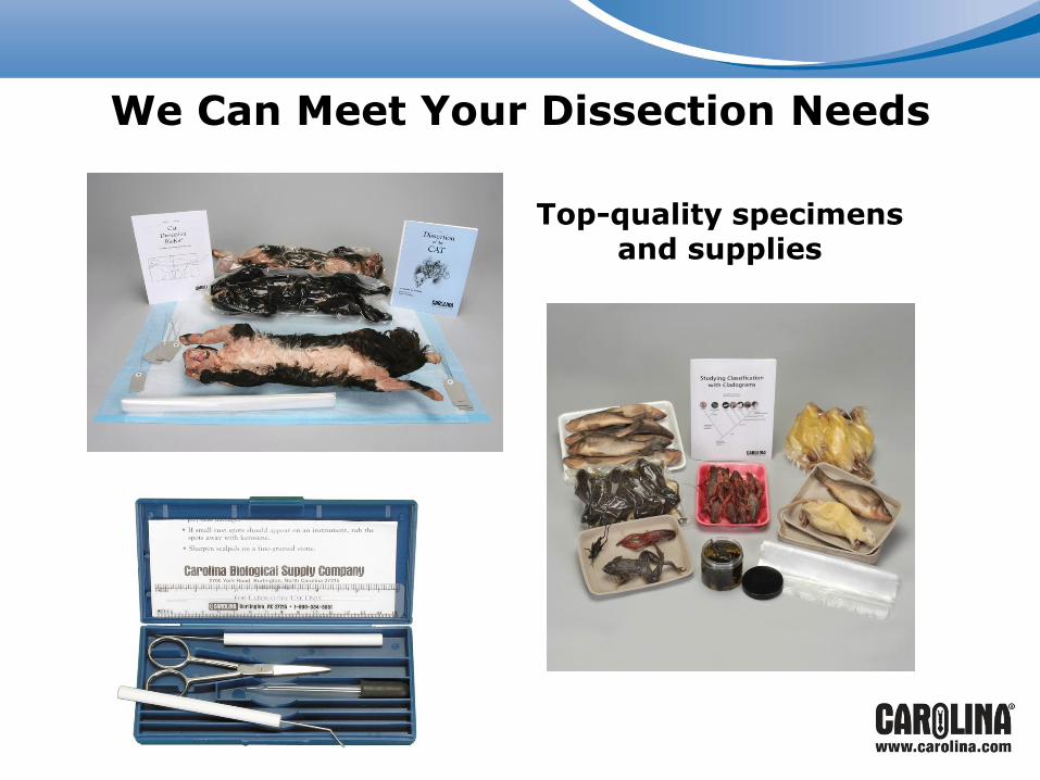
We Can Meet Your Dissection Needs
Top-quality specimens and supplies
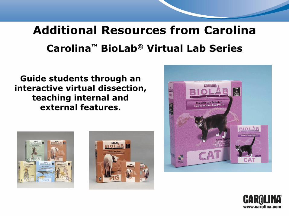
Additional Resources from Carolina
Carolina™ BioLab® Virtual Lab Series
Guide students through an interactive virtual dissection,
teaching internal and external features.

Carolina offers many free resources to support teachers.
Carolina’s Free Resources