Embed Size (px)
Citation preview
www.elsevier.com/locate/vetmic
Veterinary Microbiology 106 (2005) 49–60
Experimental reproduction of postweaning multisystemic
wasting syndrome (PMWS) in pigs in Sweden and Denmark
with a Swedish isolate of porcine circovirus type 2
F. Hasslung a,*, P. Wallgren b, A.-S. Ladekjær-Hansen c, A. Bøtner c, J. Nielsen c,E. Wattrang a, G.M. Allan d, F. McNeilly d, J. Ellis e, S. Timmusk a,
K. Belak b, T. Segall b, L. Melin b, M. Berg a, C. Fossum a
a Department of Molecular Biosciences, Section of Veterinary Immunology and Virology, Swedish University of Agricultural Sciences,
Biomedical Centre, PO Box 588, SE-751 23 Uppsala, Swedenb National Veterinary Institute, SE-751 89 Uppsala, Sweden
c Department of Virology, Danish Institute for Food and Veterinary Research, Lindholm, DK-4771 Kalvehave, Denmarkd Veterinary Sciences Division, Virology Section, Department of Agriculture and Rural Development for Northern Ireland,
Stormont, Belfast BT4 3SD, UKe Department of Veterinary Microbiology, Western College of Veterinary Medicine, University of Saskatchewan,
Saskatoon, SK, Canada S7N 5B4
Received 1 July 2004; received in revised form 19 November 2004; accepted 1 December 2004
Abstract
An experimental model using 3-day-old snatch-farrowed colostrum-deprived piglets co-infected with porcine circovirus type
2 (PCV2) and porcine parvovirus (PPV) is at present one of the best methods to study factors affecting development of
postweaning multisystemic wasting syndrome (PMWS). A Swedish isolate of PCV2 (S-PCV2) retrieved in 1993 from a healthy
pig has been used in this model to reproduce PMWS in pigs from Northern Ireland. This virus has been present in the Swedish
pig population for at least a decade without causing any known PMWS disease problems, despite its potential pathogenicity. The
reasons for this are unknown, but could be related to genetics, absence of triggers for PCV2 upregulation (infectious agent and/or
management forms) within Swedish pig husbandry. In order to confirm the pathogenicity of S-PCV2, Swedish and Danish pigs
were experimentally infected with this isolate according to the established model. Swedish pigs were also infected with a
reference isolate of PCV2 (PCV2-1010) to compare the severity of disease caused by the two isolates in Swedish pigs. Both
Danish and Swedish pigs developed PMWS after the experimental infection with S-PCV2. Antibodies to PCV2 developed later
and reached lower levels in serum from pigs infected with S-PCV2 than in pigs inoculated with PCV2-1010. In general, pigs
infected with S-PCV2 showed more severe clinical signs of disease than pigs infected with PCV2-1010, but pigs from all PCV2-
inoculated groups displayed gross and histological lesions consistent with PMWS. All pigs inoculated with PPV, alone or in
combination with PCV2, displayed interleukin-10 responses in serum while only pigs infected with PPV in combination with
* Corresponding author. Tel.: +46 18 471 46 93; fax: +46 18 471 43 82.
E-mail address: [email protected] (F. Hasslung).
0378-1135/$ – see front matter # 2005 Elsevier B.V. All rights reserved.
doi:10.1016/j.vetmic.2004.12.011
F. Hasslung et al. / Veterinary Microbiology 106 (2005) 49–6050
PCV2 showed interferon-a in serum on repeated occasions. Thus, the pathogenicity of S-PCV2 was confirmed and a role for
cytokines in the etiology of PMWS was indicated.
# 2005 Elsevier B.V. All rights reserved.
Keywords: PCV2; PMWS; Experimental infection; Sweden; Denmark
1. Introduction
Porcine circovirus type 2 (PCV2) is now accepted
as the causal agent of postweaning multisystemic
wasting syndrome (PMWS). However, it is also
recognised that other additional infectious (Allan
et al., 1999; Allan et al., 2000a; Krakowka et al., 2000)
or non-infectious factors (Rose et al., 2003) are
necessary for the full clinical expression of the
disease. PMWS was first observed in high health herds
in Canada in 1991 (Allan et al., 1998; Ellis et al.,
1998), and has since then rapidly become a major
problem in many pig-producing countries throughout
the world. Retrospective studies have demonstrated
that a PCV2 virus has been present in pigs for many
years prior to the recognition of PMWS without being
associated with any specific disease syndrome
(Rodriguez-Arrioja et al., 2003; Walker et al.,
2000). The global epizootic spread of PMWS since
1996 suggests that the PCV2 virus in pigs may have
mutated to a more pathogenic form or that another
agent in combination with PCV2 is necessary for the
development of PMWS. Alternatively, it has also been
suggested that the susceptibility of the host to PCV2-
associated clinical disease has, in some way, changed
due to alterations in the pig industry. Efforts to identify
new pathogenic genotypes of PCV2 (de Boisseson
et al., 2004) or new common co-infecting micro-
organism (Ellis et al., 2004) have, to date, failed and
epidemiological studies including numerous aspects
of husbandry forms have not yet revealed any
particular factor(s) that predispose for PMWS
(Larochelle et al., 2003; Pogranichniy et al., 2002;
Rose et al., 2003).
Dual infection with PCV2 and porcine parvovirus
(PPV) or porcine reproductive and respiratory
syndrome virus (PRRSV), as well as immuno-
modulators, have been used to successfully reproduce
PMWS experimentally in pigs (for review see Allan
et al., 2004) and one of the most reproducible infection
models involves co-infection with PCV2 and PPV
in 3-day-old snatch-farrowed colostrum-deprived
(SFCD) piglets (Allan et al., 1999). This model was
recently used in Northern Ireland to demonstrate the
potential pathogenicity of a Swedish isolate of PCV2
from 1993 (Allan et al., 2003). The Swedish PCV2 (S-
PCV2) was isolated from a clinically healthy pig,
which was raised in a SPF-herd that seroconverted to
PCV2 at that time (Wattrang et al., 2002).
Sweden remained free from PMWS until Decem-
ber 2003, and as of October 2004, only 12 farms have
been diagnosed as affected by the disease. It has been
suggested that differences in pig husbandry practices,
animal genetics and/or viral pathogenesis could have
contributed to relative freedom of Swedish pigs from
disease. The present experimental infection with S-
PCV2 and PPV was conducted using Swedish and
Danish pigs. To allow comparison between various
PCV2 isolates, one group of Swedish pigs was also
infected with a reference isolate of PCV2 (Imp. 1010).
Clinical manifestations of disease, histological
lesions, levels of virus antigen in affected tissues
and development of antibodies to PCV2 were recorded
and the IL-10 and interferon-a responses were
determined in serum obtained from Swedish pigs.
2. Materials and methods
2.1. Experimental model and virus
An experimental model, using a dual infection with
porcine circovirus type 2 (PCV2) and porcine
parvovirus (PPV) for induction of PMWS was used
as previously described (Allan et al., 1999). Two
isolates of PCV2 were used: PCV2-1010 (PCV2
Stoon) isolated from an outbreak of PMWS in a high
health herd in Canada (Ellis et al., 1998) and S-PCV2
isolated from a lymph node collected in 1993 from a
clinically healthy pig reared in a Swedish SPF-herd
(Allan et al., 2003; Wattrang et al., 2002). For co-
infection, PPV (isolate 1005 pool 7) recovered from
F. Hasslung et al. / Veterinary Microbiology 106 (2005) 49–60 51
PMWS-affected pigs was used (Allan et al., 2000b;
Krakowka et al., 2000). All viruses were propagated in
a PCV2-free cell line (PK/15A) as previously
described (Allan et al., 2003).
2.2. Experimental animals
The experimental infections were performed in two
sets, one using Swedish pigs and the other using
Danish pigs. In the first set, Swedish pigs were
snatched-farrowed (SF) and hand-reared on colostrum
and milk substitutes (snatch-farrowed colostrum-
deprived (SFCD)), whereas the Danish pigs were
caesarean-derived colostrum-deprived (CDCD). The
Swedish pigs were obtained from a conventional
piglet-producing herd. To date, the farm has not
reported any PCV2-associated disease problems and
the herd is free from infections with Salmonella spp,
Sarcoptes scabei, Serpulina hyodysenteriae and toxin
producing strains of Pasteurella multocida. The sows
were vaccinated against Escherichia coli, Erysipelo-
thrix rhusiopatiae and PPV. The piglets, originating
from five litters (crossed Hampshire, Yorkshire and
Swedish Landrace) designated A–E. The piglets were
transported within 1 h after birth to the Animal
Department at the National Veterinary Institute (NVI),
Uppsala, Sweden, and distributed into four groups that
were housed in separate rooms with individual
ventilation in a clean but not sterile environment.
During the first days of life, the piglets were hand
reared on bovine colostrum substitute (Calf Volos-
trum, Volac International Ltd., UK) and subsequently
on commercial pig milk substitute (Piggi milk,
Manufacturer No. 2077, UK) and pellets (Primary
Elite, Primary Diets Ltd., UK). All animals were
treated with intramuscular injections of antibiotics
(Nuflor, Florfenicol 30 g/100 ml 0.2 ml/pig, day) for
the first 3 days, and thereafter orally (Baytril,
enrofloxacin 25 mg/ml 0.2 ml/pig, day) once daily
throughout the experimental period. Pigs showing
severe signs of disease were euthanised and 35 of the
initially 42 piglets were included in the experiment,
which was approved by the Ethical Committee for
Animal Experiments, Uppsala, Sweden.
In the second set, eight Danish piglets (nos. 1–8)
Danish Landrace–Yorkshire crossbreds were derived
from two sows originating from the SPF-herd at the
Danish Institute for Food and Veterinary Research
(DFVF), Kalvehave, Denmark. This herd is free from
PCV1 and PCV2, PRRSV, swine influenza virus
(SIV), porcine respiratory coronavirus (PRCV), as
well as mycoplasms. The sows were vaccinated
against PPV. The experimental infection of the CDCD
piglets was carried out at the isolation facilities of
DFVF and the pigs were housed in a separate section
of the building. In all other aspects, rearing of the pigs
including colostrum and milk substitutes used and
antibiotic treatments was the same as described for the
Swedish SFCD pigs.
2.3. Experimental infection
On the 3rd day of life, all piglets were inoculated
with 1.0 ml per nare of cell lysates from indicated cell
culture. Pigs in Swedish group 1 (n = 8) were mock
inoculated with cell culture supernatant from unin-
fected PK/15A cells; pigs in Swedish group 2 (n = 8)
were inoculated with PPV 107.0TCID50 alone; pigs in
Swedish group 3 (n = 10) were inoculated with a
mixture of PCV2-1010 105.5TCID50 and PPV
107.0TCID50; and pigs in Swedish group 4 (n = 9),
as well as the Danish pigs in group 5 (n = 8), were
inoculated with S-PCV2 105.5TCID50 and PPV
107.0TCID50.
2.4. Sampling procedures
The health status of all individual pigs was assessed
twice daily and pigs developing signs of severe disease
were euthanised and necropsied (see below). For
Swedish pigs, weights and rectal temperatures were
measured twice every week. Blood samples were
collected from vena cava cranialis in evacuated test
tubes prior to infection, and on days 8, 15, 22 and 28
(groups 1–4) or 4, 7, 14, 21 and 27 post infection
(group 5). At blood sampling, individual faecal
samples were collected from pigs in groups 1–4,
and samples of faeces were collected from the floor of
each of these groups daily.
At the end of the trial, 4 weeks post infection, all
remaining pigs were sacrificed, autopsied and gross
lesions recorded. General bacteriological examination
of samples from spleen, liver and lung tissues was
performed by routine methods at the Bacteriological
Departments at NVI and DFVF, respectively. Tissue
samples were collected from lymph nodes and tonsils,
F. Hasslung et al. / Veterinary Microbiology 106 (2005) 49–6052
lungs, heart, liver, kidneys, spleen, small intestine and
any other tissues showing gross pathological changes.
Tissues were fixed in 4% paraformaldehyde or
buffered formalin for histopathological analysis or
rapidly frozen in isopropanol on dry ice and stored in
liquid nitrogen for immunohistochemical staining.
2.5. Histopathological examinations and
immunostaining of cryostat sections
Sections of fixed, paraffin-embedded tissue were
stained with hematoxylin and eosin for morphological
evaluation. The severity of lesions was scored as none
visible (�), mild (+), moderate (++) or severe (+++).
Blocks of tissue from spleen, liver and lymph node
were stained with PCV2-specific polyclonal (Ellis
et al., 1998) or monoclonal antibodies (McNeilly
et al., 2001) as previously described. The presence of
PCV2 was determined on cryostat sections and scored
as negative (�) or containing minimal (+) up to
abundant (+++++) amount of PCV2 antigen.
2.6. Quantitative PCR for estimation of viral DNA
in serum
Levels of PCV DNA were determined in serum
samples collected from piglets in group 5 by
quantitative PCR (Q-PCR) as described elsewhere
(Ladekjaer-Mikkelsen et al., 2002). Results were
presented as mean values of duplicate reactions.
2.7. PCR for detection of PCV2 in organs
TwosetsofPCRmethods fordetection ofPCV2were
designed. One that detected both PCV2 isolates used in
this study, and one that discriminates between PCV2-
1010 and S-PCV2. For the first set, PCR primers were
selected based on sequence alignments of the genomes
of several PCV isolates, and two primers, 50 CAG CAA
GAA GAA TGG AAG 30 and 50 TAT GTG GTT TCC
GGG TCT 30 were selected for the initial PCR product.
For the discriminative PCR, primers were chosen
according to sequence differences within the ORF2
regions, 50 AAG TAA TCA ATA GTT CTA 30 being
specific for S-PCV2 and 50 AAG TAATCA ATA GTG
GAG 30 being specific for PCV2-1010. These primers
were used in combination with an ORF2 full-length
primer. The specificity of both PCR methods was
verified using purified viral DNA from each isolate as
control. Samples of DNA were purified from splenic
tissue of experimental pigs according to standard
protocols,usingproteinase-Kdigestion,phenol–chisam
extraction and ethanol precipitation.
2.8. Sequence analysis of PCV2 isolates
The full-length DNA sequence of the S-PCV2
isolate used in the experimental infection was
compared to the sequence of PCV2 from the original
lymph node preserved in liquid nitrogen since 1993.
Both genomes were amplified by PCR using ORF1 R
(50 AAA GGATCC TCA GTA ATT TAT TTC ATA 30)and ORF2 R (50 TTT AAG CTT CCA TGA CGT ATC
CAA GGA GG 30) primers. The PCR products were
subsequently ligated into the T-Vector (pGEM-T Easy
Vector System I, Promega) according to the manu-
facturer’s instructions.
Sequence analysis of the PCR products was
performed by standard procedures used at the Depart-
ment of Animal Breeding and Genetics, Section of
Disease Genetics, at the Swedish University of
AgriculturalSciences, Uppsala, andatUppsalaGenome
Centre, Uppsala University, Sweden. Alignments were
made using DNASTAR and pairwise comparison of
nucleotide and amino acid sequence identities was
performed using MEGALIGN software version 1.13
(DNASTAR, Wisconsin). The nucleotides were num-
bered in analogy with Meehan et al., 1998.
2.9. Detection of antibodies to PCV2 and PPV in
serum
Antibodies to PCV2 were detected in an immuno-
peroxidase monolayer assay (IPMA) as described
elsewhere (Allan et al., 2000b; Ladekjaer-Mikkelsen
et al., 2002). Sera were examined for antibodies to
PPV by blocking ELISA (Madsen et al., 1997) or by a
competetive ELISA (Svanovir1 PPV-Ab, Svanova
Biotech, Uppsala, Sweden).
2.10. Detection of IFN-a and IL-10 in serum
Serum IFN-a was determined in samples from the
piglets in groups 1–4 by a dissociation-enhanced
lanthanide fluoroimmunoassay (DELFIA), as described
earlier (Artursson et al., 1995). The DELFIA, which is
F. Hasslung et al. / Veterinary Microbiology 106 (2005) 49–60 53
based on two mAbs directed to porcine IFN-a, had a
lower detection limit of 0.3 U IFN-a/ml. Presence of IL-
10 was determined in serum samples diluted 1:2 by
ELISA (Biosource, Camarillo, CA) according to the
manufacturers description. The sensitivity of the ELISA
is 3 pg/ml which corresponded to an absorbance value
(A450 nm) of approximately 0.2.
3. Results
3.1. Clinical observations and gross lesions at
necropsy
Among the Swedish pigs, 10 animals in total
belonging to all four experimental groups developed
transient diarrhoea within the first 4 days post
infection (DPI). Five of these pigs were sacrificed
within the first 5 DPI due to moribund conditions.
From three of these animals, enrofloxacin and
florphenicol resistant Klebsiella pneumoniae were
isolated from liver, intestine, spleen and peritoneal
cavity. From one pig, Pseudomonas aeruginosa
intermediate sensitive to enrofloxacin and florphenicol
resistant were isolated from lungs, intestine, liver and
spleen. Among pigs in group 5, no pathogenic bacteria
were detected at necropsy, except for lung tissue of
piglet 4 from which non-haemolytic E. coli was
isolated. Apart from these early manifestations of
disease, no pigs in groups 1 (mock) or 2 (PPV)
developed signs of clinical disease during the
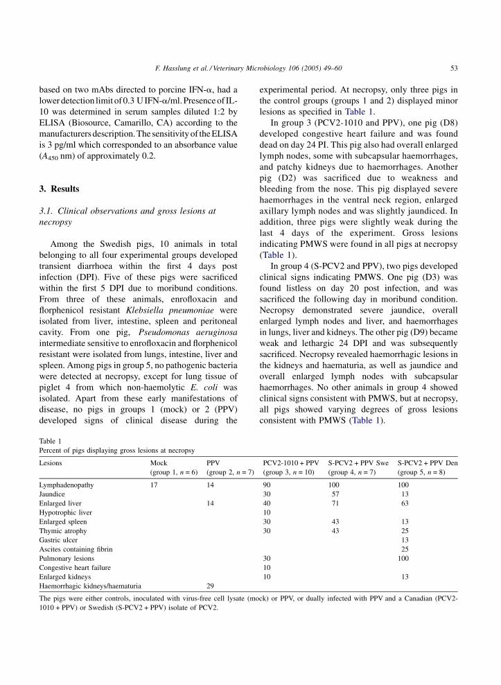
Table 1
Percent of pigs displaying gross lesions at necropsy
Lesions Mock
(group 1, n = 6)
PPV
(group 2, n = 7)
Lymphadenopathy 17 14
Jaundice
Enlarged liver 14
Hypotrophic liver
Enlarged spleen
Thymic atrophy
Gastric ulcer
Ascites containing fibrin
Pulmonary lesions
Congestive heart failure
Enlarged kidneys
Haemorrhagic kidneys/haematuria 29
The pigs were either controls, inoculated with virus-free cell lysate (mo
1010 + PPV) or Swedish (S-PCV2 + PPV) isolate of PCV2.
experimental period. At necropsy, only three pigs in
the control groups (groups 1 and 2) displayed minor
lesions as specified in Table 1.
In group 3 (PCV2-1010 and PPV), one pig (D8)
developed congestive heart failure and was found
dead on day 24 PI. This pig also had overall enlarged
lymph nodes, some with subcapsular haemorrhages,
and patchy kidneys due to haemorrhages. Another
pig (D2) was sacrificed due to weakness and
bleeding from the nose. This pig displayed severe
haemorrhages in the ventral neck region, enlarged
axillary lymph nodes and was slightly jaundiced. In
addition, three pigs were slightly weak during the
last 4 days of the experiment. Gross lesions
indicating PMWS were found in all pigs at necropsy
(Table 1).
In group 4 (S-PCV2 and PPV), two pigs developed
clinical signs indicating PMWS. One pig (D3) was
found listless on day 20 post infection, and was
sacrificed the following day in moribund condition.
Necropsy demonstrated severe jaundice, overall
enlarged lymph nodes and liver, and haemorrhages
in lungs, liver and kidneys. The other pig (D9) became
weak and lethargic 24 DPI and was subsequently
sacrificed. Necropsy revealed haemorrhagic lesions in
the kidneys and haematuria, as well as jaundice and
overall enlarged lymph nodes with subcapsular
haemorrhages. No other animals in group 4 showed
clinical signs consistent with PMWS, but at necropsy,
all pigs showed varying degrees of gross lesions
consistent with PMWS (Table 1).
PCV2-1010 + PPV
(group 3, n = 10)
S-PCV2 + PPV Swe
(group 4, n = 7)
S-PCV2 + PPV Den
(group 5, n = 8)
90 100 100
30 57 13
40 71 63
10
30 43 13
30 43 25
13
25
30 100
10
10 13
ck) or PPV, or dually infected with PPV and a Canadian (PCV2-
F. Hasslung et al. / Veterinary Microbiology 106 (2005) 49–6054
In group 5 (S-PCV2 and PPV), three pigs (nos. 2, 4
and 8) either died or were euthanised during the
experimental period. Piglet 4 appeared pale and
lethargic 5 DPI, experienced severe respiratory
distress, and was euthanised 8 DPI. Piglet 2 was
found dead on 19 DPI. Prior to this, it had suffered
from respiratory distress and appeared lethargic with
intermittently observed tremors. It was also pale and
anorexic 2–3 days prior to dying. Pig 8 was euthanised
19 DPI in a moribund condition after showing
pronounced respiratory distress since almost 2 weeks.
In addition, this pig showed inappetence, appeared
pale and lethargic and suffered from tremor attacks 2
days prior to euthanasia. The remaining five piglets
(nos. 1, 3, 5, 6 and 7) showed mild clinical symptoms
primarily characterized by periods of respiratory
distress and lethargic appearance intermittently during
the entire experimental period. All pigs in group 5
displayed gross lesions consistent with PMWS at
necropsy (Table 1).
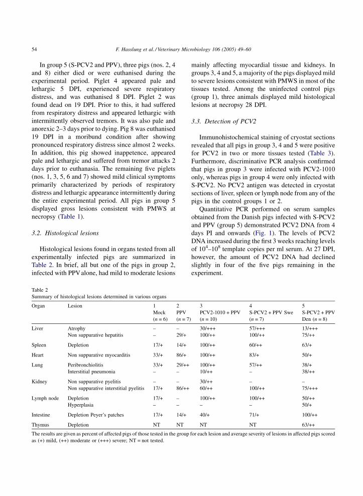
3.2. Histological lesions
Histological lesions found in organs tested from all
experimentally infected pigs are summarized in
Table 2. In brief, all but one of the pigs in group 2,
infected with PPValone, had mild to moderate lesions
Table 2
Summary of histological lesions determined in various organs
Organ Lesion 1 2
Mock
(n = 6)
PPV
(n = 7)
Liver Atrophy – –
Non suppurative hepatitis – 29/+
Spleen Depletion 17/+ 14/+
Heart Non suppurative myocarditis 33/+ 86/+
Lung Peribronchiolitis 33/+ 29/++
Interstitial pneumonia – –
Kidney Non suppurative pyelitis – –
Non suppurative interstitial pyelitis 17/+ 86/++
Lymph node Depletion 17/+ –
Hyperplasia – –
Intestine Depletion Peyer’s patches 17/+ 14/+
Thymus Depletion NT NT
The results are given as percent of affected pigs of those tested in the group
as (+) mild, (++) moderate or (+++) severe; NT = not tested.
mainly affecting myocardial tissue and kidneys. In
groups 3, 4 and 5, a majority of the pigs displayed mild
to severe lesions consistent with PMWS in most of the
tissues tested. Among the uninfected control pigs
(group 1), three animals displayed mild histological
lesions at necropsy 28 DPI.
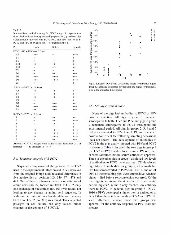
3.3. Detection of PCV2
Immunohistochemical staining of cryostat sections
revealed that all pigs in group 3, 4 and 5 were positive
for PCV2 in two or more tissues tested (Table 3).
Furthermore, discriminative PCR analysis confirmed
that pigs in group 3 were infected with PCV2-1010
only, whereas pigs in group 4 were only infected with
S-PCV2. No PCV2 antigen was detected in cryostat
sections of liver, spleen or lymph node from any of the
pigs in the control groups 1 or 2.
Quantitative PCR performed on serum samples
obtained from the Danish pigs infected with S-PCV2
and PPV (group 5) demonstrated PCV2 DNA from 4
days PI and onwards (Fig. 1). The levels of PCV2
DNA increased during the first 3 weeks reaching levels
of 104–108 template copies per ml serum. At 27 DPI,
however, the amount of PCV2 DNA had declined
slightly in four of the five pigs remaining in the
experiment.
3 4 5
PCV2-1010 + PPV
(n = 10)
S-PCV2 + PPV Swe
(n = 7)
S-PCV2 + PPV
Den (n = 8)
30/+++ 57/+++ 13/+++
100/++ 100/++ 75/++
100/++ 60/++ 63/+
100/++ 83/+ 50/+
100/++ 57/++ 38/+
10/++ – 38/++
30/++ – –
60/++ 100/++ 75/+++
100/++ 100/++ 50/++
– – 50/+
40/+ 71/+ 100/++
NT NT 63/++
for each lesion and average severity of lesions in affected pigs scored
F. Hasslung et al. / Veterinary Microbiology 106 (2005) 49–60 55
Table 3
Immunohistochemical staining for PCV2 antigen in cryostat sec-
tions obtained from liver, spleen and lymph nodes (Ly node) of pigs
experimentally infected with PCV2-1010 and PPV (no. 3) or S-
PCV2 and PPV in Sweden (no. 4) or Denmark (no. 5)
Pig Liver Spleen Ly node
PCV2-1010 + PPV (no. 3 Swe)
A7 +++ + ++++
B1 + � +
B6 + +/� ++
B11 ++ ++ +++
B14 + +/� ++
C1 + � +
D1 + ++ +++
D2 +++ ++++ +++
D8 +++ ++++ ++++
E5 + + ++
S-PCV2 + PPV (no. 4 Swe)
A3 +++ ++ +++
B7 + ++ +++
B8 ++ ++ +++
C3 + � +
D3 + +++ ++
D9 +++ +++ +++
E3 ++ +++ +++
S-PCV2 + PPV (no.5 Den)
1 + ++ ++
2 + ++ ++++
3 + ++ ++
4 � + +
5 ++ +++ ++++
6 ++ ++++ ++++
7 ++ ++++ ++++
8 + ++ +++
Amounts of PCV2 antigen were scored as not detectable (�), or
minimal (+/�) to abundant (+++++).
Fig. 1. Levels of PCV2 viral DNA found in sera from Danish pigs in
group 5, expressed as number of viral template copies for individual
pigs at the indicated time points.
3.4. Sequence analysis of S-PCV2
Sequence comparison of the genome of S-PCV2
used in the experimental infection and PCV2 retrieved
from the original lymph node revealed differences in
five nucleotides at position 103, 346, 374, 478 and
491. One of these exchanges caused a substitution of
amino acids (no. 27) located in ORF1. In ORF2, only
one exchange of nucleotides (no. 103) was found, not
leading to any change in amino acid sequence. In
addition, an intronic nucleotide deletion between
ORF1 and ORF2 (no. 315) was found. Thus, repeated
passages in cell culture had only caused minor
changes in the genome of S-PCV2.
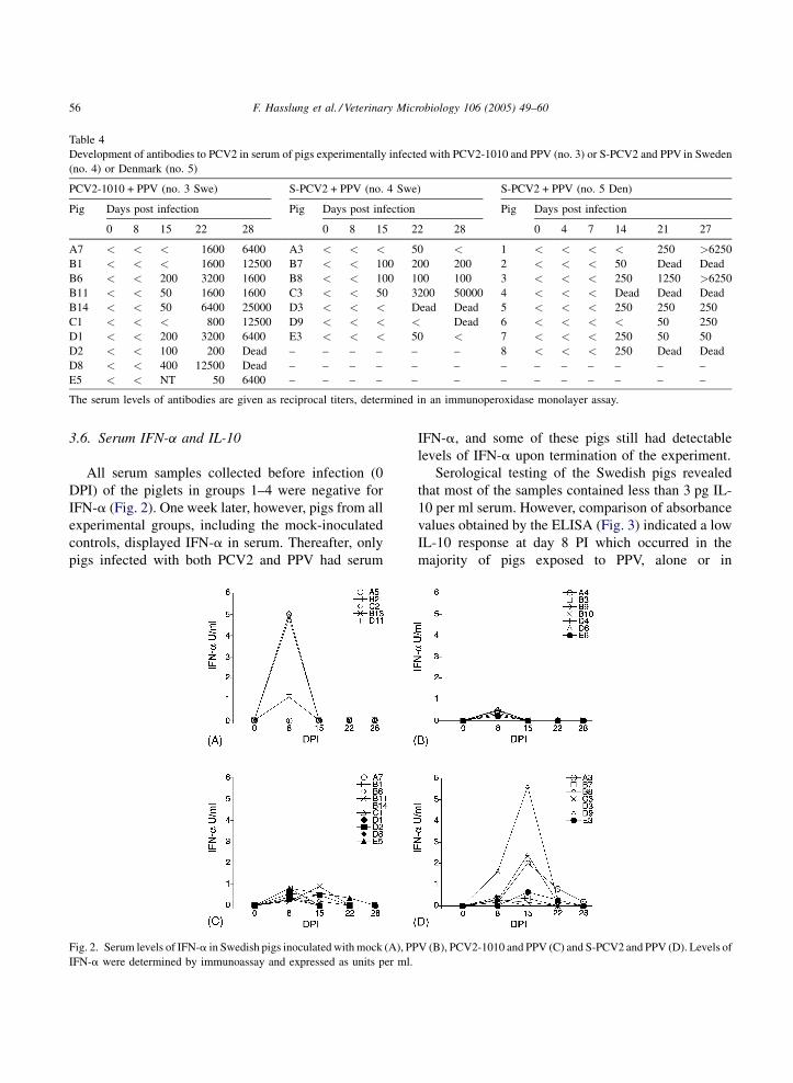
3.5. Serologic examinations
None of the pigs had antibodies to PCV2 or PPV
prior to infection. All pigs in group 1 remained
seronegative to both PCV2 and PPV, and pigs in group
2 remained seronegative to PCV2 throughout the
experimental period. All pigs in groups 2, 3, 4 and 5
had seroconverted to PPV 1 week PI, and remained
positive for PPV at the following sampling occassions
(data not shown). The development of antibodies to
PCV2 in the pigs dually infected with PPV and PCV2
is shown in Table 4. In brief, the two pigs in group 4
(S-PCV2 + PPV) that developed clinical PMWS, died
or were sacrificed before serum antibodies appeared.
Three of the other pigs in group 4 displayed low levels
of antibodies to PCV2, whereas one (C3) developed
high titers of antibodies. In group 5, all animals but
two had seroconverted to PCV2 on 14 DPI, and on 21
DPI, all the remaining pigs were seropositive, whereas
piglet 4 died before seroconversion occurred. Of the
five piglets surviving the 4 weeks of experimental
period, piglets 5, 6 and 7 only reached low antibody
titers to PCV2. In general, pigs in group 3 (PCV2-
1010 + PPV) developed a higher titer of antibodies to
PCV2 than those infected with S-PCV2 and PPV. No
such difference between these two groups was
apparent for the antibody response to PPV (data not
shown).
F. Hasslung et al. / Veterinary Microbiology 106 (2005) 49–6056
Table 4
Development of antibodies to PCV2 in serum of pigs experimentally infected with PCV2-1010 and PPV (no. 3) or S-PCV2 and PPV in Sweden
(no. 4) or Denmark (no. 5)
PCV2-1010 + PPV (no. 3 Swe) S-PCV2 + PPV (no. 4 Swe) S-PCV2 + PPV (no. 5 Den)
Pig Days post infection Pig Days post infection Pig Days post infection
0 8 15 22 28 0 8 15 22 28 0 4 7 14 21 27
A7 < < < 1600 6400 A3 < < < 50 < 1 < < < < 250 >6250
B1 < < < 1600 12500 B7 < < 100 200 200 2 < < < 50 Dead Dead
B6 < < 200 3200 1600 B8 < < 100 100 100 3 < < < 250 1250 >6250
B11 < < 50 1600 1600 C3 < < 50 3200 50000 4 < < < Dead Dead Dead
B14 < < 50 6400 25000 D3 < < < Dead Dead 5 < < < 250 250 250
C1 < < < 800 12500 D9 < < < < Dead 6 < < < < 50 250
D1 < < 200 3200 6400 E3 < < < 50 < 7 < < < 250 50 50
D2 < < 100 200 Dead – – – – – – 8 < < < 250 Dead Dead
D8 < < 400 12500 Dead – – – – – – – – – – – – –
E5 < < NT 50 6400 – – – – – – – – – – – – –
The serum levels of antibodies are given as reciprocal titers, determined in an immunoperoxidase monolayer assay.
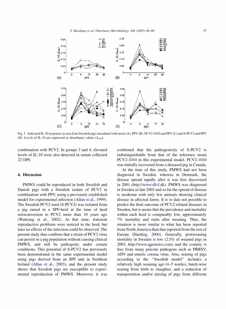
3.6. Serum IFN-a and IL-10
All serum samples collected before infection (0
DPI) of the piglets in groups 1–4 were negative for
IFN-a (Fig. 2). One week later, however, pigs from all
experimental groups, including the mock-inoculated
controls, displayed IFN-a in serum. Thereafter, only
pigs infected with both PCV2 and PPV had serum
Fig. 2. Serum levels of IFN-a in Swedish pigs inoculated with mock (A), PP
IFN-a were determined by immunoassay and expressed as units per ml.
IFN-a, and some of these pigs still had detectable
levels of IFN-a upon termination of the experiment.
Serological testing of the Swedish pigs revealed
that most of the samples contained less than 3 pg IL-
10 per ml serum. However, comparison of absorbance
values obtained by the ELISA (Fig. 3) indicated a low
IL-10 response at day 8 PI which occurred in the
majority of pigs exposed to PPV, alone or in
V (B), PCV2-1010 and PPV (C) and S-PCV2 and PPV (D). Levels of
F. Hasslung et al. / Veterinary Microbiology 106 (2005) 49–60 57
Fig. 3. Indicated IL-10 responses in sera from Swedish pigs inoculated with mock (A), PPV (B), PCV2-1010 and PPV (C) and S-PCV2 and PPV
(D). Levels of IL-10 are expressed as absorbance values (A450).
combination with PCV2. In groups 3 and 4, elevated
levels of IL-10 were also detected in serum collected
22 DPI.
4. Discussion
PMWS could be reproduced in both Swedish and
Danish pigs with a Swedish isolate of PCV2 in
combination with PPV, using a previously established
model for experimental infection (Allan et al., 1999).
The Swedish PCV2 used (S-PCV2) was isolated from
a pig raised in a SPF-herd at the time of herd
seroconversion to PCV2 more than 10 years ago
(Wattrang et al., 2002). At that time, transient
reproductive problems were noticed in the herd, but
later no effects of the infection could be observed. The
present study thus confirms that a strain of PCV2 virus
can persist in a pig population without causing clinical
PMWS, and still be pathogenic under certain
conditions. This potential of S-PCV2 has previously
been demonstrated in the same experimental model
using pigs derived from an SPF unit in Northern
Ireland (Allan et al., 2003), and the present study
shows that Swedish pigs are susceptible to experi-
mental reproduction of PMWS. Moreover, it was
confirmed that the pathogenicity of S-PCV2 is
indistinguishable from that of the reference strain
PCV2-1010 in this experimental model. PCV2-1010
was initially recovered from a diseased pig in Canada.
At the time of this study, PMWS had not been
diagnosed in Sweden, whereas in Denmark, the
disease spread rapidly after it was first discovered
in 2001 (http://www.dfvf.dk). PMWS was diagnosed
in Sweden in late 2003 and so far the spread of disease
is moderate with only few animals showing clinical
disease in affected farms. It is to date not possible to
predict the final outcome of PCV2-related diseases in
Sweden, but it seems that the prevalence and mortality
within each herd is comparably low, approximately
7% mortality and runts after weaning. Thus, the
situation is more similar to what has been reported
from North America than that reported from the rest of
Europe (Harding, 2004). Generally, postweaning
mortality in Sweden is low (2.5% of weaned pigs in
2003, http://www.qgenetics.com) and the country is
free from many porcine pathogens such as PRRSV,
ADV and enteric corona virus. Also, rearing of pigs
according to the ‘‘Swedish model’’ includes a
relatively high weaning age (4–5 weeks), batch-wise
rearing from birth to slaughter, and a reduction of
transportation and/or mixing of pigs from different
F. Hasslung et al. / Veterinary Microbiology 106 (2005) 49–6058
litters. All of these factors may contribute to the
relative resistance of the Swedish pig industry to
PCV2-related diseases, although it is now clear that
Swedish pigs can develop PWMS both experimentally
and naturally. Notably, the first PMWS outbreaks in
Sweden occurred in herds where high production
intensity had resulted in deviations from this
‘‘Swedish model’’.
The clinical and pathological evidence of disease in
S-PCV2 and PPV co-infected animals was similar in
the Swedish and Danish experiments presented herein.
To further evaluate the pathogenicity of S-PCV2, one
group of Swedish pigs was infected with a reference
strain of PCV2 (Imp. 1010, Stoon) in combination
with PPV. No pigs in this group died from PMWS, and
no animal showed clinical signs of disease related to
PMWS during the experimental period. At necropsy,
however, all pigs from this group displayed gross
lesions consistent with PMWS. The combined
manifestation of clinical disease and pathological
lesions caused by the two isolates indicates that the
pathogenicity of the Swedish isolate is equal to or even
higher than that of PCV2-1010. In two previous
studies applying the same experimental model,
however, the proportions of pigs developing clinical
PMWS was somewhat larger, both concerning PCV2-
1010 and S-PCV2 (Allan et al., 2003; Allan et al.,
1999; Allan et al., 2004). This discrepancy could
reflect a diminished pathogenicity of PCV2-1010,
possibly caused by several in vitro passages of the
virus, but variations in the experimental conditions
cannot be excluded.
To rule out that S-PCV2 had undergone major
changes during the isolation procedure, a comparison
of the genomes of S-PCV2 before and after repeated
passages in cell culture was performed. This revealed
only minor changes, indicating that the virus used in
the experimental infection was very similar to the
Swedish field isolate from 1993. As previously
described, this isolate differs from PCV2-1010 in
the nucleotide sequence of ORF2 (Allan et al., 2003)
causing eight amino acid substitutions. Nucleotide
sequence analysis revealed several differences
between S-PCV2 used in the experimental infection
and PCV2 isolated in December 2003 from the first
natural cases of PMWS in Sweden (data not shown).
The occurrence of IFN-a in serum samples
collected on day 8 from pigs in groups 1–4 indicates
that some pigs experienced infections unrelated to the
experimental infections. This was also evident from
early deaths among pigs in all experimental groups,
which in most cases were related to navel infections,
and could be expected for pigs snatched-farrowed in a
conventional farm. On later sampling occasions,
however, IFN-a could only be detected in sera from
pigs in groups 3 and 4 that had been inoculated with
PCV2. In contrast, elevated serum IL-10 was indicated
in pigs infected with PPValone or in combination with
PCV2. This suggests that PPV induces a systemic
response of this cytokine, which might be of
importance for the development of PMWS in the
experimental co-infection with PCV2. In natural cases
of PMWS, an over-expression of IL-10 mRNA has
been found in PBMC (Sipos et al., 2004) and
thymocytes (Darwich et al., 2003b), and PBMC from
PMWS affected pigs preferentially produced IL-10
and IFN-g at in vitro re-exposure to PCV2 (Darwich
et al., 2003a). If an altered cytokine profile is caused
by progressing PMWS or induced by other factors that
might predispose for PMWS, remains however to be
determined. Interestingly, PRRSV infections, often
considered as a co-factor for development of PMWS,
induced increased expression of IL-10 mRNA in
PBMC, lung tissue and bronchoalveolar cells (Chung
and Chae, 2003; Johnsen et al., 2002; Suradhat and
Thanawongnuwech, 2003). Thus, if IL-10 expression
is important for PMWS development, the PRRSV free
status of Sweden may well explain the lower incidence
of PMWS recorded within Swedish herds.
The impact of PPV infections in the pathogenesis of
PMWS was recently demonstrated under field con-
ditions where PMWS was induced in segregated early-
weaned 6–7 week old pigs co-infected with PPV and
PCV2 (Opriessnig et al., 2004), which further justifies
the current experimental model. It should, however, be
noted that the permanent treatment with broad-
spectrum antibiotics severely affected the normal
bacterial flora of the gut. Pen floor samples revealed
less than 102 CFU coliforms per gram faeces (data not
shown) compared to 108–1010 in samples from non-
treated pigs of the same age category (Melin et al.,
2000). The importance of a normal microflora for the
development of both mucosal and systemic immunity
has been described for several species, including
gnotobiotic piglets (Tlaskalova-Hogenova et al.,
2004). In addition to the antibiotic treatment, piglets
F. Hasslung et al. / Veterinary Microbiology 106 (2005) 49–60 59
were handfed with cow-milk based colostrum sub-
stitutes during the first days of life to avoid passive
transfer of porcine immunoglobulins. This treatment
might further affect the function of the gut associated
lymphoid tissue of the experimental pigs. Since large
amounts of PCV2 are secreted through faeces during
PMWS, the gut and surrounding tissues are likely to be
involved in the replication of PCV2 as well as of PPV.
Thus, several non-physiological effects are caused by
the present experimental model, which have to be
considered for a better understanding of co-factors
that contribute to the development of PMWS. Never-
theless, the results presented here clearly show that a
virus strain that persisted for 10 years in a SPF-herd
without causing clinical PMWS was able to induce
this disease under experimental conditions in pigs of
different origin. Further studies concerning the effects
of husbandry forms as well as genetics of the animals
and microorganisms are needed to elucidate why the
spread of PMWS differs between regions.
Acknowledgements
This work was supported by FORMAS, AgriFun-
Gen and the Programme for Biology of Infection at the
Veterinary Faculty, Swedish University of Agricul-
tural Sciences. Financial support was also provided by
the European Union (Project QLK2-CT-1999-00445),
Danish Research Agency (No. 23-02-0079) and
(Canadian) Natural Sciences and Engineering
Research Council-Collaborative Research Opportu-
nities grant 234281-00. The authors also wish to thank
Barbro Hogberg and Per Carlsson for invaluable help
with the animals, as well as Lisbeth Fuxler and Maria
Persson for excellent laboratory work.
References
Allan, G.M., McNeilly, F., Kennedy, S., Daft, B., Clarke, E.G., Ellis,
J.A., Haines, D.M., Meehan, B.M., Adair, B.M., 1998. Isolation
of porcine circovirus-like viruses from pigs with a wasting
disease in the USA and Europe. J. Vet. Diagn. Invest. 10, 3–10.
Allan, G.M., Kennedy, S., McNeilly, F., Foster, J.C., Ellis, J.A.,
Krakowka, S.J., Meehan, B.M., Adair, B.M., 1999. Experimen-
tal reproduction of severe wasting disease by co-infection of pigs
with porcine circovirus and porcine parvovirus. J. Comp. Pathol.
121, 1–11.
Allan, G.M., McNeilly, F., Ellis, J., Krakowka, S., Meehan, B.,
McNair, I., Walker, I., Kennedy, S., 2000a. Experimental infec-
tion of colostrum deprived piglets with porcine circovirus 2
(PCV2) and porcine reproductive and respiratory syndrome
virus (PRRSV) potentiates PCV2 replication. Arch. Virol.
145, 2421–2429.
Allan, G.M., McNeilly, F., Meehan, B.M., Ellis, J.A., Connor, T.J.,
McNair, I., Krakowka, S., Kennedy, S., 2000b. A sequential
study of experimental infection of pigs with porcine circovirus
and porcine parvovirus: immunostaining of cryostat sections and
virus isolation. J. Vet. Med. B Infect. Dis. Vet. Public Health 47,
81–94.
Allan, G., McNeilly, F., Meehan, B., McNair, I., Ellis, J., Krakowka,
S., Fossum, C., Wattrang, E., Wallgren, P., Adair, B., 2003.
Reproduction of post weaning multisystemic wasting syndrome
in pigs experimentally inoculated with a Swedish porcine cir-
covirus 2 isolate. J. Vet. Diagn. Invest. 15, 553–560.
Allan, G.M., McNeilly, F., Ellis, J., Krakowka, S., Botner, A.,
McCullough, K., Nauwynck, H., Kennedy, S., Meehan, B.,
Charreyre, C., 2004. PMWS: experimental model and co-infec-
tions. Vet. Microbiol. 98, 165–168.
Artursson, K., Lindersson, M., Varela, N., Scheynius, A., Alm, G.V.,
1995. Interferon-alpha production and tissue localization of
interferon-alpha/beta producing cells after intradermal admin-
istration of Aujeszky’s disease virus-infected cells in pigs.
Scand. J. Immunol. 41, 121–129.
Chung, H.K., Chae, C., 2003. Expression of interleukin-10 and
interleukin-12 in piglets experimentally infected with porcine
reproductive and respiratory syndrome virus (PRRSV). J. Comp.
Pathol. 129, 205–212.
Darwich, L., Balasch, M., Plana-Duran, J., Segales, J., Domingo,
M., Mateu, E., 2003a. Cytokine profiles of peripheral blood
mononuclear cells from pigs with postweaning multisystemic
wasting syndrome in response to mitogen, superantigen or recall
viral antigens. J. Gen. Virol. 84, 3453–3457.
Darwich, L., Pie, S., Rovira, A., Segales, J., Domingo, M., Oswald,
I.P., Mateu, E., 2003b. Cytokine mRNA expression profiles in
lymphoid tissues of pigs naturally affected by postweaning
multisystemic wasting syndrome. J. Gen. Virol. 84, 2117–2125.
de Boisseson, C., Beven, V., Bigarre, L., Thiery, R., Rose, N., Eveno,
E., Madec, F., Jestin, A., 2004. Molecular characterization of
Porcine circovirus type 2 isolates from post-weaning multi-
systemic wasting syndrome-affected and non-affected pigs. J.
Gen. Virol. 85, 293–304.
Ellis, J., Hassard, L., Clark, E., Harding, J., Allan, G., Willson, P.,
Strokappe, J., Martin, K., McNeilly, F., Meehan, B., Todd, D.,
Haines, D., 1998. Isolation of circovirus from lesions of pigs
with postweaning multisystemic wasting syndrome. Can. Vet. J.
39, 44–51.
Ellis, J., Clark, E., Haines, D., West, K., Krakowka, S., Kennedy, S.,
Allan, G.M., 2004. Porcine circovirus-2 and concurrent infec-
tions in the field. Vet. Microbiol. 98, 159–163.
Harding, J.C., 2004. The clinical expression and emergence of
porcine circovirus 2. Vet. Microbiol. 98, 131–135.
Johnsen, C.K., Botner, A., Kamstrup, S., Lind, P., Nielsen, J., 2002.
Cytokine mRNA profiles in bronchoalveolar cells of piglets
experimentally infected in utero with porcine reproductive
F. Hasslung et al. / Veterinary Microbiology 106 (2005) 49–6060
and respiratory syndrome virus: association of sustained expres-
sion of IFN-gamma and IL-10 after viral clearance. Viral
Immunol. 15, 549–556.
Krakowka, S., Ellis, J.A., Meehan, B., Kennedy, S., McNeilly, F.,
Allan, G., 2000. Viral wasting syndrome of swine: experimental
reproduction of postweaning multisystemic wasting syndrome
in gnotobiotic swine by coinfection with porcine circovirus 2
and porcine parvovirus. Vet. Pathol. 37, 254–263.
Ladekjaer-Mikkelsen, A.S., Nielsen, J., Stadejek, T., Storgaard, T.,
Krakowka, S., Ellis, J., McNeilly, F., Allan, G., Botner, A., 2002.
Reproduction of postweaning multisystemic wasting syndrome
(PMWS) in immunostimulated and non-immunostimulated 3-
week-old piglets experimentally infected with porcine circovirus
type 2 (PCV2). Vet. Microbiol. 89, 97–114.
Larochelle, R., Magar, R., D’Allaire, S., 2003. Comparative ser-
ologic and virologic study of commercial swine herds with and
without postweaning multisystemic wasting syndrome. Can. J.
Vet. Res. 67, 114–120.
Madsen, E.S., Madsen, K.G., Nielsen, J., Jensen, M.H., Lei, J.C.,
Have, P., 1997. Detection of antibodies against porcine parvo-
virus nonstructural protein NS1 may distinguish between vac-
cinated and infected pigs. Vet. Microbiol. 54, 1–16.
McNeilly, F., McNair, I., Mackie, D.P., Meehan, B.M., Kennedy, S.,
Moffett, D., Ellis, J., Krakowka, S., Allan, G.M., 2001. Produc-
tion, characterisation and applications of monoclonal antibodies
to porcine circovirus 2. Arch. Virol. 146, 909–922.
Meehan, B.M., McNeilly, F., Todd, D., Kennedy, S., Jewhurst, V.A.,
Ellis, J.A., Hassard, L.E., Clark, E.G., Haines, D.M., Allan,
G.M., 1998. Characterization of novel circovirus DNAs asso-
ciated with wasting syndromes in pigs. J. Gen. Virol. 79 (Pt 9),
2171–2179.
Melin, L., Katouli, M., Lindberg, A., Fossum, C., Wallgren, P., 2000.
Weaning of piglets. Effects of an exposure to a pathogenic strain
of Escherichia coli. J. Vet. Med. B Infect. Dis. Vet. Public Health
47, 663–675.
Opriessnig, T., Fenaux, M., Yu, S., Evans, R.B., Cavanaugh, D.,
Gallup, J.M., Pallares, F.J., Thacker, E.L., Lager, K.M., Meng,
X.J., Halbur, P.G., 2004. Effect of porcine parvovirus vaccina-
tion on the development of PMWS in segregated early weaned
pigs coinfected with type 2 porcine circovirus and porcine
parvovirus. Vet. Microbiol. 98, 209–220.
Pogranichniy, R.M., Yoon, K.J., Harms, P.A., Sorden, S.D., Daniels,
M., 2002. Case-control study on the association of porcine
circovirus type 2 and other swine viral pathogens with post-
weaning multisystemic wasting syndrome. J. Vet. Diagn. Invest.
14, 449–456.
Rodriguez-Arrioja, G.M., Segales, J., Rosell, C., Rovira, A., Pujols,
J., Plana-Duran, J., Domingo, M., 2003. Retrospective study on
porcine circovirus type 2 infection in pigs from 1985 to 1997 in
Spain. J. Vet. Med. B Infect. Dis. Vet. Public Health 50, 99–101.
Rose, N., Larour, G., Le Diguerher, G., Eveno, E., Jolly, J.P.,
Blanchard, P., Oger, A., Le Dimna, M., Jestin, A., Madec, F.,
2003. Risk factors for porcine post-weaning multisystemic
wasting syndrome (PMWS) in 149 French farrow-to-finish
herds. Prev. Vet. Med. 61, 209–225.
Sipos, W., Duvigneau, J.C., Willheim, M., Schilcher, F., Hartl, R.T.,
Hofbauer, G., Exel, B., Pietschmann, P., Schmoll, F., 2004.
Systemic cytokine profile in feeder pigs suffering from natural
postweaning multisystemic wasting syndrome (PMWS) as
determined by semiquantitative RT-PCR and flow cytometric
intracellular cytokine detection. Vet. Immunol. Immunopathol.
99, 63–71.
Suradhat, S., Thanawongnuwech, R., 2003. Upregulation of inter-
leukin-10 gene expression in the leukocytes of pigs infected with
porcine reproductive and respiratory syndrome virus. J. Gen.
Virol. 84, 2755–2760.
Tlaskalova-Hogenova, H., Stepankova, R., Hudcovic, T., Tuckova,
L., Cukrowska, B., Lodinova-Zadnikova, R., Kozakova, H.,
Rossmann, P., Bartova, J., Sokol, D., Funda, D.P., Borovska,
D., Rehakova, Z., Sinkora, J., Hofman, J., Drastich, P., Koke-
sova, A., 2004. Commensal bacteria (normal microflora), muco-
sal immunity and chronic inflammatory and autoimmune
diseases. Immunol. Lett. 93, 97–108.
Walker, I.W., Konoby, C.A., Jewhurst, V.A., McNair, I., McNeilly,
F., Meehan, B.M., Cottrell, T.S., Ellis, J.A., Allan, G.M., 2000.
Development and application of a competitive enzyme-linked
immunosorbent assay for the detection of serum antibodies to
porcine circovirus type 2. J. Vet. Diagn. Invest. 12, 400–405.
Wattrang, E., McNeilly, F., Allan, G.M., Greko, C., Fossum, C.,
Wallgren, P., 2002. Exudative epidermitis and porcine circo-
virus-2 infection in a Swedish SPF-herd. Vet. Microbiol. 86,
281–293.