Embed Size (px)
Citation preview
IntroductionObesity is a serious disease, resulting in significant morbidityand mortality [1]. There are a number of measures to facilitateweight loss, ranging from dietary changes to endoscopic andsurgical procedures [2 –3].
Intragastric balloons (IGBs) have been in use since the 1980s[4–5]. During the evolution of IGB use, complications arosewith the earlier, air-filled models, including leakage and migra-tion of the device from the stomach into the small intestine, re-sulting in intestinal obstruction. Complications that are moreserious, such as bleeding and gastric perforation, can also oc-cur, especially during insertion or removal of the device [6–7].
Here, we report three cases of late gastric perforation afterIGB insertion that were successfully treated using an exclusivelyendoscopic approach. This is the first report of use of this ap-proach.
All procedures performed in this study were in accordancewith the ethical standards of the institutional and/or national
research committee and with the 1964 Helsinki declarationand its later amendments or comparable ethical standards. In-formed consent was obtained from all individual participantsincluded in the study.
Case reportsPatient 1
A 36-year-old woman with a body mass index (BMI) of 34 kg/m2
(class I obesity) who had undergone placement of an IGB(Spatz3; Spatz FGIA, Inc., Great Neck, New York, United States)5 months prior presented to the emergency room with moder-ate epigastric pain. She had not previously undergone gastricmanipulation. She had stopped taking the prescribed protonpump inhibitor, of her own volition, 3 months prior to seekingtreatment. An x-ray of the abdomen revealed no abnormalities.After analgesia, she presented improvement and was dis-charged.
Exclusively endoscopic approach to treating gastric perforationcaused by an intragastric balloon: case series and literaturereview
Authors
Sérgio Alexandre Barrichello Junior1, Igor Braga Ribeiro2, Ricardo José Fittipaldi-Fernandez3, Ana Carolina Hoff3,
Diogo Turiani Hourneaux de Moura2, Mauricio Kazuyoshi Minata2, Thiago Ferreira de Souza2, Manoel dos Passos
Galvão Neto4, Eduardo Guimarães Hourneaux de Moura2
Institutions
1 Endoscopy Unit, Gastro Obeso Center, São Paulo, Brazil
2 Gastrointestinal Endoscopy Unit, Hospital das Clínicas,
University of Sao Paulo School of Medicine, São Paulo,
Brazil
3 Endoscopy Unit, Endogastro Rio, São Paulo, Brazil
4 Florida International University, Miami, Florida, United
States
submitted 14.6.2018
accepted after revision 31.7.2018
Bibliography
DOI https://doi.org/10.1055/a-0743-5520 |
Endoscopy International Open 2018; 06: E1322–E1329
© Georg Thieme Verlag KG Stuttgart · New York
ISSN 2364-3722
Corresponding author
Igor Braga Ribeiro, MD, Av. Dr. Enéas de Carvalho Aguiar,
255 – Instituto Central, Prédio dos Ambulatórios –
Pinheiros, CEP: 05403-000 São Paulo, SP, Brazil
Fax: +55-1130697579
ABSTRACT
Background and study aims Obesity is a serious disease,
resulting in significant morbidity and mortality. Intragastric
balloons (IGBs) have been in use since the 1980s. After the
insertion of an IGB, complications such as migration of the
device and even severe gastric perforation can occur, re-
quiring laparoscopic surgery. Here, we report three cases
of gastric perforation after IGB insertion. In all three cases,
the perforation was successfully repaired through an exclu-
sively endoscopic approach.
Case report
E1322 Barrichello Junior Sérgio Alexandre et al. Exclusively endoscopic approach… Endoscopy International Open 2018; 06: E1322–E1329

The woman returned to the emergency room 6 hours laterbecause her pain had worsened. Physical examination revealedintense upper abdominal pain without peritoneal irritation. Shewas not febrile, and her heart rate was within normal limits.Computed tomography (CT) of the abdomen showe pneumo-peritoneum in the subdiaphragmatic and subhepatic regions,
without free fluid in the abdominal cavity. Laboratory testsshowed a white blood cell (WBC) count of 12,000/µL, withoutelevated proportions of band or segmented neutrophils, and aC-reactive protein (CRP) level of 2mg/dL.
Antibiotic therapy, water/electrolyte replacement, and an-algesia were started. During upper gastrointestinal endoscopy,which was performed in the operating room, without CO2 insuf-flation, the IGB was removed. A deep ulcer, with a diameter ofapproximately 1 cm, was identified in the anterior wall of thegastric body (▶Fig. 1). The orifice was closed with two hemo-clips (Resolution; Boston Scientific, Natick, Massachusetts, Uni-ted States), and the final appearance was satisfactory (▶Fig. 2).There was no need for intensive care unit (ICU) admission.
On post-procedure day 3, CT showed a slight increase in thepneumoperitoneum, without leakage of fluid into the cavity. Atthat time, the WBC count was 14,000/µL, still without elevatedproportions of band or segmented neutrophils, and the CRPlevel was down to 1.2mg/dL. The patient was still afebrile andshowed no abdominal pain on palpation. She was started on aliquid diet, which was well accepted. On post-procedure day 5,the patient was discharged with a prescription for an oral anti-biotic, the liquid diet being maintained.
Patient 2
A 31-year-old woman with a BMI of 31 kg/m2 (class I obesity)who had undergone placement of an IGB (Corporea; Medicone,Cachoeirinha, Brazil) 6 days prior and was taking a proton pumpinhibitor presented to the emergency room with a 6-hour his-tory of mild but progressively increasing pain in her left should-er. She had no history of gastric surgery.
The patient was in good general condition and afebrile, witha heart rate of 86 bpm and a blood pressure of 120/75mmHg.Physical examination revealed a flaccid, painless abdomenwithout signs of peritoneal irritation. An abdominal x-ray re-vealed no indication of pneumoperitoneum and showed theIGB within the gastric pouch. Her pain worsened, migrating tothe left subcostal region.
After 12 hours of observation, the pain persisted, despitetreatment with opioids, and a non-contrast-enhanced CT scanrevealed a discrete left subdiaphragmatic liquid layer contain-ing a small amount of air, which was also present in the perihe-patic region (▶Fig. 3). The IGB was seen to be compressedagainst the anterior wall of the gastric body (▶Fig. 4). There-fore, the patient was admitted. At admission, her leukocytecount was 12,900 cells/mm3, with no left shift, and her CRP lev-el was 3mg/dL.
We opted for introduction of broad-spectrum antibiotictherapy, to be followed by endoscopic management. Duringthe endoscopy, which was performed with minimal insufflationof the gastric pouch, the IGB was removed. Insufflation withCO2 was not used.
Two shallow fibrin-coated ulcers (3mm and 7mm in diame-ter, respectively), together with a perforating lesion (approxi-mately 3mm in diameter), were observed in the greater curva-ture of the stomach, extending toward the anterior gastric wall(▶Fig. 5). The lesion was closed with two hemoclips (Instinct;Cook Medical, Winston-Salem, North Carolina, United States),
▶ Fig. 1 Perforation.
▶ Fig. 2 Closure of the ulcer with clips.
Barrichello Junior Sérgio Alexandre et al. Exclusively endoscopic approach… Endoscopy International Open 2018; 06: E1322–E1329 E1323

and two more Instinct hemoclips were applied to the ulcers toprevent bleeding (▶Fig. 6). Post-procedure admission to theICU was not necessary.
On post-procedure Day 1, the patient’s leukocyte count was15,900 cells/mm3, with 2% rods, her CRP level was 3mg/dL,and there was significant improvement in her abdominal pain.
At 48 hours after the procedure, oral contrast-enhanced CTshowed a reduction in the pneumoperitoneum that was re-stricted to the left hypochondrium and epigastrium. Therewas no extravasation of the oral contrast agent.
At 72 hours after the procedure, the patient was free ofcomplaints. She was started on a liquid diet, which was well ac-cepted. Her leukocyte count was 14,900 cells/mm3, with noshift, and her CRP level was 18mg/dL. The patient was dis-charged on Day 5 after admission. At this writing, she is in out-patient treatment and is still asymptomatic.
▶ Fig. 3 Abdominal CT showing pneumoperitoneum.
▶ Fig. 4 Graphic representation of a balloon compressed againstthe anterior gastric wall.
▶ Fig. 5 Site of perforation in the anterior wall of the gastric body.
E1324 Barrichello Junior Sérgio Alexandre et al. Exclusively endoscopic approach… Endoscopy International Open 2018; 06: E1322–E1329
Case report

Patient 3
A 26-year-old woman with a BMI of 38 kg/m2 (class II obesity)who had undergone placement of an IGB (ORBERA; Apollo En-dosurgery, Austin, Texas, United States) 5 months prior andhad discontinued use of the prescribed proton pump inhibitorin the second post-procedure month developed severe upperabdominal pain, which prompted her to seek treatment in theemergency room. During the 5 months since the procedure,she had lost 30 kg. She had no history of gastric surgery. Ondeep palpation, she showed pain in the upper abdomen, al-though without painful decompression and normal peristalsis.She was afebrile. Her leukocyte count was 9,000 cells/mm3
and her CRP level was 7mg/dL.The patient underwent a CT scan of the abdomen, which
showed that the IGB was still intact within the gastric body,there was a large amount of residual food in her stomach, andthere was pneumoperitoneum (▶Fig. 7), although no collec-tions were seen. The IGB was removed by upper gastrointesti-
nal endoscopy, which revealed a perforated ulcer of approxi-mately 1 cm in diameter in the anterior wall of the gastric body(▶Fig. 8), into which the balloon was nestled. The decision wasmade to close the lesion with three metal clips (Resolution; Bos-ton Scientific), as depicted in ▶Fig. 9.
After the procedure, the patient was admitted to the ICU,where she received no food or liquid by mouth and was startedon intravenous antibiotic therapy. On post-procedure Day 4,her leukocyte count was 7,800 cells/mm3 and her CRP levelwas 4mg/dL. Oral contrast-enhanced CT revealed no leakageinto the abdominal cavity. The patient remained afebrile andher pain had lessened. On post-procedure Day 4, she was start-ed on a liquid diet, which she tolerated well. On post-procedureDay 7, the patient was discharged with instructions to remainon a soft diet until post-discharge Day 10, when she could be-
▶ Fig. 7 CT showing pneumoperitoneum.
▶ Fig. 8 Perforated ulcer.
▶ Fig. 9 Perforation.
▶ Fig. 6 Exclusively endoscopic treatment with use of metal clips.
Barrichello Junior Sérgio Alexandre et al. Exclusively endoscopic approach… Endoscopy International Open 2018; 06: E1322–E1329 E1325
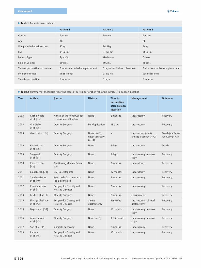
▶ Table 1 Patient characteristics.
Patient 1 Patient 2 Patient 3
Gender Female Female Female
Age 36 31 26
Weight at balloon insertion 87 kg 74,5 kg 94 kg
BMI 34 kg/m2 31 kg/m2 38 kg/m2
Balloon Type Spatz 3 Medicone Orbera
Balloon volume 500mL 600mL 600mL
Time of perforation occurence 5 months after balloon placement 6 days after balloon placement 5 Months after balloon placement
PPI discontinued Third month Using PPI Second month
Time to perforation 5 months 6 days 5 months
▶ Table 2 Summary of 15 studies reporting cases of gastric perforation following intragastric balloon insertion.
Year Author Journal History Time to
perforation
after balloon
insertion
Management Outcome
2003 Roche-Nagleet al. [33]
Annals of the Royal Collegeof Surgeons of England
None 2 months Laparotomy Recovery
2003 Giardielloet al. [35]
Obesity Surgery Fundoplication 18 days Laparotomy Recovery
2005 Genco et al. [24] Obesity Surgery None (n = 1);gastric surgery(n =4)
– Laparotomy (n =3);and laparoscopy (n =2)
Death (n =2); andrecovery (n =3)
2009 Koutelidakiset al. [36]
Obesity Surgery None 2 days Laparotomy Death
2009 Śmigielskiet al. [37]
Obesity Surgery None 9 days Laparoscopy+ endos-copy
Recovery
2010 Knoetze et al.[38]
Continuing Medical Educa-tion
None 7 months Laparotomy Recovery
2011 Baigel et al. [39] BMJ Case Reports None 22 months Laparotomy Recovery
2011 Sánchez-Pérezet al. [40]
Revista de Gastroentero-logía de México
None 2 months Laparoscopy Recovery
2012 Charalambouset al. [41]
Surgery for Obesity andRelated Diseases
None 2 months Laparoscopy Recovery
2014 Bekheit et al. [34] Obesity Surgery None 2 months Conservative Recovery
2015 El Hage Chehadeet al. [42]
Surgery for Obesity andRelated Diseases
Sleevegastrectomy
Same day Laparotomy/subtotalgastrectomy
Recovery
2016 Dayan et al. [32] Obesity Surgery None 10 months Laparoscopy+ endos-copy
Recovery
2016 Abou Husseinet al. [43]
Obesity Surgery None (n = 3) 3,6,7 months Laparoscopy+ endos-copy
Recovery
2017 Yoo et al. [44] Clinical Endoscopy None 2 months Laparoscopy Recovery
2018 Rahmanet al. [45]
Surgery for Obesity andRelated Diseases
None 13 months Laparoscopy Recovery
E1326 Barrichello Junior Sérgio Alexandre et al. Exclusively endoscopic approach… Endoscopy International Open 2018; 06: E1322–E1329
Case report
gin a solid diet. At 30 days after the procedure, upper gastroin-testinal endoscopy showed complete closure of the orifice, withthree clips still in place.
DiscussionConcern regarding obesity has been growing worldwide [8].The first steps in treatment of obesity are always lifestylechanges, focusing on a balanced diet and increased physical ac-tivity. However, diet and pharmacological therapy are limited intheir potential for achieving sustained weight loss, being effec-tive in fewer than 5% of cases [9].
In contrast, bariatric surgery provides the most effective andprolonged response, in terms of weight loss, with excellentcontrol of obesity-associated comorbidities [9–13]. However,indications for bariatric surgery are quite specific and it is notwithout risks [7, 14, 15].
Use of endoscopic procedures to control obesity can providesome of the benefits of bariatric surgery [16–17]. Such proce-dures have the advantages of often being reversible, having alower risk profile, and being applicable in patients who are notcandidates for laparoscopic or open surgery or who are at highsurgical risk.
The first IGBs were introduced in the 1980 s, and IGBs of onetype or another have been used in clinical practice ever since[4–5]. Insertion of an IGB is expected to increase the sensationof satiety and to reduce oral food intake. The IGBs availablehave evolved significantly in recent years. Early models were in-flated with air and had a limited (200–220mL) final volume.Over time, it became apparent that use of IGBs had some po-tential complications. Due to their low resistance to the effectsof gastric acid, earlier versions of IGBs lasted no more than 3 to4 months [18]. The most common side effects associated withuse of those IGBs were nausea, difficulty in inflating or deflatingthe balloon, unexpected deflation, and migration, any of whichcould lead to serious complications [19–21]. To address thisconcern, specialists organized a conference to determine theideal characteristics of safe and effective IGB practice [19].The recommended procedure is one in which the IGB is intro-duced endoscopically, after which it is filled with 400 to 700mL of saline and methylene blue; the methylene blue is addedbecause it changes the color of the urine if the balloon ruptures[5, 22–23]. The current generation of IGBs includes deviceswith capacities of up to 960mL.
The number of adverse events (AEs) associated with IGB in-sertion varies across studies. In one clinical trial of IGB use, Gen-co et al. [24] observed no such events. In another such trial,Ponce et al. [25] reported 28 AEs. Of the eight randomized con-trolled trials of IGBs conducted to date, only six reported AEs,with a weighted mean incidence of 28.2%. The weighted meanreported incidence of serious AEs was 10.5%, initial removal ofthe device being required in three studies [20, 26–27]. SevereAEs include persistent vomiting, abdominal pain, gastroesoph-ageal reflux disease, deep ulcers, and perforation during endos-copy. Ulcers were reported in the studies conducted by Ponceet al. [25], Mohammed et al. [28], and Shelby et al. [29]. Al-though classified as a moderate AE, an ulcer, left untreated,
can become life-threatening, especially if there is perforation.[30]
The mechanism by which IGB-induced perforation occurs isnot well known. It is believed that an IGB causes perforation be-cause it exerts constant pressure on and is in continuous con-tact with the gastric wall. One of the most common risk factorsfor perforation is poor adherence to or discontinuation of treat-ment with the PPI prescribed. Perforation can lead to peritonealcomplications, which can be lethal [31]. Because there is lim-ited evidence in the literature regarding the durability and effi-cacy of IGBs, the available studies having certain limitations, itis recommended that an IGB not be left in place for more than 6months [3, 23, 32]. However, that time frame is not absolute,because most reported cases of IGB-induced perforation haveoccurred during the first 6 months after insertion of the device[32], as in all three of the cases described in the current study.
The first case of gastric perforation after IGB insertion wasreported in 2003 [33].
In our case series, a different type of IGB was used in each ofthe three patients and scheduled to remain for 6 months.Against medical advice, two of the patients had discontinueduse of the PPI, which could also be considered a major risk fac-tor for IGB-induced perforation (▶Table 1). All three patientswere found to have an ulcer in the anterior gastric wall. Hospitalstays did not exceed 7 days.
The usual treatment for IGB-induced gastric perforations islaparotomy for device removal and repair of the perforation.To our knowledge, this is the first report of such perforationsbeing treated exclusively through endoscopy. In their case re-port, Mohamed et al. [34] also applied conservative treatment,although the treatment did not involve endoscopic interven-tions such as clip placement. In our review of the cases in theliterature (▶Table2), we observed that, of the 21 patients
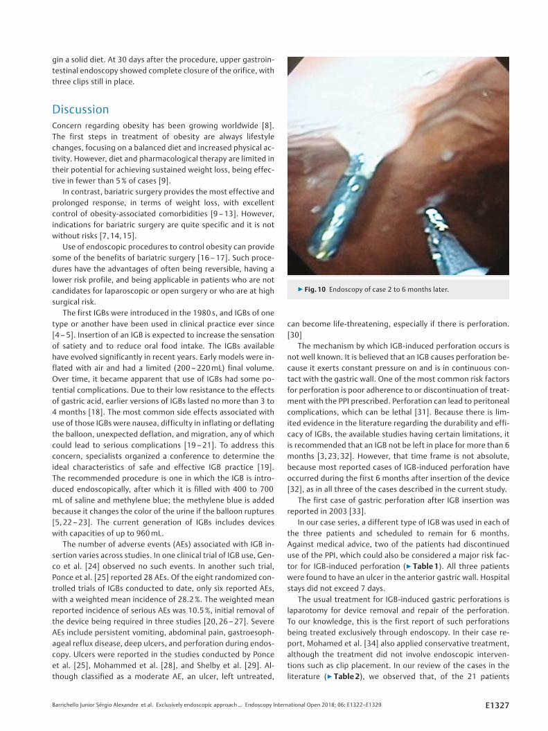
▶ Fig. 10 Endoscopy of case 2 to 6 months later.
Barrichello Junior Sérgio Alexandre et al. Exclusively endoscopic approach… Endoscopy International Open 2018; 06: E1322–E1329 E1327

who developed gastric perforation after IGB insertion, 3(14.2%) evolved to death.
In our patients, exclusive treatment by endoscopy was suc-cessful in removing the IGBs and effective in management ofperforations. This was of great benefit, especially because amajor operation (laparotomy) under general anesthesia couldbe avoided and early hospital discharge and consequently rapidreturn of the patients to their normal activities was faciitated.
All patients were followed up clinically monthly and endo-scopically after 1 year. No symptoms were identified duringthe clinic follow-up. Two patients have had a normal upper gas-trointestinal exam, with no scars. One patient underwent endo-scopic sleeve gastroplasty after 1 year. During the pre-proce-dure endoscopy, one hemoclip was still in place (▶Fig. 10).
ConclusionExclusively endoscopic therapy for gastric perforations that oc-cur after insertion of an IGB is possible in selected cases. use ofthis approach precludes the need for emergency surgery insuch cases.
Competing interests
None
References
[1] Allison DB, Downey M, Atkinson RL et al. Obesity as a disease: A whitepaper on evidence and arguments commissioned by the council ofthe obesity society. Obesity 2008; 16: 1161–1177
[2] Espinet-Coll E, Nebreda-Durán J, Gómez-Valero JA et al. Currentendoscopic techniques in the treatment of obesity. Rev Esp Enferme-dades Dig 2012; 104: 72–87
[3] Kumar N, Sullivan S, Thompson CC. The role of endoscopic therapy inobesity management: Intragastric balloons and aspiration therapy.Diabetes, Metab Syndr Obes Targets Ther 2017; 10: 311–316
[4] Gyring Nieben O, Harboe H. Intragastric balloon as an artificial bezoarfor treatment of obesity. Lancet 1982; 319: 198–199
[5] Moura D, Oliveira J, De Moura EGH et al. Effectiveness of intragastricballoon for obesity: A systematic review and meta-analysis based onrandomized control trials. Surg Obes Relat Dis 2016; 12: 420–429
[6] Saber AA, Shoar S, Almadani MW et al. Efficacy of first-time intragas-tric balloon in weight loss: a systematic review and meta-analysis ofrandomized controlled trials. Obes Surg 2017; 27: 277–287
[7] Ribeiro IB, Gestic MA, Utrini MP et al. Drain amylase levels may indi-cate gastrojejunostomy leaks after Roux-en-Y gastric bypass. ArqGastroenterol 2018; 55: 66–72
[8] Lerner H, Whang J, Nipper R. Benefit-risk paradigm for clinical trialdesign of obesity devices: FDA proposal. Surg Endosc 2013; 27: 702–707
[9] Kumar N, Thompson CC. Endoscopic solutions for weight loss. CurrOpin Gastroenterol 2011; 27: 407–411
[10] Lee W-J, Lee Y-C, Ser K-H et al. Improvement of insulin resistance afterobesity surgery: a comparison of gastric banding and bypass proce-dures. Obes Surg 2008; 18: 1119–1125
[11] Sjöström L, Lindroos A-K, Peltonen M et al. Lifestyle, diabetes, andcardiovascular risk factors 10 years after bariatric surgery. N Engl JMed 2004; 351: 2683–2693
[12] Meek CL, Lewis HB, Reimann F et al. The effect of bariatric surgery ongastrointestinal and pancreatic peptide hormones. Peptides 2016;77: 28–37
[13] Ribeiro IB, Bernardo WM, Martins C et al. Colonic stent versus emer-gency surgery as treatment of malignant colonic obstruction in thepalliative setting: a systematic review and meta-analysis. Endosc IntOpen 2018; 5: E1– E10
[14] Flum DR, Dellinger EP. Impact of gastric bypass operation on survival:A population-based analysis. J Am Coll Surg 2004; 199: 543–551
[15] Kakarla VR, Nandipati K, Lalla M et al. Are laparoscopic bariatric pro-cedures safe in superobese (BMI ≥50 kg/m2) patients? An NSQIP dataanalysis Surg Obes Relat Dis7: 452–458
[16] Kumar N, Thompson CC. Endoscopic solutions for weight loss. CurrOpin Gastroenterol 2011; 27: 407–411
[17] Abu Dayyeh BK, Edmundowicz SA, Jonnalagadda S et al. Endoscopicbariatric therapies. Gastrointest Endosc 2015; 81: 1073–1086
[18] Gleysteen JJ. A history of intragastric balloons. Surg Obes Relat Dis2016; 12: 430–435
[19] Neto MG, Silva LB, Grecco E et al. Brazilian Intragastric Balloon Con-sensus Statement (BIBC): practical guidelines based on experience ofover 40,000 cases. Surg Obes Relat Dis 2018; 14: 151–159
[20] Mathus-Vliegen EMH, Tytgat GNJ. Intragastric balloon for treatment-resistant obesity: safety, tolerance, and efficacy of 1-year balloontreatment followed by a 1-year balloon-free follow-up. GastrointestEndosc 2005; 61: 19–27
[21] Madruga NetoAC, Bernardo WM, de Moura DT et al. The effectivenessof endoscopic gastroplasty for obesity treatment according to FDAthresholds: Systematic review and meta-analysis based on random-ized controlled trials. Gastrointest Endosc 2018; 87: AB601
[22] Bernante P, Francini F, Zangrandi F et al. Green urine after intragastricballoon placement for the treatment of morbid obesity. Obes Surg2003; 13: 951–953
[23] Tate CM, Geliebter A. Intragastric balloon treatment for obesity: re-view of recent studies. Adv Ther 2017; 34: 1859–1875
[24] Genco A, Cipriano M, Bacci V et al. BioEnterics® Intragastric Balloon(BIB®): a short-term, double-blind, randomised, controlled, cross-over study on weight reduction in morbidly obese patients. Int J Obes2006; 30: 129–133
[25] Ponce J, Woodman G, Swain J et al. The REDUCE pivotal trial: A pro-spective, randomized controlled pivotal trial of a dual intragastricballoon for the treatment of obesity. Surg Obes Relat Dis 2015; 11:874–881
[26] Fuller NR, Pearson S, Lau NS et al. An intragastric balloon in the treat-ment of obese individuals with metabolic syndrome: A randomizedcontrolled study. Obesity 2013; 21: 1561–1570
[27] Abu Dayyeh BK, Eaton LL, Woodman G et al. 444 A randomized, multi-center study to evaluate the safety and effectiveness of an intragas-tric balloon as an adjunct to a behavioral modification program, incomparison with a behavioral modification program alone in theweight management of obese subjects. Gastrointest Endosc 2015;81: AB147
[28] Mohammed MA, Anwar R, Mansour AH et al. Effects of intragastricballoon versus conservative therapy on appetite regulatory hormonesin obese subjects. Trends Med Res 2014; 9: 58–80
[29] Sullivan S, Swain JM, Woodman G et al. The obalon swallowable 6-month balloon system is more effective than moderate intensity life-style therapy alone: Results from a 6-month randomized sham con-trolled trial. Gastroenterology 2016; 150: S1267
E1328 Barrichello Junior Sérgio Alexandre et al. Exclusively endoscopic approach… Endoscopy International Open 2018; 06: E1322–E1329
Case report

[30] Ribeiro IB, Rezende DT, Madruga Neto AC et al. Endoscopic dual ther-apy for giant peptic ulcer hemorrhage. Endoscopy 2018: doi:10.1055/a-0665-4142 [Epub ahead of print]
[31] Genco A, Bruni T, Doldi SB et al. BioEnterics intragastric balloon: TheItalian experience with 2,515 patients. Obes Surg 2005; 15: 1161–1164
[32] Dayan D, Sagie B, Fishman S. Late Intragastric Balloon Induced GastricPerforation. Obes Surg 2016; 26: 1138–1140
[33] Roche-Nagle G, Mulligan E, Connolly E et al. Unusual cause of a per-forated stomach. Ann R Coll Surg Engl 2003; 85: 396–397
[34] Bekheit M, Abdelsalam WN, Sgromo B et al. Is conservative manage-ment for gastric perforation secondary to intragastric balloon possi-ble? Case report and review of literature Obes Surg 2014; 24: 968–970
[35] Giardiello C, Cristiano S, Cerbone MR et al. Gastric perforation in anobese patient with an intragastric balloon, following previous fundo-plication. Obes Surg 2003; 13: 658–660
[36] Koutelidakis I, Dragoumis D, Papaziogas B et al. Gastric perforationand death after the insertion of an intragastric balloon. Obes Surg2009; 19: 393–396
[37] Smigielski JA, Szewczyk T, Modzelewski B et al. Gastric perforation asa complication after BioEnterics intragastric balloon bariatric treat-ment in obese patients – synergy of endoscopy and videosurgery.Obes Surg 2010; 20: 1597–1599
[38] Knoetze R, van Mollendorff V, Yuen O. Gastric perforation as a com-plication of an intragastric balloon. Continuing Medical Education2010; 28: 392
[39] Baigel R, Rashid F, Shrestha D etal. Peritonitis following a bariatricprocedure in a young woman. BMJ Case Rep 2011 Feb 2011: 14
[40] Sánchez-Pérez MA, Muñoz-Juárez M, Cordera-González De Cosío F etal. Gastric perforation and subarachnoid hemorrhage secondary tointragastric balloon device. Rev Gastroenterol Mex 2011; 76: 264–269
[41] Charalambous MP, Thompson J, Efthimiou E. Late gastric perforationafter insertion of intragastric balloon for weight lossvideo case reportand literature review. Surg Obes Relat Dis 2012; 8: 121–123
[42] El Hage Chehade HH, El Khatib ZO, Abtar HK. What could happen ifyou insert a BioEnterics intragastric balloon after sleeve gastrectomy?Surg Obes Relat Dis 2015; 11: e39– e41
[43] Abou Hussein BM, Khammas AA, Al Ani AM et al. Gastric perforationfollowing intragastric balloon insertion: combined endoscopic andlaparoscopic approach for management: case series and review ofliterature. Obes Surg 2016; 26: 1127–1132
[44] Yoo IK, Chun HJ, Jeen YT. Gastric perforation caused by an intragastricballoon: endoscopic findings. Clin Endosc 2017; 50: 602–604
[45] Rahman AA, Loi K. Gastric perforation as a complication of intragas-tric balloon. Surg Obes Relat Dis 2018: 1–4
Barrichello Junior Sérgio Alexandre et al. Exclusively endoscopic approach… Endoscopy International Open 2018; 06: E1322–E1329 E1329