Embed Size (px)
Citation preview

Contents lists available at ScienceDirect
J Ped Surg Case Reports 10 (2016) 29e31
Journal of Pediatric Surgery CASE REPORTS
journal homepage: www.jpscasereports .com
Excision of an intrapericardial immature teratoma in a 26-weekpremature neonate
Robert B. Hawkins a,*, Laura H. Rosenberger b, Julia C. Swanson c, James J. Gangemi a,Eugene D. McGahren d
aDivision of Thoracic & Cardiovascular Surgery, Department of Surgery, University of Virginia, Charlottesville, VA, USAbDepartment of Surgery, Memorial Sloan Kettering Cancer Center, New York, NY, USAcDivision of Congenital Heart Surgery, Department of Surgery, Baylor College of Medicine, Houston, TX, USAdDivision of Pediatric Surgery, Department of Surgery, University of Virginia, Charlottesville, VA, USA
a r t i c l e i n f o
Article history:Received 3 March 2016Accepted 26 April 2016
Key words:Cardiac tumorsPrematurityNeonate
* Corresponding author. Department of Surgery, Un800679, Charlottesville, VA 22908, USA. Tel.: þ1 3033885.
E-mail address: [email protected] (R.B. Hawkins
2213-5766/� 2016 The Authors. Published by Elsevierhttp://dx.doi.org/10.1016/j.epsc.2016.04.026
a b s t r a c t
We present a case of a 26-week premature newborn with an immature intrapericardial teratoma. Thepatient was transferred from an outside hospital for management of a large mediastinal mass causingrespiratory insufficiency. The newborn was supported with the help of a large interdisciplinary teamuntil day of life 22 when he underwent surgical excision. On follow up the infant is doing very well and isone of the youngest survivors to date.� 2016 The Authors. Published by Elsevier Inc. This is an open access article under the CC BY-NC-ND
license (http://creativecommons.org/licenses/by-nc-nd/4.0/).
Intrapericardial teratoma is a germ cell tumor that typicallyarises from the base of the heart [1]. While usually benign,intrapericardial teratomas can become relatively very large andcause symptomatic compression or fetal hydrops in utero [2].Surgical excision is curative, even for most immature variants.However, infants with this condition may require significantsupport and multiple interventions prior to surgery. Advances incritical care, surgery and interdisciplinary management haveresulted in dramatic improvement in treatment outcomes overrecent decades.
1. Case report
A 26-week and 3 days old male neonate was delivered bycesarean section at an outside center after an ultrasound performedfor premature labor demonstrated a large chest mass in the babyin utero. His birth weight was 1212 g and he required intubationfor respiratory distress. After stabilization he underwent chestcomputed tomography (CT) revealing amediastinalmassmeasuring5 � 3 � 4 cm. He was then transferred to our center. Complicating
iversity of Virginia, P.O. Box990 2192; fax: þ1 434 982
).
Inc. This is an open access article u
factors included extreme prematurity, patent ductus arteriosus(PDA), and neonatal respiratory distress syndrome. Ultrasound ofthe chest at our center demonstrated a 4.4 � 3.1 � 3.6 cm hetero-geneous mass adjacent to the right heart with compression of theright atrium and ventricle, a pericardial effusion, and a small left toright PDA. Laboratory tests revealed human chorionic gonadotropinof<0.12 and alpha-fetoprotein (AFP) of>363,000, which is elevatedeven after adjusting for prematurity.
Initially, the baby’s status was relatively stable with assistedventilation, so the decision was made to delay excision to allow forfurther growth. However, on day 7 of life he developed worseninghypercarbia and had to be placed on high frequency oscillatoryventilation and two days later on the Dräger-VG. Serial ultrasoundsdemonstrated worsening pericardial effusion, which was drainedon day 11 of life with improvement in respiratory status. He againrequired pericardiocentesis and drain placement on day 13 of life.
With continued respiratory distress requiring prone positioning,the decision to operate was made on day 22 of life. His weight wasonly 1410 g and the relatively high likelihood of an inability to go onbypass was considered. Collaboration between surgeons from theDivisions of Pediatric Surgery and Pediatric Cardiac Surgery deter-mined the risk of continued observation outweighed the risk ofsurgery at this time. The baby underwent median sternotomy andupon entry into the pericardial space straw colored fluid wasdrained. Dissection around the large teratoma revealed that it waswell-encapsulated with clean planes. There were connective tissue
nder the CC BY-NC-ND license (http://creativecommons.org/licenses/by-nc-nd/4.0/).
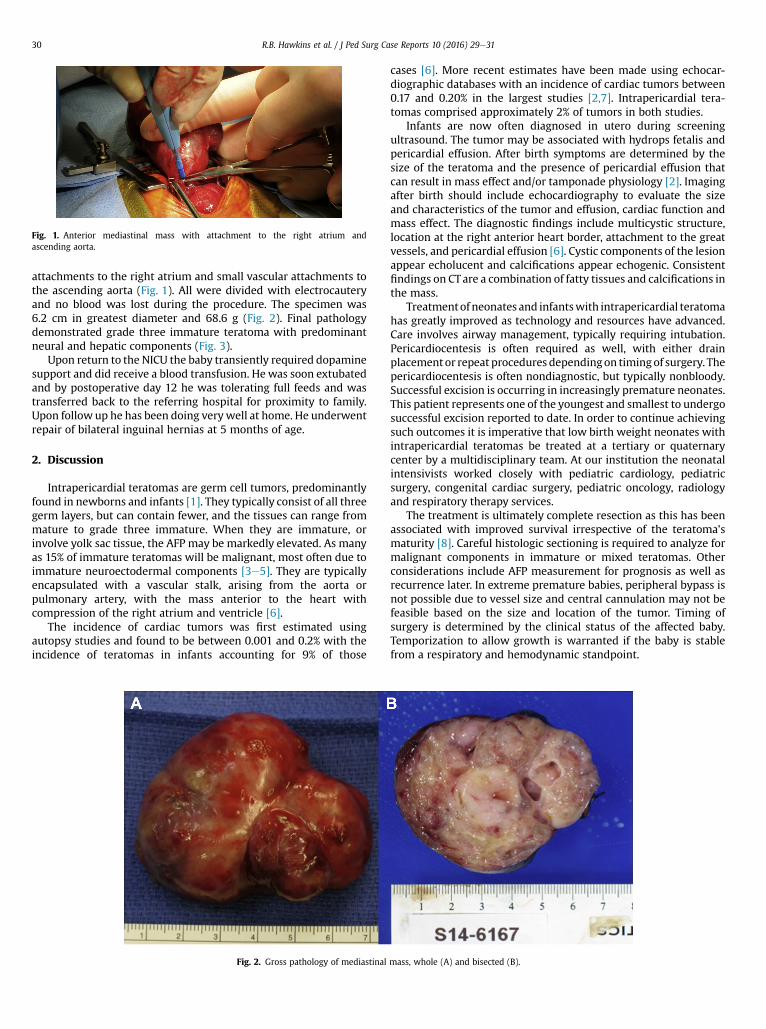
Fig. 1. Anterior mediastinal mass with attachment to the right atrium andascending aorta.
R.B. Hawkins et al. / J Ped Surg Case Reports 10 (2016) 29e3130
attachments to the right atrium and small vascular attachments tothe ascending aorta (Fig. 1). All were divided with electrocauteryand no blood was lost during the procedure. The specimen was6.2 cm in greatest diameter and 68.6 g (Fig. 2). Final pathologydemonstrated grade three immature teratoma with predominantneural and hepatic components (Fig. 3).
Upon return to the NICU the baby transiently required dopaminesupport and did receive a blood transfusion. He was soon extubatedand by postoperative day 12 he was tolerating full feeds and wastransferred back to the referring hospital for proximity to family.Upon follow up he has been doing verywell at home. He underwentrepair of bilateral inguinal hernias at 5 months of age.
2. Discussion
Intrapericardial teratomas are germ cell tumors, predominantlyfound in newborns and infants [1]. They typically consist of all threegerm layers, but can contain fewer, and the tissues can range frommature to grade three immature. When they are immature, orinvolve yolk sac tissue, the AFP may be markedly elevated. As manyas 15% of immature teratomas will be malignant, most often due toimmature neuroectodermal components [3e5]. They are typicallyencapsulated with a vascular stalk, arising from the aorta orpulmonary artery, with the mass anterior to the heart withcompression of the right atrium and ventricle [6].
The incidence of cardiac tumors was first estimated usingautopsy studies and found to be between 0.001 and 0.2% with theincidence of teratomas in infants accounting for 9% of those
Fig. 2. Gross pathology of mediastinal
cases [6]. More recent estimates have been made using echocar-diographic databases with an incidence of cardiac tumors between0.17 and 0.20% in the largest studies [2,7]. Intrapericardial tera-tomas comprised approximately 2% of tumors in both studies.
Infants are now often diagnosed in utero during screeningultrasound. The tumor may be associated with hydrops fetalis andpericardial effusion. After birth symptoms are determined by thesize of the teratoma and the presence of pericardial effusion thatcan result in mass effect and/or tamponade physiology [2]. Imagingafter birth should include echocardiography to evaluate the sizeand characteristics of the tumor and effusion, cardiac function andmass effect. The diagnostic findings include multicystic structure,location at the right anterior heart border, attachment to the greatvessels, and pericardial effusion [6]. Cystic components of the lesionappear echolucent and calcifications appear echogenic. Consistentfindings on CTare a combination of fatty tissues and calcifications inthe mass.
Treatmentof neonates and infantswith intrapericardial teratomahas greatly improved as technology and resources have advanced.Care involves airway management, typically requiring intubation.Pericardiocentesis is often required as well, with either drainplacementor repeat procedures dependingon timingof surgery. Thepericardiocentesis is often nondiagnostic, but typically nonbloody.Successful excision is occurring in increasingly premature neonates.This patient represents one of the youngest and smallest to undergosuccessful excision reported to date. In order to continue achievingsuch outcomes it is imperative that low birth weight neonates withintrapericardial teratomas be treated at a tertiary or quaternarycenter by a multidisciplinary team. At our institution the neonatalintensivists worked closely with pediatric cardiology, pediatricsurgery, congenital cardiac surgery, pediatric oncology, radiologyand respiratory therapy services.
The treatment is ultimately complete resection as this has beenassociated with improved survival irrespective of the teratoma’smaturity [8]. Careful histologic sectioning is required to analyze formalignant components in immature or mixed teratomas. Otherconsiderations include AFP measurement for prognosis as well asrecurrence later. In extreme premature babies, peripheral bypass isnot possible due to vessel size and central cannulation may not befeasible based on the size and location of the tumor. Timing ofsurgery is determined by the clinical status of the affected baby.Temporization to allow growth is warranted if the baby is stablefrom a respiratory and hemodynamic standpoint.
mass, whole (A) and bisected (B).

Fig. 3. Hematoxylin and eosin stain at 4� (A) and 20� (B) magnification.
R.B. Hawkins et al. / J Ped Surg Case Reports 10 (2016) 29e31 31
3. Conclusion
The treatment of intrapericardial teratoma is feasible even inthe setting of extreme immaturity. With multidisciplinarycare and the development of a coherent treatment plan theneonate may be able to grow and develop prior to surgicalexcision. After excision these patients tend to do very well, andour patient was transferred closer to home two weeks afterexcision.
References
[1] McAllister HA, Hall RJ, Cooley DA. Tumors of the heart and pericardium. CurrProbl Cardiol 1999;24:59e116.
[2] Allen HD, Moss AJ. Cardiac tumors. In: Moss and Adams’ heart disease ininfants, children, and adolescents: including the fetus and young adult.Lippincott Williams and Wilkins; 2007. p. 1479e94.
[3] Dehner LP. Gonadal and extragonadal germ cell neoplasia of childhood. HumPathol 1983;14:493e511.
[4] Weber HS, Kleinman CS, Hellenbrand WE, Kopf GS, Copel J. Development of abenign intrapericardial tumor between 20 and 40 weeks gestation. PediatrCardiol 1988;9:153e6.
[5] Reddy SC, Fenton KM, Ghandi Sk, Lanford LM, Pigula FA. Intrapericardial tera-toma in a neonate. Ann Thorac Surg 2003;76:626.
[6] MacKenzie S, Loken S, Kalia N, Trevenen C, Harder J, Wong A, et al. Intra-pericardial teratoma in the perinatal period. Case report and review of theliterature. J Pediatr Surg 2005;40:E13e8.
[7] Beghetti M, Gow RM, Haney I, Mawson J, Williams WG, Freedom RM. Pediatricprimary benign cardiac tumors: a 15-year review. Am Heart J 1997;134:1107e14.
[8] Heerema-McKenney A, Harrison MR, Bratton B, Farrell J, Zaloudek C. Congenitalteratoma: a clinicopathologic study of 22 fetal and neonatal tumors. Am J SurgPathol 2005;29:29e38.

![PARIPEX - INDIAN JOURNAL OF RESEARCH | Volume-8 | Issue-10 ... · teratoma is known as a monodemal teratoma.[1] Immature teratoma (IT) is a preferred term for the malignant ovarian](https://img.dokumen.tips/doc/110x75/603e5f8d2bf3bd27e47c8252/paripex-indian-journal-of-research-volume-8-issue-10-teratoma-is-known.jpg)