Embed Size (px)
Citation preview
European Journal of Pharmaceutics and Biopharmaceutics xxx (2015) xxx–xxx
Contents lists available at ScienceDirect
European Journal of Pharmaceutics and Biopharmaceutics
journal homepage: www.elsevier .com/locate /e jpb
Elastic sealants for surgical applications
http://dx.doi.org/10.1016/j.ejpb.2015.05.0220939-6411/� 2015 Elsevier B.V. All rights reserved.
⇑ Corresponding author at: Biomaterials Innovations Research Center,Department of Medicine, Brigham and Women’s Hospital, Harvard Medical School,Boston, MA, USA. Tel.: +1 617 768 8395; fax: +1 617 768 8477.
E-mail address: [email protected] (A. Khademhosseini).
Please cite this article in press as: N. Annabi et al., Elastic sealants for surgical applications, Eur. J. Pharm. Biopharm. (2015), http://dx.doi.org/1j.ejpb.2015.05.022
Nasim Annabi a,b,c,d, Kan Yue b,c, Ali Tamayol b,c, Ali Khademhosseini b,c,d,e,f,⇑a Department of Chemical Engineering, Northeastern University, Boston, MA 02115-5000, USAb Biomaterials Innovations Research Center, Department of Medicine, Brigham and Women’s Hospital, Harvard Medical School, Boston, MA, USAc Harvard-MIT Division of Health Sciences and Technology, Massachusetts Institute of Technology, Cambridge, MA, USAd Wyss Institute for Biologically Inspired Engineering, Harvard University, Boston, MA, USAe Department of Biomedical Engineering, The University of Texas at Austin, Austin, TX, USAf Department of Physics, King Abdulaziz University, Jeddah 21569, Saudi Arabia
a r t i c l e i n f o a b s t r a c t
Article history:Received 2 January 2015Revised 25 May 2015Accepted in revised form 27 May 2015Available online xxxx
Keywords:Surgical sealantsElasticityAdhesionHydrogelSurgical applicationsTissue adhesives
Sealants have emerged as promising candidates for replacing sutures and staples to prevent air and liquidleakages during and after the surgeries. Their physical properties and adhesion strength to seal thewound area without limiting the tissue movement and function are key factors in their successful imple-mentation in clinical practice. In this contribution, the advances in the development of elastic sealantsformed from synthetic and natural materials are critically reviewed and their shortcomings are pointedout. In addition, we highlight the applications in which elasticity of the sealant is critical and outline thelimitations of the currently available sealants. This review will provide insights for the development ofnovel bioadhesives with advanced functionality for surgical applications.
� 2015 Elsevier B.V. All rights reserved.
1. Introduction
Current technologies for reconnecting and sealing tissues aftersurgical procedures such as sutures, wires, and staples have severallimitations, particularly in minimally invasive procedures. Forexample, the use of suture for wound closure is time-consuming,may cause further tissue damage, result in infection, and do notprovide immediate sealing to stop body fluid and air leakages.The application of surgical adhesives is a convenient alternativemethod for wound closure because of their characteristics, suchas simple implementation procedure, shorter time, less painful topatients, and no need for removal. Toward this goal, various typesof surgical materials have been used for sealing, reconnecting tis-sues, or attaching devices to the tissues [1].
Surgical sealants are commonly used to prevent leakage of fluidand/or gas from a incision. Sealants can be formed by using naturalor synthetic polymers, or a combination of both. The market forsurgical sealants and hemostats is growing rapidly from $4 billionin 2012 to $7 billion in 2017, worldwide [2]. Although several tis-sue adhesives are commercially available, none of them are ideal
surgical sealants for repairing elastic and soft tissues such aswounded lungs, heart, and blood vessels. It is extremely challeng-ing to achieve significant adhesion to soft tissues while minimizingtoxicity, tissue damage and other side effects of the sealingmaterials. Another limitation is the low adhesion strength of themost commercially available sealants in wet and highly dynamicenvironments in the body at the presence of blood. Most of theclinically available glues and sealants do not offer both elasticityand good adhesion. For example, cyanoacrylates have high stiffnessand adhesion strength but are not elastic. On the other hand,fibrin-based sealants are more flexible with low stiffness andadhesion. There is a unmet need for surgical sealants, which canprovide flexibility without compromising strength and can stopbody fluid and air leakages in different procedures such as lungand cardiovascular surgeries.
Recently, extensive research efforts have been made to engineerbiocompatible, biodegradable, and flexible sealants for the forma-tion of leak-free closures in soft tissues [3–5]. The sealant materialsare required to be elastic and compliant to allow normal functionand movement of elastic native tissues such as lungs, skin, bloodvessel, and heart tissues. In this review, various types of elastic sur-gical sealants made of natural and synthetic polymer are discussed.These include flexible synthetic sealants based on polyurethane(PU), polyethylene glycol (PEG), polyester, and naturally occurringor composite sealants made of proteins or polysaccharides
0.1016/
2 N. Annabi et al. / European Journal of Pharmaceutics and Biopharmaceutics xxx (2015) xxx–xxx
(see Table 1). In addition, recent developments in the synthesis ofmussel inspired elastic sealants with high adhesive properties arereviewed. Finally, some clinical applications for the elastic sealantsin various surgical procedures are highlighted.
2. Synthetic polymer derived elastic sealants
Synthetic-based elastic tissue adhesives have attracted signifi-cant attention as suitable wound closure techniques for clinicalapplications due to their strong adhesive strength and tunablemechanical properties. In particular, elastic sealants based on syn-thetic polymers can be used for sutureless wound closure in elasticand soft tissues such as lungs, heart, and blood vessels. Someexamples of this class of adhesives include PU-based, PEG-based,and polyester-based adhesives.
2.1. PU-based tissue adhesives
PU-based biomaterials have been used as sealants due to theirhigh elasticity and strong adhesion to the tissues. In the prepolymerform, urethane can react with the amino groups of proteins of thetissues to create urea linkages and subsequently promote adhesionstrength to the tissues [6]. PU-based biomaterials can be synthe-sized in biodegradable forms through modification with naturalmolecules. For example, Ferreira et al. synthesized a biodegradablePU-based adhesives through modification of castor oil with iso-phorone diisocyanate (IPDI) [7]. They also formed PU-based adhe-sives through reaction between polycaprolactone (PCL) diol andIPDI or hexamethylene diisocyanate (HDI) [6]. The engineeredPU-based adhesives promoted adhesion to the tissue but inducedthrombosis as shown by hemolysis tests, which can limit its clinicalapplications [6]. To overcome this limitation, the group later devel-oped a photocrosslinkable PU-based adhesive through the modifi-cation of PCL with 2-isocyanatoerhylmethacrylate (IEMA) [8].Their experimental data showed that the synthesized materialwas slightly hemolytic (within the acceptable limit) after directlyapplying with existence of blood. Moreover, hemolysis was stoppedwhen the material was extracted with PBS solution [8].
Previous studies have shown that the use of PU-based tissueadhesives had no toxic degradation products after surgical proce-dures such as orthopedic and renal surgery, and pancreatic occlu-sion. PU-based adhesives have been also used in cosmetic surgery.For example, a sprayable PU adhesives (TissuGlu, Cohera MedicalInc., Pittsburgh, PA) was used for abdominoplasty surgery to avoidseroma formation in a canine abdominoplasty model [9]. It wasshown that TissuGlu created strong bonding between tissue layersand supported natural healing process without any toxic effects[9]. Despite significant improvements in the formation of nontoxicand biodegradable PU-based surgical materials, safety concernsstill exist for their utilization in surgical procedures.
2.2. PEG-based sealant
PEG-based biomaterials have been widely used as fluid barriersand hemostatic adhesives. There are several commercially avail-able PEG-based surgical materials including Duraseal (CovidienInc., Mansfield, MA), Coseal (Cohesion Technologies, Inc., PaloAlto, CA), FocalSeal (used to be produced by Genzyme Biosurgery,Inc., Cambridge, MA but it is currently discontinued), andAdvaSeal (Ethicon Inc., Johnson & Johnson Medical KK). Durasealis made of PEG ester and trilysine amine solutions and has beenused for neurosurgeries to prevent cerebrospinal fluid leakage aftercranial and spinal operations [10–13]. For example, Preul et al.applied Duraseal on an incision of a cranial dura and arachnoid cre-ated in dogs to seal the dural gaps. It was shown that animals
Please cite this article in press as: N. Annabi et al., Elastic sealants for surgicaj.ejpb.2015.05.022
treated by the sealant had normal dural healing process with noadverse effects on the brain. In addition, the sealant reducedcerebrospinal fluid leakage and consequently facilitated surgicalreexploration [13]. Kim et al. also performed a clinical study touse Duraseal as an adjunct spinal sealant to sutured dural repair.Their study confirmed that the sealant provided watertight closureduring spinal surgeries, which was better than the standard caretechnologies (suture only) for dural closure in spine surgeries[10]. In another clinical study, the suitability of Duraseal forreducing scar tissue and post-operative pain after lumbarmicrodiscectomy was evaluated [11]. The results on 21 patientsshowed that Duraseal was safe and reduced post-operation painas compared to the control group. In addition, the patients treatedwith the sealant had normal wound healing process with nopost-operative complications [11].
FocalSeal is another PEG-based sealant, which was developed tostop air leaks after lung surgeries. It is made from a primer and anacrylated PEG-based sealant [14–16]. The primer layer should befirst applied to provide good adhesion to the tissue, followed bythe sealant layer to provide adequate mechanical properties andmaintain the sealing during the tissue movement. Finally, the sea-lant is crosslinked by photopolymerization using visible blue light.It was shown that the engineered sealant had elastic modulus of28 kPa, which was close to human lung tissue (29.4 kPa) andextensibility up to 700% [14]. The results of clinical studiesdemonstrated that FocalSeal functions as an adjunct to the tradi-tional closure techniques to seal intra-operative pulmonary leaks[17]. In addition, the use of this sealant significantly reducedpost-operative air leaks leading to the shorter hospitalization[17]. The production of FocalSeal was discontinued due to its pooradaption by surgeons. Coseal is also a commercially availablePEG-based sealant, which is made of two 4-arm PEG withglutaryl-succinimidyl ester and thiol terminal groups [18]. Thereaction between thiol groups and the carbonyl groups of the suc-cinimidylester can form covalent bonds between PEG moleculesafter mixing [18]. Coseal has been employed in vascular surgeriesto seal suture lines and stop bleeding [19]. PEG-based sealantshave several advantages including biocompatibility, controlleddegradability, flexibility, and relatively high adhesion strength.However, high swelling ratio of PEG-based sealants may causepressure build up on the surrounding tissues when applied inclosed cavities [20].
2.3. Other synthetic polymer-based sealant
Different types of polyester-based sealants with elastic proper-ties have been developed for surgical applications to reduce theincidence of fluidic or air leaks. For example, Chen et al. developeda surgical sealant based on poly(glycerol sebacate) (PGS) and lacticacid (LA) [3]. The use of LA improved the cytocompatibility of theengineered sealant compared to PGS alone. The sealant could beapplied at 45 �C and solidified at 37 �C to form an elastic gel byexposing to a cold gas. It was shown that the fabricated sealanthad higher adhesion strength to the tissues compared to bothfibrin glues and synthetic sealants such as Pleuraseal [3].Photocurable elastomers based on PGS with ultimate strainbetween 42% and 189% and Young Modulus of 0.05–1.38 MPa werealso developed through acrylation of PGS [21]. The engineered syn-thetic elastomer had been recently used for cardiovascular surg-eries to close defects in heart and arteries and stop bleeding [22].
Sealants based on other synthetic polymers have been alsodeveloped to close wounds in different surgical procedures. Forexample, in a recent study, a polyvinyl alcohol (PVA)-based tissueadhesive for wound closure was prepared via sequential enzymaticreactions that were activated by glucose in the wound exudate[23]. The hydrogel was formed in situ after applying a mixed
l applications, Eur. J. Pharm. Biopharm. (2015), http://dx.doi.org/10.1016/
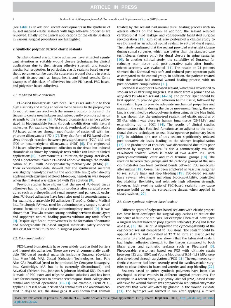
Table 1Summary of the representative surgical sealants.
Basematerials
Applications Components and products Refs.
Polyurethane – Fixation of vascular graft and bone– Abdominoplasty surgery to avoid seroma formation
– Engineered polyurethane with low hemolytic responses– Lysine-based, sprayable urethane adhesive (TissuGlu)
[6–8][9,111]
PEG – Prevent cerebrospinal fluid leakage after cranial operations andreduce scar tissue and pain after lumbar microdiscectomy
– Stop air leaks after lung surgeries– Seal suture lines and stop bleeding in vascular surgeries
– Tetra-succinimidyl PEG and tri-lysine amine (DuraSeal)– Acrylated PEG, polyester primer, and photoinitiator
(FocalSeal)– Glutaryl-succinimidyl ester and thiol terminated PEG
(Coseal)
[10–13][14–16][18,19]
Polyester – Reduce the incidence of fluidic or air leaks– Repair of vessels and heart defects
– Poly(glycerol sebacate) (PGS)– Photocrosslinkable PGS derivatives
[3,21,22][5]
Polyvinylalcohol
– Close wound in surgery – Tyramine-modified PVA [23]
Fibrin – Hemorrhage control, wound closure and tissue anastomoses – Fibrinogen and thrombin (Tisseel, Evicel, Crosseal,Hemaseel, etc.)
– Fibrinogen and a ruthenium photo-catalyst
[24–29,106][30,32,33]
Collagen – Hemostasis – Bovine collagen and thrombin [37]
Gelatin – Thoracic aortic dissections and hemostasis– Seal surgical incisions in gastrointestinal tract surgeries– Repair retinal tissues
– Gelatin-resorcinol-formaldehyde-glutaraldehyde– Gelatin and N-hydroxysuccinimide-ester functionalized
poly(L-glutamic acid) or disuccinimidyl tartrate– Photocrosslinkable gelatin adhesives– Gelatin and microbial transglutaminase
[39–41][42–44][34,35,45][46–48]
Albumin – Hemostats in vascular and cardiac surgeries– Prevent air leakage in lung surgeries
– Bovine albumin and glutaraldehyde (Bioglue)– Human albumin and a NHS-activated PEG (Progel)
[49,108][4,50]
Chitosan – Wound closure and heamostasis – Lactobionic acid and azide functionalized chitosan– Tyrosine-modified chitosan, HPR and hydrogen peroxide– Thiol-containing chitosan and maleimide containing
e-polylysine
[51–53][55][56]
Dextran – Stop air leaks after lung surgeries – Aldehyde-containing dextran and amine-containing PEGor polylysine crosslinkers
[58–62]
Chondroitinsulfate
– Seal corneal incisions– Binding to native cartilage tissue– Wound closure
– Aldehyde-bearing chondroitin sulfate and poly(vinyl alco-hol-co-vinyl amine)
– Methacrylate and aldehyde functionalized chondroitinsulfate
– NHS-activated chondroitin sulfate and amine-containingPEG
[64][66][67]
N. Annabi et al. / European Journal of Pharmaceutics and Biopharmaceutics xxx (2015) xxx–xxx 3
solution of a PVA derivative functionalized with phenolic hydroxylmoieties (PVA-Ph), together with two enzymes, i.e., glucose oxi-dase (GOx) and horseradish peroxidase (HRP) on the wound(Fig. 1). The results of mechanical testing demonstrated that theengineered PVA-based adhesives had high durability toward bothstretching and compression. In addition, the hydrogel was effectivein closing full-thickness wounds in rats as compared to commer-cially available hydrogel-based wound dressing [23].
Synthetic sealants can be easily modified and generally havehigher mechanical strength and tissue-bonding propertiescompared to naturally derived sealants. However, potentialcytotoxicity, chronic inflammation, low adherence to the wettissues and, in some cases, long curing time are some of thelimitations associated with synthetic-based sealants [6].
3. Sealants based on natural polymers
Sealants derived from natural polymers possess many advan-tages over synthetic materials including superior biocompatibility,reduced immune response, and in vivo degradability. Naturalbiopolymers can be classified into polysaccharides and polypep-tides (including proteins), which are built up by sugar or aminoacid units, respectively. Degradation of biopolymers results insmall molecular byproducts that can be easily absorbed by thebody. Therefore, developing sealants derived from natural poly-mers has been an active research area during the past two decadesand some of the developed sealant systems have been approved byFood and Drug Administrative (FDA) for certain surgical
Please cite this article in press as: N. Annabi et al., Elastic sealants for surgicaj.ejpb.2015.05.022
applications. In the following section, we will describe a briefsummary of the reported sealants containing at least one kind ofbiopolymers as the essential functioning components.
3.1. Polypeptide/protein-based sealants
3.1.1. Fibrin-based sealantsFibrin-based sealants are one of the earliest surgical glues used
for medical applications [24]. The working mechanisms offibrin-based sealants are similar to the series of bioreactions inthe final stages of blood clotting [25,26]. Typically, fibrin sealantsare composed of two major components obtained from pooledhuman plasma, fibrinogen and thrombin. Fibrinogen is a largesoluble glycoprotein existing in plasma and plays a key role inthe formation of blood clots. Thrombin activates fibrinogen andconverts it to fibrin monomers, which are further crosslinked byFactor XIII to form insoluble clots (hemostasis). Since thesebiological processes are promoted by the calcium ions, many fibrinsealants also contain a small amount of calcium ions to acceleratethe reactions [27].
Fibrin sealants can take effect in relatively short times, formcovalent connections with surrounding tissues via the amidationreactions, and also function as hemostats. Therefore, fibrin sealantshave been tested for a variety of different applications in surgeriesas both tissue sealants and hemostats. To date, fibrin sealants arecommercially available under different brand names, such asTisseel (Baxter Inc., Denmark), Evicel (Ethicon Inc., Bridgewater,NJ), Crosseal (OMRIX Biopharmaceuticals Ltd. Israel), and
l applications, Eur. J. Pharm. Biopharm. (2015), http://dx.doi.org/10.1016/
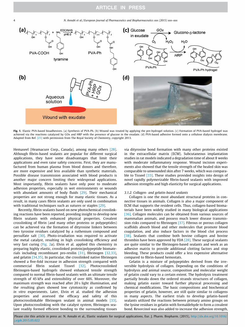
Fig. 1. Elastic PVA-based bioadhesives. (a) Synthesis of PVA-Ph. (b) Wound was treated by applying the pre-hydrogel solution. (c) Formation of PVA-based hydrogel wasachieved via the reactions catalyzed by GOx and HRP with the presence of glucose in the exudate. (d) PVA-based adhesive formed onto a cellulose dialysis membrane.Adapted from Ref. [23] with permission from The Royal Society of Chemistry, copyright 2013.
4 N. Annabi et al. / European Journal of Pharmaceutics and Biopharmaceutics xxx (2015) xxx–xxx
Hemaseel (Heamacure Corp., Canada), among many others [28].Although fibrin-based sealants are popular for different surgicalapplications, they have some disadvantages that limit theirapplications and even raise safety concerns. First, they are manu-factured from human plasma from blood donors and therefore,are more expensive and less available than synthetic materials.Possible disease transmission associated with blood products isanother major concern limiting their widespread applications.Most importantly, fibrin sealants have only poor to moderateadhesion properties, especially in wet environments or woundswith abundant amounts of body fluids [29]. Their mechanicalproperties are not strong enough for many elastic tissues. As aresult, in many cases fibrin sealants are only used in combinationwith traditional techniques such as sutures or staples [29].
Recently, fibrin sealants based on new photochemical crosslink-ing reactions have been reported, providing insight to develop newfibrin sealants with enhanced physical properties. Covalentcrosslinking of fibrin (and many other proteins or polypeptides)can be achieved via the formation of dityrosine linkers betweentwo tyrosine residues catalyzed by a ruthenium compound andpersulfate salt [30]. Photo-irradiation was required to activatethe metal catalyst, resulting in high crosslinking efficiency andvery fast curing (Fig. 2a). Elvin et al. applied this chemistry inpreparing highly elastic, crosslinked protein/polypeptide biomate-rials including recombinant pro-resilin [31], fibrinogen [32,33],and gelatin [34,35]. In particular, the crosslinked native fibrinogenshowed a five-fold increase in adhesion strength compared withcommercial fibrin sealant Tisseel [32]. Photocrosslinkedfibrinogen-based hydrogels showed enhanced tensile strengthcompared to normal fibrin-based sealants with an ultimate tensilestrength of 45 kPa and extensibility of over 60%. Moreover, themaximum strength was reached after 20 s light illumination, andthe resulting glues showed low cytotoxicity as confirmed byin vitro experiments. Later, Elvin et al. studied the adhesionproperties and assessed the efficacy and safety of thisphotocrosslinkable fibrinogen sealant in animal models [33].Upon photocrosslinking with the catalyst system, fibrinogen sea-lant readily formed efficient bonding to the surrounding tissues
Please cite this article in press as: N. Annabi et al., Elastic sealants for surgicaj.ejpb.2015.05.022
via dityrosine bond formation with many other proteins existedin the extracellular matrix (ECM). Subcutaneous implantationstudies in rat models indicated a degradation time of about 8 weekswith moderate inflammatory response. Wound incision experi-ments also showed that the tensile strength of the healed skin wascomparable to unwounded skin after 7 weeks, which was compara-ble to Tisseel [33]. These studies provided insights into design ofnovel rapidly polymerizable fibrin-based sealants with improvedadhesion strengths and high elasticity for surgical applications.
3.1.2. Collagen- and gelatin-based sealantsCollagen is one the most abundant structural proteins in con-
nective tissues in animals. Collagen is also a major component ofECM that supports the resident cells. Thus, collagen-based bioma-terials have been widely studied in many biological applications[36]. Collagen molecules can be obtained from various sources ofmammalian animals, and possess much lower disease transmis-sion risks compared to fibrinogen [37]. Fibrous or porous collagenscaffolds absorb blood and other molecules that promote bloodcoagulation, and also induce factors in the blood clot process[38]. Sealants that combine both bovine collagen and bovinethrombin have been approved by FDA [28]. These surgical sealantsare quite similar to the fibrinogen-based sealants and work as anadhesive matrix to provide additional clotting factors and stopbleeding. These products could offer a less expensive alternativecompared to fibrin-based hemostats.
Gelatin is a mixture of polypeptides derived from the irre-versible hydrolysis of collagen. Depending on the conditions ofhydrolysis and animal source, composition and molecular weightof gelatin could vary to a certain extent. The hydrolysis treatmentpartially breaks down the ordered strands structures of collagen,making gelatin easier toward further physical processing andchemical modifications. The basic compositions and biochemicalproperties of gelatin, however, are still quite similar to collagensin many aspects. The earliest trials to develop gelatin-basedsealants utilized the reactions between primary amino groups onthe lysine residues in gelatin with formaldehyde to form a covalentbond. Resorcinol was also added to increase the adhesion strength,
l applications, Eur. J. Pharm. Biopharm. (2015), http://dx.doi.org/10.1016/
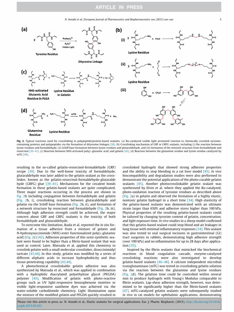
Fig. 2. Typical reactions used for crosslinking in polypeptide/protein-based sealants. (a) Ru-catalyzed visible light promoted reaction to chemically crosslink tyrosine-containing proteins and polypeptides via the formation of dityrosine linkages [30]. (b) Crosslinking mechanism of GRF or GRFG sealants, including (i) the reaction betweenlysine residues and formaldehyde, (ii) Schiff base formation between lysine residues and glutaraldehyde, and (iii) formation of the network structure from formaldehyde andresorcinol [39–41]. (c) Reaction between NHS-activated poly(L-glutamic acid) and gelatin [42]. (d) Reaction between the glutamine residue and lysine residue catalyzed bymTG [46].
N. Annabi et al. / European Journal of Pharmaceutics and Biopharmaceutics xxx (2015) xxx–xxx 5
resulting in the so-called gelatin-resorcinol-formaldehyde (GRF)recipe [39]. Due to the well-know toxicity of formaldehyde,glutaraldehyde was later added to the gelatin sealant as the cross-linker, known as the gelatin-resorcinol-formaldehyde-glutaraldehyde (GRFG) glue [39–41]. Mechanisms for the covalent bondsformation in these gelatin-based sealants are quite complicated.Three major reactions occurring in the process are shown inFig. 2b including conjugation between formaldehyde and gelatin(Fig. 2b, i), crosslinking reaction between glutaraldehyde andgelatin via the Schiff base formation (Fig. 2b, ii), and formation ofa network structure by resorcinol and formaldehyde (Fig. 2b, iii).Although high adhesion strength could be achieved, the majorconcern about GRF and GRFG sealants is the toxicity of bothformaldehyde and glutaraldehyde.
To overcome this limitation, Ikada et al. reported the in situ for-mation of a tissue adhesive from a mixture of gelatin andN-hydroxysuccinimide (NHS)-ester functionalized poly(L-glutamicacid) (Fig. 2c) [42]. Adhesion properties of this semi-synthetic sea-lant were found to be higher than a fibrin-based sealant that wasused as control. Later, Matsuda et al. applied this chemistry tocrosslink gelatin with a small molecular crosslinker, disuccinimidyltartrate [43,44]. In this study, gelatin was modified by a series ofdifferent aliphatic acids to increase hydrophobicity and thustissue-penetrating capability [43,44].
A photochemical crosslinkable gelatin adhesive was alsosynthesized by Matsuda et al., which was applied in combinationwith a hydrophilic diacrylated polyethylene glycol (PEGDA)polymer [45]. Modification of gelatin with photo-reactivegroups such as UV light-responsive benzophenone moieties orvisible light-responsive xanthene dyes was achieved via thewater-soluble carbodiimide chemistry. Light illumination uponthe mixture of the modified gelatin and PEGDA quickly resulted in
Please cite this article in press as: N. Annabi et al., Elastic sealants for surgicaj.ejpb.2015.05.022
crosslinked hydrogels that showed strong adhesive propertiesand the ability to stop bleeding in a rat liver model [45]. In vivobiocompatibility and degradation studies were also performed todemonstrate the potential applications of the photo-curable gelatinsealants [45]. Another photocrosslinkable gelatin sealant wassynthesized by Elvin et al. where they applied the Ru-catalyzed,photo-oxidation reaction of tyrosine residues as described above(Fig. 2a) in gelatin and observed the formation of a highly elastic,nontoxic gelatin hydrogel in a short time [34]. High elasticity ofthe gelatin-based sealants was demonstrated with an ultimatestrain larger than 650% and adhesive stress higher than 100 kPa.Physical properties of the resulting gelatin-based sealants couldbe tailored by changing tyrosine content of gelatin, concentration,and light exposure time. In vivo studies in a sheep model confirmedthat this gelatin-based sealant could stop blood and air leakages inlung tissue with minimal inflammatory responses [34]. This sealantwas also tested to seal surgical incisions in gastrointestinal (GI)tract surgeries in rabbits, demonstrating high adhesive strength(over 100 kPa) and no inflammation for up to 28 days after applica-tion [35].
Inspired by the fibrin sealants that mimicked the biochemicalreactions in blood coagulation cascade, enzyme-catalyzedcrosslinking reactions were also investigated to developgelatin-based sealants [46–48]. A calcium independent microbialtransglutaminase (mTG) was tested in crosslinking gelatin solutionvia the reaction between the glutamine and lysine residues(Fig. 2d). The gelation time could be controlled within severalmin to produce hydrogels with Young’s Modulus comparable tofibrin sealants. Lap-shear adhesion strength, however, was deter-mined to be significantly higher than the fibrin-based sealants[47]. mTG-catalyzed gelatin sealants were subsequently studiedin vivo in rat models for ophthalmic applications, demonstrating
l applications, Eur. J. Pharm. Biopharm. (2015), http://dx.doi.org/10.1016/
6 N. Annabi et al. / European Journal of Pharmaceutics and Biopharmaceutics xxx (2015) xxx–xxx
no cellular damage to rat retinal tissue and strong adhesion to wetretinal tissue [48].
3.1.3. Albumin-based sealantsAlbumin refers to a family of globular proteins found in blood
plasma, and can be obtained from different animal sources.Several types of albumin-based sealants have been developedbased on similar reactions as discussed above. For example,BioGlue (CryoLife Inc., Kennesaw, GA) is a commercial sealant withFDA approval as hemostats in vascular and cardiac surgeries. It ismade of bovine albumin and glutaraldehyde using a similar reac-tion as shown in Fig. 2b, ii. The adhesion strength of the glue couldbe optimized by varying the ratio and concentrations of the twocomponents [49]. Concerns about the biosafety of glutaraldehydecould arise for Bioglue for internal use.
Another commercialized albumin-based sealant is Progel(Davol Inc., Woburn, MA) developed to prevent air leakage in lungsurgeries. Progel is a composite sealant containing human albuminand a PEG crosslinker with two NHS activated ester groups [4,50].Upon mixing, the primary amine groups on lysine residues in albu-min quickly react with the succinimidyl succinate groups and forma crosslinked structure within one min, which is similar to thereaction shown in Fig. 2c. It was shown that Progel could effec-tively stop pleural air leakage and degrade relatively fast in vivowithout severe immune responses [4,50]. Its applications in lungsurgeries will be discussed in details in the following sections.
3.2. Polysaccharide-based sealants
Polysaccharides are a large family of biopolymers composed ofdifferent combinations of monosaccharide (sugar) building blocks.They vary in the structure, chemical linkage, molecular weight, andfunctional groups of the monosaccharide monomers. As a naturalpolymer, polysaccharides have been widely used in numerousmedical, pharmaceutical and food products.
3.2.1. Chitosan-based sealantsChitosan is a polysaccharide derived from chitin, which is
composed of N-acetylglucosamine building blocks via beta-1,4-glycosidic linkages and is typically obtained from the exoskele-ton of arthropods, such as crabs, lobsters and shrimps, as well asthe cell walls of fungi. Partial hydrolysis (deacetylation) of theacetyl amide groups releases some free amine groups and resultsin chitosan.
Due to the relatively poor solubility of chitosan, Ishihara et al.reported a chemically modified chitosan derivative by reactingchitosan with lactobionic acid with the presence of a watersoluble carbodiimide (Fig. 3a, i) [51,52]. Azido groups were thenintroduced to the water-soluble chitosan by reacting with4-azidobenzoic acid via the same chemistry (Fig. 3a, ii). The result-ing modified chitosan could be photocrosslinked by exposing to UVlight to form chitosan-based sealants. Under UV irradiation, theazido groups would decompose to release N2 and form highlyreactive nitrene groups, which will simultaneously dimerize togenerate azo-linkages. In vitro cell culture studies confirmed non-toxicity of the engineered chitosan sealants. In a following studyfrom the same group, this chitosan sealant was further tested ina mouse model for wound closure [53]. Significantly acceleratedwound closure and advanced granulation tissue formation wereobserved, suggesting promising application of the engineeredchitosan-based sealant for clinical applications.
Later, Moratti et al. reported the synthesis of succinylated chi-tosan by reacting chitosan with succinic anhydride (Fig. 3a, iii).This modified chitosan showed enhanced water solubility. Whenmixing with dextran aldehyde obtained from oxidation of dextran(see below for details), a gel was formed via the imine bond
Please cite this article in press as: N. Annabi et al., Elastic sealants for surgicaj.ejpb.2015.05.022
formation between the amino groups and aldehyde groups, whichshowed excellent hemostatic and adhesive properties in animalmodels [54]. Park et al. developed a synthetic procedure to grafttyrosine modified PEG chains through the amino groups on chi-tosan (Fig. 3a, iv) [55]. Grafted PEG chains largely increased the sol-ubility of chitosan and provided reactive tyrosine groups to formcrosslinked network structure. With the presence of a HPR andhydrogen peroxide, the chitosan solution quickly formed a gel viathe dityrosine bond formation. Adhesion strength of the engi-neered chitosan-PEG composite sealant was several times higherthan fibrin sealants. Moreover, in vivo studies in a rat modeldemonstrated that the fabricated chitosan-based sealant showedsuperior healing ability in skin incision comparing to suture, fibringlue and cyanoacrylate [55].
Recently, Zhao et al. reported the formation of a compositehydrogel based on thiol containing chitosan (Fig. 3a, v) and malei-mide containing e-polylysine [56]. The Michael addition reactionbetween thiol and maleimide groups led to the in situ formationof a crosslinked hydrogel upon simply mixing of the two compo-nents. This chitosan-based composite sealant showed four timeshigher adhesion strength compared with fibrin glue. Synthesis ofthiol containing chitosan (Fig. 3a, vi) was also reported by Leeet al. to chemically graft hematin, which was an Fe (III) containingporphyrin, with chitosan. The resulting chitosan was used as anenzyme-mimetic biocatalyst to accelerate the crosslinking ofcatechol-modified polymers [57].
3.2.2. Dextran-based sealantsDextran refers to a relatively complex polysaccharide with
some branched structures. Its linear part is composed of glucosebuilding blocks via the alpha-1,6-linkages. Different from chitosan,which is a polyglucosamine, there are no reactive amino groups indextran. One modification strategy reported for dextran-basedsealants relies on the selective partial oxidation of dextran tointroduce aldehyde groups (Fig. 3b), which can react with aminogroup to form imine linkages (similar to the reaction shown inFig. 2b, ii) and result in the formation of hydrogels. Othermodification methods include introducing methacrylate functionalgroups to crosslink with thiol crosslinkers [58], or preparingenzyme-crosslinkable dextran-tyramine conjugates [59]. Arakiet al. studied the aldehyde functionalized dextran-based sealantby mixing it with e-polylysine [60,61]. Gelation was achieved viathe Schiff base bond formation. Adhesion properties of thisdextran-based lung sealant were evaluated in vivo and it was foundthat it had better adhesion than the fibrin glue control.Biodegradability and histotoxicity were also tested in a dog model.Inflammatory reactions were largely reduced 4 weeks afterapplication, and the sealant could be degraded on the lung tissuewithin 3 months [60,61]. Bhatia et al. reported another tissuesealant by combining the aldehyde-containing dextran with an8-arm amine-endcapped PEG crosslinker [62]. Cytotoxicity andinflammatory properties were tested in vitro using 3T3 fibroblastcells and J774 macrophage cells. The experimental results sug-gested that this dextran-PEG tissue adhesive was non-cytotoxicand did not induce inflammatory response [62].
3.2.3. Chondroitin sulfate-based sealantsChondroitin sulfate is a sulfated polyglucosamine typically
found as the structural component in cartilage ECM and manyother tissues in the body. Due to its key biological roles in theformation and function of cartilage tissues, sealants based onchondroitin sulfate have been developed and tested to regeneratethe connections between cartilage tissue and biomaterials, as wellas in corneal wound healing. Since there are multiple reactive sitesin chondroitin sulfate, different modification strategies havebeen developed to introduce various functional groups, including
l applications, Eur. J. Pharm. Biopharm. (2015), http://dx.doi.org/10.1016/
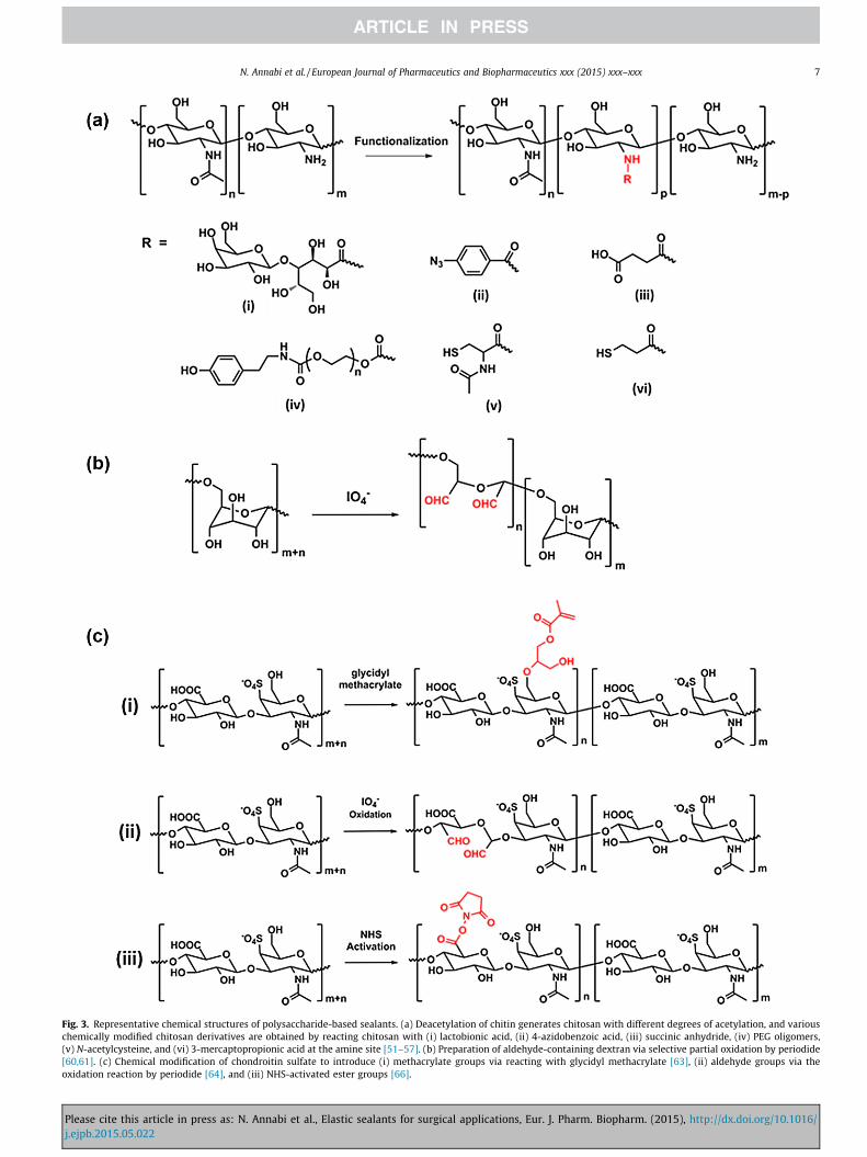
Fig. 3. Representative chemical structures of polysaccharide-based sealants. (a) Deacetylation of chitin generates chitosan with different degrees of acetylation, and variouschemically modified chitosan derivatives are obtained by reacting chitosan with (i) lactobionic acid, (ii) 4-azidobenzoic acid, (iii) succinic anhydride, (iv) PEG oligomers,(v) N-acetylcysteine, and (vi) 3-mercaptopropionic acid at the amine site [51–57]. (b) Preparation of aldehyde-containing dextran via selective partial oxidation by periodide[60,61]. (c) Chemical modification of chondroitin sulfate to introduce (i) methacrylate groups via reacting with glycidyl methacrylate [63], (ii) aldehyde groups via theoxidation reaction by periodide [64], and (iii) NHS-activated ester groups [66].
N. Annabi et al. / European Journal of Pharmaceutics and Biopharmaceutics xxx (2015) xxx–xxx 7
Please cite this article in press as: N. Annabi et al., Elastic sealants for surgical applications, Eur. J. Pharm. Biopharm. (2015), http://dx.doi.org/10.1016/j.ejpb.2015.05.022
8 N. Annabi et al. / European Journal of Pharmaceutics and Biopharmaceutics xxx (2015) xxx–xxx
methacrylate groups, aldehyde groups, and NHS-activated estergroups (Fig. 3c). Elisseeff et al. reported the synthesis of a pho-tocrosslinkable chondroitin sulfate derivative by reacting with gly-cidyl methacrylate at room temperature (Fig. 3c, i) [63]. Hydrogelswere feasibly obtained by UV crosslinking with a photoinitiator.Morphological and mechanical, and biological properties of thechondroitin sulfate-based hydrogels were studied. Later, theydeveloped a different chemical modification method of chondroitinsulfate by partially oxidizing the polysaccharide with periodide tointroduce aldehyde groups, similar to the reaction used for dextransealants [64]. A synthetic polymer, poly(vinyl alcohol-co-vinylamine), was used in combination with the aldehyde-bearing chon-droitin sulfate to form a hydrogel-based sealant. They used theengineered chondroitin sulfate-polymer sealant in a rabbit modelto seal corneal incisions. The fabricated sealant showed superiorperformances compared with the traditional suture techniques inex vivo studies [64].
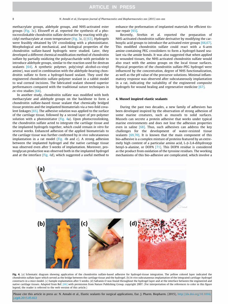
In another study, chondroitin sulfate was modified with bothmethacrylate and aldehyde groups on the backbone to form achondroitin sulfate-based tissue sealant that chemically bridgedtissue proteins and the implanted biomaterials via a two-fold cova-lent linkages [65]. The adhesive solution was applied to the surfaceof the cartilage tissue, followed by a second layer of pre-polymersolution with a photoinitiator (Fig. 4a). Upon photocrosslinking,the chondroitin sulfate acted to integrate the cartilage tissue andthe implanted hydrogels together, which could remain in vitro forseveral weeks. Enhanced adhesion of the applied biomaterials tothe cartilage tissue was further confirmed by in vivo subcutaneousimplantation in a rat model (Fig. 4b and c). A strong adhesionbetween the implanted hydrogel and the native cartilage tissuewas observed even after 5 weeks of implantation. Moreover, pro-teoglycan production was observed both in the implanted hydrogeland at the interface (Fig. 4d), which suggested a useful method to
Fig. 4. (a) Schematic diagram showing application of the chondroitin sulfate-basedchondroitin sulfate layer which served as the bridge between the cartilage tissue and theconstructs in a mice model. (c) Sample explantation after 5 weeks. (d) Safranin-O was fonative cartilage tissues. Adapted from Ref. [66] with permission from Nature Publishinglegend, the reader is referred to the web version of this article.)
Please cite this article in press as: N. Annabi et al., Elastic sealants for surgicaj.ejpb.2015.05.022
enhance the preformation of implanted materials for efficient tis-sue repair [65].
Recently, Strehin et al. reported the preparation ofNHS-activated chondroitin sulfate derivative by modifying the car-boxylic acid groups to introduce the activated ester groups [66,67].This modified chondroitin sulfate could react with a 6-armamine-containing PEG crosslinkers to form a hydrogel-based sea-lant via the amide bonds. It was also suggested that when appliedto wounded tissues, the NHS-activated chondroitin sulfate wouldalso react with the amino groups on the local tissue surfaces.Physical properties of the chondroitin sulfate-PEG hydrogel wereinfluenced by the concentration, degree of NHS functionalization,as well as the pH value of the precursor solutions. Minimal inflam-matory response was observed after subcutaneously implantationin a rat, indicating the suitability of the engineered compositehydrogels for wound healing and regenerative medicine [67].
4. Mussel inspired elastic sealants
During the past two decades, a new family of adhesives hasbeen developed inspired by the observation of strong adhesion ofsome marine creatures, such as mussels to solid surfaces.Mussels can secrete a protein adhesive that works under typicalmarine environments and does not lose the adhesion propertieseven in saline [68]. Thus, such adhesives can address the keychallenges for the development of water-resisted tissuesealants [69,70]. It is known that the main component of thisbio-adhesive is a complex mixture of proteins featured by an extre-mely high content of a particular amino acid, L-b-3,4-dihydroxyphenyl-a-alanine, or DOPA [71]. This DOPA residue is consideredas the product from oxidation of the tyrosine residues. The workingmechanisms of this bio-adhesive are complicated, which involve a
adhesive for hydrogel-tissue integration. The yellow colored layer indicated thehydrogel. (b) In vivo subcutaneous implantation of the integrated cartilage–hydrogelund throughout the hydrogel layer and at the interface between the engineered and
Group, copyright 2007. (For interpretation of the references to color in this figure
l applications, Eur. J. Pharm. Biopharm. (2015), http://dx.doi.org/10.1016/
N. Annabi et al. / European Journal of Pharmaceutics and Biopharmaceutics xxx (2015) xxx–xxx 9
series of reactions between different functional groups [72]. It isknown that diquinone intermediates are formed by oxidation ofthe DOPA residues, which could react with, for example, aminoand thiol groups via Michael addition, or certain metal ions viacoordination [73].
The unique bio-adhesive properties of mussels have attractedmuch attention to develop novel sealants for adhesion to wet sur-faces. Early trials include extracting the secreted proteins frommussels [74]. While this approach helped with understanding themechanism of the formation of this material, extraction could onlyprovide limited amount of it that was not cost-efficient. Syntheticpolypeptides with designed sequences have also been studied toinvestigate the structure-property relationships in this class of sea-lants [70,75]. In addition, recombinant DNA technology has beenused to prepare mussel inspired protein sealants [76,77].However, the expression of the key adhesive proteins typicallyresulted in low yield and purity, which limited the practicalapplications of the recombinant DNA strategy.
As a result of the difficulties in directly preparingDOPA-containing proteins, biocompatible synthetic/naturalpolymers modified with DOPA motifs are promising targets fordeveloping DOPA-based sealants. To date, the DOPA moieties havebeen successfully incorporated within a variety of polymers,including both synthetic polymers or modified natural polymers.The chemical approaches toward developing DOPA-containingpolymers differ significantly in different systems. In this section,we will briefly summarize progresses in DOPA-containingpolymers as tissue sealant materials.
In one study, Deming et al. reported the synthesis of polypep-tides with tunable DOPA contents via ring-opening polymerizationof N-carboxyanhydride (NCA) monomers prepared from DOPA andlysine by phosgenation [75]. Protected NCA monomers werecopolymerized to form high molecular weight polymers withexcellent conversions. Feasible control over DOPA content couldbe achieved by changing the feed ratio of the two monomers.Systematic studies on several factors that might influence theadhesion properties were performed, including copolymer compo-sition, concentration, selection of oxidation reagents, and curingtemperature [75]. Results showed that the synthetic polypeptidespossessed comparable adhesion properties with mussel adhesivesto several substrates under optimized crosslinking conditions[72,75,78].
Wilker et al. later reported a simplified system asmussel-mimicking adhesives by using a 3,4-dihydroxstyrenemonomer to mimic the DOPA side groups [79]. First protected bymethyl groups, anionic polymerization initiated by n-butyl lithiumsuccessfully copolymerized styrene with this monomer to form aseries of copolymers with different monomer feed ratios.Deprotection by boron tribromide released the hydroxyl groups,which were subjected to oxidation crosslinking. The authors testedand compared several different inorganic oxidative reagents,which might have limited medical applications due to toxicity.Recently, they also presented a systematic structure-propertyrelationship of the polystyrene-based sealant material [73].
In another study, Lee and Messersmith et al. synthesized amethacrylated dopamine monomer and applied free radicalpolymerization to generate DOPA-containing copolymers [80].Inspired by the adhesion phenomena of both mussels and geckos,they designed a reversible wet/dry adhesive that allowedmultiple reversible adhesion/detaching processes. A patternedpolydimethylsiloxane (PDMS) substrate was created via electronbeam lithography to mimic the structure of geckos, and theDOPA-containing copolymer was spin-coated onto the substrateto incorporate the water-resistant adhesion properties [80].
Messersmith et al. also prepared DOPA-functionalized linear ormultiple armed PEG to mimic mussel adhesive proteins [81–85].
Please cite this article in press as: N. Annabi et al., Elastic sealants for surgicaj.ejpb.2015.05.022
When 4 armed DOPA-containing PEG was treated with differentoxidative reagents, such as periodide, HRP, or mushroom tyrosi-nase, fast gelation was observed due to the crosslinking betweenthe DOPA moieties or the coupling between oxidized DOPA andfree amino groups [81]. In vivo effectiveness of this DOPA-PEG sea-lant in extrahepatic islet transplantation were reported in a murinemodel [82]. Brubaker et al. later introduced an enzyme-degradableoligopeptide linkers between the PEG chains and the DOPA moi-eties, aiming to achieve regulated degradation of the hydrogelsby enzyme. However, only relatively slow enzyme-catalyzeddegradation behaviors were observed both in vitro and in vivo[83]. Recently, Barrett et al. further investigated how the crosslink-ing of DOPA-containing multiple armed PEG sealant could be influ-enced by the addition of Fe (III) ions and pH values of the solution,revealing significant pH-regulated physical properties of theDOPA-PEG hydrogel system.
Similarly, the DOPA motif has also been introduced to otherpolymer systems, such as polyesters [86], polyallylamine [87],poly(ester amide)s [88,89], poly(propylene oxide)-poly(ethyleneoxide) block copolymers [85], and chitosan [57,90].DOPA-containing sealants provide a promising candidate for vari-ous medical applications.
5. Applications of elastic surgical sealants
According to the report from MedMarket Diligence, LLC, thereare about 114 million procedure-based wounds to occur annuallyworldwide, among which 36 million cases are from surgeries inthe US [91]. The market value for the surgical materials is expectedto grow to $4B in 2015 worldwide and may exceed $7B in 2017[92]. Although currently over two-thirds of the surgical productmarket is for hemostats, a greater sealant rate is expected due tothe lack of suitable products. Surgical sealants are required to havehigh adhesion strength and proper function in wet environments.In addition, they must be flexible to move with the tissues. A highlevel of elasticity is particularly important for surgical proceduresinvolving tissues that undergo continual expansion or contractionsuch as the heart, skin, blood vessels, and lungs. For example, inlung surgery the sealant might be applied when the lungs aredeflated, therefore, the sealant is required to have elasticity similarto the lungs to support expansion and contraction of the tissue. Incardiovascular applications, the elasticity of the sealant plays a sig-nificant role in supporting proper expansion and contraction of thetissues during blood pumping.
In lung surgery, lung tissues are sealed surgically via sutures,staples, or surgical meshes. Despite their common use in the clinic,these mechanical methods are inevitably associated with lung tis-sue damage caused by deep piercing, ischemia, and prolonged airleaks, which represent the most common complications after lungsurgeries [93]. Particularly, prolonged air leak could lead toextended chest tube drainage time that would increase the riskof developing infections and bronco-pleural fistulae in the patients,and consequently, a longer hospital stay [94,95].
A variety of complementary natural and synthetic materialshave been applied to overcome such complications including fibrinsealants, collagen-based sealants, and synthetic glues [40,96–101].However, some of these surgical sealants lack appropriate elastic-ity, adhesion strength, and burst pressure required for sealing thelung tissues. In one study, a synthetic-based absorbable biomaterialcalled FocalSeal consisting of a primer and a sealant solution wasdeveloped as a lung sealant [14,15]. The primer was first appliedto the target tissue to wet the tissue for increasing adhesion. ThePEG-based sealant solution was then injected and subsequentlyphotopolymerized to seal the lung tissues. The results of in vivo testusing a pig model showed no post-operation air leaks with intact
l applications, Eur. J. Pharm. Biopharm. (2015), http://dx.doi.org/10.1016/
10 N. Annabi et al. / European Journal of Pharmaceutics and Biopharmaceutics xxx (2015) xxx–xxx
bronchial closures. In their clinical study, it was found that 77% oftreated patients with FocalSeal remained leak free with no undesir-able side effects [14]. However, its multiple step application processdue to the use of primer and light source for crosslinking makes itsclinical applications challenging. In addition, it has been reportedthat FocalSeal may potentially enhance the rate of post-operativeempyema [16,102]. In another study, Kobayashi et al. developedan albumin-based hydrogel sealant, Progel, composed of two com-ponents including PEG disuccinimidyl succinate and human albu-min, to stop air leaks in a rat lung model [50]. The sealingproperties of the engineered lung sealant were compared to fibringlue. The average burst pressure on day 3 of surgery was about71 mmHg for Progel, which was significantly higher than fibrin glue(60 mmHg). In addition, no adverse tissue reaction was observed upto day 14 of operation [50]. The safety and effectiveness of Progel tostop air leaks after pulmonary surgery were also evaluated by test-ing the sealant on 161 patients [17]. Patients treated by Progel hadless intra-operative and post-operative leakages and shorter hospi-talization compared to the control group (65% vs 86% and 6 vs7 days) [17]. This demonstrates the suitability of Progel in lungresection surgery for closing air leaks [4]. In another study, theburst pressure values for different lung sealants were comparedin an ex vivo study using a porcine model [103]. Bioglue (V-Tech,Roskilde, Denmark) attained the highest burst pressures comparedto other tested sealants including Evicel (OMRIX biopharmaceuti-cals S.A, Belgium), Tisseel (Baxter, Denmark), TachoSil (Nycomed,Roskilde, Denmark), TissuePatchDural (Vingmed, Denmark), andPleuraseal (Covidien, Denmark). However, it was shown that therigidity of the Bioglue caused lung tissue tearing and deformation[103]. Therefore, in addition to high burst pressure and adhesionstrength, the flexibility of the lung sealants is an important param-eter for their clinical applications.
In cardiovascular surgery, one of the main challenges is theinability to reconnect tissue or attach prosthetic materials in awet and dynamic environment, such as continuous tissue contrac-tions and blood flow. Most of the currently available sealants havelow adhesion strength and mechanical properties in wetconditions. To address this limitation, recently, an elasticblood-resistant light-activated tissue adhesive based on PGS has
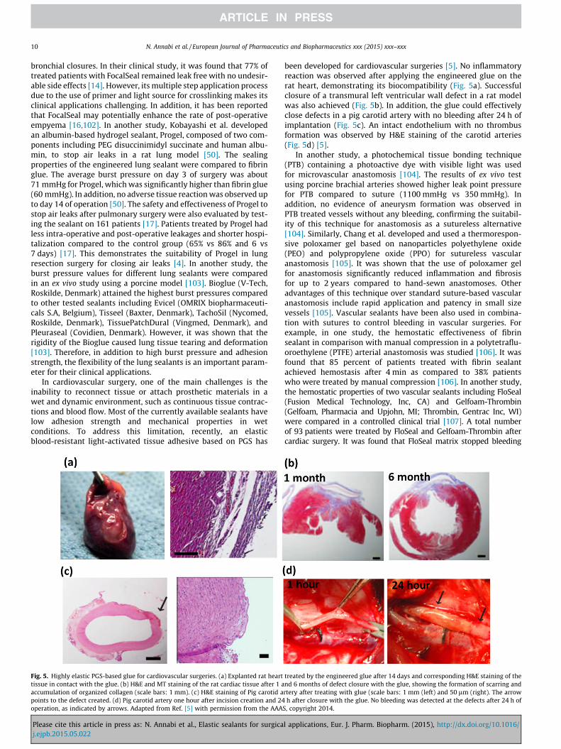
Fig. 5. Highly elastic PGS-based glue for cardiovascular surgeries. (a) Explanted rat hearttissue in contact with the glue. (b) H&E and MT staining of the rat cardiac tissue after 1 aaccumulation of organized collagen (scale bars: 1 mm). (c) H&E staining of Pig carotid apoints to the defect created. (d) Pig carotid artery one hour after incision creation and 24operation, as indicated by arrows. Adapted from Ref. [5] with permission from the AAA
Please cite this article in press as: N. Annabi et al., Elastic sealants for surgicaj.ejpb.2015.05.022
been developed for cardiovascular surgeries [5]. No inflammatoryreaction was observed after applying the engineered glue on therat heart, demonstrating its biocompatibility (Fig. 5a). Successfulclosure of a transmural left ventricular wall defect in a rat modelwas also achieved (Fig. 5b). In addition, the glue could effectivelyclose defects in a pig carotid artery with no bleeding after 24 h ofimplantation (Fig. 5c). An intact endothelium with no thrombusformation was observed by H&E staining of the carotid arteries(Fig. 5d) [5].
In another study, a photochemical tissue bonding technique(PTB) containing a photoactive dye with visible light was usedfor microvascular anastomosis [104]. The results of ex vivo testusing porcine brachial arteries showed higher leak point pressurefor PTB compared to suture (1100 mmHg vs 350 mmHg). Inaddition, no evidence of aneurysm formation was observed inPTB treated vessels without any bleeding, confirming the suitabil-ity of this technique for anastomosis as a sutureless alternative[104]. Similarly, Chang et al. developed and used a thermorespon-sive poloxamer gel based on nanoparticles polyethylene oxide(PEO) and polypropylene oxide (PPO) for sutureless vascularanastomosis [105]. It was shown that the use of poloxamer gelfor anastomosis significantly reduced inflammation and fibrosisfor up to 2 years compared to hand-sewn anastomoses. Otheradvantages of this technique over standard suture-based vascularanastomosis include rapid application and patency in small sizevessels [105]. Vascular sealants have been also used in combina-tion with sutures to control bleeding in vascular surgeries. Forexample, in one study, the hemostatic effectiveness of fibrinsealant in comparison with manual compression in a polytetraflu-oroethylene (PTFE) arterial anastomosis was studied [106]. It wasfound that 85 percent of patients treated with fibrin sealantachieved hemostasis after 4 min as compared to 38% patientswho were treated by manual compression [106]. In another study,the hemostatic properties of two vascular sealants including FloSeal(Fusion Medical Technology, Inc, CA) and Gelfoam-Thrombin(Gelfoam, Pharmacia and Upjohn, MI; Thrombin, Gentrac Inc, WI)were compared in a controlled clinical trial [107]. A total numberof 93 patients were treated by FloSeal and Gelfoam-Thrombin aftercardiac surgery. It was found that FloSeal matrix stopped bleeding
treated by the engineered glue after 14 days and corresponding H&E staining of thend 6 months of defect closure with the glue, showing the formation of scarring andrtery after treating with glue (scale bars: 1 mm (left) and 50 lm (right). The arrowh after closure with the glue. No bleeding was detected at the defects after 24 h of
S, copyright 2014.
l applications, Eur. J. Pharm. Biopharm. (2015), http://dx.doi.org/10.1016/
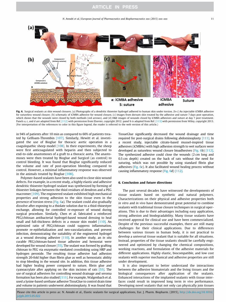
Fig. 6. Surgical sealants as skin wound closures. (a) Photographs of a dendritic thioester hydrogel adhered to human skin under torsion. (b-c) An injectable iCMBA adhesivefor sutureless wound closure; (b) schematic of iCMBA adhesive for wound closure, (c) images from dorsum skin treated by the adhesive and suture 7 days post operation,which shows that the wounds were closed by both methods (red arrows), and (d) H&E images of wounds closed by iCMBA adhesives and suture at day 7 post treatment.Panels a, c, and d are adapted from Ref. [112] with permission from Elsevier, copyright 2012; panel b is adapted from Ref. [113] with permission from Wiley, copyright 2013.(For interpretation of the references to color in this figure legend, the reader is referred to the web version of this article.)
N. Annabi et al. / European Journal of Pharmaceutics and Biopharmaceutics xxx (2015) xxx–xxx 11
in 94% of patients after 10 min as compared to 60% of patients trea-ted by Gelfoam-Thrombin [107]. Similarly, Hewitt et al. investi-gated the use of Bioglue for thoracic aortic operation in acoagulopathic sheep model [108]. In their experiments, the sheepwere first anticoagulated with heparin and then subjected toend-to-side anastomoses of a graft to a thoracic aorta. The anasto-moses were then treated by Bioglue and Surgicel (as control) tocontrol bleeding. It was found that Bioglue significantly reducedthe volume and rate of post-operation bleeding compared tocontrol. However, a minimal inflammatory response was observedin the animals treated by Bioglue [108].
Polymer-based sealants have been also used to close skin wounddefects. For example, in a recent study, a highly elastic and adhesivedendritic thioester hydrogel sealant was synthesized by forming ofthioester linkages between the thiol residues of dendron and a PEGmacromer [109]. The engineered sealant exhibited high mechanicalproperties and strong adhesion to the skin tissue even in thepresence of torsion stress (Fig. 6a). The sealant could also graduallydissolve after exposing to a thiolate solution due to a thiol-thioestyerexchange, allowing for controlled re-exposure of wound duringsurgical procedure. Similarly, Chen et al. fabricated a reinforcedPEG/chitosan antibacterial hydrogel-based wound dressing to healsmall and full-thickness defects in a mouse skin model [110]. Anoptimized ratio of PEG/chitosan could suppress inflammation,promote re-epithelialization and neo-vascularization, and preventinfection, demonstrating the suitability of the engineered hydrogelas a wound dressing adhesive [110]. In another study, an in situcurable PEG/chitosan-based tissue adhesive and hemostat weredeveloped for wound closure [55]. The sealant was formed by graftingchitosan to PEG via enzymatic-mediated crosslinking using HRP andhydrogen peroxide. The resulting tissue adhesive had adhesionstrength 20-fold higher than fibrin glue as well as hemostatic abilityto stop bleeding in the wound site. In addition, this tissue adhesivehad higher healing power compared to suture, fibrin glue andcyanoacrylate after applying on the skin incision of rats [55]. Theuse of surgical adhesives for controlling wound drainage and seromaformation has been also studied [111]. For example, Walgenbach et al.used a lysine-based urethane adhesive TissuGlue to control drain timeand volume in patients underwent abdominoplasty. It was found that
Please cite this article in press as: N. Annabi et al., Elastic sealants for surgicaj.ejpb.2015.05.022
TissueGlue significantly decreased the wound drainage and timerequired for post-surgical drains following abdominoplasty [111]. Ina recent study, injectable citrate-based mussel-inspired tissueadhesives (iCMBAs) with high adhesion strength to wet surfaces weredeveloped as sutureless wound closure bioadhesives (Fig. 6b) [112].The synthesized adhesive could close the wounds (2 cm long and0.5 cm depth) created on the back of rats without the need forsuturing, which was not possible by using standard fibrin glueadhesives (Fig. 6c). It also facilitated wound healing process withoutcausing inflammatory response (Fig. 6d) [112].
6. Conclusion and future directions
The past several decades have witnessed the developments oftissue sealants based on synthetic and natural polymers.Characterizations on their physical and adhesive properties bothin vitro and in vivo have demonstrated great potential to combinesealants with traditional tissue closure techniques in surgical oper-ations. This is due to their advantages including easy application,strong adhesion and biodegradability. Many tissue sealants havereceived approval for clinical use and have been commercialized.Despite of the previous successful examples, there are still manychallenges for their clinical applications. Due to differencesbetween various tissues in human body, it is not practical todevelop a universal tissue sealant that is suitable for all situations.Instead, properties of the tissue sealants should be carefully engi-neered and optimized by changing the chemical compositions,working reactions, and formulation of the adhesive materials fordifferent applications. Highly elastic, biocompatible, and low costsealants with superior mechanical and adhesive properties are stillunder development.
It is also important to better understand the interactionsbetween the adhesive biomaterials and the living tissues and thebiological consequences after application of the sealants.Enhanced interactions of the implanted sealants with tissue inter-faces could result in improved long term in vivo performance.Developing novel sealants that not only can physically join tissues
l applications, Eur. J. Pharm. Biopharm. (2015), http://dx.doi.org/10.1016/
12 N. Annabi et al. / European Journal of Pharmaceutics and Biopharmaceutics xxx (2015) xxx–xxx
together but also actively promote tissue growth and repair. It isanticipated that close collaboration between bioengineers, mate-rial scientists, and surgeons is required to efficiently advance theresearch of tissue sealants in future studies.
Acknowledgments
N.A. acknowledges the support from the National Health andMedical Research Council.
The authors acknowledge funding from the National ScienceFoundation (EFRI-1240443), IMMODGEL (602694), and theNational Institutes of Health (EB012597, AR057837, DE021468,HL099073, AI105024, AR063745).
References
[1] N. Annabi, A. Tamayol, S.R. Shin, A.M. Ghaemmaghami, N.A. Peppas, A.Khademhosseini, Nano Today 9 (2014) 574–589.
[2] L.W.S.S. MedMarket Diligence, Glues, Wound Closure and Anti-AdhesionMarket, Forecast to 2017.
[3] Q. Chen, S. Liang, G.A. Thouas, Soft Matter 7 (2011) 6484–6492.[4] C. Fuller, J. Cardiothor. Surg. 8 (2013) 90.[5] N. Lang, M.J. Pereira, Y. Lee, I. Friehs, N.V. Vasilyev, E.N. Feins, K. Ablasser, E.D.
O’Cearbhaill, C. Xu, A. Fabozzo, R. Padera, S. Wasserman, F. Freudenthal, L.S.Ferreira, R. Langer, J.M. Karp, P.J. del Nido, Sci. Trans. Med. 6 (2014)(218ra216).
[6] P. Ferreira, A.F.M. Silva, M.I. Pinto, M.H. Gil, J. Mater. Sci. Mater. Med. 19(2008) 111–120.
[7] P. Ferreira, R. Pereira, J.F.J. Coelho, A.F.M. Silva, M.H. Gil, Int. J. Biol. Macromol.40 (2007) 144–152.
[8] P. Ferreira, J.F.J. Coelho, M.H. Gil, Int. J. Pharm. 352 (2008) 172–181.[9] T.W. Gilbert, S.F. Badylak, J. Gusenoff, E.J. Beckman, D.M. Clower, P. Daly, J.P.
Rubin, Plast. Reconstr. Surg. 122 (2008) 95–102.[10] K.D. Kim, N.M. Wright, Spine 36 (2011) 1906–1912.[11] P. Fransen, Spine J. 10 (2010) 751–761.[12] G. Lee, C.K. Lee, M. Bynevelt, Spine 35 (2010) E1522–E1524.[13] M.C. Preul, W.D. Bichard, T.R. Muench, R.F. Spetzler, Neurosurgery 53 (2003)
1189–1199.[14] P. Macchiarini, J. Wain, S. Almy, P. Dartevelle, J. Thorac. Cardiovasc. Surg. 117
(1999) 751–758.[15] A.M. Gillinov, B.W. Lytle, J. Card. Surg. 16 (2001) 255–257.[16] J.C. Wain, L.R. Kaiser, D.W. Johnstone, S.C. Yang, C.D. Wright, J.S. Friedberg,
R.H. Feins, R.F. Heitmiller, D.J. Mathisen, M.R. Selwyn, Ann. Thorac. Surg. 71(2001) 1623–1629.
[17] M.S. Allen, D.E. Wood, R.W. Hawkinson, D.H. Harpole, R.J. McKenna, G.L.Walsh, E. Vallieres, D.L. Miller, F.C. Nichols, W.R. Smythe, R.D. Davis,M.T.M.S.S.S. Grp, Ann. Thorac. Surg. 77 (2004) 1792–1801.
[18] D.G. Wallace, G.M. Cruise, W.M. Rhee, J.A. Schroeder, J.J. Prior, J. Ju, M.Maroney, J. Duronio, M.H. Ngo, T. Estridge, G.C. Coker, J. Biomed. Mater. Res.58 (2001) 545–555.
[19] A. Hill, T.D. Estridge, M. Maroney, E. Monnet, B. Egbert, G. Cruise, G.T. Coker, J.Biomed. Mater. Res. 58 (2001) 308–312.
[20] J.C. Wheat, J.S. Wolf Jr, Urol. Clin. North Am. 36 (2009) 265–275.[21] C.L.E. Nijst, J.P. Bruggeman, J.M. Karp, L. Ferreira, A. Zumbuehl, C.J. Bettinger,
R. Langer, Biomacromolecules 8 (2007) 3067–3073.[22] N. Lang, M.J. Pereira, Y. Lee, I. Friehs, N.V. Vasilyev, E.N. Feins, K. Ablasser, E.D.
O’Cearbhaill, C. Xu, A. Fabozzo, R. Padera, S. Wasserman, F. Freudenthal, L.S.Ferreira, R. Langer, J.M. Karp, P.J. del Nido, Sci. Transl. Med. 6 (2014)(218ra216).
[23] S. Sakai, M. Tsumura, M. Inoue, Y. Koga, K. Fukano, M. Taya, J. Mater. Chem. B1 (2013) 5067–5075.
[24] M.R. Jackson, Am. J. Surg. 182 (2001) S1–S7.[25] B.M. Alving, M.J. Weinstein, J.S. Finlayson, J.E. Menitove, J.C. Fratantoni,
Transfusion 35 (1995) 783–790.[26] U. Martinowitz, R. Saltz, Curr. Opin. Hematol. 3 (1996) 395–402.[27] R.W. Busuttil, J. Am. Coll. Surg. 197 (2003) 1021–1028.[28] A.P. Duarte, J.F. Coelho, J.C. Bordado, M.T. Cidade, M.H. Gil, Prog. Polym. Sci. 37
(2012) 1031–1050.[29] S.P. Mandell, N.S. Gibran, Exp. Opin. Biol. Ther. 14 (2014) 821–830.[30] D.A. Fancy, T. Kodadek, Proc. Natl. Acad. Sci. 96 (1999) 6020–6024.[31] C.M. Elvin, A.G. Carr, M.G. Huson, J.M. Maxwell, R.D. Pearson, T. Vuocolo, N.E.
Liyou, D.C.C. Wong, D.J. Merritt, N.E. Dixon, Nature 437 (2005) 999–1002.[32] C.M. Elvin, A.G. Brownlee, M.G. Huson, T.A. Tebb, M. Kim, R.E. Lyons, T.
Vuocolo, N.E. Liyou, T.C. Hughes, J.A.M. Ramshaw, J.A. Werkmeister,Biomaterials 30 (2009) 2059–2065.
[33] C.M. Elvin, S.J. Danon, A.G. Brownlee, J.F. White, M. Hickey, N.E. Liyou, G.A.Edwards, J.A.M. Ramshaw, J.A. Werkmeister, J. Biomed. Mater. Res., Part A 93A(2010) 687–695.
[34] C.M. Elvin, T. Vuocolo, A.G. Brownlee, L. Sando, M.G. Huson, N.E. Liyou, P.R.Stockwell, R.E. Lyons, M. Kim, G.A. Edwards, G. Johnson, G.A. McFarland,J.A.M. Ramshaw, J.A. Werkmeister, Biomaterials 31 (2010) 8323–8331.
Please cite this article in press as: N. Annabi et al., Elastic sealants for surgicaj.ejpb.2015.05.022
[35] T. Vuocolo, R. Haddad, G.A. Edwards, R.E. Lyons, N.E. Liyou, J.A. Werkmeister,J.A.M. Ramshaw, C.M. Elvin, J. Gastrointest. Surg. 16 (2012) 744–752.
[36] N. Annabi, A. Tamayol, J.A. Uquillas, M. Akbari, L.E. Bertassoni, C. Cha, G.Camci-Unal, M.R. Dokmeci, N.A. Peppas, A. Khademhosseini, Adv. Mater. 26(2014) 85–124.
[37] M. Hino, O. Ishiko, K.I. Honda, T. Yamane, K. Ohta, T. Takubo, N. Tatsumi, Br. J.Haematol. 108 (2000) 194–195.
[38] R.F. Nistor, F.M. Chiari, H. Maier, K. Hehl, Skull Base Surg. 7 (1997) 23–30.[39] J.M. Albes, C. Krettek, B. Hausen, R. Rohde, A. Haverich, H.G. Borst, C.J.
Tatooles, Ann. Thorac. Surg. 56 (1993) 910–915.[40] H. Nomori, H. Horio, S. Morinaga, K. Suemasu, Ann. Thorac. Surg. 67 (1999)
212–216.[41] H. Nomori, T. Horio, K. Suemasu, Surg. Today Jpn. J. Surg. 30 (2000) 244–248.[42] H. Iwata, S. Matsuda, K. Mitsuhashi, E. Itoh, Y. Ikada, Biomaterials 19 (1998)
1869–1876.[43] M. Matsuda, M. Inoue, T. Taguchi, J. Bioact. Compat. Polym. 27 (2012)
481–498.[44] M. Matsuda, M. Ueno, Y. Endo, M. Inoue, M. Sasaki, T. Taguchi, Coll. Surf. B
Biointerf. 91 (2012) 48–56.[45] Y. Nakayama, T. Matsuda, J. Biomed. Mater. Res. 48 (1999) 511–521.[46] T.H. Chen, H.D. Embree, E.M. Brown, M.M. Taylor, G.F. Payne, Biomaterials 24
(2003) 2831–2841.[47] M.K. McDermott, T.H. Chen, C.M. Williams, K.M. Markley, G.F. Payne,
Biomacromolecules 5 (2004) 1270–1279.[48] T.H. Chen, R. Janjua, M.K. McDermott, S.L. Bernstein, S.M. Steidl, G.F. Payne, J.
Biomed. Mater. Res. Part b appl., Biomaterials 77B (2006) 416–422.[49] H.-H. Chao, D.F. Torchiana, J. Card. Surg. 18 (2003) 500–503.[50] H. Kobayashi, T. Sekine, T. Nakamura, Y. Shimizu, J. Biomed. Mater. Res. 58
(2001) 658–665.[51] K. Ono, Y. Saito, H. Yura, K. Ishikawa, A. Kurita, T. Akaike, M. Ishihara, J.
Biomed. Mater. Res. 49 (2000) 289–295.[52] M. Ishihara, Trends Glycosci. Glycotechnol. 14 (2002) 331–341.[53] M. Ishihara, K. Nakanishi, K. Ono, M. Sato, M. Kikuchi, Y. Saito, H. Yura,
T. Matsui, H. Hattori, M. Uenoyama, A. Kurita, Biomaterials 23 (2002)833–840.
[54] G. Liu, Z. Shi, T. Kuriger, L.R. Hanton, J. Simpson, S.C. Moratti, B.H. Robinson, T.Athanasiadis, R. Valentine, P.J. Wormald, S. Robinson, Macromol. Symp. 279(2009) 151–157.
[55] E. Lih, J.S. Lee, K.M. Park, K.D. Park, Acta Biomater. 8 (2012) 3261–3269.[56] W. Nie, X. Yuan, J. Zhao, Y. Zhou, H. Bao, Carbohydr. Polym. 96 (2013)
342–348.[57] J.H. Ryu, Y. Lee, M.J. Do, S.D. Jo, J.S. Kim, B.-S. Kim, G.-I. Im, T.G. Park, H. Lee,
Acta Biomater. 10 (2014) 224–233.[58] C. Hiemstra, L.J. van der Aa, Z. Zhong, P.J. Dijkstra, J. Feijen, Macromolecules
40 (2007) 1165–1173.[59] R. Jin, C. Hiemstra, Z. Zhong, J. Feijen, Biomaterials 28 (2007) 2791–2800.[60] M. Araki, H. Tao, N. Nakajima, H. Sugai, T. Sato, S.-H. Hyon, T. Nagayasu,
T. Nakamura, J. Thorac. Cardiovasc. Surg. 134 (2007) 1241–1248.[61] M. Araki, H. Tao, T. Sato, N. Nakajima, H. Sugai, S.-H. Hyon, T. Nagayasu,
T. Nakamura, J. Thorac. Cardiovasc. Surg. 134 (2007) 145–151.[62] S.K. Bhatia, S.D. Arthur, H.K. Chenault, G.K. Kodokian, Biotechnol. Lett. 29
(2007) 1645–1649.[63] Q. Li, C.G. Williams, D.D.N. Sun, J. Wang, K. Leong, J.H. Elisseeff, J. Biomed.
Mater. Res., Part A 68A (2004) 28–33.[64] J.M.G. Reyes, S. Herretes, A. Pirouzmanesh, D.A. Wang, J.H. Elisseeff, A. Jun, P.J.
McDonnell, R.S. Chuck, A. Behrens, Invest. Ophthalmol. Vis. Sci. 46 (2005)1247–1250.
[65] D.-A. Wang, S. Varghese, B. Sharma, I. Strehin, S. Fermanian, J. Gorham, D.H.Fairbrother, B. Cascio, J.H. Elisseeff, Nat. Mater. 6 (2007) 385–392.
[66] I. Strehin, W.M. Ambrose, O. Schein, A. Salahuddin, J. Elisseeff, J. CataractRefract. Surg. 35 (2009) 567–576.
[67] I. Strehin, Z. Nahas, K. Arora, T. Nguyen, J. Elisseeff, Biomaterials 31 (2010)2788–2797.
[68] J.H. Waite, Int. J. Adhes. Adhes. 7 (1987) 9–14.[69] V. Vreeland, J.H. Waite, L. Epstein, J. Phycol. 34 (1998) 1–8.[70] H. Tatehata, A. Mochizuki, T. Kawashima, S. Yamashita, H. Yamamoto, J. Appl.
Polym. Sci. 76 (2000) 929–937.[71] J.H. Waite, J. Biol. Chem. 258 (1983) 2911–2915.[72] M. Yu, J. Hwang, T.J. Deming, J. Am. Chem. Soc. 121 (1999) 5825–5826.[73] C.R. Matos-Perez, J.D. White, J.J. Wilker, J. Am. Chem. Soc. 134 (2012) 9498–
9505.[74] D.J. Crisp, G. Walker, G.A. Young, A.B. Yule, J. Colloid Interface Sci. 104 (1985)
40–50.[75] M.E. Yu, T.J. Deming, Macromolecules 31 (1998) 4739–4745.[76] D.S. Hwang, Y. Gim, H.J. Yoo, H.J. Cha, Biomaterials 28 (2007) 3560–3568.[77] A. Salerno, I. Goldberg, Appl. Microbiol. Biotechnol. 39 (1993) 221–226.[78] J. Wang, C. Liu, X. Lu, M. Yin, Biomaterials 28 (2007) 3456–3468.[79] G. Westwood, T.N. Horton, J.J. Wilker, Macromolecules 40 (2007) 3960–3964.[80] H. Lee, B.P. Lee, P.B. Messersmith, Nature 448 (2007) U334–U338.[81] B.P. Lee, J.L. Dalsin, P.B. Messersmith, Biomacromolecules 3 (2002) 1038–
1047.[82] C.E. Brubaker, H. Kissler, L.-J. Wang, D.B. Kaufman, P.B. Messersmith,
Biomaterials 31 (2010) 420–427.[83] C.E. Brubaker, P.B. Messersmith, Biomacromolecules 12 (2011) 4326–4334.[84] D.G. Barrett, D.E. Fullenkamp, L. He, N. Holten-Andersen, K.Y.C. Lee, P.B.
Messersmith, Adv. Funct. Mater. 23 (2013) 1111–1119.
l applications, Eur. J. Pharm. Biopharm. (2015), http://dx.doi.org/10.1016/
N. Annabi et al. / European Journal of Pharmaceutics and Biopharmaceutics xxx (2015) xxx–xxx 13
[85] D.G. Barrett, G.G. Bushnell, P.B. Messersmith, Adv. Healthcare Mater. 2 (2013)745–755.
[86] J.L. Murphy, L. Vollenweider, F. Xu, B.P. Lee, Biomacromolecules 11 (2010)2976–2984.
[87] M. Krogsgaard, M.A. Behrens, J.S. Pedersen, H. Birkedal, Biomacromolecules14 (2013) 297–301.
[88] I. Manolakis, B.A.J. Noordover, R. Vendamme, W. Eevers, Macromol. RapidCommun. 35 (2014) 71–76.
[89] H. Zhang, L.P. Bré, T. Zhao, Y. Zheng, B. Newland, W. Wang, Biomaterials 35(2014) 711–719.
[90] J.H. Ryu, Y. Lee, W.H. Kong, T.G. Kim, T.G. Park, H. Lee, Biomacromolecules 12(2011) 2653–2659.
[91] L. MedMarket Diligence, Report #S190, Worldwide Surgical Sealants, Glues,Wound Closure and Anti-Adhesion Markets, 2010–2017.
[92] L.R.S. MedMarket Diligence, Worldwide Surgical Sealants, Glues, WoundClosure and Anti-Adhesion Markets, 2008–2015.
[93] A. Belboul, L. Dernevik, O. Aljassim, B. Skrbic, G. Radberg, D. Roberts, Eur. J.Cardiothorac. Surg. 26 (2004) 1187–1191.
[94] A. D’Andrilli, C. Andreetti, M. Ibrahim, A.M. Ciccone, F. Venuta, U. Mansmann,E.A. Rendina, Eur. J. Cardiothorac. Surg. 35 (2009) 817–820 (discussion 820–811).
[95] L. Bertolaccini, P. Lyberis, E. Manno, J. Cardiothor. Surg. 5 (2010) 45.[96] A. Brunelli, M. Monteverde, A. Borri, M. Salati, R.D. Marasco, A. Fianchini, Ann.
Thorac. Surg. 77 (2004) 1205–1210 (discussion 1210).[97] T. Fabian, J.A. Federico, R.B. Ponn, Ann. Thorac. Surg. 75 (2003) 1587–1592.[98] G. Cardillo, F. Carleo, L. Carbone, A.R. De Massimi, A. Lococo, P.F. Santini, A.
Janni, A. Gonfiotti, Eur. J. Cardiothorac. Surg. 41 (2012) 657–662.[99] A. Gonfiotti, P.F. Santini, M. Jaus, A.J. Phd, A. Lococo, A.R. De Massimi, A.
D’Agostino, F. Carleo, M. Di Martino, V. Larocca, G. Cardillo, Ann. Thorac. Surg.92 (2011) 1217–1224.
Please cite this article in press as: N. Annabi et al., Elastic sealants for surgicaj.ejpb.2015.05.022
[100] C. Moser, I. Opitz, W. Zhai, V. Rousson, E.W. Russi, W. Weder, D. Lardinois, J.Thorac. Cardiovasc. Surg. 136 (2008) 843–849.
[101] U. Anegg, V. Matzi, J. Smolle, A. Maier, F. Smolle-Juettner, Eur. J. Cardiothorac.Surg. 31 (2007) 198–202.
[102] H.L. Porte, T. Jany, R. Akkad, M. Conti, P.A. Gillet, A. Guidat, A.J. Wurtz, Ann.Thorac. Surg. 71 (2001) 1618–1622.
[103] T.B. Pedersen, J.L. Honge, H.K. Pilegaard, J.M. Hasenkam, Ann. Thorac. Surg. 94(2012) 234–240.
[104] A.C. O’Neill, J.M. Winograd, J.L. Zeballos, T.S. Johnson, M.A. Randolph, K.E.Bujold, I.E. Kochevar, R.W. Redmond, Lasers Surg. Med. 39 (2007) 716–722.
[105] E.I. Chang, M.G. Galvez, J.P. Glotzbach, C.D. Hamou, S. El-ftesi, C.T. Rappleye,K.-M. Sommer, J. Rajadas, O.J. Abilez, G.G. Fuller, M.T. Longaker, G.C. Gurtner,Nat. Med. 17 (2011) U1147–U1160.
[106] R.T. Chalmers, R.C. Darling Iii, J.T. Wingard, I. Chetter, B. Cutler, J.A. Kern, J.C.Hart, Br. J. Surg. 97 (2010) 1784–1789.
[107] M.C. Oz, D.M. Cosgrove, B.R. Badduke, J.D. Hill, M.R. Flannery, R. Palumbo, N.Topic, G. Fusion, Matrix Study Ann. Thorac. Surg. 69 (2000) 1376–1382.
[108] C.W. Hewitt, S.W. Marra, B.R. Kann, H.S. Tran, M.M. Puc, F.A. Chrzanowski Jr.,J.L. Tran, S.D. Lenz, J.H. Cilley Jr., V.A. Simonetti, A.J. DelRossi, Ann. Thorac.Surg. 71 (2001) 1609–1612.
[109] C. Ghobril, K. Charoen, E.K. Rodriguez, A. Nazarian, M.W. Grinstaff, Angew.Chem. Int. Ed. 52 (2013) 14070–14074.
[110] S.-H. Chen, C.-T. Tsao, C.-H. Chang, Y.-T. Lai, M.-F. Wu, C.-N. Chuang, H.-C.Chou, C.-K. Wang, K.-H. Hsieh, Mater. Sci. Eng., C 33 (2013) 2584–2594.
[111] K.J. Walgenbach, H. Bannasch, S. Kalthoff, J.P. Rubin, Aesthetic Plast. Surg. 36(2012) 491–496.
[112] M. Mehdizadeh, H. Weng, D. Gyawali, L. Tang, J. Yang, Biomaterials 33 (2012)7972–7983.
[113] M. Mehdizadeh, J. Yang, Macromol. Biosci. 13 (2013) 271–288.
l applications, Eur. J. Pharm. Biopharm. (2015), http://dx.doi.org/10.1016/