Embed Size (px)
Citation preview

Genes, Brain and Behavior (2011) 10: 765–777 doi: 10.1111/j.1601-183X.2011.00717.x
Etiology of a genetically complex seizure disorderin Celf4 mutant mice
J. L. Wagnon†, C. L. Mahaffey†, W. Sun†,Y. Yang†, H.-T. Chao‡ and W. N. Frankel∗,†
†The Jackson Laboratory, Bar Harbor, ME, USA, and‡Department of Neuroscience, Baylor College of Medicine,Houston, TX, USA*Corresponding author: W. N. Frankel, The Jackson Laboratory,600 Main Street, Bar Harbor, ME 04609-1500, USA. E-mail:[email protected]
Mice deficient for the gene encoding the RNA-binding
protein CELF4 (CUGBP, ELAV-like family member 4) have
a complex seizure phenotype that includes both con-
vulsive and non-convulsive seizures, depending upon
gene dosage and strain background, modeling geneti-
cally complex epilepsy. Invertebrate CELF is associated
with translational control in fruit fly ovary epithelium
and with neurogenesis and neuronal function in the
nematode. Mammalian CELF4 is expressed widely dur-
ing early development, but is restricted to the central
nervous system in adults. To better understand the eti-
ology of the seizure disorder of Celf4 deficient mice, we
studied seizure incidence with spatial and temporal con-
ditional knockout Celf4 alleles. For convulsive seizure
phenotypes, it is sufficient to delete Celf4 in adult-
hood at the age of 7 weeks. This timing is in contrast
to absence-like non-convulsive seizures, which require
deletion before the end of the first postnatal week.
Interestingly, selective deletion of Celf4 from cerebral
cortex and hippocampus excitatory neurons, but not
from inhibitory neurons, is sufficient to lower seizure
threshold and to promote spontaneous convulsions. Cor-
respondingly, Celf4 deficient mice have altered excita-
tory, but not inhibitory, neurotransmission as measured
by patch-clamp recordings of cortical layer V pyramidal
neurons. Finally, immunostaining in conjunction with
an inhibitory neuron-specific reporter shows that CELF4
is expressed predominantly in excitatory neurons. Our
results suggest that CELF4 plays a specific role in regulat-
ing excitatory neurotransmission. We posit that altered
excitatory neurotransmission resulting from Celf4 defi-
ciency underlies the complex seizure disorder in Celf4
mutant mice.
Keywords: Epilepsy, genetics, mice, neurotransmission,RNA-binding proteins
Received 26 April 2011, revised 22 June 2011, accepted forpublication 30 June 2011
Common idiopathic epilepsy is thought to be largely geneticbut with complex inheritance (Ottman 2005; Ottman et al.1997). Rare, Mendelian idiopathic epilepsies are usuallycaused by mutations in ion channel or neurotransmitterreceptor subunits (Gardiner 2005). Unfortunately, up to 30%of epilepsy patients do not achieve full seizure control withcurrent drug therapies, which primarily modulate ion channelor γ -aminobutyric acid (GABA) activity (Bialer & White 2010).These pharmacoresistant epilepsies could be due, in part, tomore diverse underlying mechanisms.
Indeed, for known epilepsy genes, some exceptions tothe ‘channelopathy’ rule exist, including LGI1 (Kalachikovet al. 2002) and EFHC1 (Suzuki et al. 2004). LGI1, a neu-ronal secreted protein, interacts with many synaptic proteins(Fukata et al. 2006; Kunapuli et al. 2009; Schulte et al. 2006)and regulates excitatory synaptic development and transmis-sion (Fukata et al. 2006,. 2010; Yu et al. 2010; Zhou et al.2009). EFHC1 is a microtubule-associated protein that regu-lates cell division and neuronal migration during development(de Nijs et al. 2009). These cases highlight the roles of mod-ulatory proteins that are one or more steps removed fromneurotransmission itself.
One such mechanism for common epilepsy may involvedysregulation of RNA-binding proteins (RBPs) as implicatedin humans and mice deficient for JRK/JH8 (Moore et al.2001; Toth et al. 1995) and fragile X mental retardationprotein (FMRP) (Musumeci et al. 2000; Wisniewski et al.1985). Ribonucleoprotein (RNP) complexes, comprised ofproteins, messenger RNA (mRNA) and non-coding RNA,perform essential roles in RNA metabolism (Glisovic et al.2008). RNA-binding proteins are key RNP components thatrecognize and bind specific subsets of RNAs. RNA-bindingprotein dysregulation can affect the expression of manygenes, often leading to disease. Significantly, RBPs havebeen implicated in many other complex neurological andneuropsychiatric disorders including amyotrophic lateral scle-rosis and frontotemporal dementia (Lagier-Tourenne et al.2010), mental retardation (Garber et al. 2008), autism (Kauf-mann et al. 2004; Martin et al. 2007; Stein et al. 2006) andschizophrenia (Aberg et al. 2006).
Mice hypomorphic for CELF4 (CUGBP, ELAV-like familymember 4), a brain-specific RBP, have a complex seizuredisorder influenced by genetic background, making theman interesting model for common idiopathic epilepsy (Yanget al. 2007). Celf4 is expressed widely during developmentbut is restricted to the adult central nervous system (Meinset al. 2002). Celf4 is a member of an RBP family in mam-mals, orthologous to an invertebrate RBP associated withtranslational control in Drosophila ovary epithelium and withneurogenesis and neuronal function in nematodes (Goodet al. 2000; Ladd et al. 2001). Human CELF4 protein is
© 2011 The Authors 765Genes, Brain and Behavior © 2011 Blackwell Publishing Ltd and International Behavioural and Neural Genetics Society
Wagnon et al.
99.6% identical to mouse and 47% identical to the nema-tode ortholog, UNC-75 (Loria et al. 2003; Meins et al. 2002).UNC-75 mutations cause defects in neurotransmission thatcan be rescued by expression of human CELF4 (Loriaet al. 2003), suggesting that they both regulate synaptictransmission.
To better understand the etiology of the seizure disorderin Celf4 mutant mice, here we study a gene-targeted nullconditional allele, examining the effects of both temporaland spatial deletion on seizure type as well as on synaptictransmission.
Materials and methods
AnimalsAll animals were fed standard National Institutes of Health dietcontaining 6% fat and acidified water ad libitum. All animalprocedures followed Association for Assessment and Accreditationof Laboratory Animal Care guidelines and were approved byinstitutional Animal Care and Use Committee.
Generation of Celf4 gene-targeted null conditional
alleleA 7-kb NheI/BsaA1 fragment of a 129S1/SvImJ BAC containing 4.2 kb5′ and 2.2 kb 3′ to Celf4 exon 1 was subcloned into pBluescript. A loxPsite was introduced 2.1 kb upstream and a loxP-frt-neo–frt fragmentwas inserted 444 bp downstream of exon 1 by recombineering(Celf4 reference sequence NM_001146292). R1 ES cells (129 strainbackground) were used for homologous recombination (Nagy et al.1993), and ES cell clones were screened by polymerase chainreaction then confirmed by Southern blot before microinjectingtwo targeted ES cell clones into C57BL/6J blastocysts to makechimeras. The neomycin cassette was removed by mating to amouse strain expressing flippase (FLPe). The mice were backcrossedat least 10 generations to C57BL/6J (B6J) or 129S1/SvImJ (129S1) tominimize variability in strain background that could affect phenotypicresults. To distinguish the floxed and deleted alleles from wildtype, the following primers were used and gave the followingproducts:
Primer 1: 5′AAGAGGAGATACTAGACACCTAGG 3′Primer 2: 5′ AAGCATTTGCTACTACCAGAAGGG 3′Primer 3: 5′ GATGCATGCTTTGCATACTTCTGC 3′Primers 1 and 2: 259 bp (wild-type allele) and 329 bp (floxedallele with 5′ loxP insert)Primers 1 and 3: 459 bp (deleted allele).
Generation of temporal and spatial conditional Celf4
mutantsGermline deletion of Celf4 on the B6J and 129S1 back-grounds was achieved by mating mice with the Celf4 null con-ditional allele to EIIA-cre (B6.FVB-Tg(EIIa-cre)C5379Lmgd/J) andMeox-cre (129S4-Meox2tm1(cre)Sor/J) mice, respectively. Tem-poral and spatial conditional knockouts were made by matingmice with the Celf4 null conditional allele on the B6J backgroundto ER-cre (B6.Cg-Tg(CAG-cre/Esr1*)5Amc/J), Emx1-cre (B6.129S2-Emx1tm1(cre)Krj/J), CamK2a-cre (B6.Cg-Tg(Camk2a-cre)1Lfr/Mmcd),Pvalb-cre (B6.129P2-Pvalbtm1(cre)Arbr/J) or Viaat-cre (FVB-Tg(Slc32a1-cre)/Frk) mice. Pvalb-cre mice were incipient congenics back-crossed at least three generations to B6J when they were used.Mice to be tested for timing of absence seizure generation by Celf4deletion at P7 and earlier were generated by crossing B6J mice withthe Celf4 null conditional allele with Ubc-Cre mice (B6.Cg-Tg(UBC-cre/ERT2)1Ejb/J). These tamoxifen studies utilized Ubc-Cre insteadof ER-Cre because Ubc-Cre has stronger, more uniform expressionin the brain (our unpublished results).
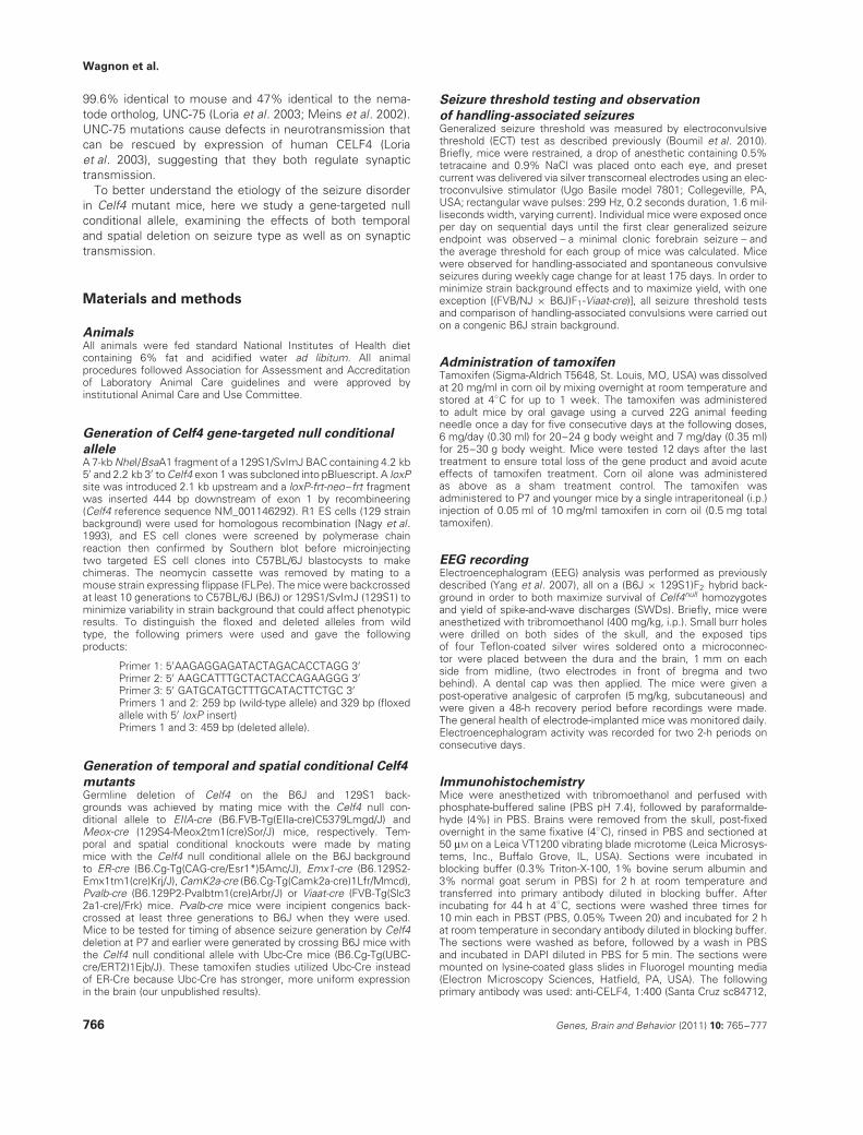
Seizure threshold testing and observation
of handling-associated seizuresGeneralized seizure threshold was measured by electroconvulsivethreshold (ECT) test as described previously (Boumil et al. 2010).Briefly, mice were restrained, a drop of anesthetic containing 0.5%tetracaine and 0.9% NaCl was placed onto each eye, and presetcurrent was delivered via silver transcorneal electrodes using an elec-troconvulsive stimulator (Ugo Basile model 7801; Collegeville, PA,USA; rectangular wave pulses: 299 Hz, 0.2 seconds duration, 1.6 mil-liseconds width, varying current). Individual mice were exposed onceper day on sequential days until the first clear generalized seizureendpoint was observed – a minimal clonic forebrain seizure – andthe average threshold for each group of mice was calculated. Micewere observed for handling-associated and spontaneous convulsiveseizures during weekly cage change for at least 175 days. In order tominimize strain background effects and to maximize yield, with oneexception [(FVB/NJ × B6J)F1-Viaat-cre)], all seizure threshold testsand comparison of handling-associated convulsions were carried outon a congenic B6J strain background.
Administration of tamoxifenTamoxifen (Sigma-Aldrich T5648, St. Louis, MO, USA) was dissolvedat 20 mg/ml in corn oil by mixing overnight at room temperature andstored at 4◦C for up to 1 week. The tamoxifen was administeredto adult mice by oral gavage using a curved 22G animal feedingneedle once a day for five consecutive days at the following doses,6 mg/day (0.30 ml) for 20–24 g body weight and 7 mg/day (0.35 ml)for 25–30 g body weight. Mice were tested 12 days after the lasttreatment to ensure total loss of the gene product and avoid acuteeffects of tamoxifen treatment. Corn oil alone was administeredas above as a sham treatment control. The tamoxifen wasadministered to P7 and younger mice by a single intraperitoneal (i.p.)injection of 0.05 ml of 10 mg/ml tamoxifen in corn oil (0.5 mg totaltamoxifen).
EEG recordingElectroencephalogram (EEG) analysis was performed as previouslydescribed (Yang et al. 2007), all on a (B6J × 129S1)F2 hybrid back-ground in order to both maximize survival of Celf4null homozygotesand yield of spike-and-wave discharges (SWDs). Briefly, mice wereanesthetized with tribromoethanol (400 mg/kg, i.p.). Small burr holeswere drilled on both sides of the skull, and the exposed tipsof four Teflon-coated silver wires soldered onto a microconnec-tor were placed between the dura and the brain, 1 mm on eachside from midline, (two electrodes in front of bregma and twobehind). A dental cap was then applied. The mice were given apost-operative analgesic of carprofen (5 mg/kg, subcutaneous) andwere given a 48-h recovery period before recordings were made.The general health of electrode-implanted mice was monitored daily.Electroencephalogram activity was recorded for two 2-h periods onconsecutive days.
ImmunohistochemistryMice were anesthetized with tribromoethanol and perfused withphosphate-buffered saline (PBS pH 7.4), followed by paraformalde-hyde (4%) in PBS. Brains were removed from the skull, post-fixedovernight in the same fixative (4◦C), rinsed in PBS and sectioned at50 μM on a Leica VT1200 vibrating blade microtome (Leica Microsys-tems, Inc., Buffalo Grove, IL, USA). Sections were incubated inblocking buffer (0.3% Triton-X-100, 1% bovine serum albumin and3% normal goat serum in PBS) for 2 h at room temperature andtransferred into primary antibody diluted in blocking buffer. Afterincubating for 44 h at 4◦C, sections were washed three times for10 min each in PBST (PBS, 0.05% Tween 20) and incubated for 2 hat room temperature in secondary antibody diluted in blocking buffer.The sections were washed as before, followed by a wash in PBSand incubated in DAPI diluted in PBS for 5 min. The sections weremounted on lysine-coated glass slides in Fluorogel mounting media(Electron Microscopy Sciences, Hatfield, PA, USA). The followingprimary antibody was used: anti-CELF4, 1:400 (Santa Cruz sc84712,
766 Genes, Brain and Behavior (2011) 10: 765–777
Etiology of seizure disorder in Celf4 mutant mice
Santa Cruz, CA, USA). The secondary antibodies were goat anti-rabbitAlexa Fluor 488, 1:1000 (Invitrogen A11070, Carlsbad, CA, USA) forCELF4 alone and goat anti-rabbit Alexa Fluor 647, 1:1000 (InvitrogenA21244) for CELF4 in conjunction with tdTomato.
Western blotCortical or hippocampal tissue from wild-type and Celf4null homozy-gous (germline deletion with Meox-cre) mice was homogenizedin lysis buffer [150 mM NaCl, 50 mM Tris–HCl (pH 8), 1% NP-40,0.5% sodium deoxycholate and 0.1% sodium dodecyl sulfate (SDS)]supplemented with protease inhibitors (complete mini; Roche, Indi-anapolis, IN, USA) and incubated for 30 min at 4◦C. The lysate wascentrifuged at 4500 g for 5 min at 4◦C. Protein in the supernatant wasquantified using the Bradford reagent (Bio-Rad, Hercules, CA, USA).Protein (300 μg from hippocampus and 500 μg from cortex) wassubjected to SDS–polyacrylamide gel electrophoresis, transferredto nitrocellulose and probed with anti-CELF4 antibody, 1:200 (SantaCruz sc84712) and goat anti-rabbit peroxidase-conjugated secondaryantibody, 1:5000 (Bio-Rad). Signal was detected with the ECL-pluskit (GE Healthcare, Pittsburgh, PA, USA). The blot was strippedwith Restore stripping buffer (Thermo Scientific, Rockford, IL, USA)and reprobed with anti-β-tubulin antibody, 1:2000 (Sigma-AldrichT4026) and goat anti-mouse peroxidase-conjugated secondary anti-body, 1:5000 (Thermo Scientific 31430).
Whole-cell patch-clamp recording and data analysisAcute brain slices were prepared from Celf4null heterozygotes andhomozygotes, and wild-type littermates between P14 and P21.To maximize the yield of homozygotes while minimizing strainbackground effects, all patch-clamp recording studies were car-ried out on the 129S1 strain background. Mice were anesthetizedwith tribromoethanol (250 mg/kg, i.p.) and decapitated. Brains werequickly removed and transferred into ice-cold solution containing210 mM sucrose, 3.0 mM KCl, 1.0 mM CaCl2, 3.0 mM MgSO4, 1.0 mM
NaH2PO4, 26 mM NaHCO3 and 10 mM glucose, saturated with 95%O2 and 5% CO2. Coronal slices were cut at 300 μm on a LeicaVT 1200 vibrating blade microtome (Leica Microsystems, Inc.) andkept in artificial cerebral spinal fluid (ACSF) containing 124 mM NaCl,3.0 mM KCl, 1.5 mM CaCl2, 1.3 mM MgSO4, 1.0 mM NaH2PO4, 26 mM
NaHCO3 and 20 mM glucose, saturated with 95% O2 and 5% CO2at room temperature (21–23◦C). Slices were allowed to recoverfor at least 1 h before any recording. Each slice was transferred to asubmerge-type chamber where it was continuously exposed to ACSFheated to 31–32◦C, saturated with 95% O2 and 5% CO2, and flow-ing at a rate of ∼2 ml/min. Whole-cell patch-clamp recordings weremade at the soma of layer V pyramidal neurons of the visual cortexusing a × 40 water immersion objective (× 40/0.80W; Zeiss). Patchpipettes were pulled from thick-wall borosilicate glass (1.5/0.86 mm;Sutter Instruments, Novato, CA, USA) on a horizontal puller (P-97; Sut-ter Instruments). Resistance of electrodes was between 2 and 4 M�.The pipette solution contained 100 mM CsCH3SO3, 4 mM ATP-Mg,20 mM HEPES, 15 mM CsCl and 0.5 mM EGTA, pH 7.2 with CsOH.Liquid junction potential was not corrected. Recordings were madewith a Multiclamp 700B amplifier (Molecular Devices, Sunnyvale, CA,USA). The series resistance (Rs), usually between 8 and 15 M�, wasmonitored throughout the recording, and data were not included infurther analysis when Rs varied by 20% or more during recording.The quantal excitatory postsynaptic currents (EPSCs) and inhibitorypostsynaptic currents (IPSCs) were recorded in the presence oftetrodotoxin (TTX, 1–3 μM) on reverse potentials of GABA and gluta-mate receptors, respectively. Data were filtered at 2 kHz, sampled at10 kHz and digitized by an ITC-18 interface (HEKA Instruments, Inc.,Bellmore, NY, USA). All analysis was performed using consumer rout-ings in MATLAB version 7.7.0 (MathWorks, Natick, MA, USA). QuantalEPSCs were detected and thresholded at 3.5 times the standarddeviation of baseline noise and quantal IPSCs were thresholded at 2times of the standard deviation of baseline noise. The average of allisolated events in each cell was normalized and used as a template tofit each single event. Two hundred events in first 400 good fits wererandomly selected and then averaged to give the mean response foreach cell.
Results
Celf4null mice have a complex seizure phenotype
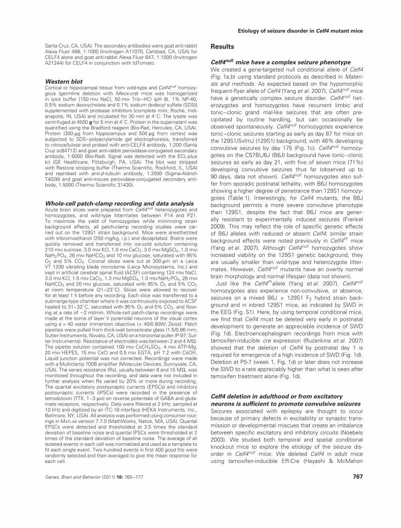
We created a gene-targeted null conditional allele of Celf4(Fig. 1a,b) using standard protocols as described in Materi-als and methods. As expected based on the hypomorphicfrequent-flyer allele of Celf4 (Yang et al. 2007), Celf4null micehave a genetically complex seizure disorder. Celf4null het-erozygotes and homozygotes have recurrent limbic andtonic–clonic grand mal-like seizures that are often pre-cipitated by routine handling, but can occasionally beobserved spontaneously. Celf4null homozygotes experiencetonic–clonic seizures starting as early as day 87 for mice onthe 129S1/SvImJ (129S1) background, with 46% developingconvulsive seizures by day 175 (Fig. 1c). Celf4null homozy-gotes on the C57BL/6J (B6J) background have tonic–clonicseizures as early as day 21, with five of seven mice (71%)developing convulsive seizures thus far (observed up to90 days, data not shown). Celf4null homozygotes also suf-fer from sporadic postnatal lethality, with B6J homozygotesshowing a higher degree of penetrance than 129S1 homozy-gotes (Table 1). Interestingly, for Celf4 mutants, the B6Jbackground permits a more severe convulsive phenotypethan 129S1, despite the fact that B6J mice are gener-ally resistant to experimentally induced seizures (Frankel2009). This may reflect the role of specific genetic effectsof B6J alleles with reduced or absent Celf4; similar strainbackground effects were noted previously in Celf4Ff mice(Yang et al. 2007). Although Celf4null homozygotes showincreased viability on the 129S1 genetic background, theyare usually smaller than wild-type and heterozygote litter-mates. However, Celf4null mutants have an overtly normalbrain morphology and normal lifespan (data not shown).
Just like the Celf4Ff allele (Yang et al. 2007), Celf4null
homozygotes also experience non-convulsive, or absence,seizures on a mixed B6J × 129S1 F2 hybrid strain back-ground and in inbred 129S1 mice, as indicated by SWD inthe EEG (Fig. S1). Here, by using temporal conditional mice,we find that Celf4 must be deleted very early in postnataldevelopment to generate an appreciable incidence of SWD(Fig. 1d). Electroencephalogram recordings from mice withtamoxifen-inducible cre expression (Ruzankina et al. 2007)showed that the deletion of Celf4 by postnatal day 1 isrequired for emergence of a high incidence of SWD (Fig. 1d).Deletion at P5-7 (week 1, Fig. 1d) or later does not increasethe SWD to a rate appreciably higher than what is seen aftertamoxifen treatment alone (Fig. 1d).
Celf4 deletion in adulthood or from excitatory
neurons is sufficient to promote convulsive seizures
Seizures associated with epilepsy are thought to occurbecause of primary defects in excitability or synaptic trans-mission or developmental miscues that create an imbalancebetween specific excitatory and inhibitory circuits (Noebels2003). We studied both temporal and spatial conditionalknockout mice to explore the etiology of the seizure dis-order in Celf4null mice. We deleted Celf4 in adult miceusing tamoxifen-inducible ER-Cre (Hayashi & McMahon
Genes, Brain and Behavior (2011) 10: 765–777 767
Wagnon et al.
(a) (b)
(c) (d)
Figure 1: Summary of Celf4null allele construction and seizure incidence. (a) Schematic representation of targeting construct forgeneration of the Celf4 null conditional allele. A loxP site was introduced upstream of Celf4 exon 1, and a loxP-frt-neo-frt site wasintroduced downstream, creating the Celf4flox allele. The neo cassette was removed by mating to an FLPe mouse strain. Celf4flox
mice were backcrossed at least 10 generations to either C57BL/6J or 129S1. (b) CELF4 protein expression is abolished in a Celf4null
homozygote (129S1 background strain shown). Cortices from wild-type or Celf4null homozygous mice were homogenized in lysisbuffer, and the total protein was extracted and processed for western blot. The blot was probed with CELF4 antibody and signal wasdetected using a peroxidase-conjugated secondary antibody. CELF4 protein is observed as an ∼50 kDa band in the wild-type lanesonly. A non-specific band of 65 kDa is observed in all lanes. The blot was stripped and probed with β-tubulin as a loading control.(c) Celf4null homozygotes exhibit handling-associated spontaneous seizures. Mice were observed for spontaneous convulsive seizuresafter weekly cage change for at least 175 days. A curve showing the fraction of mice (129S1) with at least one observed seizure ateach timepoint is shown (64 mice total). (d) Celf4 must be deleted early to generate the non-convulsive (absence) seizure phenotype.EEG recordings were made from mice receiving tamoxifen by i.p. injection (day 1, week 1) or oral gavage (week 3+). Experimentalmice were Celf4flox/flox and cre+, and control mice were either Celf4+/+ with or without cre expression or Celf4flox/flox without creexpression. The average number of SWD per hour ± SEM is shown (total number of mice recorded: day 1, n = 5; week 1, n = 16;week 3+, n = 12). The average number of SWD per hour for a germline Celf4null homozygote or compound heterozygote (Celf4Ff/null )is shown for comparison (total number of mice recorded: 6). In Celf4null mutants, SWD are not observed on strain backgrounds thatare predominantly B6J; all mice shown here are on the (B6J × 129S1)F2 hybrid background.
Table 1: Survival of Celf4null homozygotes∗
Celf4 genotype 129S1 C57BL/6J
+/+ 112 30+/− 224 56−/− 74 7(%) (18) (7.5)
∗The Celf4 gene-targeted null conditional allele was generatedon the 129S1 background, and mice were backcrossed 10generations to 129S1 or C57BL/6J. For deletion, 129S1 andC57BL/6J null conditional mice were crossed with Meox-creor EIIa-cre mice, respectively. The number of animals with aspecific genotype (examined at weaning age) is shown. Thepercent that were homozygous null is shown in the bottom rowin parentheses (expect 25% from Mendelian law).
2002) and assessed their seizure susceptibility by mea-suring ECT – often a component of epilepsy susceptibility(Frankel 2009) – and by observing grand mal-like convulsions
after routine handling. Control mice for these experimentsconsisted of a combination of wild-type mice with or withoutcre expression, mice expressing floxed Celf4 but not cre ormice that were sham treated (Table S1 specifies the num-bers and genotypes of the controls for each group undergoingECT testing). We also tested mice with a germline deletionof Celf4 generated with EIIA-Cre (Lakso et al. 1996), as wellas mice with excitatory or inhibitory neuron-specific somaticdeletions.
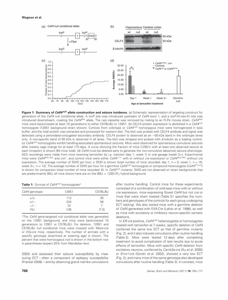
In ER-cre positive, Celf4null heterozygotes or homozygotestreated with tamoxifen at 7 weeks, specific deletion of Celf4conferred the same low ECT as that of germline mutants(Fig. 2), and it also induced convulsions after routine handling(Table 2). Mice were tested 12 days after completingtreatment to avoid complication of test results due to acuteeffects of tamoxifen. Mice with specific Celf4 deletion fromexcitatory neurons, conferred by Camk2a-cre (Xu et al. 2000)or Emx1-cre (Gorski et al. 2002), showed a very low ECT(Fig. 2), and many mice of the same genotype also developedconvulsions after routine handling (Table 2). In contrast, mice
768 Genes, Brain and Behavior (2011) 10: 765–777
Etiology of seizure disorder in Celf4 mutant mice
Figure 2: Reduced seizure threshold in excitatory neuron-specific or adult conditional Celf4null mice. Generalized seizurethreshold was measured by ECT test in conditional knockout mice. The average seizure threshold in milliamps (mA) ± SEM forindividual mice in each group is shown; threshold values for female (left) and male (right) mice are shown separately becausefemale mice typically have a lower standard threshold (Frankel et al. 2001). The number of mice in each test group is indicatedin parentheses beside the group description on the x-axis. Experimental mice were cre+ and either Celf4flox/flox homozygotes orCelf4flox/+ heterozygotes. Experimental ER-Cre mice and littermate controls were treated with tamoxifen (tam) by oral gavage at theage of 7 weeks for 5 sequential days and tested 12 days after completion of treatment. Control mice for each experiment were eitherCelf4flox/flox homozygotes or Celf4flox/+ heterozygotes that were cre−, Celf4+/+ with or without cre expression, or a combination ofthese groups (Table S1). All mice were on a C57BL/6J strain background, except for the Viaat-Cre mice which were tested on the(C57BL/6J × FVB/NJ)F1 hybrid background, which has a significantly lower seizure threshold than C57BL/6J itself as seen in the figure.A regression fit model was used to assess statistical significance incorporating both sex and treatment (experimental vs. control) asindependent covariates. The reduced ECTs of the germline, the adult and the excitatory neuron-specific deletions were statisticallysignificant. P-values were germline (EIIa-Cre, P < 0.0001), adult (ER-Cre, P < 0.0001), excitatory (Camk2a-Cre, P < 0.01; Emx1-Cre,P < 0.001) and inhibitory (Pvalb-Cre, P > 0.5; Viaat-Cre, P < 0.01).
with inhibitory neuron-specific deletion, conferred by Pvalb-cre (Hippenmeyer et al. 2005) or Viaat-cre (Chao et al. 2010),showed no ECT decrease (Fig. 2) nor have any handling-associated convulsions been observed to date (Table 2).
Celf4 is expressed predominantly in excitatory
neurons
We hypothesized that the deletion of CELF4 from excitatoryneurons in cerebral cortex and hippocampus was primarilyresponsible for the low seizure threshold and routinehandling-associated seizures seen in the CamK2a-cre andEmx1-cre mice as the Viaat-cre mice were unaffected. Onepossibility for this excitatory neuron-specific effect is thatCelf4 may be expressed only in excitatory neurons, at leastin cerebral cortex and hippocampus, and thus may only exerta function in that neuron type. Alternatively, Celf4 may beexpressed in several types of neurons, including inhibitoryneurons, reflecting a selective function in each neurontype. Immunohistochemistry was performed to discriminatebetween these possibilities.
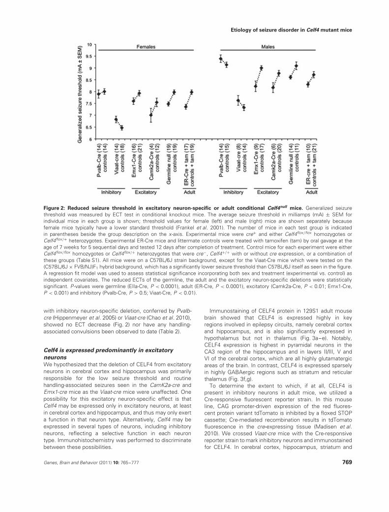
Immunostaining of CELF4 protein in 129S1 adult mousebrain showed that CELF4 is expressed highly in keyregions involved in epilepsy circuits, namely cerebral cortexand hippocampus, and is also significantly expressed inhypothalamus but not in thalamus (Fig. 3a–e). Notably,CELF4 expression is highest in pyramidal neurons in theCA3 region of the hippocampus and in layers II/III, V andVI of the cerebral cortex, which are all highly glutamatergicareas of the brain. In contrast, CELF4 is expressed sparselyin highly GABAergic regions such as striatum and reticularthalamus (Fig. 3f,g).
To determine the extent to which, if at all, CELF4 ispresent in inhibitory neurons in adult mice, we utilized aCre-responsive fluorescent reporter strain. In this mouseline, CAG promoter-driven expression of the red fluores-cent protein variant tdTomato is inhibited by a floxed STOPcassette; Cre-mediated recombination results in tdTomatofluorescence in the cre-expressing tissue (Madisen et al.2010). We crossed Viaat-cre mice with the Cre-responsivereporter strain to mark inhibitory neurons and immunostainedfor CELF4. In cerebral cortex, hippocampus, striatum and
Genes, Brain and Behavior (2011) 10: 765–777 769
Wagnon et al.
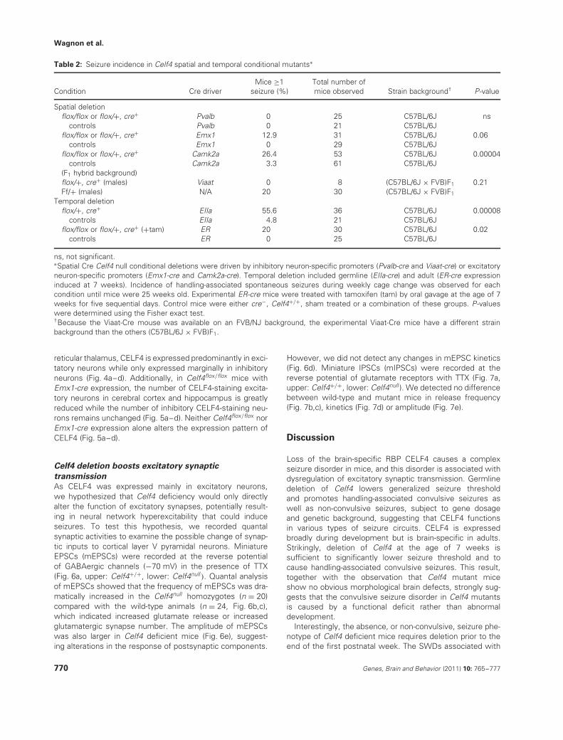
Table 2: Seizure incidence in Celf4 spatial and temporal conditional mutants∗
Condition Cre driverMice ≥1
seizure (%)Total number ofmice observed Strain background† P-value
Spatial deletionflox/flox or flox/+, cre+ Pvalb 0 25 C57BL/6J ns
controls Pvalb 0 21 C57BL/6Jflox/flox or flox/+, cre+ Emx1 12.9 31 C57BL/6J 0.06
controls Emx1 0 29 C57BL/6Jflox/flox or flox/+, cre+ Camk2a 26.4 53 C57BL/6J 0.00004
controls Camk2a 3.3 61 C57BL/6J(F1 hybrid background)flox/+, cre+ (males) Viaat 0 8 (C57BL/6J × FVB)F1 0.21Ff/+ (males) N/A 20 30 (C57BL/6J × FVB)F1
Temporal deletionflox/+, cre+ EIIa 55.6 36 C57BL/6J 0.00008
controls EIIa 4.8 21 C57BL/6Jflox/flox or flox/+, cre+ (+tam) ER 20 30 C57BL/6J 0.02
controls ER 0 25 C57BL/6J
ns, not significant.∗Spatial Cre Celf4 null conditional deletions were driven by inhibitory neuron-specific promoters (Pvalb-cre and Viaat-cre) or excitatoryneuron-specific promoters (Emx1-cre and Camk2a-cre). Temporal deletion included germline (EIIa-cre) and adult (ER-cre expressioninduced at 7 weeks). Incidence of handling-associated spontaneous seizures during weekly cage change was observed for eachcondition until mice were 25 weeks old. Experimental ER-cre mice were treated with tamoxifen (tam) by oral gavage at the age of 7weeks for five sequential days. Control mice were either cre−, Celf4+/+, sham treated or a combination of these groups. P-valueswere determined using the Fisher exact test.†Because the Viaat-Cre mouse was available on an FVB/NJ background, the experimental Viaat-Cre mice have a different strainbackground than the others (C57BL/6J × FVB)F1.
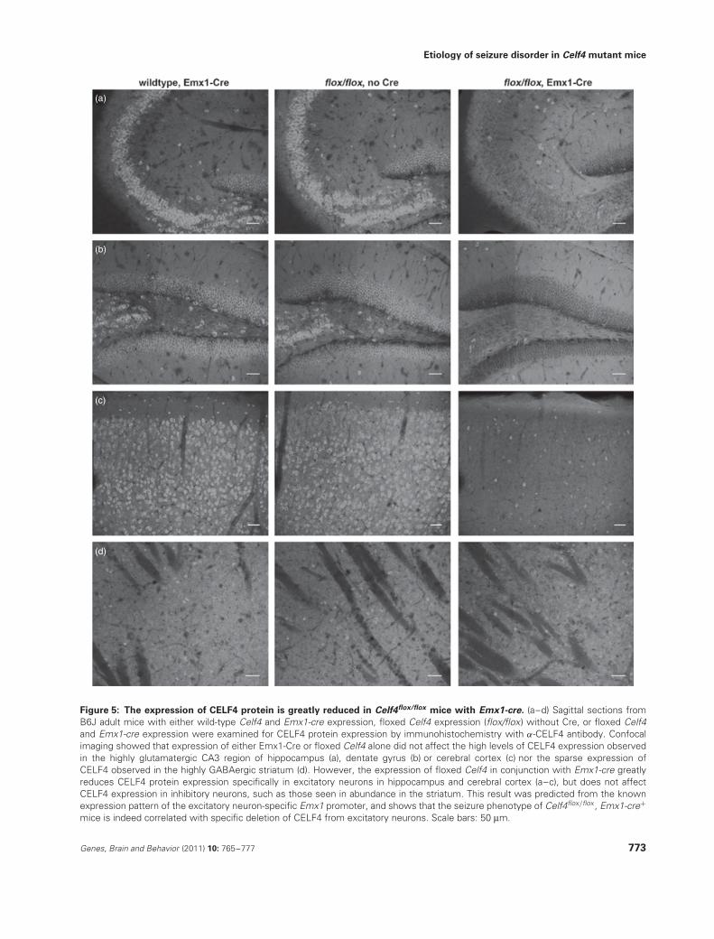
reticular thalamus, CELF4 is expressed predominantly in exci-tatory neurons while only expressed marginally in inhibitoryneurons (Fig. 4a–d). Additionally, in Celf4flox/flox mice withEmx1-cre expression, the number of CELF4-staining excita-tory neurons in cerebral cortex and hippocampus is greatlyreduced while the number of inhibitory CELF4-staining neu-rons remains unchanged (Fig. 5a–d). Neither Celf4flox/flox norEmx1-cre expression alone alters the expression pattern ofCELF4 (Fig. 5a–d).
Celf4 deletion boosts excitatory synaptic
transmission
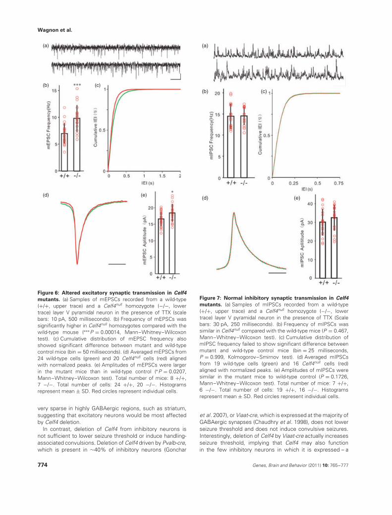
As CELF4 was expressed mainly in excitatory neurons,we hypothesized that Celf4 deficiency would only directlyalter the function of excitatory synapses, potentially result-ing in neural network hyperexcitability that could induceseizures. To test this hypothesis, we recorded quantalsynaptic activities to examine the possible change of synap-tic inputs to cortical layer V pyramidal neurons. MiniatureEPSCs (mEPSCs) were recorded at the reverse potentialof GABAergic channels (−70 mV) in the presence of TTX(Fig. 6a, upper: Celf4+/+, lower: Celf4null ). Quantal analysisof mEPSCs showed that the frequency of mEPSCs was dra-matically increased in the Celf4null homozygotes (n = 20)compared with the wild-type animals (n = 24, Fig. 6b,c),which indicated increased glutamate release or increasedglutamatergic synapse number. The amplitude of mEPSCswas also larger in Celf4 deficient mice (Fig. 6e), suggest-ing alterations in the response of postsynaptic components.
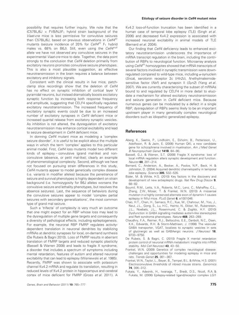
However, we did not detect any changes in mEPSC kinetics(Fig. 6d). Miniature IPSCs (mIPSCs) were recorded at thereverse potential of glutamate receptors with TTX (Fig. 7a,upper: Celf4+/+, lower: Celf4null ). We detected no differencebetween wild-type and mutant mice in release frequency(Fig. 7b,c), kinetics (Fig. 7d) or amplitude (Fig. 7e).
Discussion
Loss of the brain-specific RBP CELF4 causes a complexseizure disorder in mice, and this disorder is associated withdysregulation of excitatory synaptic transmission. Germlinedeletion of Celf4 lowers generalized seizure thresholdand promotes handling-associated convulsive seizures aswell as non-convulsive seizures, subject to gene dosageand genetic background, suggesting that CELF4 functionsin various types of seizure circuits. CELF4 is expressedbroadly during development but is brain-specific in adults.Strikingly, deletion of Celf4 at the age of 7 weeks issufficient to significantly lower seizure threshold and tocause handling-associated convulsive seizures. This result,together with the observation that Celf4 mutant miceshow no obvious morphological brain defects, strongly sug-gests that the convulsive seizure disorder in Celf4 mutantsis caused by a functional deficit rather than abnormaldevelopment.
Interestingly, the absence, or non-convulsive, seizure phe-notype of Celf4 deficient mice requires deletion prior to theend of the first postnatal week. The SWDs associated with
770 Genes, Brain and Behavior (2011) 10: 765–777
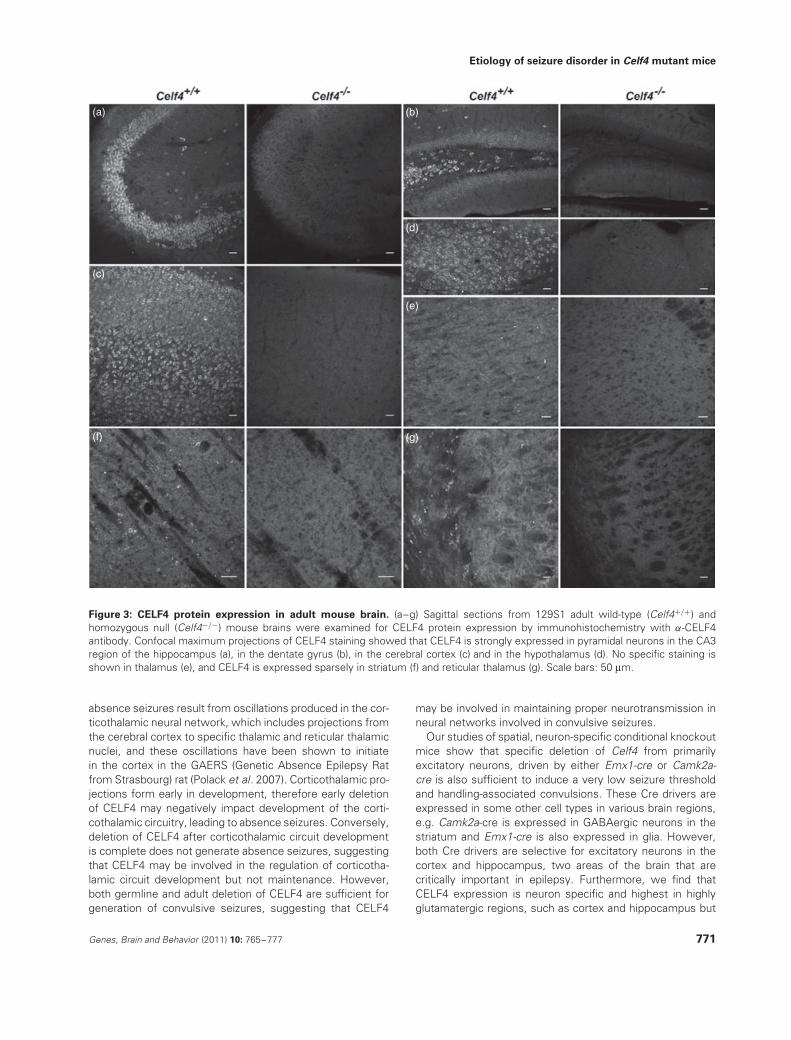
Etiology of seizure disorder in Celf4 mutant mice
(a) (b)
(d)
(e)
(g)
(c)
(f)
Figure 3: CELF4 protein expression in adult mouse brain. (a–g) Sagittal sections from 129S1 adult wild-type (Celf4+/+) andhomozygous null (Celf4−/−) mouse brains were examined for CELF4 protein expression by immunohistochemistry with α-CELF4antibody. Confocal maximum projections of CELF4 staining showed that CELF4 is strongly expressed in pyramidal neurons in the CA3region of the hippocampus (a), in the dentate gyrus (b), in the cerebral cortex (c) and in the hypothalamus (d). No specific staining isshown in thalamus (e), and CELF4 is expressed sparsely in striatum (f) and reticular thalamus (g). Scale bars: 50 μm.
absence seizures result from oscillations produced in the cor-ticothalamic neural network, which includes projections fromthe cerebral cortex to specific thalamic and reticular thalamicnuclei, and these oscillations have been shown to initiatein the cortex in the GAERS (Genetic Absence Epilepsy Ratfrom Strasbourg) rat (Polack et al. 2007). Corticothalamic pro-jections form early in development, therefore early deletionof CELF4 may negatively impact development of the corti-cothalamic circuitry, leading to absence seizures. Conversely,deletion of CELF4 after corticothalamic circuit developmentis complete does not generate absence seizures, suggestingthat CELF4 may be involved in the regulation of corticotha-lamic circuit development but not maintenance. However,both germline and adult deletion of CELF4 are sufficient forgeneration of convulsive seizures, suggesting that CELF4
may be involved in maintaining proper neurotransmission inneural networks involved in convulsive seizures.
Our studies of spatial, neuron-specific conditional knockoutmice show that specific deletion of Celf4 from primarilyexcitatory neurons, driven by either Emx1-cre or Camk2a-cre is also sufficient to induce a very low seizure thresholdand handling-associated convulsions. These Cre drivers areexpressed in some other cell types in various brain regions,e.g. Camk2a-cre is expressed in GABAergic neurons in thestriatum and Emx1-cre is also expressed in glia. However,both Cre drivers are selective for excitatory neurons in thecortex and hippocampus, two areas of the brain that arecritically important in epilepsy. Furthermore, we find thatCELF4 expression is neuron specific and highest in highlyglutamatergic regions, such as cortex and hippocampus but
Genes, Brain and Behavior (2011) 10: 765–777 771
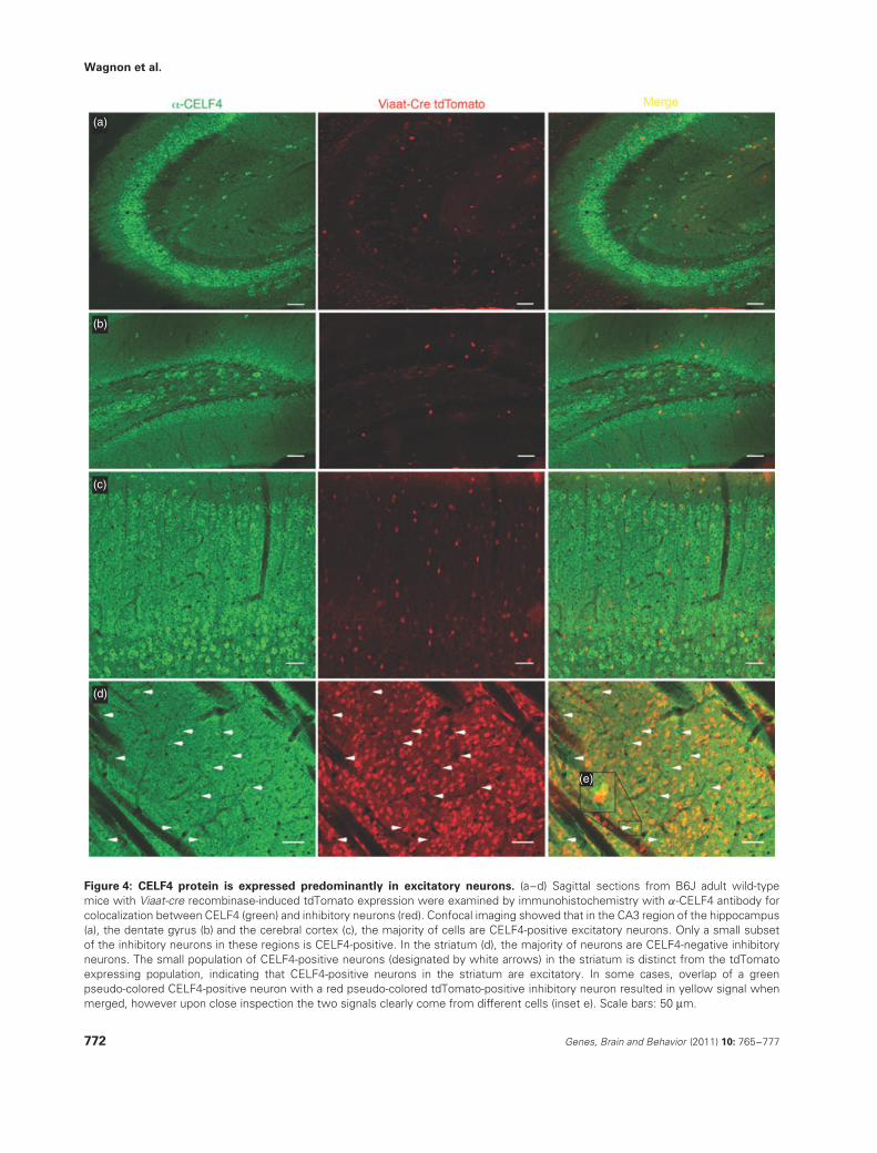
Wagnon et al.
(a)
(b)
(c)
(d)
(e)
Figure 4: CELF4 protein is expressed predominantly in excitatory neurons. (a–d) Sagittal sections from B6J adult wild-typemice with Viaat-cre recombinase-induced tdTomato expression were examined by immunohistochemistry with α-CELF4 antibody forcolocalization between CELF4 (green) and inhibitory neurons (red). Confocal imaging showed that in the CA3 region of the hippocampus(a), the dentate gyrus (b) and the cerebral cortex (c), the majority of cells are CELF4-positive excitatory neurons. Only a small subsetof the inhibitory neurons in these regions is CELF4-positive. In the striatum (d), the majority of neurons are CELF4-negative inhibitoryneurons. The small population of CELF4-positive neurons (designated by white arrows) in the striatum is distinct from the tdTomatoexpressing population, indicating that CELF4-positive neurons in the striatum are excitatory. In some cases, overlap of a greenpseudo-colored CELF4-positive neuron with a red pseudo-colored tdTomato-positive inhibitory neuron resulted in yellow signal whenmerged, however upon close inspection the two signals clearly come from different cells (inset e). Scale bars: 50 μm.
772 Genes, Brain and Behavior (2011) 10: 765–777
Etiology of seizure disorder in Celf4 mutant mice
(a)
(b)
(c)
(d)
Figure 5: The expression of CELF4 protein is greatly reduced in Celf4flox/flox mice with Emx1-cre. (a–d) Sagittal sections fromB6J adult mice with either wild-type Celf4 and Emx1-cre expression, floxed Celf4 expression (flox/flox) without Cre, or floxed Celf4and Emx1-cre expression were examined for CELF4 protein expression by immunohistochemistry with α-CELF4 antibody. Confocalimaging showed that expression of either Emx1-Cre or floxed Celf4 alone did not affect the high levels of CELF4 expression observedin the highly glutamatergic CA3 region of hippocampus (a), dentate gyrus (b) or cerebral cortex (c) nor the sparse expression ofCELF4 observed in the highly GABAergic striatum (d). However, the expression of floxed Celf4 in conjunction with Emx1-cre greatlyreduces CELF4 protein expression specifically in excitatory neurons in hippocampus and cerebral cortex (a–c), but does not affectCELF4 expression in inhibitory neurons, such as those seen in abundance in the striatum. This result was predicted from the knownexpression pattern of the excitatory neuron-specific Emx1 promoter, and shows that the seizure phenotype of Celf4flox/flox , Emx1-cre+
mice is indeed correlated with specific deletion of CELF4 from excitatory neurons. Scale bars: 50 μm.
Genes, Brain and Behavior (2011) 10: 765–777 773
Wagnon et al.
(a)
(b)
(d) (e)
(c)
Figure 6: Altered excitatory synaptic transmission in Celf4
mutants. (a) Samples of mEPSCs recorded from a wild-type(+/+, upper trace) and a Celf4null homozygote (−/−, lowertrace) layer V pyramidal neuron in the presence of TTX (scalebars: 10 pA, 500 milliseconds). (b) Frequency of mEPSCs wassignificantly higher in Celf4null homozygotes compared with thewild-type mouse (∗∗∗P = 0.00014, Mann–Whitney–Wilcoxontest). (c) Cumulative distribution of mEPSC frequency alsoshowed significant difference between mutant and wild-typecontrol mice (bin = 50 milliseconds). (d) Averaged mEPSCs from24 wild-type cells (green) and 20 Celf4null cells (red) alignedwith normalized peaks. (e) Amplitudes of mEPSCs were largerin the mutant mice than in wild-type control (∗P = 0.0207,Mann–Whitney–Wilcoxon test). Total number of mice: 8 +/+,7 −/−. Total number of cells: 24 +/+, 20 −/−. Histogramsrepresent mean ± SD. Red circles represent individual cells.
very sparse in highly GABAergic regions, such as striatum,suggesting that excitatory neurons would be most affectedby Celf4 deletion.
In contrast, deletion of Celf4 from inhibitory neurons isnot sufficient to lower seizure threshold or induce handling-associated convulsions. Deletion of Celf4 driven by Pvalb-cre,which is present in ∼40% of inhibitory neurons (Gonchar
(a)
(b)
(d) (e)
(c)
Figure 7: Normal inhibitory synaptic transmission in Celf4
mutants. (a) Samples of mIPSCs recorded from a wild-type(+/+, upper trace) and a Celf4null homozygote (−/−, lowertrace) layer V pyramidal neuron in the presence of TTX (Scalebars: 30 pA, 250 milliseconds). (b) Frequency of mIPSCs wassimilar in Celf4null compared with the wild-type mice (P = 0.467,Mann–Whitney–Wilcoxon test). (c) Cumulative distribution ofmIPSC frequency failed to show significant difference betweenmutant and wild-type control mice (bin = 25 milliseconds,P = 0.999, Kolmogorov–Smirnov test). (d) Averaged mIPSCsfrom 19 wild-type cells (green) and 16 Celf4null cells (red)aligned with normalized peaks. (e) Amplitudes of mIPSCs weresimilar in the mutant mice to wild-type control (P = 0.1726,Mann–Whitney–Wilcoxon test). Total number of mice: 7 +/+,6 −/−. Total number of cells: 19 +/+, 16 −/−. Histogramsrepresent mean ± SD. Red circles represent individual cells.
et al. 2007), or Viaat-cre, which is expressed at the majority ofGABAergic synapses (Chaudhry et al. 1998), does not lowerseizure threshold and does not induce convulsive seizures.Interestingly, deletion of Celf4 by Viaat-cre actually increasesseizure threshold, implying that Celf4 may also functionin the few inhibitory neurons in which it is expressed – a
774 Genes, Brain and Behavior (2011) 10: 765–777
Etiology of seizure disorder in Celf4 mutant mice
possibility that requires further inquiry. We note that the(C57BL/6J × FVB/NJ)F1 hybrid strain background of theViaat-cre mice is less permissive for convulsive seizuresthan C57BL/6J, based on previous observations in Celf4Ff
mutants (seizure incidence of 20% for Celf4Ff F1 hybridmales vs. 68% on B6J). Still, even using the Celf4null
allele we have not observed any convulsive seizures in theexperimental Viaat-cre mice to date. Together, the data pointstrongly to the conclusion that Celf4 deletion primarily fromexcitatory neurons promotes convulsive seizure phenotypes.This is also a most plausible suggestion, as effectiveneurotransmission in the brain requires a balance betweenexcitatory and inhibitory signals.
Consistent with the clinical results in live mice, patch-clamp slice recordings show that the deletion of Celf4has no effect on synaptic inhibition of cortical layer Vpyramidal neurons, but instead dramatically boosts excitatorysynaptic function by increasing both response frequencyand amplitude, suggesting that CELF4 specifically regulatesexcitatory neurotransmission. The increased frequency ofexcitatory synaptic events could be due to an increasednumber of excitatory synapses in Celf4 deficient mice orincreased quantal release from excitatory synaptic vesicles.As inhibition is not altered, the dysregulation of excitatoryneurotransmission may enhance cortical excitability and leadto seizure development in Celf4 deficient mice.
In donning Celf4 mutant mice as modeling a ‘complexseizure disorder’, it is useful to be explicit about the multipleways in which the term ‘complex’ applies to this particularanimal model. First, Celf4 loss mutants model two differentkinds of epilepsy – convulsive (grand mal-like) and non-convulsive (absence, or petit mal-like); clearly an exampleof phenomenological complexity. Second, although we havenot focused on pursuing strain differences in this study,Celf4 mutants appear to model genetically complex disease(i.e. variants in modifier alleles) because the penetrance ofseizure and survival phenotypes is highly dependent on strainbackground (i.e. homozygosity for B6J alleles worsens theconvulsive seizure and lethality phenotypes, but resolves theabsence seizures). Last, the sequence of behaviors duringthe convulsive seizures appear to model ‘complex partialseizures with secondary generalizations’, the most commontype of grand mal seizure.
Such a ‘trifecta’ of complexity is very much an outcomethat one might expect for an RBP whose loss may lead tothe dysregulation of multiple gene targets and consequentlya diversity of pathological effects, including epileptogenesis.For example, the neuronal RBP FMRP regulates activity-dependent translation in neuronal dendrites by stabilizingmRNAs at dendritic synapses for local, on-demand synthesis(De Rubeis & Bagni 2010). Loss of FMRP results in aberranttranslation of FMRP targets and reduced synaptic plasticity(Bassell & Warren 2008) and leads to fragile X syndrome,a disorder that includes a spectrum of symptoms includingmental retardation, features of autism and altered neuronalexcitability that can lead to epilepsy (Wisniewski et al. 1985).Recently, FMRP was shown to associate with potassiumchannel Kv4.2 mRNA and regulate its translation, resulting inreduced levels of Kv4.2 protein in hippocampus and cerebralcortex of mice deficient for FMRP (Gross et al. 2011). A
Kv4.2 loss-of-function truncation has been identified in ahuman case of temporal lobe epilepsy (TLE) (Singh et al.2006) and decreased Kv4.2 expression is associated withincreased neuronal excitability in animal models of TLE(Bernard et al. 2004).
Our finding that Celf4 deficiency leads to enhanced exci-tatory neurotransmission underscores the importance ofmRNA transcript regulation in the brain, including the contri-bution of RBPs to neurological function. Microarray analysisusing Celf4Ff homozygotes showed that mRNA transcripts ofseveral factors involved in synaptic transmission were down-regulated compared to wild-type mice, including α-synuclein(Snca), serotonin receptor 2c (Htr2c), N-ethylmaleimide-sensitive factor (Nsf ) and synapsin II (Syn2) (Yang et al.2007). We are currently characterizing the subset of mRNAsbound to and regulated by CELF4 in more detail to eluci-date a mechanism for altered excitatory neurotransmissionand seizure generation in Celf4 deficient mice. Becausenumerous genes can be modulated by a defect in a singleRBP, dysregulation of RBPs seems likely to be an importantupstream player in many genetically complex neurologicaldisorders such as idiopathic generalized epilepsy.
References
Aberg, K., Saetre, P., Lindholm, E., Ekholm, B., Pettersson, U.,Adolfsson, R. & Jazin, E. (2006) Human QKI, a new candidategene for schizophrenia involved in myelination. Am J Med GenetB Neuropsychiatr Genet 141B, 84–90.
Bassell, G.J. & Warren, S.T. (2008) Fragile X syndrome: loss oflocal mRNA regulation alters synaptic development and function.Neuron 60, 201–214.
Bernard, C., Anderson, A., Becker, A., Poolos, N.P., Beck, H. &Johnston, D. (2004) Acquired dendritic channelopathy in temporallobe epilepsy. Science 305, 532–535.
Bialer, M. & White, H.S. (2010) Key factors in the discovery anddevelopment of new antiepileptic drugs. Nat Rev Drug Discov 9,68–82.
Boumil, R.M., Letts, V.A., Roberts, M.C., Lenz, C., Mahaffey, C.L.,Zhang, Z.W., Moser, T. & Frankel, W.N. (2010) A missensemutation in a highly conserved alternate exon of dynamin-1 causesepilepsy in fitful mice. PLoS Genet 6, e1001046.
Chao, H.T., Chen, H., Samaco, R.C., Xue, M., Chahrour, M., Yoo, J.,Neul, J.L., Gong, S., Lu, H.C., Heintz, N., Ekker, M., Rubenstein,J.L., Noebels, J.L., Rosenmund, C. & Zoghbi, H.Y. (2010)Dysfunction in GABA signalling mediates autism-like stereotypiesand Rett syndrome phenotypes. Nature 468, 263–269.
Chaudhry, F.A., Reimer, R.J., Bellocchio, E.E., Danbolt, N.C., Osen,K.K., Edwards, R.H. & Storm-Mathisen, J. (1998) The vesicularGABA transporter, VGAT, localizes to synaptic vesicles in setsof glycinergic as well as GABAergic neurons. J Neurosci 18,9733–9750.
De Rubeis, S. & Bagni, C. (2010) Fragile X mental retardationprotein control of neuronal mRNA metabolism: insights into mRNAstability. Mol Cell Neurosci 43, 43–50.
Frankel, W.N. (2009) Genetics of complex neurological disease:challenges and opportunities for modeling epilepsy in mice andrats. Trends Genet 25, 361–367.
Frankel, W.N., Taylor, L., Beyer, B., Tempel, B.L. & White, H.S. (2001)Electroconvulsive thresholds of inbred mouse strains. Genomics74, 306–312.
Fukata, Y., Adesnik, H., Iwanaga, T., Bredt, D.S., Nicoll, R.A. &Fukata, M. (2006) Epilepsy-related ligand/receptor complex LGI1
Genes, Brain and Behavior (2011) 10: 765–777 775
Wagnon et al.
and ADAM22 regulate synaptic transmission. Science 313,1792–1795.
Fukata, Y., Lovero, K.L., Iwanaga, T., Watanabe, A., Yokoi, N.,Tabuchi, K., Shigemoto, R., Nicoll, R.A. & Fukata, M. (2010)Disruption of LGI1-linked synaptic complex causes abnormalsynaptic transmission and epilepsy. Proc Natl Acad Sci U S A107, 3799–3804.
Garber, K.B., Visootsak, J. & Warren, S.T. (2008) Fragile X syndrome.Eur J Hum Genet 16, 666–672.
Gardiner, M. (2005) Genetics of idiopathic generalized epilepsies.Epilepsia 46 (Suppl. 9), 15–20.
Glisovic, T., Bachorik, J.L., Yong, J. & Dreyfuss, G. (2008) RNA-binding proteins and post-transcriptional gene regulation. FEBSLett 582, 1977–1986.
Gonchar, Y., Wang, Q. & Burkhalter, A. (2007) Multiple distinctsubtypes of GABAergic neurons in mouse visual cortex identifiedby triple immunostaining. Front Neuroanat 1, 3.
Good, P.J., Chen, Q., Warner, S.J. & Herring, D.C. (2000) A familyof human RNA-binding proteins related to the Drosophila Brunotranslational regulator. J Biol Chem 275, 28583–28592.
Gorski, J.A., Talley, T., Qiu, M., Puelles, L., Rubenstein, J.L. & Jones,K.R. (2002) Cortical excitatory neurons and glia, but not GABAergicneurons, are produced in the Emx1-expressing lineage. J Neurosci22, 6309–6314.
Gross, C., Yao, X., Pong, D.L., Jeromin, A. & Bassell, G.J. (2011)Fragile X mental retardation protein regulates protein expressionand mRNA translation of the potassium channel Kv4.2. J Neurosci31, 5693–5698.
Hayashi, S. & McMahon, A.P. (2002) Efficient recombination indiverse tissues by a tamoxifen-inducible form of Cre: a tool fortemporally regulated gene activation/inactivation in the mouse.Dev Biol 244, 305–318.
Hippenmeyer, S., Vrieseling, E., Sigrist, M., Portmann, T., Laengle,C., Ladle, D.R. & Arber, S. (2005) A developmental switch in theresponse of DRG neurons to ETS transcription factor signaling.PLoS Biol 3, e159.
Kalachikov, S., Evgrafov, O., Ross, B., Winawer, M., Barker-Cummings, C., Martinelli Boneschi, F., Choi, C., Morozov, P.,Das, K., Teplitskaya, E., Yu, A., Cayanis, E., Penchaszadeh, G.,Kottmann, A.H., Pedley, T.A., Hauser, W.A., Ottman, R. & Gilliam,T.C. (2002) Mutations in LGI1 cause autosomal-dominant partialepilepsy with auditory features. Nat Genet 30, 335–341.
Kaufmann, W.E., Cortell, R., Kau, A.S., Bukelis, I., Tierney, E., Gray,R.M., Cox, C., Capone, G.T. & Stanard, P. (2004) Autism spectrumdisorder in fragile X syndrome: communication, social interaction,and specific behaviors. Am J Med Genet A 129A, 225–234.
Kunapuli, P., Jang, G.F., Kazim, L. & Cowell, J.K. (2009) Massspectrometry identifies LGI1-interacting proteins that are involvedin synaptic vesicle function in the human brain. J Mol Neurosci 39,137–143.
Ladd, A.N., Charlet, N. & Cooper, T.A. (2001) The CELF family of RNAbinding proteins is implicated in cell-specific and developmentallyregulated alternative splicing. Mol Cell Biol 21, 1285–1296.
Lagier-Tourenne, C., Polymenidou, M. & Cleveland, D.W. (2010)TDP-43 and FUS/TLS: emerging roles in RNA processing andneurodegeneration. Hum Mol Genet 19, R46–R64.
Lakso, M., Pichel, J.G., Gorman, J.R., Sauer, B., Okamoto, Y., Lee,E., Alt, F.W. & Westphal, H. (1996) Efficient in vivo manipulationof mouse genomic sequences at the zygote stage. Proc Natl AcadSci U S A 93, 5860–5865.
Loria, P.M., Duke, A., Rand, J.B. & Hobert, O. (2003) Twoneuronal, nuclear-localized RNA binding proteins involved insynaptic transmission. Curr Biol 13, 1317–1323.
Madisen, L., Zwingman, T.A., Sunkin, S.M., Oh, S.W., Zariwala, H.A.,Gu, H., Ng, L.L., Palmiter, R.D., Hawrylycz, M.J., Jones, A.R.,Lein, E.S. & Zeng, H. (2010) A robust and high-throughput Crereporting and characterization system for the whole mouse brain.Nat Neurosci 13, 133–140.
Martin, C.L., Duvall, J.A., Ilkin, Y., Simon, J.S., Arreaza, M.G., Wilkes,K., Alvarez-Retuerto, A., Whichello, A., Powell, C.M., Rao, K.,
Cook, E. & Geschwind, D.H. (2007) Cytogenetic and molecularcharacterization of A2BP1/FOX1 as a candidate gene for autism.Am J Med Genet B Neuropsychiatr Genet 144B, 869–876.
Meins, M., Schlickum, S., Wilhelm, C., Missbach, J., Yadav,S., Glaser, B., Grzmil, M., Burfeind, P. & Laccone, F. (2002)Identification and characterization of murine Brunol4, a newmember of the elav/bruno family. Cytogenet Genome Res 97,254–260.
Moore, T., Hecquet, S., McLellann, A., Ville, D., Grid, D., Picard, F.,Moulard, B., Asherson, P., Makoff, A.J., McCormick, D., Nashef,L., Froguel, P., Arzimanoglou, A., LeGuern, E. & Bailleul, B. (2001)Polymorphism analysis of JRK/JH8, the human homologue ofmouse jerky, and description of a rare mutation in a case of CAEevolving to JME. Epilepsy Res 46, 157–167.
Musumeci, S.A., Bosco, P., Calabrese, G., Bakker, C., De Sarro,G.B., Elia, M., Ferri, R. & Oostra, B.A. (2000) Audiogenic seizuressusceptibility in transgenic mice with fragile X syndrome. Epilepsia41, 19–23.
Nagy, A., Rossant, J., Nagy, R., Abramow-Newerly, W. & Roder,J.C. (1993) Derivation of completely cell culture-derived mice fromearly-passage embryonic stem cells. Proc Natl Acad Sci U S A 90,8424–8428.
de Nijs, L., Leon, C., Nguyen, L., Loturco, J.J., Delgado-Escueta, A.V.,Grisar, T. & Lakaye, B. (2009) EFHC1 interacts with microtubulesto regulate cell division and cortical development. Nat Neurosci12, 1266–1274.
Noebels, J.L. (2003) The biology of epilepsy genes. Annu RevNeurosci 26, 599–625.
Ottman, R. (2005) Analysis of genetically complex epilepsies.Epilepsia 46 (Suppl. 10), 7–14.
Ottman, R., Hauser, W.A., Barker-Cummings, C., Lee, J.H. & Risch,N. (1997) Segregation analysis of cryptogenic epilepsy and anempirical test of the validity of the results. Am J Hum Genet 60,667–675.
Polack, P.O., Guillemain, I., Hu, E., Deransart, C., Depaulis, A. &Charpier, S. (2007) Deep layer somatosensory cortical neuronsinitiate spike-and-wave discharges in a genetic model of absenceseizures. J Neurosci 27, 6590–6599.
Ruzankina, Y., Pinzon-Guzman, C., Asare, A., Ong, T., Pontano, L.,Cotsarelis, G., Zediak, V.P., Velez, M., Bhandoola, A. & Brown,E.J. (2007) Deletion of the developmentally essential gene ATR inadult mice leads to age-related phenotypes and stem cell loss. CellStem Cell 1, 113–126.
Schulte, U., Thumfart, J.O., Klocker, N., Sailer, C.A., Bildl, W.,Biniossek, M., Dehn, D., Deller, T., Eble, S., Abbass, K., Wangler,T., Knaus, H.G. & Fakler, B. (2006) The epilepsy-linked Lgi1 proteinassembles into presynaptic Kv1 channels and inhibits inactivationby Kvbeta1. Neuron 49, 697–706.
Singh, B., Ogiwara, I., Kaneda, M., Tokonami, N., Mazaki, E., Baba, K.,Matsuda, K., Inoue, Y. & Yamakawa, K. (2006) A Kv4.2 truncationmutation in a patient with temporal lobe epilepsy. Neurobiol Dis24, 245–253.
Stein, J.M., Bergman, W., Fang, Y., Davison, L., Brensinger, C.,Robinson, M.B., Hecht, N.B. & Abel, T. (2006) Behavioral andneurochemical alterations in mice lacking the RNA-binding proteintranslin. J Neurosci 26, 2184–2196.
Suzuki, T., Delgado-Escueta, A.V. & Aguan, K. et al. (2004) Mutationsin EFHC1 cause juvenile myoclonic epilepsy. Nat Genet 36,842–849.
Toth, M., Grimsby, J., Buzsaki, G. & Donovan, G.P. (1995) Epilepticseizures caused by inactivation of a novel gene, jerky, related tocentromere binding protein-B in transgenic mice. Nat Genet 11,71–75.
Wisniewski, K.E., French, J.H., Fernando, S., Brown, W.T.,Jenkins, E.C., Friedman, E., Hill, A.L. & Miezejeski, C.M. (1985)Fragile X syndrome: associated neurological abnormalities anddevelopmental disabilities. Ann Neurol 18, 665–669.
Xu, B., Zang, K., Ruff, N.L., Zhang, Y.A., McConnell, S.K., Stryker,M.P. & Reichardt, L.F. (2000) Cortical degeneration in the absence
776 Genes, Brain and Behavior (2011) 10: 765–777
Etiology of seizure disorder in Celf4 mutant mice
of neurotrophin signaling: dendritic retraction and neuronal lossafter removal of the receptor TrkB. Neuron 26, 233–245.
Yang, Y., Mahaffey, C.L., Berube, N., Maddatu, T.P., Cox, G.A. &Frankel, W.N. (2007) Complex seizure disorder caused by Brunol4deficiency in mice. PLoS Genet 3, e124.
Yu, Y.E., Wen, L., Silva, J., Li, Z., Head, K., Sossey-Alaoui, K., Pao,A., Mei, L. & Cowell, J.K. (2010) Lgi1 null mutant mice exhibitmyoclonic seizures and CA1 neuronal hyperexcitability. Hum MolGenet 19, 1702–1711.
Zhou, Y.D., Lee, S., Jin, Z., Wright, M., Smith, S.E. & Anderson, M.P.(2009) Arrested maturation of excitatory synapses in autosomaldominant lateral temporal lobe epilepsy. Nat Med 15, 1208–1214.
Acknowledgments
Viaat-cre mice were generated in the laboratory of Huda Y.Zoghbi, to whom we are grateful for sharing prior to publication.We thank Barbara Beyer, Verity Letts, Satoko Tokuda, CarolyneDunbar and Nathalie Berube for assistance with mouse studiesand EEG recordings. We also thank Rebecca Boumil, GregoryCox, Verity Letts, and Zhong-wei Zhang for helpful comments andadvice. The Jackson Laboratory’s Cell Biology and Microinjectionservices, Gene Expression and Sequencing services and ImagingSciences service were subsidized by an NCI core grant (5 P30CA034196-27). This work was supported by grants from theNational Institutes of Health (NS061971 and NS061971-2Z toWNF).
Supporting Information
Additional Supporting Information may be found in the onlineversion of this article:
Figure S1: SWD of Celf4flox/flox, ER-cre positive, tamoxi-fen-treated animal.
Shown are example SWD episodes from mutant, cre-positive Celf4 mice with SWD in the day 1 tamoxifen-treatedgroup, described as part of Fig. 1d. On the left is an SWD thatoccurred during active wake phase and is associated with abrief behavioral arrest, and is flanked by typical movementartifacts in the EEG upon cessation and resumption of normallocomotor activity. On the right is an SWD that occurredduring quiet wake – no movement artifact but still fast, low-voltage interictal EEG. 1s, 1 second; differential electrodemontage abbreviations are BL, back-left; BR, back-right;FL, front-left; FR, front-right as described in Materials andMethods.
Table S1: Control groups for ECT testinga.As a service to our authors and readers, this journal
provides supporting information supplied by the authors.Such materials are peer-reviewed and may be re-organizedfor online delivery, but are not copy-edited or typeset.Technical support issues arising from supporting information(other than missing files) should be addressed to the authors.
Genes, Brain and Behavior (2011) 10: 765–777 777