Embed Size (px)
Citation preview
Etiological Role of Ferritin and Vitamin D in Patients with TelogenEffluviumNoha Ezzat Mohammad1*, Radawa Said Ibrahium2, Mohamad Hassan Mohammed1, Sara Ahmed Galal2, Rehab Maher2 and Hanan Ali Darwish2
1Dermatology and Venereology, Faculty of Medicine, Elfayoum University, Egypt2Faculty of Medicine for Girls, Alazhar University, Egypt*Corresponding author: Noha Ezzat Mohammad, Dermatology and Venereology, Faculty of Medicine, Elfayoum University, Egypt, Tel: +201005413367; E-mail:[email protected]
Received date: September 26, 2017; Accepted date: October 25, 2017; Published date: October 31, 2017
Copyright: ©2017 Mohammad NM, et al. This is an open-access article distributed under the terms of the Creative Commons Attribution License, which permitsunrestricted use, distribution, and reproduction in any medium, provided the original author and source are credited.
Abstract
Introduction: Telogen effluvium (TE) is an abnormality of hair cycling that result in an abrupt onset of generalizedshedding of telogen hairs from the scalp, with or without an entifiable trigger. Ferritin and vitamin D play an importantrole in hair follicle differentiation.
Aim: The work was to investigate level of serum ferritin and vitamin D levels in patients with acute/chronic TE.
Subject and methods: This case control study was conducted on 30 patients with TE. They were 25 femalesand 5 males and they were also classified clinically into 15 patients (50%) suffered from acute form of the disease,they aged (18 to 48 years) and 15 patients (50%) have chronic form, aged (24 to 49 years). Thirty healthy personswere included as a control group
Result: There was a significant difference between patients and control groups as regard serum ferritin whilehigher significant difference regarding vitamin D level in patients than in control group. There were no significantdifference regarding serum ferritin and vitamin D with age, sex or duration of the disease.
Conclusion and recommendation: Significant relation was detected between TE and decreased serum ferritinlevels. However large scale prospective studies should be performed on a large number of people to clarify therelation between serum ferritin and vitamin D with TE and routine investigations of serum ferritin and vitamin D in allpatients with TE should be done.
Keywords: Telogen effluvium (TE); Ferritin; Vitamin D
IntroductionTelogen Effluvium is a very common and distressing disease in
which sudden and diffuse hair loss caused by normal hair growth cycleinterruption due to many factors [1].
Many mechanisms are impeded as severe fever, pregnancy, chronicsystemic diseases, horrible hemorrhage, crash diet, sudden starvation,nutritional deficiency, accidental trauma, surgical operations andsevere emotional stress [2].
If hair shedding lasts less than 6 months so it is acute TE, whilechronic TE is hair shedding lasting longer than 6 months. In somepatients, hair shedding continues to be intermittently or continuouslygreater than normal for long time [3].
Hair on all parts of the body can be affected in TE but, only scalphair loss is triggering for medical consultation [4].
Ferritin the main iron-binding protein in nonerythroid cells is ahighly conserved protein complex that plays an important role in ironstorage [5].
Iron is a vital metal to the normal physiological processes includingmany metabolic processes; it works as an oxygen transporter and in
DNA synthesis. The Iron stores in body are reflected by serum ferritinlevel. Low serum ferritin levels are associated with diseases as TE, irondeficiency anemia, and bone mineral density [6].
The epidermal keratinocytes have the required enzymes tometabolize vitamin D, and receptor for 1, 25 (OH) 2D3 [7] so,epidermis is the main site for synthesis of 25(OH) D3, mediated bysunlight [8].
Healthy hair follicles contain vitamin D instate of unhealthy one [9].
Role of vitamin D on the prevention of diseases, such as telogeneffluvium, cardiovascular disease, MetS and anemia have been provedrecently [6].
Vitamin D regulates hair follicle cycles so a shorter life span of hairfollicles is associated with its deficiency. Absorption of calcium that isessential for hair health needs presence of vitamin D. Omega 3 fattyacids contained in vitamin D help to protect against mico-organismsand promotion of scalp circulation [10].
Aim of the StudyAim of the work was to investigate level of serum ferritin and
vitamin D levels in patients with acute/chronic TE.Journa
l of C
linic
al & Experimental Derm
atology Research
ISSN: 2155-9554
Journal of Clinical & ExperimentalDermatology Research
Mohammad et al., J Clin Exp Dermatol Res 2017,8:6
DOI: 10.4172/2155-9554.1000431
Research Article Open Access
J Clin Exp Dermatol Res, an open access journalISSN:2155-9554
Volume 8 • Issue 6 • 1000431
Subjects and Methods:This study [a case-control study) included 30 patients with TE
attending the outpatient clinic of dermatology at Al-Haud Al-Marsoudand Al Zahraa and El-fayoum University hospitals during period fromFebruary to July 2017. There were 5 males (16.7%) and 25 females(83.3%) their ages ranged from 18 to 49 years [mean ± SD=32.33 ±9.46).
Thirty age and sex matched healthy subjects were selected as acontrol group, they were also 5 males (16.7%) and 25 females (83.3%)their ages ranged from 18 to 49 years (mean ± SD=32.30 ± 9.40).
All selected individuals were enrolled in the study after an informedconsent after taking the approval of Research Ethics Committee of theFaculty of Medicine for Girls, Al-Azhar University and Faculty ofMedicine-El fayoum University.
We excluded drug abusers, patients exposed to recent severehemorrhage, patients who are on calcium modifying drugs, femaleswith recent pregnancy or labour, Patients with systemic diseases asautoimmune diseases and those on immunosuppressive drugs.
All members of the study were subjected to: Full history taking,clinical examination to exclude any systemic illness that may cause TE.Local examination was done to exclude other causes of hair loss.
Gentle pulling test: Approximately, 60 hairs were grasped betweenthumb, index and middle fingers and gently pulled. A negative test [≤ 6hairs obtained) indicates normal shedding, whereas a positive test [>6hairs obtained) indicates active hair shedding. Laboratoryinvestigations: Sample collection: 3 ml of venous blood was collectedfrom all studied groups for estimation of 25 hydroxy vitamin D andserum ferritin by ELISA technique.
Statistical analysisAll statistical calculations were done using computer programs SPSS
(Statistical Package for the Social Science; SPSS Inc., Chicago, IL, USA)version 15 for Microsoft Windows.
Data was summarized using mean, standard deviation and range forquantitative variables and number and percent for qualitative variables.
ResultsThis case control study was conducted on 30 patients with telogen
effluvium. The patients were classified as 25 females and 5 males andthey were also classified clinically into 15 patients (50%) presentedwith acute form of the disease, there ages ranged from (18 to 48 years)and also 15 patients (50%) with chronic form, there ages ranged from(24 to 49 years).
Patient Control
Age Range 18-49 18-49
Mean ± SD 32.3 ± 9.5 32.3 ± 9.4
Vitamin D ng/ml range 0.36-116.9 0.03-3.09
Mean ± SD 19.7 ± 37.7 0.83 ± 0.7
Ferritin ng/ml range 16.3-278.0 27.64228-239.02
Mean ± SD 76.0 ± 81.8 123.7 ± 60.7
Sex Male 5 (16.7%) 5 (16.7%)
Female 25 (83.3%) 25 (83.3%)
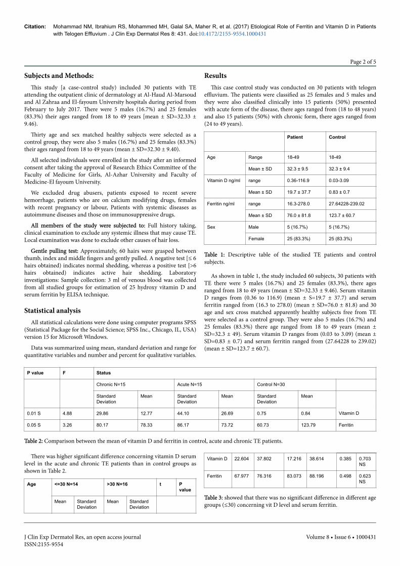
Table 1: Descriptive table of the studied TE patients and controlsubjects.
As shown in table 1, the study included 60 subjects, 30 patients withTE there were 5 males (16.7%) and 25 females (83.3%), there agesranged from 18 to 49 years (mean ± SD=32.33 ± 9.46). Serum vitaminD ranges from (0.36 to 116.9) (mean ± S=19.7 ± 37.7) and serumferritin ranged from (16.3 to 278.0) (mean ± SD=76.0 ± 81.8) and 30age and sex cross matched apparently healthy subjects free from TEwere selected as a control group. They were also 5 males (16.7%) and25 females (83.3%) there age ranged from 18 to 49 years (mean ±SD=32.3 ± 49). Serum vitamin D ranges from (0.03 to 3.09) (mean ±SD=0.83 ± 0.7) and serum ferritin ranged from (27.64228 to 239.02)(mean ± SD=123.7 ± 60.7).
P value F Status
Chronic N=15 Acute N=15 Control N=30
StandardDeviation
Mean StandardDeviation
Mean StandardDeviation
Mean
0.01 S 4.88 29.86 12.77 44.10 26.69 0.75 0.84 Vitamin D
0.05 S 3.26 80.17 78.33 86.17 73.72 60.73 123.79 Ferritin
Table 2: Comparison between the mean of vitamin D and ferritin in control, acute and chronic TE patients.
There was higher significant difference concerning vitamin D serumlevel in the acute and chronic TE patients than in control groups asshown in Table 2.
Age <=30 N=14 >30 N=16 t Pvalue
Mean StandardDeviation
Mean StandardDeviation
Vitamin D 22.604 37.802 17.216 38.614 0.385 0.703NS
Ferritin 67.977 76.316 83.073 88.196 0.498 0.623NS
Table 3: showed that there was no significant difference in different agegroups (≤30) concerning vit D level and serum ferritin.
Citation: Mohammad NM, Ibrahium RS, Mohammed MH, Galal SA, Maher R, et al. (2017) Etiological Role of Ferritin and Vitamin D in Patientswith Telogen Effluvium . J Clin Exp Dermatol Res 8: 431. doi:10.4172/2155-9554.1000431
Page 2 of 5
J Clin Exp Dermatol Res, an open access journalISSN:2155-9554
Volume 8 • Issue 6 • 1000431
There was a significant difference between mean of ferritin in acuteand chronic TE patients with the mean of ferritin in control as shownin Table 3.
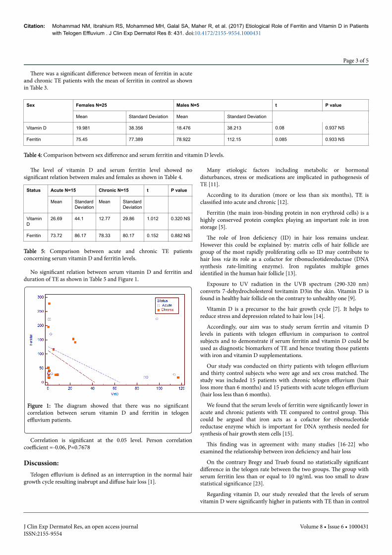
Sex Females N=25 Males N=5 t P value
Mean Standard Deviation Mean Standard Deviation
Vitamin D 19.981 38.356 18.476 38.213 0.08 0.937 NS
Ferritin 75.45 77.389 78.922 112.15 0.085 0.933 NS
Table 4: Comparison between sex difference and serum ferritin and vitamin D levels.
The level of vitamin D and serum ferritin level showed nosignificant relation between males and females as shown in Table 4.
Status Acute N=15 Chronic N=15 t P value
Mean StandardDeviation
Mean StandardDeviation
VitaminD
26.69 44.1 12.77 29.86 1.012 0.320 NS
Ferritin 73.72 86.17 78.33 80.17 0.152 0.882 NS
Table 5: Comparison between acute and chronic TE patientsconcerning serum vitamin D and ferritin levels.
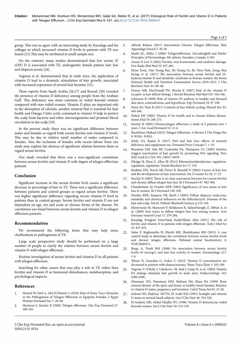
No significant relation between serum vitamin D and ferritin andduration of TE as shown in Table 5 and Figure 1.
Figure 1: The diagram showed that there was no significantcorrelation between serum vitamin D and ferritin in telogeneffluvium patients.
Correlation is significant at the 0.05 level. Person correlationcoefficient =-0.06, P=0.7678
Discussion:Telogen effluvium is defined as an interruption in the normal hair
growth cycle resulting inabrupt and diffuse hair loss [1].
Many etiologic factors including metabolic or hormonaldisturbances, stress or medications are implicated in pathogenesis ofTE [11].
According to its duration (more or less than six months), TE isclassified into acute and chronic [12].
Ferritin (the main iron-binding protein in non erythroid cells) is ahighly conserved protein complex playing an important role in ironstorage [5].
The role of Iron deficiency (ID) in hair loss remains unclear.However this could be explained by: matrix cells of hair follicle aregroup of the most rapidly proliferating cells so ID may contribute tohair loss via its role as a cofactor for ribonucleotidereductase (DNAsynthesis rate-limiting enzyme). Iron regulates multiple genesidentified in the human hair follicle [13].
Exposure to UV radiation in the UVB spectrum (290-320 nm)converts 7-dehydrocholesterol tovitamin D3in the skin. Vitamin D isfound in healthy hair follicle on the contrary to unhealthy one [9].
Vitamin D is a precursor to the hair growth cycle [7]. It helps toreduce stress and depression related to hair loss [14].
Accordingly, our aim was to study serum ferrtin and vitamin Dlevels in patients with telogen effluvium in comparison to controlsubjects and to demonstrate if serum ferritin and vitamin D could beused as diagnostic biomarkers of TE and hence treating those patientswith iron and vitamin D supplementations.
Our study was conducted on thirty patients with telogen effluviumand thirty control subjects who were age and sex cross matched. Thestudy was included 15 patients with chronic telogen effluvium (hairloss more than 6 months) and 15 patients with acute telogen effluvium(hair loss less than 6 months).
We found that the serum levels of ferritin were significantly lower inacute and chronic patients with TE compared to control group. Thiscould be argued that iron acts as a cofactor for ribonucleotidereductase enzyme which is important for DNA synthesis needed forsynthesis of hair growth stem cells [15].
This finding was in agreement with: many studies [16-22] whoexamined the relationship between iron deficiency and hair loss
On the contrary Bregy and Trueb found no statistically significantdifference in the telogen rate between the two groups. The group withserum ferritin less than or equal to 10 ng/mL was too small to drawstatistical significance [23].
Regarding vitamin D, our study revealed that the levels of serumvitamin D were significantly higher in patients with TE than in control
Citation: Mohammad NM, Ibrahium RS, Mohammed MH, Galal SA, Maher R, et al. (2017) Etiological Role of Ferritin and Vitamin D in Patientswith Telogen Effluvium . J Clin Exp Dermatol Res 8: 431. doi:10.4172/2155-9554.1000431
Page 3 of 5
J Clin Exp Dermatol Res, an open access journalISSN:2155-9554
Volume 8 • Issue 6 • 1000431
group. This was in agree with an interesting study by Karadage and hiscolluges in which increased vitamin D levels in patients with TE wasfound [21].This may be attributed to small sample size.
On the contrary many studies demonstrated that low serum 25(OH) D is associated with TE, androgenitic female pattern hair lossand alopecia areata [24].
Vegesna et al. demonstrated that in nude mice, the application ofvitamin D lead to a dramatic stimulation of hair growth, associatedwith increased expression of several hair keratins [25].
Three reports from Saudi Arabia [26,27] and Kuwait [28] revealedthe presence of vitamin D deficiency among people from the ArabianGulf. This deficiency was more common in veiled Kuwaiti womencompared with non-veiled women. Vitamin D plays an important rolein the absorption of calcium, another mineral that is essential for hairhealth and Omega 3 fatty acids contained in vitamin D help to protectthe scalp from bacteria and other microorganisms and promote bloodcirculation in the scalp [10].
In the present study there was no significant difference betweenmales and females as regard both serum ferritin and vitamin D levels.This may be due to relative small number of males compared tofemales. Also the exclusion of females with recent labour from ourstudy may explain the absence of significant relation between them asregard serum ferritin.
Our study revealed that there was a non-significant correlationbetween serum ferritin and vitamin D with degree of telogen effluviumpatients
ConclusionSignificant increase in the serum ferritin level causes a significant
decrease in percentage of hair in TE. There was a significant differencebetween patients and control groups as regard serum ferritin. Therewas higher significant difference regarding serum vitamin D level inpatients than in control groups. Serum ferritin and vitamin D are notdependent on age, sex and acute or chronic forms of the disease. Nocorrelation was found between serum ferritin and vitamin D in telogeneffluvium patients.
RecommendationWe recommend the following items that may help more
clarifications to pathogenesis of TE:
Large scale prospective study should be performed on a largenumber of people to clarify the relation between serum ferritin andvitamin D with telogen effluvium.
Routine investigation of serum ferritin and vitamin D in all patientswith telogen effluvium.
Searching for other causes that may play a role in TE rather thanferritin and vitamin D as hormonal disturbances, malabsorption, andpsychological impacts.
References1. Hamad W, Said A, Abd El Hamid A (2010) Role of Some Trace Elements
in the Pathogenesis of Telogen Effluvium in Egyptian Females. J EgyptWomen Dermatol Soc 7: 44-48.
2. Harrison S, Sinclair R (2002) Telogen effluvium. Clin Exp Dermatol 27:389-395.
3. Alfredo Rebora (2017) Intermittent Chronic Telogen Effluvium. SkinAppendage Disord 3: 36-38.
4. Marks JG, Miller J (2006) Telogeneffluvium. InLookingbill and Marks'Principles of Dermatology, 4th edition, Saunders, Canada, 4-263.
5. Arosio P, Levi S (2002) Ferritin, iron homeostatis, and oxidative damage.Free Radic Biol Med 33: 457-463.
6. Hyun Yoon, Nan Young Bae, Mi Young Gi, Bu Yeon Park, Jeong MinSeong, et al. (2017) The association between serum ferritin and 25-hydroxyvitamin D and metabolic syndrome in Korean women: the KoreaNational Health and Nutrition Examination Survey 2010–2012. J ClinBiochem Nutr. 61: 60–66.
7. Demay MB, MacDonald PN, Skorija K (2007) Role of the vitamin Dreceptor in hair follicle biology. J Steroid Biochem Mol Biol 103: 344-346.
8. Lehmann B (2009) Role of vitamin D3 pathway in healthy and diseasedskin-facts, contradictions, and hypotheses. Exp Dermatol 18: 97-108.
9. Stenn KS, Paus R (2011) Controls of hair follicle cycling. Physiol Rev 81:449-494.
10. Holick MF (2005) Vitamin D for health and in chronic kidney disease.Semin Dial 18: 266-275.
11. Sinclair R (2005) Chronictelogen effluvium: a study of 5 patients over 7years. J Am Acad Dermatol 52: 12-6.
12. Shashikant Malkud (2015) Telogen Effluvium: A Review J Clin Diagn Res9: WE01–WE03.
13. Emily LG, Rajani K (2017) Diet and hair loss: effects of nutrientdeficiency and supplement use. Dermatol Pract Concept 7: 1–10.
14. Beaudoin GM, Sisk JM, Coulombe PA, Thompson CC (2005) Hairlesstriggers reactivation of hair growth by promoting Wnt signaling. ProcNatl Acad Sci USA 102: 14653-14658.
15. Elledge SJ, Zhou Z, Allen JB (2012) Ribonucleotidereductase: regulation,regulation, regulation. Trends Biochem Sci 17: 119.
16. Rushton DH, Norris MJ, Dover R, Busuttil N (2002) Causes of hair lossand the developments in hair rejuvenation. Int J Cosmet Sci 24: 17-23.
17. Sinclair R (2002) There is no clear association between low serum ferritinand chronic diffuse telogen hair loss. Br J Dermatol 147: 982-984.
18. Chamberlain AJ, Dwaber RPR (2003) Significance of iron status in hairloss in women. Br J Dermatol 149: 428.
19. Dawber RPR, Simpson NB, Barth J (2003) Diffuse alopecia: endocrine,metabolic and chemical influences on the follicularcycle. Diseases of thehair and scalp, 3rd ed. Oxford: Blackwell Science; p.123-150.
20. Moeinvaziri M, Mansoori P, Holakooee K, SafaeeNaraghi Z, Abbasi A, etal. (2009) Iron status in diffuse telogen hair loss among women. ActaDermato venerol Croat 17: 279-284.
21. Karadağ, Ertuğrul, EmreTutal, KadirOkhan Akin (2011) The role offerritin and vitamin D in patients with telogen effluvium. Turk J Med Sci41: 827-833.
22. Sakar P, Raghunatha H, Harish MR, Shashikumar BM (2013) A casecontrol study to determine the correlation between serum ferritin levelsand chronic telogen effluvium. Webmed central biochemistry 4:WMC0040331.
23. Bregy A, Trueb RM (2008) No association between serum ferritinlevels>10 microg/L and hair loss activity in women. Dermatology 217:1-6.
24. Yilmaz N, Serarslan G, Gokce C (2012) Vitamin D concentration aredecreased in patients with alopeaciaareata. Vitam Trace Elem 1: 105-109.
25. Vegesna V, O’Kelly J, Uskokovic, M, Said J, Lemp N, et al. (2002) VitaminD3 analogs stimulate hair growth in nude mice. Endocrinology 143:4389-4396.
26. Ghannam NN, Hammani MM, Bakheet SM, Khan BA (1999) Bonemineral density of the spine and femur in healthy Saudi females: Relationto vitamin D status, pregnancy and lactation. Calcif Tissue Int 65: 23-28.
27. Sedrani SH, Elidrissy AWTH, El Arabi KM (1987) Sunlight and vitaminD status in normal Saudi subjects. Am J Clin Nutr 46: 324-328.
28. El-Sonbaty MR, Abdul-Ghaffar NU (1996) Vitamin D deficiencyin veiledKuwaiti women. Eur J Clin Nutr 50: 315-318.
Citation: Mohammad NM, Ibrahium RS, Mohammed MH, Galal SA, Maher R, et al. (2017) Etiological Role of Ferritin and Vitamin D in Patientswith Telogen Effluvium . J Clin Exp Dermatol Res 8: 431. doi:10.4172/2155-9554.1000431
Page 4 of 5
J Clin Exp Dermatol Res, an open access journalISSN:2155-9554
Volume 8 • Issue 6 • 1000431