Embed Size (px)
Citation preview

Establishing a patient safety program in Interventional Radiology
A. Kyle Jones, Ph.D., DABRAssistant Professor
MD Anderson Cancer Center

Disclosure
• I am co‐owner of Fluoroscopic Safety, LLC, a company that provides training for physicians in the safe use of fluoroscopy
• Fluoroscopic Safety is not discussed in this presentation
A. Kyle Jones, Ph.D. AAPM 2013 WE‐A‐144‐1 2

Why?
• We are practicing in an era of increased emphasis on quality and patient safety
• Reimbursement models are changing with emphasis on outcomes and minimizing/managing complications
• Ethical considerations• Accreditation and regulatory considerations
A. Kyle Jones, Ph.D. AAPM 2013 WE‐A‐144‐1 3

Updates to TAC 289.227
• Requires creation of a Fluoroscopy Radiation Protocol Committee– RSC for fluoroscopy
• Required tasks– Meet– Restrict use of fluoroscopy
• Radiologist, R.O., or physician completing 8 hrs Cat. 1 CME + 1 hr hands on training
• aka privileges– Record dose descriptors– Establish reference levels and review process
A. Kyle Jones, Ph.D. AAPM 2013 WE‐A‐144‐1 4

RISKS OF FLUOROSCOPICALLY GUIDED PROCEDURES
A. Kyle Jones, Ph.D. AAPM 2013 WE‐A‐144‐1 5

Who is at risk?
• Physicians• Nurses• Technologists• Anesthesiologists• Patients• Facilities
A. Kyle Jones, Ph.D. AAPM 2013 WE‐A‐144‐1 6

Risks to the operator and staff
• Infection• Back injury• Falls• Heavy objects• Litigation• Radiation‐induced cataracts• Radiation‐induced cancer
A. Kyle Jones, Ph.D. AAPM 2013 WE‐A‐144‐1 7

Risks to the patient
• Death• Puncture of vessel• Contrast reaction• Hematoma• Infection• Stochastic effects from radiation exposure• Tissue effects from radiation exposure
A. Kyle Jones, Ph.D. AAPM 2013 WE‐A‐144‐1 8

Stochastic effects
• Stochastic effect – risk ↑ linearly with dose• Risk depends on
1. Volume of tissue irradiated2. Type of tissue irradiated3. Total dose delivered to tissue4. Age of patient5. Patient genetics
• There is always a risk of stochastic effects if we use ionizing radiation, but these risks can be managed
A. Kyle Jones, Ph.D. AAPM 2013 WE‐A‐144‐1 9

Stochastic effects• In some cases the risk to the patient can be reduced
• Procedural planning and situational awareness are key
Vañó E, Arranz L, Sastre JM, et al. Dosimetric and radiation protection considerations based on some cases of patient skin injuries in interventional cardiology. Br J Radiol 1998;71:510‐516
A. Kyle Jones, Ph.D. AAPM 2013 WE‐A‐144‐1 10
Copyrighted image
removed

Tissue effects
• Tissue effects have a threshold– Risk = 0 below a certain dose*– Severity increases with increasing dose above threshold dose
• Our understanding of tissue effects has been evolving
• In most cases, can be prevented– Training of operators– Safety program– QC of equipment
A. Kyle Jones, Ph.D. AAPM 2013 WE‐A‐144‐1 11

Threshold doses for tissue effects
• For many years, hard thresholds for various types of tissue effects were quoted
• It has become apparent that these “thresholds” can vary widely between patients
• Depends on1. Patient genetics2. Prior skin irradiation3. Disease state/treatment
A. Kyle Jones, Ph.D. AAPM 2013 WE‐A‐144‐1 12

Balter et al. Fluoroscopically guided interventional procedures: A review of radiation effects on patients’ skin and hair. Radiology 254:326‐341, 2010.
A. Kyle Jones, Ph.D. AAPM 2013 WE‐A‐144‐1 13

Radiation injuries
• Radiation‐induced skin injuries are particularly troublesome for several reasons– Patient does not experience any sensations– Latent period means that cause and effect may not be connected by patient or physician
A. Kyle Jones, Ph.D. AAPM 2013 WE‐A‐144‐1 14

6‐8 weeks post procedure 16‐21 weeks post procedure 18‐21 months post procedure
Multiple coronary angiography, angioplasty, and bypass graft on a single day – estimated peak skin dose > 20 Gy
Shope TB . Radiation-induced skin injuries from fluoroscopy. http://www.fda.gov/RadiationEmittingProducts/RadiationEmittingProductsandProcedures/MedicalImagi
ng/MedicalX-Rays/ucm116682.htm U.S. Food and Drug Administration.
A. Kyle Jones, Ph.D. 15AAPM 2013 WE‐A‐144‐1

Radiation injuries
• Radiation injuries can be particularly gruesome and, depending on severity, may never completely heal
Wagner LK, Archer BR. Minimizing Risks from Fluoroscopic X Rays: Bioeffects, Instrumentation, and Examination, 3rd edition; Houston, TX; R. M. Partnership, 2000
Wagner LK, McNesse MD, Marx MV, Siegel EL. Severe skin reactions from interventional fluoroscopy: case report and review of literature. Radiology 1999;213:773‐776
A. Kyle Jones, Ph.D. AAPM 2013 WE‐A‐144‐1 16
Copyrighted image
removed
Copyrighted image
removed

What can we do?
• Reduce the risk of stochastic effects for operator, staff, and patient
• Prevent most tissue effects such as radiation‐induced cataracts and skin injuries
• Recognize situations where a high probability for injury exists so the patient can be appropriately medically managed
A. Kyle Jones, Ph.D. AAPM 2013 WE‐A‐144‐1 17

Three‐pronged approach
• Pre‐procedure actions• Intra‐procedure actions• Post‐procedure actions
A. Kyle Jones, Ph.D. AAPM 2013 WE‐A‐144‐1 18

A. Kyle Jones, Ph.D. AAPM 2013 WE‐A‐144‐1 19

PRE‐PROCEDURE ELEMENTS
A. Kyle Jones, Ph.D. 20AAPM 2013 WE‐A‐144‐1

Informed consent
“Informed consent is a patient's right to be presented with sufficient information, by
either the physician or their representative, to allow the patient to make an informed decision regarding whether or not to consent to a treatment or procedure.”
http://www.med‐ed.virginia.edu/courses/rad/consent/
A. Kyle Jones, Ph.D. AAPM 2013 WE‐A‐144‐1 21

Informed consent
“Informed consent is a patient's right to be presented with sufficient information, by
either the physician or their representative, to allow the patient to make an informed decision regarding whether or not to consent to a treatment or procedure.”
http://www.med‐ed.virginia.edu/courses/rad/consent/
A. Kyle Jones, Ph.D. AAPM 2013 WE‐A‐144‐1 22

A. Kyle Jones, Ph.D. 23AAPM 2013 WE‐A‐144‐1
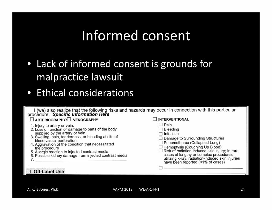
Informed consent
• Lack of informed consent is grounds for malpractice lawsuit
• Ethical considerations
A. Kyle Jones, Ph.D. 24AAPM 2013 WE‐A‐144‐1

Patient education
• PA/physician must have the tools and knowledge to simply explain the risks to the patient without inducing panic
• One approach to this is a pamphlet/handout– Mechanisms of injury– How we prevent injuries– Decisions made during the case
A. Kyle Jones, Ph.D. AAPM 2013 WE‐A‐144‐1 25

Identify high‐risk patients• Certain conditions are suspected to pre‐dispose patients to radiation induced skin injuries– Diabetes mellitus – Connective tissue disorders
– Ataxia telangiectasia– Drug interactions
• Also, a recent high dose procedure can result in the induction of effects at lower doses in the future
Wagner et al, Severe skin reactions from interventional fluoroscopy: Case report and review of the literature. Radiology;213:773‐776, 1999.
Koenig TR, et al. Skin injuries from fluoroscopically guided procedures: Part I, characteristics of radiation injury. AJR 177:3‐11, 2001.
Balter et al. Fluoroscopically guided interventional procedures: A review of radiation effects on patients’ skin and hair. Radiology 254:326‐341, 2010.
A. Kyle Jones, Ph.D. AAPM 2013 WE‐A‐144‐1 26

Identify high‐risk patients
• Most easily done during the consenting process
• The RIS can be used to automatically identify and flag these patients
• High‐risk patients can be routed to a dose sparing protocol, physician can be advised– Fewer acquisition runs, more storing of fluoro– Alternate Ka,r thresholds– Postpone procedure?
A. Kyle Jones, Ph.D. AAPM 2013 WE‐A‐144‐1 27

Multiple and repeated procedures• Two scenarios
1. By performing a very complex case in multiple sessions, fxcan be used to reduce late effects
2. If a procedure is repeated, an unexpected skin reaction may occur as the Biologically Equivalent Dose from the two procedures is greater than the dose from either individual procedure
• We can look to radiobiology for guidance on managing multiple irradiations of the skin
Balter et al. Fluoroscopically guided interventional procedures: A review of radiation effects on patients’ skin and hair. Radiology Appendix E1, 2010.
A. Kyle Jones, Ph.D. AAPM 2013 WE‐A‐144‐1 28

Training of physicians and staff• Physicians performing fluoroscopically‐guided procedures should be
trained in the safe use of fluoroscopic equipment– NCRP 168– State regulations
• Continuing education• Understand dose saving features of each type of equipment on
which they work– Hands‐on component
NCRP 168
A. Kyle Jones, Ph.D. AAPM 2013 WE‐A‐144‐1 29

Training resources
A. Kyle Jones, Ph.D. AAPM 2013 WE‐A‐144‐1 30
http://www.aapm.org/education/ERG/
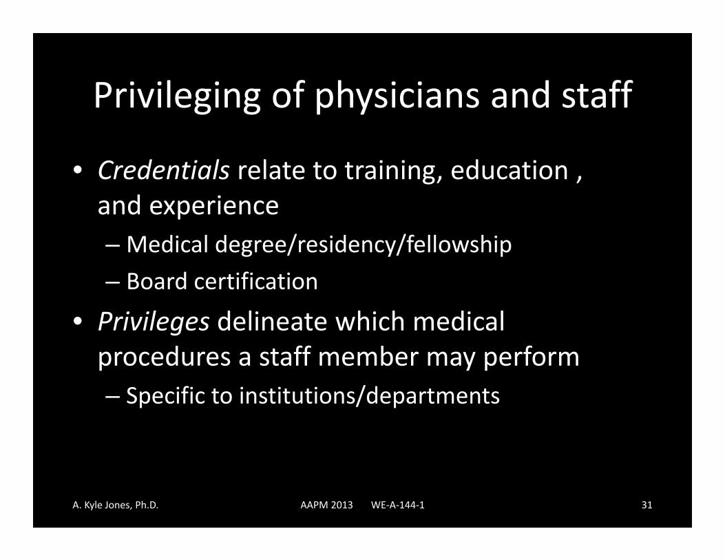
Privileging of physicians and staff
• Credentials relate to training, education , and experience– Medical degree/residency/fellowship– Board certification
• Privileges delineate which medical procedures a staff member may perform– Specific to institutions/departments
A. Kyle Jones, Ph.D. AAPM 2013 WE‐A‐144‐1 31

Privileging of physicians and staff
• AAPM Task Group 124 has published their report:
which is the definitive resource
A. Kyle Jones, Ph.D. AAPM 2013 WE‐A‐144‐1 32

INTRA‐PROCEDURE ELEMENTS
A. Kyle Jones, Ph.D. 33AAPM 2013 WE‐A‐144‐1

NCRP 168
A. Kyle Jones, Ph.D. AAPM 2013 WE‐A‐144‐1 34

Reference air kerma (Ka,r) thresholds
• All equipment manufactured after June 2006 is required by law to display the Ka,r
• Alerting the physician at certain points guarantees there are no surprises at the end of a case
• Decisions can be made based on medical management at each threshold– Pace of procedure– Good practice (YDNKWIHUYKWIH)
A. Kyle Jones, Ph.D. AAPM 2013 WE‐A‐144‐1 35

Establishing Ka,r thresholds
A. Kyle Jones, Ph.D. AAPM 2013 WE‐A‐144‐1 36

NCRP 168
A. Kyle Jones, Ph.D. AAPM 2013 WE‐A‐144‐1 37

Setting notification levels• Use a representative phantom and perform a “procedure” on the phantom– Measure ESD (cannot used lead‐backed dosimeter)– Correct geometry– Distribution of acquisition/fluoroscopy
• Apply f‐factor, use ratio of Ka,r/PSD to determine notification levels– VIR ~ 1– Cardiology ~ 0.7‐0.8– Neuro ~ 0.8‐1.0
• Some manufacturers allow a number of notification levels to be programmed in the system
A. Kyle Jones, Ph.D. AAPM 2013 WE‐A‐144‐1 38

A. Kyle Jones, Ph.D. AAPM 2013 WE‐A‐144‐1 39

A very important piece of information
• Estimating the PSD with any degree of accuracy REQUIRES knowledge of the source‐to‐patient distance (SPD)
• If this information is not available in DICOM headers or the RDSR, it must be recorded manually
• Provide instructions to the technologist or other staff for measuring and recording the SPD when the SRDL is reached
A. Kyle Jones, Ph.D. AAPM 2013 WE‐A‐144‐1 40

A. Kyle Jones, Ph.D. AAPM 2013 WE‐A‐144‐1 41
0
2
4
6
8
10
12
14
16
18
50 55 60 65 70 75 80 85 90 95 100
PSD (G
y)
SPD (cm)
IRP = 63.5 cm

Situational awareness
• For patients who have undergone a recent high dose procedure, use a different projection to reduce the cumulative skin dose– Reduce 95% area load– May not reduce PSD
• May not be able to completely eliminate overlap, but for angled projections can have large benefit– Importance of tight collimation
A. Kyle Jones, Ph.D. AAPM 2013 WE‐A‐144‐1 42

A. Kyle Jones, Ph.D. 43AAPM 2013 WE‐A‐144‐1

A. Kyle Jones, Ph.D. 44AAPM 2013 WE‐A‐144‐1

A. Kyle Jones, Ph.D. AAPM 2013 WE‐A‐144‐1 45
SU‐E‐I‐24

POST‐PROCEDURE ELEMENTS
A. Kyle Jones, Ph.D. 46AAPM 2013 WE‐A‐144‐1

Record dose descriptors
• Medical record has been suggested– Perhaps difficult– May not be searchable
• Dictated• Scanned
– What is and what is not part of the medical record?• DICOM Structured Dose Reporting is here
– Recently installed our first system (VC14)– Legacy systems may never support– Third party options are available
• DICOM headers contain some information
A. Kyle Jones, Ph.D. AAPM 2013 WE‐A‐144‐1 47

NCRP 168
A. Kyle Jones, Ph.D. AAPM 2013 WE‐A‐144‐1 48

A. Kyle Jones, Ph.D. 49AAPM 2013 WE‐A‐144‐1

A. Kyle Jones, Ph.D. AAPM 2013 WE‐A‐144‐1 50

A. Kyle Jones, Ph.D. AAPM 2013 WE‐A‐144‐1 51

A. Kyle Jones, Ph.D. AAPM 2013 WE‐A‐144‐1 52

DICOM SR
A. Kyle Jones, Ph.D. AAPM 2013 WE‐A‐144‐1 53

Record dose descriptors
• Other possibilities include RIS or logbooks– Would like it to be searchable
• Tracking• Practice improvement• Identify/prevent sentinel events
• We went with the RIS– Manual entry into designated fields– Reports can be generated, already linked with procedure (accession number)
– Automatic analysis of data/entry into database
A. Kyle Jones, Ph.D. AAPM 2013 WE‐A‐144‐1 54

Record dose descriptors
• What we record:– Ka,r– KAP– Number of rotational angiography runs (DynaCT)– Fluoroscopy time
• Track repeated or multiple procedures
A. Kyle Jones, Ph.D. AAPM 2013 WE‐A‐144‐1 55

KAP meter calibration
• FDA (2006)/IEC 60601‐2‐43 (2010) requirement for accuracy (Ka,r): +/‐ 35% IEC 60580 (2000) requires +/‐ 25% accuracy under specific conditions for KAP
• Take the time to measure a single‐ or double‐point calibration factor for the KAP meter in each of your interventional labs
• NEMA XR‐27 – facility to store calibration factor(s) in the RDSR
• AAPM TG 190 standardizing measurement procedures
A. Kyle Jones, Ph.D. AAPM 2013 WE‐A‐144‐1 56

A. Kyle Jones, Ph.D. AAPM 2013 WE‐A‐144‐1 57
Shrimptom PC and Wall BF. An evaluation of the Diamentor transmission ionisation chamber in indicating exposure‐area product (R cm2) during diagnostic radiological examinations. Phys Med Biol 27:871‐878, 1982
My experience (1 manufacturer) for Ka,r accuracy:
• Generally +/‐ 15% across all FOV and rates (FLU, ACQ, CINE)• Generally less accurate as FOV is reduced• Taking advantage of lenient limits?

Automated solutions
• Clinical Microsystems• GE DoseWatch• Siemens CareAnalytics• Radimetrics
A. Kyle Jones, Ph.D. AAPM 2013 WE‐A‐144‐1 58

Flagging and follow up
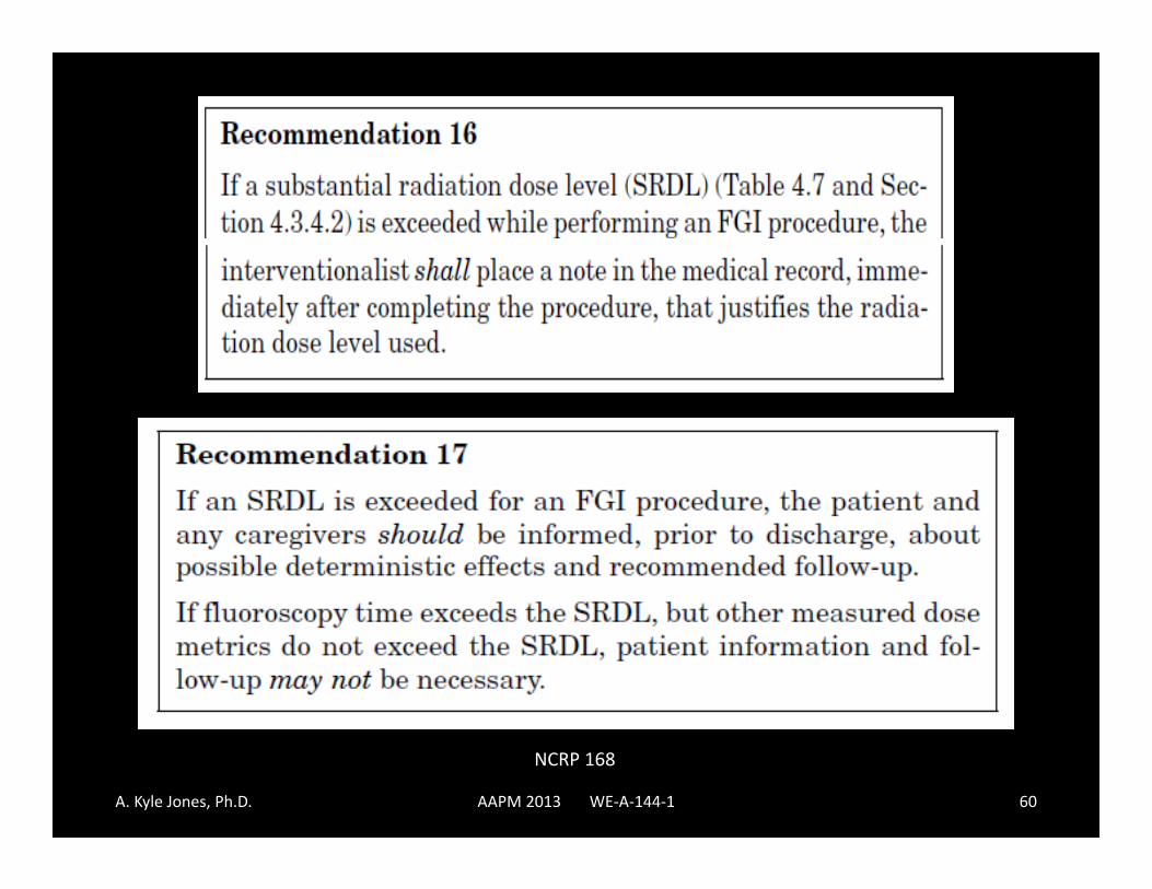
• A case where our substantial radiation dose level (SRDL) is reached is flagged by the technologist, triggering our f/u protocol (PA):– Patient informed that SRDL (>= 5 Gy) was reached and why
– Patient instruction provided (one sheet)• Signs/symptoms (red area the size of your hand)• Instructions (do not scratch or itch)• Actions (call us)
– Telephone or in‐person f/u scheduled for 4 weeks– Verify that dose report is archived
A. Kyle Jones, Ph.D. AAPM 2013 WE‐A‐144‐1 59
NCRP 168
A. Kyle Jones, Ph.D. AAPM 2013 WE‐A‐144‐1 60

Steele JS, Jones AK, Ninan EP. Quality initiatives: establishing an interventional radiology patient radiation safety program. Radiographics 32(1):277‐87, 2012.
A. Kyle Jones, Ph.D. AAPM 2013 WE‐A‐144‐1 61

Medical management of skin reactions
• Acute skin doses greater than a few Gy are only seen in medicine as a result of fluoroscopically guided procedures
• Knowing who to consult regarding skin reactions is tricky
• Dermatologists may never have seen these types of reactions
• Radiation oncologists seem to be the best to discuss these matters with– Typically do not see late/acute effects of same severity
A. Kyle Jones, Ph.D. AAPM 2013 WE‐A‐144‐1 62

PRACTICE AND QUALITY IMPROVEMENT
A. Kyle Jones, Ph.D. 63AAPM 2013 WE‐A‐144‐1

Practice improvement
• Dose information collected can be used to calculate dose metrics that can be used to accomplish data driven practice improvement– Compare dose metrics to national averages
• RAD‐IR study
– Identify procedures/physicians/etc. for improvement
– Control charts for identifying exceptional variation• Discuss SRDL/reactions at QI conference or M+M
A. Kyle Jones, Ph.D. AAPM 2013 WE‐A‐144‐1 64

NCRP 168
A. Kyle Jones, Ph.D. AAPM 2013 WE‐A‐144‐1 65

Radiation dose audits
Balter S et al. Patient radiation dose audits for fluoroscopically guided interventional procedures. Med Phys 38(3):1611‐18, 2011.
A. Kyle Jones, Ph.D. AAPM 2013 WE‐A‐144‐1 66

Don’t audit only “high dose” cases…
A. Kyle Jones, Ph.D. AAPM 2013 WE‐A‐144‐1 67

Hepatic chemoembolization
Fluoro time
KAP (cGy‐cm2)
Ka,r(mGy)
MEAN 22 40713 1809MEDIAN 19 34898 1604RAD‐IR MEAN 16.8 28232 1406
A. Kyle Jones, Ph.D. AAPM 2013 WE‐A‐144‐1 68

Pelvic arterial embolization (tumor)
Fluoro time
KAP (cGy‐cm2)
Ka,r(mGy)
MEAN 17 39023 1836MEDIAN 19 32309 1964RAD‐IR MEAN 28.4 30284 1846
A. Kyle Jones, Ph.D. AAPM 2013 WE‐A‐144‐1 69

Nephrostomy placement
Fluorotime
KAP (cGy‐cm2)
Ka,r(mGy)
MEAN 6 1838 115MEDIAN 4 1024 50RAD‐IR MEAN 10.5 2555 257
A. Kyle Jones, Ph.D. AAPM 2013 WE‐A‐144‐1 70

In certain circumstances
• It is necessary and appropriate to administer a high dose of radiation during a fluoroscopically guided procedure
• One must still be conscious of how much radiation has been used
• The risk of injury must be commensurate with the benefits of the procedure
• Medical management after the procedure must be appropriate
A. Kyle Jones, Ph.D. AAPM 2013 WE‐A‐144‐1 71

A. Kyle Jones, Ph.D. AAPM 2013 WE‐A‐144‐1 72

Acknowledgements
• Lou Wagner, Ph.D.• Joseph Steele, M.D.• Alex Pasciak, Ph.D.
A. Kyle Jones, Ph.D. AAPM 2013 WE‐A‐144‐1 73

Further reading• Miller et al., Quality improvement guidelines for recording patient radiation dose
in the medical record. J Vasc Interv Radiol 15:423–429, 2004.– SIR Standards of Practice Committee
• Miller DL, Balter S, Noonan PT, Georgia JD, Minimizing radiation‐induced skin injury in interventional radiology procedures. Radiology 225:329–336, 2002 .
• Stecker et al., Guidelines for patient radiation dose management. J Vasc IntervRadiol 20:S263–S273, 2009.
– SIR Safety and Health Committee– Discharge/consenting examples
• Archer BR and Wagner LK, Protecting patients by training physicians in fluoroscopic radiation management. J Appl Clin Med Phys 1:32‐37, 2000.
• Wagner LK and Archer BR, Minimizing Risks from Fluoroscopic X Rays, 2nd ed., R.M. Partnership, The Woodlands, TX.
• Balter S, et al. Fluoroscopically guided interventional procedures: A review of radiation effects on patients’ skin and hair. Radiology, 254:326‐341
• NCRP Report 168
A. Kyle Jones, Ph.D. AAPM 2013 WE‐A‐144‐1 74