Embed Size (px)
Citation preview
ESC GUIDELINES
ESC Guidelines for the management of acutemyocardial infarction in patients presentingwith ST-segment elevationThe Task Force on the management of ST-segment elevation acutemyocardial infarction of the European Society of Cardiology (ESC)
Authors/Task Force Members: Ph. Gabriel Steg (Chairperson) (France)*,Stefan K. James (Chairperson) (Sweden)*, Dan Atar (Norway), Luigi P. Badano(Italy), Carina Blomstrom-Lundqvist (Sweden), Michael A. Borger (Germany),Carlo Di Mario (United Kingdom), Kenneth Dickstein (Norway), Gregory Ducrocq(France), Francisco Fernandez-Aviles (Spain), Anthony H. Gershlick (UnitedKingdom), Pantaleo Giannuzzi (Italy), Sigrun Halvorsen (Norway), Kurt Huber(Austria), Peter Juni (Switzerland), Adnan Kastrati (Germany), Juhani Knuuti(Finland), Mattie J. Lenzen (Netherlands), Kenneth W. Mahaffey (USA),Marco Valgimigli (Italy), Arnoud van ’t Hof (Netherlands), Petr Widimsky(Czech Republic), Doron Zahger (Israel)ESC Committee for Practice Guidelines (CPG): Jeroen J. Bax (Chairman) (Netherlands), Helmut Baumgartner(Germany), Claudio Ceconi (Italy), Veronica Dean (France), Christi Deaton (UK), Robert Fagard (Belgium),Christian Funck-Brentano (France), David Hasdai (Israel), Arno Hoes (Netherlands), Paulus Kirchhof(Germany UK), Juhani Knuuti (Finland), Philippe Kolh (Belgium), Theresa McDonagh (UK), Cyril Moulin (France),Bogdan A. Popescu (Romania), Zeljko Reiner (Croatia), Udo Sechtem (Germany), Per Anton Sirnes (Norway),Michal Tendera (Poland), Adam Torbicki (Poland), Alec Vahanian (France), Stephan Windecker (Switzerland).
Document Reviewers: David Hasdai (CPG Review Coordinator) (Israel), Felicity Astin (UK), Karin Astrom-Olsson(Sweden), Andrzej Budaj (Poland), Peter Clemmensen (Denmark), Jean-Philippe Collet (France), Keith A. Fox(UK), Ahmet Fuat (UK), Olivija Gustiene (Lithuania), Christian W. Hamm (Germany), Petr Kala (Czech Replublic),Patrizio Lancellotti (Belgium), Aldo Pietro Maggioni (Italy), Bela Merkely (Hungary), Franz-Josef Neumann(Germany), Massimo F. Piepoli (Italy), Frans Van de Werf (Belgium), Freek Verheugt (Netherlands),Lars Wallentin (Sweden)
Stefan K. James (Chairperson), Department of Medical Sciences / Uppsala Clinical Research Center, Uppsala University and Department of Cardiology Uppsala University Hospital,75185 Uppsala, Sweden. Tel: +46 705 944 404, Fax: +46 18 506 638, Email: [email protected]
* Corresponding authors: Ph. Gabriel Steg (Chairperson), AP-HP, Hopital Bichat / Univ Paris Diderot, Sorbonne Paris-Cite / INSERM U-698, Paris, France. Tel: +33 1 40 25 86 68,Fax: +33 1 40 25 88 65, Email: [email protected]
Disclaimer. The ESC Guidelines represent the views of the ESC and were arrived at after careful consideration of the available evidence at the time they were written. Healthprofessionals are encouraged to take them fully into account when exercising their clinical judgement. The guidelines do not, however, override the individual responsibility of healthprofessionals to make appropriate decisions in the circumstances of the individual patients, in consultation with that patient, and where appropriate and necessary the patient’sguardian or carer. It is also the health professional’s responsibility to verify the rules and regulations applicable to drugs and devices at the time of prescription.
& The European Society of Cardiology 2012. All rights reserved. For permissions please email: [email protected]
† Other ESC entities having participated in the development of this document:
Associations: European Association of Echocardiography (EAE), European Association for Cardiovascular Prevention (EACPR), European Heart Rhythm Association (EHRA), Euro-pean Association of Percutaneous Cardiovascular Interventions (EAPCI), Heart Failure Association (HFA)
Working Groups: Acute Cardiac care, Cardiovascular Pharmacology and Drug Therapy, Thrombosis
Councils: Cardiovascular Imaging, Cardiovascular Nursing and Allied Professions, Primary Cardiovascular Care, Cardiovascular Surgery
The content of these European Society of Cardiology (ESC) Guidelines has been published for personal and educational use only. No commercial use is authorized. No part of theESC Guidelines may be translated or reproduced in any form without written permission from the ESC. Permission can be obtained upon submission of a written request to OxfordUniversity Press, the publisher of the European Heart Journal and the party authorized to handle such permissions on behalf of the ESC.
European Heart Journal (2012) 33, 2569–2619doi:10.1093/eurheartj/ehs215
Downloaded from https://academic.oup.com/eurheartj/article-abstract/33/20/2569/447818by gueston 04 February 2018
The disclosure forms of the authors and reviewers are available on the ESC website www.escardio.org/guidelines
Online publish-ahead-of-print 24 August 2012
- - - - - - - - - - - - - - - - - - - - - - - - - - - - - - - - - - - - - - - - - - - - - - - - - - - - - - - - - - - - - - - - - - - - - - - - - - -- - - - - - - - - - - - - - - - - - - - - - - - - - - - - - - - - - - - - - - - - - - - - - - - - - - - - - - - - - - - - - - - - - - - - - - - - - -Keywords Guidelines † Acute myocardial infarction † ST-segment elevation † Acute coronary syndromes
Ischaemic heart disease † Reperfusion therapy † Primary percutaneous coronary interventionAntithrombotic therapy † Secondary prevention
Table of ContentsAbbreviations and Acronyms . . . . . . . . . . . . . . . . . . . . . . . 2570
1. Preamble . . . . . . . . . . . . . . . . . . . . . . . . . . . . . . . . . . . 2572
2. Introduction . . . . . . . . . . . . . . . . . . . . . . . . . . . . . . . . . 2573
2.1. Definition of acute myocardial infarction . . . . . . . . . . 2573
2.2. Epidemiology of ST-segment elevation myocardial
infarction . . . . . . . . . . . . . . . . . . . . . . . . . . . . . . . . . . 2573
3. Emergency care . . . . . . . . . . . . . . . . . . . . . . . . . . . . . . 2574
3.1. Initial diagnosis . . . . . . . . . . . . . . . . . . . . . . . . . . . 2574
3.2. Relief of pain, breathlessness and anxiety . . . . . . . . . . 2576
3.3. Cardiac arrest . . . . . . . . . . . . . . . . . . . . . . . . . . . 2576
3.4. Pre-hospital logistics of care . . . . . . . . . . . . . . . . . . 2577
3.4.1. Delays . . . . . . . . . . . . . . . . . . . . . . . . . . . . . . 2577
3.4.2. Emergency medical system . . . . . . . . . . . . . . . . 2578
3.4.3. Networks . . . . . . . . . . . . . . . . . . . . . . . . . . . 2578
3.4.4. General practitioners . . . . . . . . . . . . . . . . . . . . 2579
3.4.5. Admission procedures . . . . . . . . . . . . . . . . . . . 2579
3.4.6. Logistics . . . . . . . . . . . . . . . . . . . . . . . . . . . . 2579
3.5. Reperfusion therapy . . . . . . . . . . . . . . . . . . . . . . . 2580
3.5.1. Restoring coronary flow and myocardial tissue
reperfusion . . . . . . . . . . . . . . . . . . . . . . . . . . . . . . . 2580
3.5.2. Selection of a strategy for reperfusion . . . . . . . . . 2581
3.5.3. Primary percutaneous coronary intervention . . . . 2582
3.5.4. Fibrinolysis and subsequent interventions . . . . . . . 2586
3.5.5. Coronary bypass surgery and multivessel coronary
revascularization . . . . . . . . . . . . . . . . . . . . . . . . . . . 2590
3.5.6. Non-reperfused patients . . . . . . . . . . . . . . . . . . 2590
3.6. Management of hyperglycaemia in the acute phase of ST-
segment elevation myocardial infarction . . . . . . . . . . . . . . 2592
4. Management during hospitalization and at discharge . . . . . . 2593
4.1. Coronary care unit logistics and monitoring . . . . . . . . 2593
4.1.1. Coronary care unit . . . . . . . . . . . . . . . . . . . . . 2593
4.1.2. Monitoring . . . . . . . . . . . . . . . . . . . . . . . . . . . 2593
4.1.3. Ambulation . . . . . . . . . . . . . . . . . . . . . . . . . . 2593
4.1.4. Length of stay . . . . . . . . . . . . . . . . . . . . . . . . . 2593
4.2. Risk assessment and imaging . . . . . . . . . . . . . . . . . . 2594
4.2.1. Indications and timing . . . . . . . . . . . . . . . . . . . .2594
4.3. Assessment of myocardial viability . . . . . . . . . . . . . . 2595
4.4. Long-term therapies for ST-segment elevation
myocardial infarction . . . . . . . . . . . . . . . . . . . . . . . . . . 2595
4.4.1. Lifestyle interventions and risk factor control . . . . 2595
4.4.2. Antithrombotic therapy . . . . . . . . . . . . . . . . . . 2596
4.4.3. Beta-blockers . . . . . . . . . . . . . . . . . . . . . . . . . 2597
4.4.4. Lipid-lowering therapy . . . . . . . . . . . . . . . . . . . 2598
4.4.5. Nitrates . . . . . . . . . . . . . . . . . . . . . . . . . . . . . 2598
4.4.6. Calcium antagonists . . . . . . . . . . . . . . . . . . . . . 2598
4.4.7. Angiotensin-converting enzyme inhibitors and
angiotensin receptor blockers . . . . . . . . . . . . . . . . . . 2598
4.4.8. Aldosterone antagonists . . . . . . . . . . . . . . . . . . 2598
4.4.9. Magnesium, glucose–insulin–potassium, lidocaine . 2598
5. Complications following ST-segment elevation myocardial
infarction . . . . . . . . . . . . . . . . . . . . . . . . . . . . . . . . . . . . 2600
5.1. Haemodynamic disturbances . . . . . . . . . . . . . . . . . . 2600
5.1.1. Heart failure . . . . . . . . . . . . . . . . . . . . . . . . . . 2600
5.1.2. Management of heart failure following ST-segment
elevation myocardial infarction (Table 23) . . . . . . . . . . . 2601
5.1.3. Arrhythmias and conduction disturbances in the
acute phase . . . . . . . . . . . . . . . . . . . . . . . . . . . . . . 2603
5.2. Cardiac complications . . . . . . . . . . . . . . . . . . . . . . 2606
5.2.1. Mitral valve regurgitation . . . . . . . . . . . . . . . . . 2606
5.2.2. Cardiac rupture . . . . . . . . . . . . . . . . . . . . . . . 2607
5.2.3. Ventricular septal rupture . . . . . . . . . . . . . . . . . 2607
5.2.4. Right ventricular infarction . . . . . . . . . . . . . . . . 2607
5.2.5. Pericarditis . . . . . . . . . . . . . . . . . . . . . . . . . . . 2607
5.2.6. Left ventricular aneurysm . . . . . . . . . . . . . . . . . 2607
5.2.7. Left ventricular thrombus . . . . . . . . . . . . . . . . . 2607
6. Gaps in the evidence and areas for future research . . . . . . . 2608
Abbreviations and Acronyms
ACE angiotensin-converting enzymeACS acute coronary syndromeADP adenosine diphosphateAF atrial fibrillationAMI acute myocardial infarctionAV atrioventricularAIDA-4 Abciximab Intracoronary vs. intravenously
Drug ApplicationAPACHE II Acute Physiology Aand Chronic Health Evalu-
ation IIATOLL Acute myocardial infarction Treated with
primary angioplasty and inTravenous enOxa-parin or unfractionated heparin to Lower is-chaemic and bleeding events at short- andLong-term follow-upAcute Myocardial Infarc-tion Treated with Primary Angioplasty andIntravenous Enoxaparin or UnfractionatedHeparin to Lower Ischemic and BleedingEvents at Short- and Long-term Follow-up
ESC Guidelines2570
Downloaded from https://academic.oup.com/eurheartj/article-abstract/33/20/2569/447818by gueston 04 February 2018

aPTT activated partial thromboplastin timeARB angiotensin receptor blockerASSENT 3 ASssessment of the Safety and Efficacy of a
New Thrombolytic 3ATLAS ACS (etc.) Anti-Xa Therapy to Lower cardiovascular
events in Addition to Standard therapy in sub-jects with Acute Coronary Syndrome–Thrombolysis In Myocardial Infarction 51
b.i.d. bis in die (twice daily)BMI body mass indexBMS bare-metal stentBNP B-type natriuretic peptideBRAVE-3 Bavarian Reperfusion Alternatives
Evaluation-3CAD coronary artery diseaseCAPITAL-AMI Combined Angioplasty and Pharmacological
Intervention vs. Thrombolytics ALlone inAcute Myocardial Infarction
CHA2DS2-VASc Cardiac failure, Hypertension, Age ≥75[Doubled], Diabetes, Stroke [Doubled] –VASascular disease, Age 65–74 and Sex cat-egory [Female])
CHADS2 Cardiac failure, Hypertension, Age, Diabetes,Stroke (Doubled)
CK-MB creatine kinase myocardial bandCLARITY-TIMI 2828
CLlopidogrel as Adjunctive ReperfusionTherapy–Thrombolysis Iin Myocardial Infarc-tion 28
COMMIT Clopidogrel and Metoprolol in Myocardial In-farction Trial
CPG Committee for Practice GuidelinesCRISP AMI Counterpulsation to Reduce Infarct Size
Pre-PCI-Acute Myocardial InfarctionCRT cardiac resynchronization therapyCVLPRIT Complete Versus Lesion-only PRIimary PCI
TrialCT computed tomographyDAPT dual antiplatelet therapyDES drug-eluting stentDIGAMI Diabetes, Insulin Glucose Infusion in Acute
Myocardial InfarctionEAPCI European Association of Percutaneous Car-
diovascular InterventionsECG electrocardiogramEMS emergency medical systemEPHESUS Eplerenone Post-AMI Heart failure Efficacy
and SUrvival StudyESC European Society of CardiologyExTRACT-TIMI 25 Enoxaparin and Thrombolysis Reperfusion for
ACute myocardial infarction Treatment—Thrombolysis In Myocardial Infarction 25
FINESSE Facilitated INtervention with Enhanced reper-fusion Speed to Stop Events
FMC first medical contactGP glycoprotein
GRACIA GRupo de Analisis de la Cardiopatıa Isque-mica Aguda
GUSTO Global Utilization of Streptokinase and Tissueplasminogen activator for Occluded coronaryarteries
HbA1c haemoglobin A1cHORIZONS–AMI Harmonizing Outcomes with RevascularIZa-
tiON and Stents in Acute MyocardialInfarction
i.c. intracoronaryi.v. intravenousIABP intra-aortic balloon pumpINFUSE–AMI Intracoronary abciximab iNFUsion and aspir-
ation thrombectomy for anterior ST-segmentElevAtion Myocardial Infarction
IRA infarct-related arteryISIS-2 Second International Study of Infarct SurvivalLab catheterization laboratoryLBBB left bundle branch blockLDL low-density lipoproteinLV left ventricularLVAD left ventricular assist deviceNORDISTEMI NORwegian study on DIstrict treatment of
ST-Elevation Myocardial InfarctionNRMI National Registry of Myocardial InfarctionNSTE-ACS non-ST-segment elevation acute coronary
syndromesOASIS Optimal Antiplatelet Strategy for
InterventionSOAT Occluded Artery TrialON-TIME 2 ONgoing Tirofiban In Myocardial infarction
EvaluationOPTIMAAL OPtimal Therapy In Myocardial infarction
with the Angiotensin II Antagonist Losartanp.o. per osPAMI-II Primary Angioplasty in Myocardial Infarction IIPET positron emission tomographyPCI percutaneous coronary interventionPLATO PLATelet inhibition and patient OutcomesPRAMI PReventive Angioplasty in Myocardial Infarc-
tion trialPRIMARY PCI primary percutaneous coronary interventionPROVE IT-TIMI 22 PRavastatin Or atorVastatin Evaluation and In-
fection Therapy–Thrombolysis In MyocardialInfarction 22
RBBB right bundle branch blockr-PA reteplaseRIFLE-STEACS RadIal Vs. FemoraL randomized investigation
in ST elevation Acute Coronary SyndromeRIVAL RadIal Vs. femorAL access for coronary
interventionSBP systolic blood pressureSHOCK SHould we emergently revascularize
Occluded coronaries for CardiogenicshocK
ESC Guidelines 2571
Downloaded from https://academic.oup.com/eurheartj/article-abstract/33/20/2569/447818by gueston 04 February 2018
STEMI ST-segment elevation myocardial infarctionSTREAM STrategic Reperfusion Early After Myocardial
infarctiont-PA tissue plasminogen activatorTACTICS Treat angina with Aggrastat and determine
Cost of Therapy with an Invasive or Conser-vative Strategy
TAPAS Thrombus Aspiration during Percutaneouscoronary intervention in Acute myocardialinfarction
TIA transient ischaemic attackTNK-tPA tenecteplaseTRANSFER Trial of Routine ANgioplasty and Stenting
after Fibrinolysis to Enhance Reperfusion inacute myocardial infarction
TRITON—TIMI 38 TRial to assess Improvement in TherapeuticOutcomes by optimizing platelet InhibitioNwith prasugrel—Thrombolysis in MyocardialInfarction 38
UFH unfractionated heparinVALIANT VALsartan In Acute myocardial iNfarction
TrialVF ventricular fibrillationVT ventricular tachycardia
1. PreambleGuidelines summarize and evaluate all available evidence—at thetime of the writing process—on a particular issue, with the aimof assisting physicians in selecting the best management strategiesfor an individual patient with a given condition, taking intoaccount the impact on outcome, as well as the risk–benefit ratioof particular diagnostic or therapeutic means. Guidelines are not
substitutes but are complements for textbooks and cover theESC Core Curriculum topics. Guidelines and recommendationsshould help physicians to make decisions in their daily practice.However, the final decisions concerning an individual patientmust be made by the responsible physician(s).
A great number of guidelines have been issued in recent years bythe European Society of Cardiology (ESC), as well as by other so-cieties and organizations. Because of their impact on clinical prac-tice, quality criteria for the development of guidelines have beenestablished, in order to make all decisions transparent to theuser. The recommendations for formulating and issuing ESC guide-lines can be found on the ESC web site (http://www.escardio.org/guidelines-surveys/esc-guidelines/about/Pages/rules-writing.aspx).ESC guidelines represent the official position of the ESC on a giventopic and are regularly updated.
Members of this Task Force were selected by the ESC to repre-sent professionals involved with the medical care of patients withthis condition. Selected experts in the field undertook a compre-hensive review of the published evidence for diagnosis, manage-ment and/or prevention of a given condition, according to ESCCommittee for Practice Guidelines (CPG) policy. A critical evalu-ation of diagnostic and therapeutic procedures was performed, in-cluding assessment of the risk–benefit ratio. Estimates of expectedhealth outcomes for larger populations were included, where dataexist. The levels of evidence and the strengths of recommendationof particular treatment options were weighed and gradedaccording to predefined scales, as outlined in Tables 1 and 2.
The experts of the writing and reviewing panels filled in Declar-ation of Interest forms, in order to identify what might be per-ceived as real or potential sources of conflicts of interest. Theseforms were compiled into a single file and can be found on theESC web site (http://www.escardio.org/guidelines). Any changesin declarations of interest that arise during the writing periodmust be notified to the ESC and updated. The Task Force received
Table 1 Classes of recommendations
Classes of recommendations
Definition Suggested wording to use
Class I Evidence and/or general agreement that a given treatment or procedure is beneficial, useful, effective.
Is recommended/is indicated
Class II Conflicting evidence and/or a divergence of opinion about the usefulness/efficacy of the given treatment or procedure.
Class IIa Weight of evidence/opinion is in favour of usefulness/efficacy.
Should be considered
Class IIb Usefulness/efficacy is less well established by evidence/opinion.
May be considered
Class III Evidence or general agreement that the given treatment or procedure is not useful/effective, and in some cases may be harmful.
Is not recommended
ESC Guidelines2572
Downloaded from https://academic.oup.com/eurheartj/article-abstract/33/20/2569/447818by gueston 04 February 2018
its entire financial support from the ESC, without any involvementfrom the healthcare industry.
The ESC CPG supervises and co-ordinates the preparation ofnew guidelines produced by task forces, expert groups or consen-sus panels. The Committee is also responsible for the endorse-ment process of these Guidelines. The ESC Guidelines undergoextensive review by the CPG and external experts. After appropri-ate revisions, it is approved by all the experts involved in the TaskForce. The finalized document is approved by the CPG for publi-cation in the European Heart Journal.
The task of developing ESC Guidelines covers not only theintegration of the most recent research, but also the creation ofeducational tools and implementation programmes for the recom-mendations. To implement the guidelines, condensed pocket guide-lines editions, summary slides, booklets with essential messages, andelectronic versions for digital applications (smartphones, etc.) areproduced. These versions are abridged and, thus, if needed, oneshould always refer to the full text version, which is freely availableon the ESC web site. The national societies of the ESC are encour-aged to endorse, translate and implement the ESC Guidelines.Implementation programmes are needed because it has beenshown that the outcome of disease may be favourably influencedby the thorough application of clinical recommendations.
Surveys and registries are needed to verify that real-life dailypractice is in keeping with what is recommended in the guidelines,thus completing the loop between clinical research, writing ofguidelines, and implementing them into clinical practice.
The guidelines do not, however, override the individual respon-sibility of health professionals to make appropriate decisionsaccording to the circumstances of individual patient, in consultationwith that patient and, where appropriate and necessary, thepatient’s guardian or carer. It is also the health professional’sresponsibility to verify the rules and regulations applicable todrugs and devices at the time of prescription.
2. Introduction
2.1 Definition of acute myocardialinfarctionThe management of acute myocardial infarction continues toundergo major changes. Good practice should be based on soundevidence, derived from well-conducted clinical trials. Because of
the great number of trials on new treatments performed in recentyears, and in view of new diagnostic tests, the ESC decided that itwas opportune to upgrade the previous guidelines and appointeda Task Force. It must be recognized that, even when excellent clinicaltrials have been undertaken, their results are open to interpretationand that treatment options may be limited by resources. Indeed,cost-effectiveness is becoming an increasingly important issuewhen deciding upon therapeutic strategies.
Owing to major changes in the biomarkers available for diagno-sis, criteria for acute myocardial infarction have been revised. Thecurrent international consensus definition states that the term‘acute myocardial infarction’ (AMI) should be used when there isevidence of myocardial necrosis in a clinical setting consistentwith myocardial ischaemia.2 Under these conditions, any one ofthe criteria described in Table 3 meets the diagnosis for spontan-eous myocardial infarction. The present guidelines pertain topatients presenting with ischaemic symptoms and persistentST-segment elevation on the electrocardiogram (ECG). Most ofthese patients will show a typical rise in biomarkers of myocardialnecrosis and progress to Q-wave myocardial infarction. Separateguidelines have recently been developed by another Task Forceof the ESC for patients presenting with ischaemic symptoms butwithout persistent ST-segment elevation and for patients undergo-ing myocardial revascularization in general.3,4
2.2 Epidemiology of ST-segmentelevation myocardial infarctionWorldwide, coronary artery disease (CAD) is the single most fre-quent cause of death. Over seven million people every year diefrom CAD, accounting for 12.8% of all deaths.5 Every sixth manand every seventh woman in Europe will die from myocardial in-farction. The incidence of hospital admissions for AMI withST-segment elevations (STEMI) varies among countries that
Table 2 Levels of evidence
Level of evidence A
Data derived from multiple randomized clinical trials or meta-analyses.
Level of evidence B
Data derived from a single randomized clinical trial or large non-randomized studies.
Level of evidence C
Consensus of opinion of the experts and/or small studies, retrospective studies, registries.
Table 3 Universal definition of myocardial infarctiona
Detection of rise and/or fall of cardiac biomarker values (preferably troponin) with at least one value above the 99th percentile of the upper reference limit and with at least one of the following:
Symptoms of ischaemia; New or presumably new significant ST-T changes or new LBBB; Development of pathological Q waves in the ECG; Imaging evidence of new loss of viable myocardium, or new
regional wall motion abnormality; Identification of an intracoronary thrombus by angiography or
autopsy.
Cardiac death with symptoms suggestive of myocardial ischaemia, and presumably new ECG changes or new LBBB, but death occurring before blood cardiac biomarkers values are released or before cardiac biomarker values would be increased.
Stent thrombosis associated with MI when detected by coronaryangiography or autopsy in the setting of myocardial ischaemia and witha rise and/or fall of cardiac biomarker values with at least one valueabove the 99th percentile URL.
ECG ¼ electrocardiogram; LBBB ¼ left bundle branch block.aExcluding myocardial infarction associated with revascularization procedures orcriteria for prior myocardial infarction.
ESC Guidelines 2573
Downloaded from https://academic.oup.com/eurheartj/article-abstract/33/20/2569/447818by gueston 04 February 2018

belong to the ESC.6 The most comprehensive STEMI registry isprobably in Sweden, where the incidence is 66 STEMI/100 000/year. Similar figures were also reported in the Czech Republic,7
Belgium,6 and the USA: 8 the incidence rates (per 100 000) ofSTEMI decreased between 1997 and 2005 from 121 to 77,whereas the incidence rates of non-STEMI increased slightlyfrom 126 to 132. Thus, the incidence of STEMI appears to be de-clining, while there is a concomitant increase in the incidence ofnon-STEMI.9 The mortality of STEMI is influenced by manyfactors, among them: age, Killip class, time delay to treatment,mode of treatment, history of prior myocardial infarction, diabetesmellitus, renal failure, number of diseased coronary arteries, ejec-tion fraction, and treatment. The in-hospital mortality of unse-lected STEMI patients in the national registries of the ESCcountries varies between 6% and 14%.10 Several recent studieshave highlighted a fall in acute and long-term mortality followingSTEMI, in parallel with greater use of reperfusion therapy, primarypercutaneous coronary intervention (primary PCI), modern antith-rombotic therapy and secondary prevention treatments.6,8,11,12 Still,mortality remains substantial with approximately 12% of patientsdead within 6 months,13 but with higher mortality rates in higher-riskpatients,14 which justifies continuous efforts to improve quality ofcare, adherence to guidelines and research.
3. Emergency care
3.1 Initial diagnosisManagement—including both diagnosis and treatment—of AMIstarts at the point of first medical contact (FMC), defined as thepoint at which the patient is either initially assessed by a paramedicor physician or other medical personnel in the pre-hospital setting,or the patient arrives at the hospital emergency department— andtherefore often in the outpatient setting.15 A working diagnosis ofmyocardial infarction must first be made. This is usually based on ahistory of chest pain lasting for 20 min or more, not responding tonitroglycerine. Important clues are a history of CAD and radiationof the pain to the neck, lower jaw or left arm. The pain may not besevere. Some patients present with less-typical symptoms, such asnausea/vomiting, shortness of breath, fatigue, palpitations orsyncope. These patients tend to present later, are more likely tobe women, diabetic or elderly patients, and less frequentlyreceive reperfusion therapy and other evidence-based therapiesthan patients with a typical chest pain presentation. Registriesshow that up to 30% of patients with STEMI present with atypicalsymptoms.16 Awareness of these atypical presentations and aliberal access to acute angiography for early diagnosis mightimprove outcomes in this high-risk group.
Timely diagnosis of STEMI is key to successful management.ECG monitoring should be initiated as soon as possible in allpatients with suspected STEMI to detect life-threatening arrhyth-mias and allow prompt defibrillation if indicated. A 12-lead ECGshould be obtained and interpreted as soon as possible at thepoint of FMC (Table 4).17 Even at an early stage, the ECG isseldom normal. Typically, ST-segment elevation in acute myocar-dial infarction, measured at the J point, should be found in two con-tiguous leads and be ≥0.25 mV in men below the age of 40 years,
≥0.2 mV in men over the age of 40 years, or ≥0.15 mV in womenin leads V2–V3 and/or ≥0.1 mV in other leads (in the absence ofleft ventricular (LV) hypertrophy or left bundle branch block(LBBB).2 In patients with inferior myocardial infarction, it isadvisable to record right precordial leads (V3R and V4R) seekingST elevation, in order to identify concomitant right ventricularinfarction.2,18 Likewise, ST-segment depression in leads V1–V3
suggests myocardial ischaemia, especially when the terminalT-wave is positive (ST-elevation equivalent), and may be confirmedby concomitant ST elevation ≥0.1 mV recorded in leads V7–V9.
2
The ECG diagnosis may be more difficult in some cases(Table 5), which nevertheless deserve prompt management.Among these:
† BBB: in the presence of LBBB, the ECG diagnosis of acutemyocardial infarction is difficult, but often possible if markedST abnormalities are present. Somewhat complex algorithmshave been offered to assist the diagnosis,22 but they do notprovide diagnostic certainty.23 The presence of concordant STelevation (i.e. in leads with positive QRS deflections) appearsto be one of the best indicators of ongoing myocardial infarctionwith an occluded infarct artery.24 Previous data from thromb-olysis trials have shown that reperfusion therapy is beneficialoverall in patients with LBBB and suspected myocardial infarc-tion. However, most LBBB patients evaluated in the emergency
Table 4 Recommendations for initial diagnosis
Recommendations Class a Level b Ref C
A 12-lead ECG must be obtained as soon as possible at the point of FMC, with a target delay of ≤10 min.
I B 17, 19
ECG monitoring must be initiated as soon as possible in all patients with suspected STEMI.
I B 20, 21
Blood sampling for serum markers is recommended routinely in the acute phase but one should not wait for the results before initiating reperfusion treatment.
I C -
The use of additional posterior chest wall leads (V7–V9 ≥0.05 mV) in patients with high suspicion of infero-basal myocardial infarction (circumflex occlusion) should be considered.
IIa C -
Echocardiography may assist in making the diagnosis in uncertain cases but should not delay transfer for angiography.
IIb C -
ECG ¼ electrocardiogram; FMC ¼ first medical contact; STEMI ¼ ST-segmentelevation myocardial infarction.aClass of recommendation.bLevel of evidence.cReference
ESC Guidelines2574
Downloaded from https://academic.oup.com/eurheartj/article-abstract/33/20/2569/447818by gueston 04 February 2018

department do not have an acute coronary occlusion, nor dothey require primary PCI. A previous ECG may be helpful in de-termining whether the LBBB is new (and, therefore, the suspi-cion of ongoing myocardial infarction is high). Importantly, inpatients with clinical suspicion of ongoing myocardial ischaemiawith new or presumed new LBBB, reperfusion therapy shouldbe considered promptly, preferably using emergency coronaryangiography with a view to primary PCI or, if unavailable, intra-venous (i.v.) thrombolysis. A positive point-of-care troponin test1–2 h after symptom onset in patients with BBB of uncertainorigin may help decide whether to perform emergency angiog-raphy with a view to primary PCI. Patients with myocardial in-farction and RBBB also have a poor prognosis,25 althoughRBBB usually will not hamper interpretation of ST-segment ele-vation. Prompt management should be considered when per-sistent ischaemic symptoms occur in the presence of RBBB,regardless of whether or not the latter is previously known.
† Ventricular pacing may also prevent interpretation of ST-segmentchanges and may require urgent angiography to confirm diagnosisand initiate therapy. Reprogramming the pacemaker—allowing anevaluation of ECG changes during intrinsic heart rhythm—may beconsidered in patients known not to be dependent on ventricularpacing, without delaying invasive investigation.
† Patients without diagnostic ECG: some patients with acute cor-onary occlusion may have an initial ECG without ST-segmentelevation, sometimes because they are seen very early aftersymptom onset (in which case, one should look for hyper-acuteT waves, which may precede ST-segment elevation). It is im-portant to repeat the ECG or monitor the ST segment. In add-ition, there is a concern that some patients with genuine acuteocclusion of a coronary artery and ongoing myocardial infarc-tion (such as those with an occluded circumflex coronaryartery,26,27 acute occlusion of a vein graft, or left maindisease), may present without ST-segment elevation and bedenied reperfusion therapy, resulting in larger infarction andworse outcomes. Extending the standard 12-lead ECG withV7–V9 leads, while useful, does not always identify thesepatients. In any case, ongoing suspicion of myocardial ischae-mia—despite medical therapy—is an indication for emergencycoronary angiography with a view to revascularization, even inpatients without diagnostic ST-segment elevation.3
† Isolated posterior myocardial infarction: Acute myocardial infarc-tion of the infero-basal portion of the heart, often correspond-ing to the left circumflex territory in which isolated ST-depression≥0.05 mV in leads V1 through V3 represents the dominantfinding, should be treated as a STEMI. The use of additionalposterior chest wall leads [V7–V9 ≥0.05 mV (≥0.1 mV inmen ,40 years old)] is recommended to detect ST elevationconsistent with infero-basal myocardial infarction.
† Left main coronary obstruction—lead aVR ST elevation and infero-lateral ST depression: The presence of ST-depression .0.1 mVin eight or more surface leads, coupled with ST elevation inaVR and/or V1 but an otherwise unremarkable ECG, suggestsischaemia due to multivessel or left main coronary artery ob-struction, particularly if the patient presents with haemodynamiccompromise.28
In patients with a suspicion of myocardial ischaemia andST-segment elevation or new or presumed new LBBB, reperfusiontherapy needs to be initiated as soon as possible. However, theECG may be equivocal in the early hours and, even in proven in-farction, may never show the classical features of ST-segment ele-vation and new Q waves. If the ECG is equivocal or does not showevidence to support the clinical suspicion of myocardial infarction,ECGs should be repeated and, when possible, the current ECGshould be compared with previous tracings. Additional recordingsof, for example, lead V7, V8 and V9 may be helpful in making thediagnosis in selected cases.
Blood sampling for serum markers is routinely carried out in theacute phase but one should not wait for the results before initiatingreperfusion treatment. Troponin (T or I) is the biomarker ofchoice, given its high sensitivity and specificity for myocardialnecrosis. In patients who have both a clinically low or intermediatelikelihood of ongoing myocardial ischaemia and a long priorduration of symptoms, a negative troponin test may help toavoid unnecessary emergency angiography in some patients.
If in doubt regarding the possibility of acute evolving myocardialinfarction, emergency imaging (as opposed to waiting for the bio-markers to become elevated) allows the provision of timely reper-fusion therapy to these patients. If locally available, emergencycoronary angiography is the modality of choice, as it can be fol-lowed immediately by primary PCI if the diagnosis is confirmed.In hospitals or settings in which coronary angiography is notimmediately available—provided it does not delay transfer—rapid confirmation of segmental wall-motion abnormalities by two-dimensional echocardiography may assist in making a decision foremergency transfer to a PCI centre, since regional wall-motionabnormalities occur within minutes following coronary occlusion,well before necrosis. However, wall-motion abnormalities arenot specific to acute myocardial infarction and may be due toother causes such as ischaemia, an old infarction or ventricularconduction defects. Two-dimensional echocardiography is of par-ticular value for the diagnosis of other causes of chest pain, suchas pericardial effusion, massive pulmonary embolism or dissectionof the ascending aorta (Table 4). The absence of wall-motion ab-normalities excludes major myocardial infarction. In the emergencysetting, the role of computed tomography (CT) scan should be
Table 5 Atypical ECG presentations that deserveprompt management in patients with signs andsymptoms of ongoing myocardial ischaemia
• LBBB
• Ventricular paced rhythm
• Patients without diagnostic ST-segment elevation but with persistentischaemic symptoms
• Isolated posterior myocardial infarction
• ST-segment elevation in lead aVR
ECG ¼ electrocardiogram; LBBB ¼ left bundle branch block.
ESC Guidelines 2575
Downloaded from https://academic.oup.com/eurheartj/article-abstract/33/20/2569/447818by gueston 04 February 2018
confined to differential diagnosis of acute aortic dissection orpulmonary embolism.
Stress-induced (Takotsubo) cardiomyopathy is a recently recog-nized syndrome, which may be difficult to differentiate from STEMIas symptoms and findings, ranging from slight chest pain to cardio-genic shock, may mimic an acute myocardial infarction but theECG changes at presentation are usually modest and do notcorrelate with the severity of ventricular dysfunction. It is oftentriggered by physical or emotional stress and characterized in itstypical form by transient apical or mid-left ventricular dilationand dysfunction. Because there is no specific test to rule out myo-cardial infarction in this setting, emergency angiography should notbe delayed and, in the absence of myocardial infarction, will showneither significant culprit coronary artery stenosis nor intracoron-ary thrombi. The diagnosis is confirmed by the finding, on imaging,of transient apical- to mid-ventricular ballooning with compensa-tory basal hyperkinesis, and by disproportionately low plasmalevels of cardiac biomarkers with respect to the severity of ven-tricular dysfunction and, eventually, by recovery of left ventricularfunction.29
3.2 Relief of pain, breathlessnessand anxietyRelief of pain is of paramount importance, not only for humanereasons but because the pain is associated with sympathetic activa-tion that causes vasoconstriction and increases the workload ofthe heart. Titrated i.v. opioids (e.g. morphine) are the analgesicsmost commonly used in this context (Table 6). Intramuscular injec-tions should be avoided. Repeated doses may be necessary. Side-effects include nausea and vomiting, hypotension with bradycardia,and respiratory depression. Anti-emetics may be administeredconcurrently with opioids to minimize nausea. The hypotensionand bradycardia will usually respond to atropine and the respira-tory depression to naloxone (0.1–0.2 mg i.v. every 15 min whenindicated), which should always be available.
Oxygen (by mask or nasal prongs) should be administered tothose who are breathless, hypoxic, or who have heart failure.Whether oxygen should be systematically administered to patientswithout heart failure or dyspnoea is at best uncertain.30 Non-invasive
monitoring of blood oxygen saturation greatly helps when decidingon the need to administer oxygen or ventilatory support.
Anxiety is a natural response to the pain and the circumstancessurrounding a heart attack. Reassurance of patients and thoseclosely associated with them is of great importance. If the patientbecomes excessively disturbed, it may be appropriate to adminis-ter a tranquillizer, but opioids are frequently all that is required.
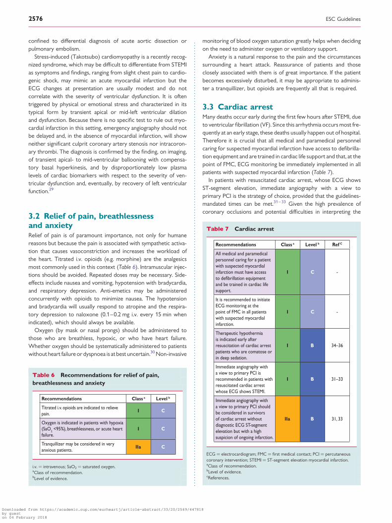
3.3 Cardiac arrestMany deaths occur early during the first few hours after STEMI, dueto ventricular fibrillation (VF). Since this arrhythmia occurs most fre-quently at an early stage, these deaths usually happen out of hospital.Therefore it is crucial that all medical and paramedical personnelcaring for suspected myocardial infarction have access to defibrilla-tion equipment and are trained in cardiac life support and that, at thepoint of FMC, ECG monitoring be immediately implemented in allpatients with suspected myocardial infarction (Table 7).
In patients with resuscitated cardiac arrest, whose ECG showsST-segment elevation, immediate angiography with a view toprimary PCI is the strategy of choice, provided that the guidelines-mandated times can be met.31– 33 Given the high prevalence ofcoronary occlusions and potential difficulties in interpreting the
Table 6 Recommendations for relief of pain,breathlessness and anxiety
Recommendations Class a Level b
Titrated i.v. opioids are indicated to relievepain.
I C
Oxygen is indicated in patients with hypoxia (SaO2 <95%), breathlessness, or acute heart failure.
I C
Tranquillizer may be considered in veryanxious patients.
IIa C
i.v. ¼ intravenous; SaO2 ¼ saturated oxygen.aClass of recommendation.bLevel of evidence.
Table 7 Cardiac arrest
Recommendations Class a Level b Ref C
All medical and paramedical personnel caring for a patient with suspected myocardial infarction must have access to defibrillation equipmentand be trained in cardiac life support.
I C -
It is recommended to initiate ECG monitoring at the point of FMC in all patients with suspected myocardial infarction.
I C -
Therapeutic hypothermiais indicated early after resuscitation of cardiac arrest patients who are comatose or in deep sedation.
I B 34–36
Immediate angiography with a view to primary PCI isrecommended in patients with resuscitated cardiac arrest whose ECG shows STEMI.
I B 31–33
Immediate angiography with a view to primary PCI shouldbe considered in survivors of cardiac arrest without diagnostic ECG ST-segmentelevation but with a high suspicion of ongoing infarction.
IIa B 31, 33
ECG ¼ electrocardiogram; FMC ¼ first medical contact; PCI ¼ percutaneouscoronary intervention; STEMI ¼ ST-segment elevation myocardial infarction.aClass of recommendation.bLevel of evidence.cReferences.
ESC Guidelines2576
Downloaded from https://academic.oup.com/eurheartj/article-abstract/33/20/2569/447818by gueston 04 February 2018
ECG in patients after cardiac arrest, immediate angiography shouldbe considered in survivors of cardiac arrest having a high index ofsuspicion of ongoing infarction (such as the presence of chest painbefore arrest, history of established CAD, and abnormal or uncer-tain ECG results).31,33 Additionally, there is evidence that survivorsof out-of-hospital cardiac arrest who are comatose have improvedneurological outcomes when cooling is provided early after resus-citation. Therefore, these patients should rapidly receive thera-peutic hypothermia.34– 36 The optimal sequence of cooling andprimary PCI in these patients is unclear.
The implementation of local/regional protocols to optimallymanage out-of-hospital cardiac arrest is pivotal to providingprompt cardiopulmonary resuscitation, early defibrillation (ifneeded), and effective advanced cardiac life support. Availability ofautomated external defibrillators is a key factor in increasing sur-vival. Prevention and improved treatment of out-of-hospitalcardiac arrest is key to reductions in mortality related to CAD.For a more detailed discussion of these issues, refer to the recentEuropean Resuscitation Council Guidelines for Resuscitation.37
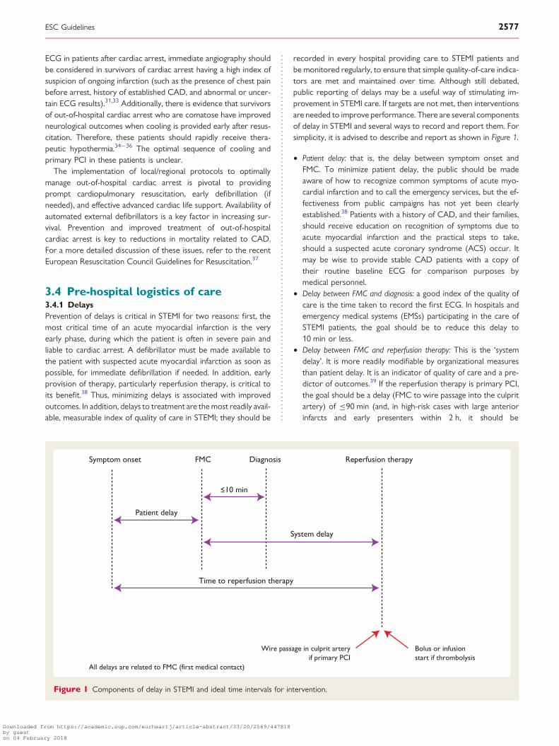
3.4 Pre-hospital logistics of care3.4.1 DelaysPrevention of delays is critical in STEMI for two reasons: first, themost critical time of an acute myocardial infarction is the veryearly phase, during which the patient is often in severe pain andliable to cardiac arrest. A defibrillator must be made available tothe patient with suspected acute myocardial infarction as soon aspossible, for immediate defibrillation if needed. In addition, earlyprovision of therapy, particularly reperfusion therapy, is critical toits benefit.38 Thus, minimizing delays is associated with improvedoutcomes. In addition, delays to treatment are the most readily avail-able, measurable index of quality of care in STEMI; they should be
recorded in every hospital providing care to STEMI patients andbe monitored regularly, to ensure that simple quality-of-care indica-tors are met and maintained over time. Although still debated,public reporting of delays may be a useful way of stimulating im-provement in STEMI care. If targets are not met, then interventionsare needed to improve performance. There are several componentsof delay in STEMI and several ways to record and report them. Forsimplicity, it is advised to describe and report as shown in Figure 1.
† Patient delay: that is, the delay between symptom onset andFMC. To minimize patient delay, the public should be madeaware of how to recognize common symptoms of acute myo-cardial infarction and to call the emergency services, but the ef-fectiveness from public campaigns has not yet been clearlyestablished.38 Patients with a history of CAD, and their families,should receive education on recognition of symptoms due toacute myocardial infarction and the practical steps to take,should a suspected acute coronary syndrome (ACS) occur. Itmay be wise to provide stable CAD patients with a copy oftheir routine baseline ECG for comparison purposes bymedical personnel.
† Delay between FMC and diagnosis: a good index of the quality ofcare is the time taken to record the first ECG. In hospitals andemergency medical systems (EMSs) participating in the care ofSTEMI patients, the goal should be to reduce this delay to10 min or less.
† Delay between FMC and reperfusion therapy: This is the ‘systemdelay’. It is more readily modifiable by organizational measuresthan patient delay. It is an indicator of quality of care and a pre-dictor of outcomes.39 If the reperfusion therapy is primary PCI,the goal should be a delay (FMC to wire passage into the culpritartery) of ≤90 min (and, in high-risk cases with large anteriorinfarcts and early presenters within 2 h, it should be
Symptom onset FMC Diagnosis Reperfusion therapy
10 min
Patient delay
System delay
Wire passage in culprit arteryif primary PCI
Bolus or infusionstart if thrombolysis
Time to reperfusion therapy
All delays are related to FMC (first medical contact)
Figure 1 Components of delay in STEMI and ideal time intervals for intervention.
ESC Guidelines 2577
Downloaded from https://academic.oup.com/eurheartj/article-abstract/33/20/2569/447818by gueston 04 February 2018

≤60 min).40,41 If the reperfusion therapy is fibrinolysis, the goalis to reduce this delay (FMC to needle) to ≤30 min.
† In PCI-capable hospitals, the goal should be to achieve a‘door-to-balloon’ delay ≤60 min between presentation in the hos-pital and primary PCI (defined as wire passage into the culpritartery). This delay reflects the organization and performanceof the PCI-capable hospital.
† From the patient’s perspective, the delay between symptom onsetand provision of reperfusion therapy (either starting fibrinolysis orpassing a wire through the culprit vessel) is possibly the mostimportant, since it reflects total ischaemic time. It should bereduced as much as possible.
3.4.2 Emergency medical systemAn EMS with an easily remembered and well publicized uniquetelephone number for medical emergencies is important inorder to avoid transportation delays. A teleconsultationbetween the EMS and a reference cardiology centre is ideal,but is only available in a limited number of countries. Therefore,a well-trained EMS and an updated and shared, written STEMImanagement protocol are critically important. Although the useof an EMS decreases the delay and is the preferred mode ofinitial care for patients with suspected STEMI, it is under-utilizedin many countries and, not infrequently, patients self-present tothe emergency department. The ambulance service has a criticalrole in the management of acute myocardial infarction and shouldbe considered not only a mode of transport but also a place forinitial diagnosis, triage and treatment. Pre-hospital diagnosis, triageand initial emergency treatment in the ambulance has beenshown to be associated with greater use of reperfusion therapies,reduced delays and improved clinical outcomes.39,42 In addition,EMS transportation allows for the diagnosis and treatment ofcardiac arrest. The quality of the care given depends on the train-ing of the staff concerned. All ambulance personnel should betrained to recognize the symptoms of an AMI, administeroxygen, relieve pain and provide basic life support (Table 8).All emergency ambulances should be equipped with ECG recor-ders, defibrillators, and at least one person on board trained inadvanced life support. There is evidence that properly trainedparamedical personnel can effectively identify AMI and providetimely reperfusion, and that physician-manned ambulances—which are available in only a few countries—are not necessaryfor effective pre-hospital management of AMI.43 Paramedicstrained to administer thrombolytics do so safely and effectively.Since pre-hospital thrombolysis is an attractive therapeuticoption in patients presenting early after symptom onset, especial-ly when transfer time is prolonged,40,44,45 ongoing training ofparamedics to undertake these functions is recommended, evenin the era of primary PCI. In specific regions, air ambulancesystems further reduce transportation delays and improve out-comes.46 Ambulance staff should be able to record an ECG fordiagnostic purposes and either interpret it or transmit it, sothat it can be reviewed by experienced staff in a coronary careunit or elsewhere. The recording, interpretation and, sometimes,teletransmission of an ECG before hospital admission can greatlyaccelerate in-hospital management and increase the probability oftimely reperfusion therapy.
3.4.3 NetworksOptimal treatment of STEMI should be based on the implementa-tion of networks between hospitals with various levels of technol-ogy, connected by an efficient ambulance service. The goal of thesenetworks is to provide optimal care while minimizing delays, inorder to improve clinical outcomes. Cardiologists should activelycollaborate with all stakeholders, particularly emergency physi-cians, in establishing such networks. The main features of such anetwork are:
† Clear definition of geographical areas of responsibility† Shared protocols, based on risk stratification and transportation
by trained paramedic staff in appropriately equipped ambulancesor helicopters
† Pre-hospital triage of STEMI patients to the appropriate institu-tions, bypassing non-PCI hospitals whenever primary PCI can beimplemented within the recommended time limits
† On arrival at the appropriate hospital, the patient should imme-diately be taken to the catheterization laboratory, bypassing theemergency department
† Patients presenting to a non-PCI-capable hospital and awaitingtransportation for primary or rescue PCI must be attended inan appropriately monitored and staffed area
† If the diagnosis of STEMI has not been made by the ambulancecrew, and the ambulance arrives at a non-PCI-capable hospital,the ambulance should await the diagnosis and, if STEMI is con-firmed, should continue to a PCI-capable hospital.
To maximize staff experience, primary PCI centres should performthe procedure systematically on a twenty-four hours, seven days aweek (24/7) basis for all STEMI patients. Other models, althoughnot ideal, may include weekly or daily rotation of primary PCIcentres or multiple primary PCI centres in the same region. Hos-pitals that cannot offer a 24/7 service for primary PCI should beallowed to perform primary PCI in patients already admitted foranother reason, who develop STEMI during their hospital stay.These hospitals should, however, be discouraged from initiating aservice limited to daytime- or within-hours primary PCI, sincethis generates confusion with the EMS operators and is unlikelyto match the door-to-balloon time and quality of intervention offocussed 24/7 true-primary PCI centres. The current catchmentpopulation for network systems in European countries that offerprimary PCI to the majority of their population is 0.3–1.0million.6 In small service areas the experience may be suboptimal,due to an insufficient number of STEMI patients. However, theoptimal size of the catchment area is not clear. Geographicalareas where the expected transfer time to the primary PCIcentre makes it impossible to achieve the maximal allowabledelays indicated in the recommendations below (see section3.4.6) should develop systems for rapid thrombolysis, preferablyin-ambulance/out-of-hospital, with subsequent immediate transferto primary PCI centres.
Such networks reduce treatment delays and increase the pro-portion of patients receiving reperfusion.47– 49 In each network,the quality of care, time delays and patient outcomes should bemeasured and compared at regular intervals and appropriate mea-sures taken to bring about improvement. In a large survey in theUSA, several strategies were associated with shorter delays
ESC Guidelines2578
Downloaded from https://academic.oup.com/eurheartj/article-abstract/33/20/2569/447818by gueston 04 February 2018

before primary PCI, including the ability to activate the catheteriza-tion laboratory by a single call, preferably while the patient is enroute to hospital, expecting laboratory staff to arrive in the cath-eterization laboratory within 20 min of being paged, having a cardi-ologist on site, and using real-time data feedback between theupstream care and the catheterization laboratory.50 The most ef-fective strategies for increasing the proportion of patients receivingeffective reperfusion and reduce delays to primary PCI may differin other healthcare systems. In order to address the issue ofaccess to primary PCI and effective implementation of networksacross Europe,6 the ESC working group on acute cardiac care,the European Association of Percutaneous Cardiovascular Inter-ventions (EAPCI), and EuroPCR, have joined forces in the Stentfor Life initiative, to improve access to timely, effective primaryPCI through focussed implementation programmes, tailored toeach specific national healthcare setting and attempting to learnfrom success.51 Experience acquired through this initiative,across various European systems of care, is published regularlyand provides tips and resources to increase and improve the imple-mentation of primary PCI (www.stentforlife.com).52
3.4.4 General practitionersIn some countries, general practitioners play a major role in theearly care of acute myocardial infarction and are often the firstto be contacted by patients. If general practitioners respondquickly they can be very effective, since they usually know thepatient and can perform and interpret the ECG. Their first task
after the ECG diagnosis should be to alert the EMS. But they arealso able to administer opioids and antithrombotic drugs (includingfibrinolytics if that is the management strategy), and can undertakedefibrillation if needed. In most settings, however, consultationwith a general practitioner—instead of a direct call to theEMS—increases pre-hospital delay. Therefore, in general, thepublic should be educated to call the EMS, rather thanthe primary care physician, for symptoms suggestive of myocardialinfarction.
3.4.5 Admission proceduresThe processing of patients once they arrive in hospital must bespeedy, particularly with regard to the diagnosis and administrationof fibrinolytic agents or the performance of primary PCI, if indi-cated. Candidates for primary PCI should, as often as possible,be admitted directly to the catheterization laboratory, bypassingthe emergency department and/or intensive coronary care unit,while patient candidates for fibrinolysis must be treated directlyin the pre-hospital setting, in the emergency department or inthe coronary care unit.53,54
3.4.6 LogisticsIn the optimal situation (Figure 2), the patient calls a central EMSnumber for help as soon as possible after the onset of chestpain. The EMS dispatches a fully equipped ambulance with person-nel trained to perform and interpret a 12-lead ECG. Once theECG reveals ST-segment elevation or new (or presumed new)
Table 8 Logistics of pre-hospital care
Recommendations Class a Level b Ref C
Ambulance teams must be trained and equipped to identify STEMI (with use of ECG recorders and telemetry asnecessary) and administer initial therapy, including thrombolysis where applicable.
I B 43
The prehospital management of STEMI patients must be based on regional networks designed to deliver reperfusiontherapy expeditiously and effectively, with efforts made to make primary PCI available to as many patients as possible.
I B 47
Primary PCI-capable centres must deliver a 24/7 service and be able to start primary PCI as soon as possible butalways within 60 min from the initial call.
I B 6, 52, 55
All hospitals and EMSs participating in the care of patients with STEMI must record and monitor delay times and workto achieve and maintain the following quality targets: • first medical contact to first ECG ≤10 min; • first medical contact to reperfusion therapy; • for fibrinolysis ≤30 min; • for primary PCI ≤90 min (≤60 min if the patient presents within 120 min of symptom onset or directly to a PCI-
capable hospital).
I B 56, 57
All EMSs, emergency departments, and coronary care units must have a written updated STEMI management protocol,preferably shared within geographic networks.
I C
Patients presenting to a non-PCI-capable hospital and awaiting transportation for primary or rescue PCI must beattended in an appropriately monitored area.
I C
Patients transferred to a PCI-capable centre for primary PCI should bypass the emergency department and betransferred directly to the catheterization laboratory.
IIa B 41, 50, 58
ECG ¼ electrocardiogram; EMS ¼ emergency medical system; PCI ¼ percutaneous coronary intervention; 24/7 ¼ 24 hours a day, seven days a week; STEMI ¼ ST-segmentelevation myocardial infarction.aClass of recommendation.bLevel of evidence.cReferences.
ESC Guidelines 2579
Downloaded from https://academic.oup.com/eurheartj/article-abstract/33/20/2569/447818by gueston 04 February 2018
LBBB, the nearest PCI hospital is informed of the expected time ofpatient arrival. During the ambulance transfer, the catheterizationlaboratory is prepared and staff summoned, if necessary, allowingdirect transfer of the patient to the catheterization laboratorytable (bypassing the emergency department and coronary careunit). In cases where the diagnostic ECG has been done elsewhere(e.g. in a non-PCI hospital, at a physician’s office, etc.), the EMS iscalled for transfer and the above chain followed. This scenario isbest accomplished in a regional network with one high-volumePCI centre, several surrounding non-PCI hospitals and a single re-gional EMS. Such regional networks should have predefined man-agement protocols for STEMI patients.
3.5 Reperfusion therapy3.5.1 Restoring coronary flow and myocardial tissuereperfusionFor patients with the clinical presentation of STEMI within 12 hof symptom onset and with persistent ST-segment elevation ornew or presumed new LBBB, early mechanical (PCI) or pharma-cological reperfusion should be performed as early as possible(Table 9).
There is general agreement that reperfusion therapy should beconsidered if there is clinical and/or electrocardiographic evidenceof ongoing ischaemia, even if, according to the patient, symptomsstarted .12 h before as the exact onset of symptoms is oftenunclear, or when the pain and ECG changes have been stuttering.59
There is, however, no consensus as to whether PCI is also bene-ficial in patients presenting .12 h from symptom onset in theabsence of clinical and/or electrocardiographic evidence ofongoing ischaemia. In such asymptomatic late-comers, a small (n ¼347) randomized study has shown myocardial salvage and improved4-year survival resulting from primary PCI, compared with conserva-tive treatment alone, in patients without persistent symptoms 12–48 h after symptom onset.60,61 However, in stable patients with per-sistent occlusion of the infarct-related artery, the large (n ¼ 2166)Occluded Artery Trial (OAT) revealed no clinical benefit fromroutine coronary intervention with medical management,62,63
beyond that from medical management alone, when the occlusionwas identified 3–28 days after acute myocardial infarction, includingin the subgroup of 331 patients randomized between 24 and 72 hafter onset of infarction.64 A meta-analysis of trials, testingwhether late re-canalization of an occluded infarct artery is benefi-cial, provided results consistent with those from OAT.51
Yes No
No
Preferably<60 min
Immediately
Preferably 3–24 h
Preferably≤90 min(≤60 min in early presenters) Preferably
≤30 min
aThe time point the diagnosis is confirmed with patienthistory and ECG ideally within 10 min from the firstmedical contact (FMC).All delays are related to FMC (first medical contact).
Immediate transferto PCI center
Immediate transferto PCI center
Yes
STEMI diagnosisa
Primary-PCI capable center
Primary-PCI
Coronary angiography
Rescue PCI
EMS or non primary-PCIcapable center
Immediatefibrinolysis
Successfulfibrinolysis?
PCI possible <120 min?
Cath = catheterization laboratory; EMS = emergency medical system; FMC = first medical contact; PCI = percutaneous coronary intervention; STEMI = ST-segment elevation myocardial infarction.
Figure 2 Prehospital and in-hospital management, and reperfusion strategies within 24 h of FMC (adapted from Wijns et al.).4
ESC Guidelines2580
Downloaded from https://academic.oup.com/eurheartj/article-abstract/33/20/2569/447818by gueston 04 February 2018

3.5.2 Selection of a strategy for reperfusionPrimary PCI—defined as an emergent percutaneous catheterintervention in the setting of STEMI, without previous fibrinolytictreatment—is the preferred reperfusion strategy in patients withSTEMI, provided it can be performed expeditiously (i.e. withinguideline-mandated times), by an experienced team, and regardlessof whether the patient presents to a PCI-capable hospital(Figure 1). If FMC is via an EMS or at a non-PCI-capable centre,transfer via the EMS to the catheterization laboratory for PCIshould be implemented immediately. An experienced teamincludes not only interventional cardiologists, but also skilledsupport staff. This means that only hospitals with an establishedinterventional cardiology programme (available 24/7) should useprimary PCI as a routine treatment. Lower mortality ratesamong patients undergoing primary PCI are observed in centreswith a high volume of PCI procedures. Primary PCI is effective insecuring and maintaining coronary artery patency and avoidssome of the bleeding risks of fibrinolysis. Randomized clinicaltrials comparing timely primary PCI with in-hospital fibrinolytictherapy in high-volume, experienced centres have repeatedlyshown that primary PCI is superior to hospital fibrinolysis.68– 71
(In these trials there was no routine follow-up rescue PCI or angi-ography.) In settings where primary PCI cannot be performedwithin 120 min of FMC by an experienced team, fibrinolysis
should be considered, particularly if it can be given pre-hospital(e.g. in the ambulance)45,72,73 and within the first 120 min ofsymptom onset (Figure 2).40,74 It should be followed by consider-ation of rescue PCI or routine angiography.
Both randomized studies and registries have indicated that longdelays to primary PCI are associated with worse clinical outcomes.Time delay to reperfusion is defined in section 3.4.1, above. The‘PCI-related delay’ is the theoretical difference between the timeof FMC to balloon inflation, minus the time from FMC to start of fi-brinolytic therapy (i.e. ‘door-to-balloon’ minus ‘door-to-needle’).The extent to which the PCI-related delay diminishes the advantagesof PCI over fibrinolysis has been the subject of many analyses anddebates. Because no specifically designed study has addressed thisissue, caution is needed when interpreting the results of thesepost-hoc analyses. From randomized trials, it was calculated that thePCI-related delay that may mitigate the benefit of mechanical inter-vention varies between 60 and 110 min. In another analysis of thesetrials, a benefit of primary PCI over fibrinolytic therapy was calcu-lated, up to a PCI-related delay of 120 min.66 In 192 509 patientsincluded in the US National Registry of Myocardial Infarction(NRMI) 2–4 registry,41 the mean PCI-related time delay, where mor-tality rates of the two reperfusion strategies were comparable, wascalculated at 114 min. This study also indicated that this delayvaried considerably according to age, symptom duration andinfarct location: from ,1 h for an anterior infarction in a patient,65 years of age presenting ,2 h after symptom onset, to almost3 h for a non-anterior infarction in a patient .65 years of age pre-senting .2 h after symptom onset. Although these results werederived from a post-hoc analysis of a registry and reported delaysare sometimes inaccurate, this study suggests that an individualized,rather than a uniform, approach for selecting the optimal reperfusionmodality could be more appropriate when PCI cannot be performed
Table 9 Recommendations for reperfusion therapy
Recommendations Class a Level b Ref C
Reperfusion therapy is indicated in all patients with symptoms of <12 h duration and persistent ST-segment elevation or (presumed) new LBBB.
I A 65, 66
Reperfusion therapy (preferably primary PCI) is indicated if there is evidence of ongoing ischaemia, even if symptoms may have started >12 h beforehand or if pain and ECG changes have been stuttering.
I C 67
Reperfusion therapy with primary PCI may be considered in stable patients presenting 12–24 h after symptom onset.
IIb B 60, 61
Routine PCI of a totally occluded artery >24 h after symptom onset in stable patients without signs of ischaemia (regardless of whether fibrinolysis was given or not) is not recommended.
III A 62–64
ECG ¼ electrocardiogram; i.v. ¼ intravenous; LBBB ¼ left bundle branch block;PCI ¼ percutaneous coronary intervention.aClass of recommendation.bLevel of evidence.cReferences.
Table 10 A summary of important delays andtreatment goals in the management of acuteST-segment elevation myocardial infarction
Delay Target
Preferred for FMC to ECG and diagnosis ≤10 min
Preferred for FMC to fibrinolysis (‘FMC to needle’)
≤30 min
Preferred for FMC to primary PCI (‘door to balloon’) in primary PCI hospitals
≤60 min
Preferred for FMC to primary PCI ≤90 min (≤60 min if early presenter with large area at risk)
Acceptable for primary PCI rather than fibrinolysis
≤120 min(≤90 min if early presenter with large area at risk)if this target cannot be met, consider fibrinolysis.
Preferred for successful fibrinolysis to angiography
3–24 h
FMC ¼ first medical contact; PCI ¼ percutaneous coronary intervention.
ESC Guidelines 2581
Downloaded from https://academic.oup.com/eurheartj/article-abstract/33/20/2569/447818by gueston 04 February 2018

expeditiously. Taking into account the studies and registries men-tioned above, a target for quality assessment is that primary PCI(wire passage) should be performed within 90 min after FMC in allcases. In patients presenting early, with a large amount of myocar-dium at risk, the delay should be shorter (,60 min). In patients pre-senting directly in a PCI-capable hospital, the goal should also be toachieve primary PCI within 60 min of FMC. Although no specificstudies have been performed, a maximum delay of only 90 minafter FMC seems a reasonable goal in these patients. Note thatthese target delays for implementation of primary PCI are qualityindicators and that they differ from the maximal PCI-related delayof 120 min, which is useful in selecting primary PCI over immediatethrombolysis as the preferred mode of reperfusion (Table 10).
3.5.3 Primary percutaneous coronary intervention3.5.3.1 Procedural aspects of primary percutaneous coronaryintervention (Table 11)Approximately 50% of STEMI patients have significant multivesseldisease. Only the infarct-related artery should be treated duringthe initial intervention. There is no current evidence to supportemergency intervention in non-infarct-related lesions.75,76 Theonly exceptions, when multivessel PCI during acute STEMI is jus-tified, are in patients with cardiogenic shock in the presence ofmultiple, truly critical (≥90% diameter) stenoses or highly un-stable lesions (angiographic signs of possible thrombus orlesion disruption), and if there is persistent ischaemia after PCIof the supposed culprit lesion. However, in patients with multi-vessel disease and cardiogenic shock, non-culprit lesionswithout critical stenoses should not routinely be stented.77 Seealso section 3.5.4.9.
Because of the need for potent antithrombotic and antiplateletagents, bleeding is more frequent when PCI is performed duringACS (and STEMI in particular) when compared with bleeding oc-curring during an elective procedure. Use of drugs with a morepotent antithrombotic effect is often accompanied by an increasein the risk of bleeding, mostly related to the arterial puncturesite. The radial approach has been shown to reduce the inci-dence of acute bleeding events, especially in ACS; in the RadIalvs. femorAL (RIVAL) access for coronary intervention trial,using radial rather than femoral access actually reduced mortalityin the subset of STEMI patients.78 Similar findings were alsoobserved in the RIFLE STEACS trial.79 In RIVAL there was,however, an interaction between benefit of the radial accessroute and operator experience, suggesting that the benefit ofradial access over femoral depends upon the radial expertiseof operators.
In primary PCI, drug-eluting stents (DES) reduce the risk ofrepeated target vessel revascularization, compared with bare-metalstents (BMS).80 There have been concerns about increased risks ofvery late stent thrombosis and reinfarction with DES, comparedwith BMS.80 However, use of DES has not been associated withan increased risk of death, myocardial infarction or stent throm-bosis on long-term follow up.82 An issue with the routine use ofDES in this setting is that it is often difficult to determine reliablythe ability of patients to comply with or tolerate the protracteduse of dual antiplatelet therapy (DAPT). Whether newer genera-tions of DES provide improved clinical outcomes—comparedwith older generation DES or BMS—following primary PCI iscurrently being tested.
Table 11 Primary PCI: indications and procedural aspects
Recommendations Class a Levelb Ref C
Indications for primary PCI
Primary PCI is the recommended reperfusion therapy over fibrinolysis if performed by an experienced team within 120 min of FMC.
I A 69, 99
Primary PCI is indicated for patients with severe acute heart failure or cardiogenic shock, unless the expected PCI related delay is excessive and the patient presents early after symptom onset.
I B 100
Procedural aspects of primary PCI
Stenting is recommended (over balloon angioplasty alone) for primary PCI. I A 101, 102
Primary PCI should be limited to the culprit vessel with the exception of cardiogenic shock and persistent ischaemia after PCI of the supposed culprit lesion.
IIa B75, 103–
105
If performed by an experienced radial operator, radial access should be preferred over femoral access. IIa B 78, 79
If the patient has no contraindications to prolonged DAPT (indication for oral anticoagulation, or estimated high long-term bleeding risk) and is likely to be compliant, DES should be preferred over BMS.
IIa A80, 82, 106,
107
Routine thrombus aspiration should be considered. IIa B 83–85
Routine use of distal protection devices is not recommended. III C 86, 108
Routine use of IABP (in patients without shock) is not recommended. III A 97, 98
BMS ¼ bare-metal stent; DAPT ¼ dual antiplatelet therapy; DES ¼ drug-eluting stent; IABP ¼ intra-aortic balloon pump; PCI ¼ percutaneous coronary intervention.aClass of recommendation.bLevel of evidence.cReferences.
ESC Guidelines2582
Downloaded from https://academic.oup.com/eurheartj/article-abstract/33/20/2569/447818by gueston 04 February 2018
One single-centre randomized trial, the Thrombus Aspirationduring Percutaneous coronary intervention in Acute myocardialinfarction (TAPAS) trial,83 showed improvement in indices of myo-cardial reperfusion (ST-segment resolution and myocardial blush)from routine use of manual thrombus aspiration before a balloonor a stent is introduced into the coronary artery. One-year follow-up from that trial found a reduction in mortality with thrombus as-piration as a secondary endpoint.84 A meta-analysis of TAPAS andseveral smaller trials found similar results.85 Mechanical thrombec-tomy or embolic protection devices have not been found toprovide similar benefits. However, the difference in clinical impactbetween the various models is still unclear.86 In the recent INtracor-onary abciximab inFUsion and aSpiration thrombEctomy in patientsundergoing percutaneous coronary intervention for Anterior STsegment elevation Myocardial Infarction (INFUSE-AMI) randomizedtrial, thrombus aspiration did not affect infarct size.87 Several large,randomized trials have been initiated to attempt to confirm theresults of TAPAS.88,89
Operators performing primary PCIs in STEMI should be awareof the importance of selecting an appropriate stent size. Mostpatients with STEMI have some degree of coronary spasm and,thus, intracoronary administration of nitrates is recommendedbefore starting the coronary angiographic sequence used forstent size selection. The presence of thrombus can also lead tostent under-sizing (or otherwise suboptimal deployment), whichis a frequent cause of re-stenosis or stent thrombosis in real-lifepractice.
Preliminary clinical studies have explored the value of myocar-dial pre- and post-conditioning to improve myocardial salvage.A small, randomized trial tested the value of remote conditioningusing intermittent arm ischaemia through four cycles of 5 min infla-tions and deflation of a blood pressure cuff.90 This was associatedwith improvement in surrogate markers of myocardial salvage,measured by myocardial perfusion imaging at 30 days. It isunknown whether this is associated with clinical benefits. Therole of post-conditioning has been explored by small trials, usingeither repeated balloon inflations or cyclosporine infusions. Theresults are conflicting.91 –95 Given the preliminary nature of thesefindings and the small size of the trials, confirmation of a clinicalbenefit of myocardial pre- and post-conditioning by ongoing ran-domized trials is warranted before these procedures can berecommended in routine clinical practice.
The Counterpulsation to Reduce Infarct Size Pre-PCI-Acute Myocar-dial Infarction (CRISP AMI) trial showed no benefit from aroutine intra-aortic balloon pump (IABP) in anterior myocardial in-farction without shock,97 and did show increased bleeding, which isconsistent with data available regarding the role of IABPs inpatients with acute myocardial infarction without cardiogenicshock.(98)
3.5.3.2 Periprocedural pharmacotherapy (Table 12)Patients undergoing primary PCI should receive a combination ofDAPT with aspirin and an adenosine diphosphate (ADP) receptorblocker, as early as possible before angiography, and a parenteralanticoagulant. No trials to date have evaluated the commencement
of DAPT prior to hospital admission, rather than in hospital, nor itsuse before, rather than during, angiography in the setting of STEMI,but this is common practice in Europe and is consistent with thepharmacokinetic data for oral antithrombotic agents, suggestingthat the earliest administration would be preferable to achieveearly efficacy.
Aspirin should preferably be given orally (preferably 150–300 mg) including chewing, to ensure complete inhibition ofTXA2-dependent platelet aggregation, but may be given intraven-ously in patients who are unable to swallow. There is little clinicaldata on the optimal i.v. dosage, but pharmacological data suggestthat a lower dose range than orally may avoid inhibition of prosta-cyclin and therefore a bolus dose range of 80–150 mg should bepreferred for i.v. aspirin.
The preferred ADP-receptor blockers are prasugrel [60 mg peros (p.o.) loading dose, 10 mg maintenance dose] or ticagrelor[180 mg p.o. loading dose, 90 mg maintenance dose bis in die(b.i.d)]; these drugs have a more rapid onset of action andgreater potency and have proved superior to clopidogrel in largeoutcome trials.109,110 In the TRial to assess Improvement in Thera-peutic Outcomes by optimizing platelet inhibitioN–ThrombolysisIn Myocardial Infarction 38 (TRITON–TIMI 38), prasugrelreduced the composite primary endpoint (cardiovascular death,non-fatal MI, or stroke) in clopidogrel-naıve patients undergoingPCI, either primary or secondary PCI for STEMI, or moderate-to high-risk non-ST-segment elevation acute coronary syndromes(NSTE-ACS) once coronary angiography had been performed.109
In the whole cohort, there was a significant increase in the rateof non-CABG-related TIMI major bleeding. In the subset ofpatients with STEMI undergoing primary or secondary PCI, thebenefit was consistent, without significant increase innon-CABG-related bleeding risk.111 Prasugrel is contraindicatedin patients with prior stroke/transient ischaemic attack (TIA). Itsuse is generally not recommended in patients aged ≥75 years orin patients with lower body weight (,60 kg) as it was not asso-ciated with net clinical benefit in these subsets. The Europeanlabel indicates that, if used in these patients, a similar loadingdose but a reduced maintenance dose of 5 mg should be consid-ered, but no outcome data are available with this dose and thereare alternative ADP receptor blockers in this setting.112 In thePLATelet inhibition and patient Outcomes (PLATO) trial, ticagrelorreduced the composite primary endpoint (cardiovascular death,non-fatal MI, or stroke) and also reduced cardiovascular mortalityin clopidogrel naıve or pretreated patients with either STEMI(planned for primary PCI) or moderate-to-high risk NSTE-ACS(planned for either conservative or invasive management).109,110
Although there was no significant difference in overall PLATO-defined major bleeding rates between the clopidogrel and ticagre-lor groups, PLATO-defined and TIMI-defined major bleeding thatwas unrelated to CABG surgery was increased with ticagrelor. Inthe subset of patients with STEMI, the benefit was consistent.113
Ticagrelor may cause transient dyspnoea at the onset of therapy,which is not associated with morphological or functional lung ab-normalities, and which rarely leads to discontinuation.114 InPLATO, patients experiencing dyspnoea had a mortality benefit
ESC Guidelines 2583
Downloaded from https://academic.oup.com/eurheartj/article-abstract/33/20/2569/447818by gueston 04 February 2018

of ticagrelor consistent with the overall trial population. Ticagrelormay also be associated with asymptomatic bradycardia in the firstweek of therapy. None of the more potent agents (prasugrel orticagrelor) should be used in patients with a previous haemorrhagicstroke or in patients with a moderate-to-severe liver disease.When neither of these agents is available (or if they are contrain-dicated), clopidogrel 600 mg p.o. should be given instead.115 Clopi-dogrel has not been evaluated against placebo in anylarge-outcome study in the setting of primary PCI, but a higherregimen of 600 mg loading dose/150 mg maintenance dose in thefirst week was superior to the 300/75 mg regimen in the subsetof patients undergoing PCI in the Optimal Antiplatelet Strategy forInterventions (OASIS) 7 trial,115 and use of high clopidogrelloading doses has been demonstrated to achieve more rapidinhibition of the ADP receptor. This is consistent with the
pharmacokinetics of clopidogrel, a pro-drug, which requires exten-sive metabolism before being active and therefore should be givenin higher doses and as early as possible for it to exert its action inthe emergency setting of primary PCI. Furthermore, pre-treatmentwith high dose clopidogrel was superior to in-laboratory treatmentin observational studies.116,117 All ADP receptor blockers shouldbe used with caution in patients at high risk of bleeding or withsignificant anaemia.
Anticoagulant options for primary PCI include unfractionatedheparin (UFH), enoxaparin and bivalirudin. Use of fondaparinuxin the context of primary PCI was associated with potentialharm in the OASIS 6 trial and is therefore not recommended.118
There have been no placebo-controlled trials evaluating UFH inprimary PCI but there is a large body of experience with thisagent. Dosage should follow standard recommendations for PCI
Table 12 Periprocedural antithrombotic medication in primary percutaneous coronary intervention
Recommendations Class a Levelb Ref C
Antiplatelet therapy
Aspirin oral or i.v. (if unable to swallow) is recommended I B 133, 134
An ADP-receptor blocker is recommended in addition to aspirin. Options are: I A 135, 136
• Prasugrel in clopidogrel-naive patients, if no history of prior stroke/TIA, age <75 years. I B 109
• Ticagrelor. I B 110
• Clopidogrel, preferably when prasugrel or ticagrelor are either not available or contraindicated. I C -
GP IIb/IIIa inhibitors should be considered for bailout therapy if there is angiographic evidence of massive thrombus, slow or no-reflow or a thrombotic complication.
IIa C -
Routine use of a GP IIb/IIIa inhibitor as an adjunct to primary PCI performed with unfractionated heparin may be considered in patients without contraindications.
IIb B 137–141
Upstream use of a GP IIb/IIIa inhibitor (vs. in-lab use) may be considered in high-risk patients undergoing transfer for primary PCI.
IIb B127, 128, 137, 142
Options for GP IIb/IIIa inhibitors are (with LoE for each agent):
• Abciximab A 137
• Eptifibatide (with double bolus) B 138, 139
• Tirofiban (with a high bolus dose) B 140, 141
Anticoagulants
An injectable anticoagulant must be used in primary PCI. I C -
Bivalirudin (with use of GP IIb/IIIa blocker restricted to bailout) is recommended over unfractionated heparin and a GP IIb/IIIa blocker.
I B 124
Enoxaparin (with or without routine GP IIb/IIIa blocker) may be preferred over unfractionated heparin. IIb B 122
Unfractionated heparin with or without routine GP IIb/IIIa blocker must be used in patients not receiving bivalirudin or enoxaparin.
I C 1
Fondaparinux is not recommended for primary PCI. III B 118
The use of fibrinolysis before planned primary PCI is not recommended. III A 127, 143
ADP ¼ adenosine diphosphate; GP ¼ glycoprotein; i.v. ¼ intravenous; lab ¼ catheterization laboratory; PCI ¼ percutaneous coronary intervention; TIA ¼ transient ischaemicattack; UFH ¼ unfractionated heparin.aClass of recommendation.bLevel of evidence.cReferences.
ESC Guidelines2584
Downloaded from https://academic.oup.com/eurheartj/article-abstract/33/20/2569/447818by gueston 04 February 2018

[i.e. initial bolus 70–100 U/kg when no glycoprotein (GP) IIb/IIIainhibitor is planned or 50–60 U/kg when the use of GP IIb/IIIa inhi-bitors is expected]. There are no solid data recommending the useof activated clotting time to tailor dose or monitor UFH and, ifactivated clotting time is used, it should not delay recanalizationof the infarct-related artery. Enoxaparin (0.5 mg/kg i.v. followedby s.c. treatment) was suggested by several non-randomizedstudies to provide benefit over UFH in primary PCI.119– 121 Itwas compared with UFH in one randomized open label trial, theAcute myocardial infarction Treated with primary angioplasty andinTravenous enOxaparin or unfractionated heparin to Lower is-chaemic and bleeding events at short- and Long-term follow-up(ATOLL) trial. The primary composite endpoint of 30-day death,complication of myocardial infarction, procedural failure andmajor bleeding was not significantly reduced (17% reduction,P ¼ 0.063), but there were reductions in the composite main sec-ondary endpoint of death, recurrent myocardial infarction or ACSor urgent revascularization, and in other secondary compositeendpoints such as death, or resuscitated cardiac arrest anddeath, or complication of myocardial infarction. Importantly,there was no indication of increased bleeding from use of enoxa-parin over UFH.122 Based on these considerations and on theconsiderable clinical experience with enoxaparin in other PCIsettings,109– 111 enoxaparin may be preferred over UFH.
One large open-label trial demonstrated the superiority of biva-lirudin over the combination of UFH + GP IIb/IIIa inhibitor,123 abenefit driven by a marked reduction in bleeding, associated withan initial increase in stent thrombosis, which disappeared after30 days.124 Importantly, that study reported a reduction inall-cause and cardiovascular mortality at 30 days, which was main-tained up to 3 years.82 A large fraction of patients in the Harmon-izing Outcomes with RevascularIZatiON and Stents in AcuteMyocardial (HORIZONS–AMI) trial, received prerandomizationUFH and approximately 10% bailout GP IIb/IIIa blockers. This isnoteworthy because the interpretation of the trial result is slightlyconfounded by an interaction between prerandomization use ofUFH, use of a 600mg loading dose of clopidogrel and reducedrisk of stent thrombosis.125
Several trials, performed before the routine use of DAPT,mostly using abciximab, had documented clinical benefits of GPIIb/IIIa inhibitors as adjuncts to primary PCI performed withUFH.126 The Facilitated INtervention with Enhanced reperfusionSpeed to Stop Events (FINESSE) trial127 found that routine up-stream use of abciximab before primary PCI did not yield clinicalbenefit but increased bleeding risk, compared with routine use inthe catheterization laboratory, suggesting that, for patients goingon to primary PCI, there does not appear to be any appreciablebenefit and only harm in starting GP IIb/IIIa inhibitors in the pre-hospital setting. A post-hoc subset analysis of the FINESSE trial, fo-cussing on patients presenting within 4 h of symptom onset tonon-PCI hospitals and requiring transfer, suggested they mightderive a survival benefit from use of abciximab.128 More recently,the ONgoing Tirofiban in Myocardial infarction Evaluation 2(ON-TIME 2) trial129 found an improvement in surrogate
markers of reperfusion from the use of tirofiban started duringthe pre-hospital phase, upstream of primary PCI, and continuedfor up to 18 h after the procedure (compared to only provisionaluse (i.e. not systematic use) in the catheterization laboratory).There was also a reduction in the composite secondary endpointof death in recurrent myocardial infarction in urgent targetvessel revascularization and thrombotic bailout. Finally, in thelarge HORIZONS–AMI trial,124 there was no clear benefit fromusing a combination of GP IIb/IIIa inhibitor +UFH, compared tobivalirudin (with a substantial fraction of patients receiving UFHbefore randomization) and the Bavarian Reperfusion AlternativesEvaluation-3 (BRAVE-3) trial did not find evidence of a reductionin infarct size from treatment with abciximab in primary PCIpatients treated with 600 mg of clopidogrel.130 Therefore, thereis no definitive answer regarding the current role of routine useof GP IIb/IIIa inhibitors in primary PCI in the era of potentDAPT, particularly when prasugrel or ticagrelor is used, and thevalue of starting upstream of the catheterization laboratory is, atbest, uncertain. Using GP IIb/IIIa inhibitors as bailout therapy inthe event of angiographic evidence of large thrombus, slow orno-reflow and other thrombotic complications is reasonable, al-though it has not been tested in a randomized trial. In conclusion,the existing data suggest that, if bivalirudin is chosen as the anti-coagulant, there is no benefit of routine addition of GP IIb/IIIablockers and a strategy of bivalirudin alone (with provisionalbailout use of GP IIb/IIIa blockers) leads to lower bleeding ratesand reduced mortality. If UFH or enoxaparin is chosen as the anti-coagulant, the role of routine—as opposed to ‘bailout’—use of GPIIb/IIIa blockers remains debatable.
Intracoronary (i.c.) rather than i.v. administration of GP IIb/IIIainhibitors has been tested in several small studies and is associatedwith some benefits.131 The Intracoronary abciximab iNFUsion andaspiration thrombectomy for anterior ST-segment ElevAtion Myo-cardial Infarction (INFUSE-AMI) trial87 randomized 452 patientsundergoing percutaneous coronary intervention with bivalirudinto local delivery of abciximab vs. no abciximab. Intracoronaryabciximab reduced the 30-day infarct size, evaluated by magneticresonance imaging, but did not improve abnormal wall motionscore, ST-segment resolution, post-PCI coronary flow or myocar-dial perfusion. The large Abciximab Intracoronary vs. intravenouslyDrug Application 4 (AIDA-4) randomized trial, which enrolled2065 patients (i.e. more than all previous studies combined)found no clinical benefit (but also no harm) in this route of admin-istration in terms of the composite of death, reinfarction and heartfailure, and found a borderline reduction in the secondary endpointof heart failure.132 Therefore, the i.c. route may be considered butthe i.v. route should remain the standard of care for administrationof GP IIb/IIIa inhibitors.
Routine post-procedural anticoagulant therapy is not indicatedafter primary PCI, except when there is a separate indication foreither full-dose anticoagulation (due, for instance, to atrial fibrilla-tion, mechanical valves or LV thrombus) or prophylactic doses forprevention of venous thromboembolism in patients requiringprolonged bed rest.
ESC Guidelines 2585
Downloaded from https://academic.oup.com/eurheartj/article-abstract/33/20/2569/447818by gueston 04 February 2018

3.5.3.3 Prevention and treatment of microvascular obstructionand no-reflowInadequate myocardial perfusion after successful mechanicalopening of the infarct-related artery is often referred to as‘no-reflow’. The diagnosis of no-reflow is usually made when post-procedural thrombolysis in myocardial infarction (TIMI) flow is,3, or in the case of a TIMI flow of 3 when myocardial blushgrade is 0 or 1, or when ST resolution within 4 h of the procedureis ,70%.144 Other non-invasive techniques are contrast echocar-diography, single-photon emission tomography, positron emissiontomography (PET), and contrast-enhanced magnetic resonanceimaging (MRI).
There have been many attempts to treat no-reflow using intra-coronary vasodilators, i.v. infusion of adenosine or abciximab, butthere is no definitive proof that these therapies affect clinical out-comes. Likewise, although it is widely used in clinical practice, thereis no firm evidence that manual thrombus aspiration reduces distalembolization.83–86,145
3.5.4 Fibrinolysis and subsequent interventions3.5.4.1 Benefit of fibrinolysisFibrinolysis is an important reperfusion strategy, particularly inthose settings where primary PCI cannot be offered to STEMIpatients within the recommended timelines. The benefit of fibrino-lytic therapy in patients with STEMI is well established:146 com-pared with placebo, approximately 30 early deaths are preventedper 1000 patients treated within 6 h after symptom onset.Overall, the largest absolute benefit is seen among patients withthe highest risk, even though the proportional benefit may besimilar. The benefit is also seen in the elderly: in a subgroup of3300 patients over the age of 75 years presenting within 12 h ofsymptom onset and with either ST-segment elevation or bundle-branch block, mortality rates were reduced significantly by fibrino-lytic therapy.147
3.5.4.2 Time to treatmentAn analysis of studies in which .6000 patients were randomizedto pre-hospital or in-hospital thrombolysis, showed a significant re-duction (17%) in early mortality with pre-hospital treatment.72 In ameta-analysis of 22 trials,65 a much larger mortality reduction wasfound in patients treated within the first 2 h than in those treatedlater. These data support pre-hospital initiation of fibrinolytic treat-ment if this reperfusion strategy is indicated. More recent post-hocanalyses of several randomized trials and data from registries haveconfirmed the clinical usefulness of pre-hospital fibrinoly-sis.40,44,47,143 Most of these studies reported outcome datasimilar to those of primary PCI, provided that early angiographyand PCI were performed in those needing intervention (especiallythose who appear to have failed lysis). However, whether pre-hospital fibrinolysis is associated with a similar or better clinicaloutcome than primary PCI in early-presenting patients has notbeen studied prospectively in an adequately sized, randomizedfashion. The ongoing STrategic Reperfusion Early After Myocardialinfarction (STREAM) study is addressing this issue.148
3.5.4.3 Hazards of fibrinolysisFibrinolytic therapy is associated with a small but significant excessof strokes,146 with all of the excess hazard appearing on the firstday after treatment. These early strokes are largely attributableto cerebral haemorrhage; later strokes are more frequently throm-botic or embolic. Advanced age, lower weight, female gender,prior cerebrovascular disease, and systolic and diastolic hyperten-sion on admission are significant predictors of intracranial haemor-rhage.149 In the latest trials, intracranial bleeding occurred in 0.9–1.0% of the total population studied.150,151 Major non-cerebralbleeds (bleeding complications requiring blood transfusion orthat are life-threatening) occur in 4–13% of the patientstreated.150 –152 Administration of streptokinase may be associatedwith hypotension, but severe allergic reactions are rare.Re-administration of streptokinase should be avoided because ofantibodies, which can impair its activity, and because of the riskof allergic reactions.
3.5.4.4 Comparison of fibrinolytic agentsIn the Global Utilization of Streptokinase and Tissue plasminogenactivator for Occluded coronary arteries (GUSTO) trial,153
tissue plasminogen activator (tPA; alteplase) with concomitant acti-vated partial thromboplastin time (aPTT)-adjusted i.v. UFHresulted in 10 fewer deaths per 1000 patients treated, when com-pared with streptokinase, at the cost of three additional strokes,only one of which led to a residual neurological deficit. Several var-iants of tPA have been studied. Double-bolus r-PA (reteplase)does not offer any advantage over accelerated tPA, exceptfor its ease of administration.151 Single-bolus weight-adjustedTNK-tPA (tenecteplase) is equivalent to accelerated tPA for30-day mortality and is associated with a significantly lower rateof non-cerebral bleedings and less need for blood transfusion.150
Bolus fibrinolytic therapy is easier to use in the pre-hospital setting.
3.5.4.5 Contraindications to fibrinolytic therapyAbsolute and relative contraindications to fibrinolytic therapy arelisted in Table 13. Successful resuscitation does not contraindicatefibrinolytic therapy. However, lytic therapy is not effective andincreases bleeding, and is not indicated in patients who are refrac-tory to resuscitation. Prolonged, or traumatic but successful, resus-citation increases bleeding risk and is a relative contraindication tofibrinolysis.154
Fibrinolytic therapy is recommended within 12 h of symptomonset if primary PCI cannot be performed within 90 min ofbeing able to administer fibrinolysis and within 120 min fromFMC (see section 3.4.6 and Figure 1) and there are no contraindica-tions (Table 14). The later the patient presents (particularly after6 h), the more consideration should be given to transfer forprimary PCI (in preference to fibrinolytic therapy) as the efficacyand clinical benefit of fibrinolysis decrease over time, which, inlater presentations, has the effect of increasing the acceptabletime delay before transfer for primary PCI.74
Where appropriate facilities exist, with trained medical or para-medical staff able to analyse on-site or to transmit the ECG to thehospital for supervision, fibrinolytic therapy should be initiated in
ESC Guidelines2586
Downloaded from https://academic.oup.com/eurheartj/article-abstract/33/20/2569/447818by gueston 04 February 2018

the pre-hospital setting. The aim is to start fibrinolytic therapywithin 30 min of FMC. For patients arriving at the hospital, arealistic aim is to initiate fibrinolysis within 30 min (door-to-needletime). A fibrin-specific agent should be preferred. The doses offibrinolytic agents are shown in Table 15.
3.5.4.6 Adjunctive antiplatelet and anticoagulant therapiesThe doses of antiplatelet and antithrombin co-therapies are givenin Table 16.
Convincing evidence of the effectiveness of aspirin in addition tofibrinolysis was demonstrated by the Second International Study ofInfarct Survival (ISIS-2), in which the benefits of aspirin and strepto-kinase were seen to be additive.133 The first dose of 150–300 mgshould be chewed or given intravenously (though at a lower doserange) and a lower dose (75–100 mg) given orally daily thereafter.In the CLopidogrel as Adjunctive Reperfusion Therapy–Thromb-olysis In Myocardial Infarction 28 (CLARITY-TIMI 28) trial, clopido-grel added to aspirin reduced the risk of cardiovascular events inpatients ≤75 years of age who had been treated withfibrinolysis, and in the Clopidogrel and Metoprolol in MyocardialInfarction Trial (COMMIT), clopidogrel reduced overall mortalityin such patients.156,157 Accordingly, there is a good case for theroutine use of clopidogrel added to aspirin as an adjunct to lytictherapy. Prasugrel and ticagrelor have not been studied as adjunctsto fibrinolysis and should not be given.
The role of GP IIb/IIIa inhibitors used in conjunction with earlyroutine post-thrombolysis PCI is unclear. In the GRupo de Analisisde la Cardiopatıa Isquemica Aguda (GRACIA-3) trial,173 436patients with STEMI, treated with tenecteplase, enoxaparin and
aspirin, were randomly assigned to receive tirofiban or no tirofi-ban. There was no evidence that administration of tirofibanimproved epicardial or myocardial perfusion.
Parenteral anticoagulation has been used extensively duringand after fibrinolysis and should preferably be given until revascu-larization (if performed). Otherwise it should be given for at least48 h or for the duration of hospital stay, up to 8 days. UFH wasfound to improve coronary patency after alteplase but not afterstreptokinase.174,175 Careful dosing and close monitoring of i.v.UFH therapy is mandatory; aPTT values .70 s are associatedwith a higher likelihood of bleeding, reinfarction and death. Inspite of an increased risk of major bleeding, the net clinicalbenefit favoured enoxaparin over UFH in more recent studies:in the ASsessment of the Safety and Efficacy of a New Thrombo-lytic 3 (ASSENT 3) trial (n ¼ 6095), a standard dose of enoxa-parin given in association with tenecteplase for a maximum of7 days reduced the risk of in-hospital reinfarction or in-hospitalrefractory ischaemia when compared with UFH.158 However, inthe ASSENT-3 PLUS trial (n ¼ 1639),159 pre-hospital administra-tion of the same dose of enoxaparin resulted in a significant in-crease in the intracranial haemorrhage rate in elderly patients. Inthe large Enoxaparin and Thrombolysis Reperfusion for ACutemyocardial infarction Treatment–Thrombolysis In Myocardial In-farction 25 (ExTRACT–TIMI 25) trial (n ¼ 20 506), a lower doseof enoxaparin was given to patients .75 years of age and tothose with impaired renal function (estimated creatinine clearance, 30 mL/min). Enoxaparin was associated with a reduction in therisk of death and reinfarction at 30 days when compared with aweight-adjusted UFH dose, but at the cost of a significant increase
Table 13 Contraindications to fibrinolytic therapy
Absolute
Previous intracranial haemorrhage or stroke of unknown origin at any time
Ischaemic stroke in the preceding 6 months
Central nervous system damage or neoplasms or atrioventricular malformation
Recent major trauma/surgery/head injury (within the preceding 3 weeks)
Gastrointestinal bleeding within the past month
Known bleeding disorder (excluding menses)
Aortic dissection
Non-compressible punctures in the past 24 h (e.g. liver biopsy, lumbar puncture)
Relative
Transient ischaemic attack in the preceding 6 months
Oral anticoagulant therapy
Pregnancy or within 1 week postpartum
Refractory hypertension (systolic blood pressure >180 mmHg and/or diastolic blood pressure >110 mmHg)
Advanced liver disease
Infective endocarditis
Active peptic ulcer
Prolonged or traumatic resuscitation
ESC Guidelines 2587
Downloaded from https://academic.oup.com/eurheartj/article-abstract/33/20/2569/447818by gueston 04 February 2018
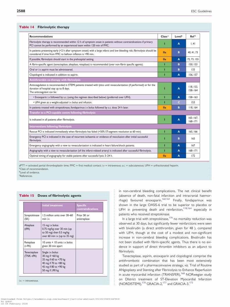
in non-cerebral bleeding complications. The net clinical benefit(absence of death, non-fatal infarction and intracranial haemor-rhage) favoured enoxaparin.160,161 Finally, fondaparinux wasshown in the large OASIS-6 trial to be superior to placebo orUFH in preventing death and reinfarction,118,164 especially inpatients who received streptokinase.
In a large trial with streptokinase,176 no mortality reduction wasobserved at 30 days, but significantly fewer reinfarctions were seenwith bivalirudin (a direct antithrombin, given for 48 ), comparedwith UFH, though at the cost of a modest and non-significantincrease in non-cerebral bleeding complications. Bivalirudin hasnot been studied with fibrin-specific agents. Thus there is no evi-dence in support of direct thrombin inhibitors as an adjunct tofibrinolysis.
Tenecteplase, aspirin, enoxaparin and clopidogrel comprise theantithrombotic combination that has been most extensivelystudied as part of a pharmacoinvasive strategy, viz. Trial of RoutineANgioplasty and Stenting after Fibrinolysis to Enhance Reperfusionin acute myocardial infarction (TRANSFER),168 NORwegian studyon DIstrict treatment of ST-Elevation Myocardial Infarction(NORDISTEMI),170 GRACIA-2,177 and GRACIA-3.173
Table 14 Fibrinolytic therapy
Recommendations Class a Levelb Ref C
Fibrinolytic therapy is recommended within 12 h of symptom onset in patients without contraindications if primary PCI cannot be performed by an experienced team within 120 min of FMC.
I A 1, 41
In patients presenting early (<2 h after symptom onset) with a large infarct and low bleeding risk, fibrinolysis should be considered if time from FMC to balloon inflation is >90 min.
IIa B 40, 41, 73
If possible, fibrinolysis should start in the prehospital setting. IIa A 72, 73, 155
A fibrin-specific agent (tenecteplase, alteplase, reteplase) is recommended (over non-fibrin specific agents). I B 150, 153
Oral or i.v. aspirin must be administered. I B 133
Clopidogrel is indicated in addition to aspirin. I A 156, 157
Antithrombin co-therapy with fibrinolysis
Anticoagulation is recommended in STEMI patients treated with lytics until revascularization (if performed) or for the duration of hospital stay up to 8 days. The anticoagulant can be:
I A118, 153, 158–164
• Enoxaparin i.v followed by s.c. (using the regimen described below) (preferred over UFH). I A 158–163
• UFH given as a weight-adjusted i.v. bolus and infusion. I C 153
In patients treated with streptokinase, fondaparinux i.v. bolus followed by s.c. dose 24 h later. IIa B 118, 164
Transfer to a PCI-capable centre following fibrinolysis
Is indicated in all patients after fibrinolysis. I A165–167, 168–171
Interventions following fibrinolysis
Rescue PCI is indicated immediately when fibrinolysis has failed (<50% ST-segment resolution at 60 min). I A 165, 166
Emergency PCI is indicated in the case of recurrent ischaemia or evidence of reocclusion after initial successful fibrinolysis.
I B 165
Emergency angiography with a view to revascularization is indicated in heart failure/shock patients. I A 167
Angiography with a view to revascularization (of the infarct-related artery) is indicated after successful fibrinolysis. I A 168–171
Optimal timing of angiography for stable patients after successful lysis: 3–24 h. IIa A 172
aPTT ¼ activated partial thromboplastin time; FMC ¼ first medical contact; i.v ¼ intravenous; s.c. ¼ subcutaneous; UFH ¼ unfractionated heparin.aClass of recommendation.bLevel of evidence.cReferences.
Table 15 Doses of fibrinolytic agents
Initial treatment Specific contraindications
Streptokinase (SK)
1.5 million units over 30–60 min i.v.
Prior SK oranistreplase
Alteplase (tPA)
15 mg i.v. bolus0.75 mg/kg over 30 min (up to 50 mg) then 0.5 mg/kg over 60 min i.v. (up to 35 mg)
Reteplase (r-PA)
10 units + 10 units i.v. bolus given 30 min apart
Tenecteplase(TNK–tPA)
Single i.v. bolus: 30 mg if <60 kg35 mg if 60 to <70 kg40 mg if 70 to <80 kg45 mg if 80 to <90 kg50 mg if ≥90 kg
i.v. ¼ intravenous.
ESC Guidelines2588
Downloaded from https://academic.oup.com/eurheartj/article-abstract/33/20/2569/447818by gueston 04 February 2018

3.5.4.7 Angiography after fibrinolysisFollowing initiation of lytic therapy, patients should be transferredto a PCI centre (see section 3.4.6). In cases of failed fibrinolysis, or ifthere is evidence of re-occlusion or reinfarction with recurrence ofST-segment elevation, the patient should undergo immediate angi-ography and rescue PCI.165 Re-administration of fibrinolysis hasnot been shown to be beneficial. Even if it is likely that fibrinolysiswill be successful (ST-segment resolution .50% at 60–90 min;typical reperfusion arrhythmia; disappearance of chest pain), a
strategy of routine early angiography is recommended if thereare no contraindications. Several randomized trials168 –171,178,179
and three contemporary meta-analyses172,180 have shown thatearly routine post-thrombolysis angiography with subsequent PCI(if required) reduced the rates of reinfarction and recurrent ischae-mia compared with a ‘watchful waiting’ strategy, in which angiog-raphy and revascularization were indicated only in patients withspontaneous or induced severe ischaemia or LV dysfunction. Thebenefits of early routine PCI after thrombolysis were seen in the
Table 16 Doses of antiplatelet and antithrombin co-therapies
Doses of antiplatelet co-therapies
With primary PCI
Aspirin Loading dose of 150–300 mg orally or of 80–150 mg i.v. if oral ingestion is not possible, followed by a maintenance dose of 75–100mg/day.
Clopidogrel Loading dose of 600 mg orally, followed by a maintenance dose of 75 mg/day.
Prasugrel Loading dose of 60 mg orally, followed by a maintenance dose of 10 mg/day.In patients with body weight <60 kg, a maintenance dose of 5 mg is recommended.In patients >75 years, prasugrel is generally not recommended, but a dose of 5 mg should be used if treatment is deemed necessary.
Ticagrelor Loading dose of 180 mg orally, followed by a maintenance dose of 90 mg b.i.d.
Abciximab Bolus of 0.25 mg/kg i.v. and 0.125 µg/kg/min infusion (maximum 10 µg/min) for 12 h.
Eptifibatide Double bolus of 180 µg/kg i.v. (given at a 10-min interval) followed by an infusion of 2.0 µg/kg/min for 18 h.
Tirofiban 25 µg/kg over 3 min i.v., followed by a maintenance infusion of 0.15 µg/kg/min for 18 h.
With fibrinolytic therapy
Aspirin Starting dose 150–500 mg orally or i.v. dose of 250 mg if oral ingestion is not possible.
Clopidogrel Loading dose of 300 mg orally if aged ≤75 years, followed by a maintenance dose of 75 mg/day.
Without reperfusion therapy
Aspirin Starting dose 150–500 mg orally.
Clopidogrel 75 mg/day orally.
Doses of antithrombin co-therapies
With primary PCI
Unfractionated heparin 70–100 U/kg i.v. bolus when no GP IIb/IIIa inhibitor is planned.50–60 U/kg i.v. bolus with GP IIb/IIIa inhibitors.
Enoxaparin 0.5 mg/kg i.v. bolus.
Bivalirudin 0.75 mg/kg i.v. bolus followed by i.v infusion of 1.75 mg/kg/h for up to 4 h after the procedure as clinically warranted. After cessation of the 1.75 mg/kg/h infusion, a reduced infusion dose of 0.25 mg/kg/h may be continued for 4–12 h as clinically necessary.
With fibrinolytic therapy
Unfractionated heparin 60 U/kg i.v. bolus with a maximum of 4000 U followed by an i.v. infusion of 12 U/kg with a maximum of 1000 U/h for 24–48 h.TargetaPTT: 50–70 s or 1.5 to 2.0 times that of control to be monitored at 3, 6, 12 and 24 h.
Enoxaparin In patients <75 years of age: 30 mg i.v. bolus followed 15 min later by 1 mg/kg s.c. every 12 h until hospital discharge for a maximum of 8 days The first two
doses should not exceed 100 mg.In patients >75 years of age: no i.v. bolus; start with first s.c. dose of 0.75 mg/kg with a maximum of 75 mg for the first two s.c. doses.In patients with creatinine clearance of <30 mL/min, regardless of age, the s.c. doses are given once every 24 h.
Fondaparinux 2.5 mg i.v. bolus followed by a s.c. dose of 2.5 mg once daily up to 8 days or hospital discharge.
Without reperfusion therapy
Unfractionated heparin Same dose as with fibrinolytic therapy.
Enoxaparin Same dose as with fibrinolytic therapy.
Fondaparinux Same dose as with fibrinolytic therapy.
aPTT ¼ activated partial thromboplastin time; b.i.d.¼ twice a day; GP ¼ glycoprotein; i.v. ¼ intravenous; PCI ¼ percutaneous coronary intervention; s.c. ¼ subcutaneous; UFH ¼unfractionated heparin.
ESC Guidelines 2589
Downloaded from https://academic.oup.com/eurheartj/article-abstract/33/20/2569/447818by gueston 04 February 2018

absence of increased risk of adverse events (stroke or major bleed-ing). Thus, early referral for angiography with subsequent PCI (ifindicated) should be the standard of care after thrombolysis: theso-called ‘pharmacoinvasive’ strategy. A crucial issue is theoptimal delay between lysis and PCI: there was a wide variationin delay in trials, from a median of 1.3 h in the Combined Angio-plasty and Pharmacological Intervention versus ThrombolyticsALone in Acute Myocardial Infarction (CAPITAL-AMI) trial to16.7 h in the GRACIA-1 trial.171,179 Based on the three mostrecent trials, all of which had a median delay between start oflysis and angiography of 2–3 h, a time window of 3–24 h aftersuccessful lysis is recommended.168 –170 The ongoing STREAM148
and GRACIA-4 trials are exploring whether lysis performedwith modern adjunctive therapies, and followed by subsequentPCI, can achieve similar or better outcomes compared withprimary PCI.
3.5.4.8 Adjunctive antithrombotic therapy for delayed percutaneouscoronary intervention after lysisFor patients undergoing PCI several hours or days after fibrinolysis,PCI should be supported by DAPT (aspirin and an ADP antagonist)and antithrombin therapy, in doses similar to those used forprimary PCI.
3.5.4.9 Revascularization strategy for ST-segment elevationmyocardial infarction with multivessel diseaseApart from patients in cardiogenic shock, and in patients withcontinuous ischaemia after opening the supposed culprit lesion,performing PCI of non-culprit vessels in the acute setting is gener-ally discouraged. The best strategy for STEMI patients with multi-vessel disease, who underwent primary PCI of the infarct-relatedartery in the acute phase with remaining multivessel disease, isstill not well established. Among the possible strategies, two thatare frequently used are either a conservative approach—whichuses medical therapy after primary PCI, and revascularization ofother arteries only if there are symptoms or evidence of ischaemiain provocative tests—or a staged revascularization approach,using PCI or coronary bypass surgery of non-infarct arteriesseveral days or weeks after primary PCI, often after confirmationof the stenosis severity with measurements of fractional flowreserve. A multidisciplinary approach is often needed, including aheart team and appropriate informed consent of the patient.
In STEMI patients with multivessel disease initially treated withprimary or post-thrombolysis culprit-artery PCI and confirmedpresence of ischaemia in non-infarcted territories, staged revascu-larization may be performed before discharge or in the days toweeks after initial PCI.181 A comparison of in-hospital completerevascularization [infarct-related artery (IRA) and non-IRA] vs.conservative approach (IRA only) is being undertaken in the Com-plete Vs. Lesion-only PRImary PCI Trial (CVLPRIT) and also in thePreventive Angioplasty in Myocardial Infarction (PRAMI) trial. Bothassess the benefit/risk of treating non-infarct-related lesions. Like-wise, the DANish study of optimal acute treatment of patients withST-elevation Myocardial Infarction 3 (DANAMI-3) trial is currentlytesting whether or not to treat non-culprit lesions in patientstreated previously with primary PCI.
3.5.5 Coronary bypass surgery and multivessel coronaryrevascularizationThe number of patients who require CABG surgery in the acutephase of STEMI is small, but CABG may be indicated in patientswith anatomy unsuitable for PCI but who have a patentinfarct-related artery, since patency of this artery provides timefor transfer to the surgical team. It may also be indicated in patientsin cardiogenic shock if the coronary anatomy is not amenable toPCI, or at the time of repair for patients with mechanical complica-tions. CABG is rarely used and its benefits are uncertain in patientswith failed PCI, coronary occlusion not amenable to PCI, and in thepresence of refractory symptoms after PCI since, in most of thesecases, time for implementation of surgical reperfusion will be longand the risks associated with surgery are maximal in this setting.
3.5.5.1 Withholding adenosine diphosphate inhibitors for surgeryThe risk of bleeding related to surgery must be balanced againstthe risk of recurrent ischaemic events related to discontinuationof therapy, bearing in mind the nature of the surgery, the ischaemicrisk and extent of CAD, the time since the acute episode, the timesince PCI and the risk of stent thrombosis. Clopidogrel is asso-ciated with an increased risk of bleeding if discontinued less than5 days before surgery. Prasugrel is also associated with a markedincrease in bleeding risk.109 With respect to ticagrelor, data fromthe PLATO trial,110 suggest that ticagrelor, discontinued 3–5days before CABG surgery, yielded similar CABG-related majorbleeding and transfusions for clopidogrel and ticagrelor. Althoughnon-fatal myocardial infarction and stroke rates in the twogroups were not significantly different in this cohort, there was ahalving of mortality in the ticagrelor group. In stabilized patients,it is reasonable to stop clopidogrel at least 5 days before surgeryand to stop prasugrel 7 days before surgery. Given the PLATOdata, ticagrelor may be discontinued 3 to 5 days before surgery.
Whether ADP receptor antagonists should be restarted afterCABG surgery has not been addressed in any specific trial andthe optimal timing of such restarting remains uncertain.However, given the reduction of the primary endpoint and mortal-ity with ticagrelor in the PLATO trial and the continued risk for is-chaemic events in patients post-CABG it is reasonable to restartDAPT as soon as considered safe in relation to bleeding risk.
In very-high-risk patients in whom cessation of antiplatelettherapy before surgery seems to carry a high risk (e.g. within thefirst weeks after stent implantation), it has been suggested toswitch, before surgery, to a short half-life and reversible antiplate-let agent, e.g. the GP IIb/IIIa receptor inhibitors tirofibanor eptifiba-tide,182 but there is no clinical evidence to support this approachbased solely on pharmacokinetic or pharmacodynamic studies. Inthe future, the use of cangrelor, an i.v. reversible ADP receptor an-tagonist, may permit platelet inhibition to be maintained up tosurgery in patients discontinuing oral antiplatelet therapy.183
3.5.6 Non-reperfused patients3.5.6.1 Antithrombotic useIn patients presenting within 12 h of symptom onset, and in whomreperfusion therapy was not given, or in patients presentingbeyond 12 h, aspirin, clopidogrel and an antithrombin agent(UFH, enoxaparin or fondaparinux) should be given as soon as
ESC Guidelines2590
Downloaded from https://academic.oup.com/eurheartj/article-abstract/33/20/2569/447818by gueston 04 February 2018

possible (see section 3.4.6).1,156,184 In OASIS-6, fondaparinux wassuperior to UFH in a subgroup of 1641 such patients and mightbe the preferred antithrombin for this indication.185 If PCI isneeded in a patient receiving fondaparinux, i.v. UFH should beadministered during the procedure, using the same doses as forprimary PCI, to minimize the risk of catheter thrombosis.186
Recommended doses are given in Table 16. None of the oralagents have been studied in this particular subset of patients, butthe benefit of clopidogrel over placebo was consistent in ACSpatients, independent of revascularization strategy, in the Clopido-grel in Unstable angina to prevent Recurrent Events (CURE)trial.187 Ticagrelor was superior to clopidogrel in ACS patientswho were randomized for an early non-invasive strategy, with asimilar trend also in those who were not revascularized duringthe index hospitalization.188
3.5.6.2 Invasive evaluation and revascularizationPatients sometimes seek medical attention too late and either donot receive reperfusion therapy, or undergo unsuccessful reperfu-sion therapy. It has been suggested that achieving late coronarypatency in either of these situations may still have a beneficialeffect by preventing adverse LV remodelling, improving LV func-tion, increasing electrical stability and inducing collateral vesselsto other coronary beds for protection against future events (the‘open artery’ hypothesis). Several trials have evaluated this hypoth-esis, of which the largest by far was the OAT trial (see above),62 inwhich 20% of the patients received fibrinolytic therapy for theindex event. PCI did not reduce the occurrence of death, reinfarc-tion or heart failure, compared to medical therapy alone. Further-more, there was a trend towards excess reinfarction during fouryears of follow-up in the invasive therapy group, compared withthe medical therapy group. A meta-analysis of all trials in thissetting provided similar results.63 These studies demonstrate thatlate PCI of an occluded infarct-related artery after myocardial in-farction in stable patients has no incremental benefit overoptimal medical therapy. Thus, in patients presenting days afterthe acute event with a completed myocardial infarction, onlythose with recurrent angina or documented residual ischaemia,and proven viability on non-invasive imaging in a large myocardialterritory, may be considered for revascularization when theinfarct artery is occluded.4
Special patient subsetsSeveral specific patient subsets deserve particular consideration
(Table 17):
† Women tend to present later and may have somewhat atypicalsymptoms more frequently than men.191 Yet myocardial infarc-tion remains the leading cause of death in women and it is there-fore important to maintain a high degree of awareness formyocardial infarction in women with potential symptoms of is-chaemia. In addition, several observational studies have shownthat women tend to undergo fewer interventions than menand that they also less frequently receive reperfusiontherapy;192 also that this may not be fully accounted for bythe age difference, i.e. women experiencing myocardial infarc-tion at a later age than men.193,194 When women are giveneffective reperfusion therapy, such as primary PCI, they
experience the same risk of death as men.195 It is thereforecrucial to provide reperfusion therapy as effectively in womenas in men. Women generally have lower body weight and aremore susceptible to bleeding, which is why antithrombotic ther-apies and their doses should be used with close attention tobleeding risk.
† Elderly patients often present with atypical or mild symptoms,which may result in delayed or missed diagnoses of myocardialinfarction (MI).189 The elderly are at particular risk of bleedingand other complications from acute therapies because bleedingrisk increases with age, because renal function tends to decreaseand because the prevalence of comorbidities is high. In addition,observational studies have shown frequent overdosing of antith-rombotic therapies.190 It is therefore key to maintain a highindex of suspicion for myocardial infarction in elderly patientswho present with atypical complaints and to pay specific atten-tion to proper dosing of antithrombotic therapies, particularly inrelation with renal function.
† Renal dysfunction is present in approximately 30–40% of patientswith ACS and is associated with a worse prognosis andincreased bleeding risk.196 Decisions on reperfusion in patientswith STEMI have to be made before any assessment of renalfunction is available, but it is important to estimate the glomeru-lar filtration rate as soon as possible after admission. ACSpatients with chronic kidney disease are frequently overdosedwith antithrombotics, leading to increased bleeding risk.190
The benefit of ticagrelor was consistent or enhanced in patientswith renal dysfunction: GFR , 60 mL/min in the PLATO trial.197
In patients with known or anticipated reduction of renal func-tion, several antithrombotic agents should be either withheldor their doses reduced appropriately (Table 18). Ensuringproper hydration during and after primary PCI, and limitingthe dose of contrast agents, are important in minimizing therisk of contrast-induced nephropathy.4
† Diabetic patients are at higher risk of death and complications, butselection of antithrombotic therapies and reperfusion therapy isthe same as in non-diabetics. The benefits of the potent oral
Table 17 Special subsets
Recommendations Class a Levelb Ref C
Both genders must bemanaged in a similar fashion.
I C -
A high index of suspicion for myocardial infarction must be maintained in women, diabetics, and elderly patients with atypical symptoms.
I B 189
Special attention must be given to proper dosing of antithrombotics in elderly and renal failure patients.
I B 190
aClass of recommendation.bLevel of evidence.cReferences.
ESC Guidelines 2591
Downloaded from https://academic.oup.com/eurheartj/article-abstract/33/20/2569/447818by gueston 04 February 2018

P2Y12 receptor inhibitors (prasugrel or ticagrelor) vs. clopidogrelare consistent or enhanced in patients with diabetes.198,199
3.6 Management of hyperglycaemiain the acute phase of ST-segmentelevation myocardial infarctionHyperglycaemia on admission is common in patients with an ACSand is a powerful predictor of mortality and in-hospital complica-tions. These elevated glucose concentrations have been associatedwith an adverse prognosis, both in diabetic and non-diabeticpatients. However, elevated glucose concentrations may also bea sign of disturbed long-term glucose metabolism, because of un-diagnosed diabetes or impaired glucose tolerance.200 It was recent-ly shown, in STEMI patients without known diabetes, thathyperglycaemia and elevated haemoglobin A1c (HbA1c) are asso-ciated with a poor prognosis through different mechanisms, withhyperglycaemia especially predicting short-term prognosis in
association with a larger infarct size, whereas elevated HbA1c
was associated with long-term effects on outcome through ahigher baseline risk.201
Although correction of hyperglycaemia by insulin may be ofbenefit, clinical trials evaluating the effect of metabolic interventionin patients with STEMI showed conflicting results.202 In particular,the benefits of tight glucose control through i.v. insulin shown inthe Diabetes, Insulin Glucose infusion in Acute Myocardial Infarction(DIGAMI) trial was not confirmed in the subsequent DIGAMI-2trial. Glucose–insulin–potassium infusions were found to be ofno value and potentially harmful in a combined analysis of twolarge randomized trials.203 Additionally, in critically ill patients,there is a high risk of hypoglycaemia-related events when using in-tensive insulin therapy.204 The definitive answer with regard toglucose management in patients with STEMI, including treatmentthresholds and glucose targets, is lacking and therefore a strategyof ‘strict, but not too strict’ glucose control in STEMI seems tobe a practical approach. In the acute phase, it is reasonable tomanage hyperglycaemia (i.e. maintain a blood glucose
Table 18 Initial dosing of antithrombotic agents inpatients with chronic kidney disease (estimatedcreatinine clearance <60 mL/min)
Recommendation
Aspirin No dose adjustment.
Clopidogrel No dose adjustment.
Prasugrel No dose adjustment. No experience with end-stagerenal disease/dialysis.
Ticagrelor No dose adjustment. No experience with end-stagerenal disease/dialysis.
Enoxaparin No adjustment of bolus dose. Following thrombolysis,in patients with creatinine clearance <30 mL/min, thes.c. doses are given once every 24 h.
Unfractionatedheparin
No adjustment of bolus dose.
Fondaparinux No dose adjustment.No experience in patients with end-stage renaldisease or dialysis patients.
Bivalirudin • In patients with moderate renal insufficiency (GFR 30–59 mL/min) a lower initial infusion rate of 1.4
mg/kg/h should be given.The bolus dose should notbe changed.
• In patients with severe renal insufficiency (GFR <30 mL/min) and in dialysis-dependent patients
bivalirudin is contraindicated.
Abciximab No specific recommendation. Careful considerationof bleeding risk.
Eptifibatide • In patients with moderate renal insufficiency (GFR≥30 to <50 mL/min), an i.v. bolus of 180 µg shouldbe administered followed by a continuous infusion
dose of 1.0 µg/kg/min for the duration of therapy.• In patients with severe renal insufficiency (GFR <30 mL/min) eptifibatide is contraindicated.
Tirofiban In patients with severe renal insufficiency (GFR<30 mL/min) the infusion dose should be reducedto 50%.
GFR ¼ glomerular filtration rate; i.v. ¼ intravenous; s.c. ¼ subcutaneous;.
Table 19 Management of hyperglycaemia inST-segment elevation myocardial infarction
Recommendations Class a Levelb Ref C
Measurement of glycaemia is indicated at initial evaluation in all patients, and should be repeated in patients with known diabetes or hyperglycaemia.
I C -
Plans for optimal outpatientglucose control and secondary prevention must be determined in patients with diabetes before discharge.
I C -
The goals of glucose controlin the acute phase should be to maintain glucose concentrations ≤11.0 mmol/L(200 mg/dL) while avoidingfall of glycaemia <5 mmol/L(<90 mg/dL). In some patients,this may require a dose-adjusted insulin infusion withmonitoring of glucose, as long as hypoglycaemia is avoided.
IIa B202, 204,
207
A measurement of fasting glucose and HbA1c and, insome cases, a post-dischargeoral glucose tolerance test should be considered in patients with hyperglycaemia but without a history of diabetes.
IIa B 208
Routine glucose-insulin-potassium infusion is not indicated.
III A 118, 203
HbA1c ¼ haemoglobin A1c.aClass of recommendation.bLevel of evidence.cReferences.
ESC Guidelines2592
Downloaded from https://academic.oup.com/eurheartj/article-abstract/33/20/2569/447818by gueston 04 February 2018

concentration ≤11.0 mmol/L) but absolutely avoid hypogly-caemia.205,206 This may require a dose-adjusted insulin infusionwith monitoring of glycaemia in some patients.
Given the frequency of unrecognised diabetes and impairedglucose metabolism in STEMI patients, it is reasonable tomeasure HbA1c and fasting blood glucose in all patients withoutknown diabetes, who developed hyperglycaemia during the acutephase (Table 19). If equivocal, an oral glucose tolerance test maybe needed after discharge. This should preferably be measured 4days after the acute phase. The best therapeutic strategy to specif-ically lower elevated HbA1c-associated mortality risk remainsuncertain, apart from strategies of secondary prevention (antiplate-let therapy, aggressive lipid control, blood pressure control, life-style modification, and cardiac rehabilitation), which should beimplemented in all survivors of acute myocardial infarction.Whether the results of more intensive, early glycaemic therapywith oral agents provides cardiovascular protection is not knownand warrants further study.207
4. Management duringhospitalization and at discharge
4.1 Coronary care unit logisticsand monitoring4.1.1 Coronary care unitSTEMI patients should be admitted to an intensive cardiac care orcoronary care unit (Table 20), or equivalent monitored unit, fol-lowing reperfusion treatment. A coronary care unit is an intensivecare unit designed to provide specialized care to patients with car-diovascular disease requiring constant monitoring. The staff shouldbe thoroughly familiar with the management of ACS, arrhythmias,heart failure, mechanical circulatory support, and complex invasive
and non-invasive haemodynamic monitoring (arterial and pulmon-ary artery pressures), respiratory monitoring (continuous positiveairway pressure and biphasic positive airway pressure), andsupport, as well as body cooling techniques. The unit should beable to manage patients with serious renal and pulmonarydisease. The desirable organization, structure and criteria of thecoronary care unit have been described in an ESC positionpaper.209
4.1.2 MonitoringECG monitoring for arrhythmias and ST-segment deviationsshould be continued for at least 24 h after symptom onset in allSTEMI patients. Further monitoring for arrhythmia depends uponperceived risk and equipment available. When a patient leavesthe coronary care unit, monitoring may be continued by telemetry.
4.1.3 AmbulationPatients with significant LV damage should initially rest in bedbefore a first assessment of infarct extent and severity is possiblefor detection of early heart failure and arrhythmias. In uncompli-cated cases, the patient can usually sit out of bed on the firstday, be allowed to use a commode and undertake self-care andself-feeding. Ambulation can often start early (particularly inpatients treated via the radial access). Patients who have experi-enced complications should be kept in bed for longer and theirphysical activity resumed as a function of symptoms and extentof myocardial damage.
4.1.4 Length of stayThe optimal length of stay in the coronary care unit and hospitalshould be determined on an individual basis, considering thepatient’s particular medical and social situation, including premor-bid health. Over the years, there has been a progressive reduction
Table 20 Logistical issues for hospital stay
Recommendations Class a Level b Ref C
All hospitals participating in the care of STEMI patients should have a coronary care unit equipped to provide allaspects of care for STEMI patients, including treatment of ischaemia, severe heart failure, arrhythmias and commoncomorbidities.
I C -
Length of stay in the coronary care unit
Patients undergoing uncomplicated successful reperfusion therapy should be kept in the coronary care unit for aminimum of 24 h, after which they may be moved to a step-down monitored bed for another 24–48 h.
I C -
Transfer back to a referring non-PCI hospital
Early transfer (same day) may be considered in selected, low-risk patients after successful primary PCI withoutobserved arrhythmia.
IIb C -
Hospital discharge
Early discharge (after approximately 72 h) is reasonable in selected low-risk patients, if early rehabilitation andadequate follow-up are arranged.
IIb B212, 215,
216
PCI ¼ percutaneous coronary intervention; STEMI ¼ ST-segment elevation myocardial infarction.aClass of recommendation.bLevel of evidence.cReferences.
ESC Guidelines 2593
Downloaded from https://academic.oup.com/eurheartj/article-abstract/33/20/2569/447818by gueston 04 February 2018

in length of stay after myocardial infarction—especially followingsuccessful primary revascularization—without any increase in sub-sequent mortality, suggesting that earlier discharge is not asso-ciated with late mortality.210,211 Moreover, the Primary Angioplastyin Myocardial Infarction II (PAMI-II) trial showed that low-riskpatients with successful primary PCI could safely be dischargedfrom hospital at day 3 without non-invasive testing.212 Overall,early discharge of low-risk patients (within 72 h) is both feasibleand safe in patients with uncomplicated STEMI and successfulprimary PCI.211 –213 To identify these low-risk patients, schemessuch as the PAMI-II criteria or the Zwolle primary PCI Index canbe helpful.212,213 The PAMI II criteria designate as low risk patientsaged ,70 years, with a left ventricular ejection fraction .45%,one- or two-vessel disease, successful PTCA and no persistentarrhythmias. Nevertheless, a short hospital stay implies limitedtime for proper patient education and up-titration of secondaryprevention treatments. Consequently, these patients should beoffered early post-discharge consultations with a cardiologist orprimary care physician and the option of a formal rehabilitationprogram, either in-hospital or on an outpatient basis.
Current practice may also include early transfer to a local hos-pital following successful primary PCI. In selected low-riskpatients—identified as being asymptomatic without any arrhyth-mia, haemodynamically stable, not requiring vasoactive or mechan-ical support and not scheduled for further revascularization—earlytransfer (same day) under adequate monitoring and supervisionappears safe and feasible.214
4.2 Risk assessment and imaging4.2.1 Indications and timing (Table 21)After reperfusion treatment, it is important to identify patients athigh risk of further events such as reinfarction or death, and hope-fully to intervene in order to prevent these events. Because the
risk of events decreases with time, early risk assessment is indi-cated. Assessment of infarct size and resting LV function, usuallyby echocardiography, should be undertaken before discharge.The timing of further investigations will depend on local facilitiesand whether angiography and PCI have been performed successful-ly. With the increasing use of primary PCI, risk assessment for is-chaemia before discharge has become less important, since it canbe assumed that the infarct-related coronary lesion has beentreated and stabilized and the presence or absence of significantlesions in other arteries has been assessed. Several risk scoreshave been developed, based on readily identifiable parameters inthe acute phasebefore reperfusion.217 – 219 Clinical indicators ofhigh risk in the acute phase include older age, fast heart rate, hypo-tension, Killip class .I, anterior infarction, previous infarction, ele-vated initial serum creatinine and history of heart failure. Malignantarrhythmias, persistent chest pain and early angina on minimalexertion are also associated with worse outcome.
If, in spite of the angiography performed in the acute phase,there are concerns about inducible ischaemia, an outpatientexercise-testing or stress-imaging test (using scintigraphy, echocar-diography or magnetic resonance imaging) within 4–6 weeks isappropriate (Table 9). Because of high availability and low cost,an exercise ECG is commonly used. However, in patients with pre-vious myocardial infarction, its accuracy is limited. Stress imagingtests are more accurate and allow localization of the ischaemia.The most-validated tests are perfusion scintigraphy and stressechocardiography. In post-myocardial infarction patients, thedetection of residual ischaemia is challenging, due to existingwall-motion abnormalities. Computed tomography angiography isa sensitive technique to detect coronary lesions but, as an anatom-ical test, it does not assess ischaemia, which remains essential fortherapeutic decisions. If the main concern is arrhythmia, additionalelectrophysiological testing may be needed before discharge, and
Table 21 Summary of indications for imaging and stress testing
Recommendations Class a Levelb Ref C
At presentation
In the acute phase, when diagnosis is uncertain, emergency echocardiography may be useful. However, if inconclusive orunavailable and persistent doubt, emergency angiography should be considered.
I C -
After the acute phase
All patients should have an echocardiography for assessment of infarct size and resting LV function, I B 220, 221
If echocardiography is not feasible, MRI may be used as an alternative. IIb C -
Before or after discharge
For patients with multivessel disease, or in whom revascularization of other vessels is considered, stress testing or imaging (e.g. using stress myocardial perfusion scintigraphy, stress echocardiography, positron emission tomography or MRI) for ischaemia and viability is indicated.
I A 4, 220, 222
Computed tomography angiography has no role in the routine management of STEMI patients. III C -
Echocardiography ¼ transthoracic echocardiography, or transoesophageal if required; LV ¼ left ventricular; MRI ¼ magnetic resonance imaging; STEMI ¼ ST-segment elevationmyocardial infarction.aClass of recommendation.bLevel of evidence.cReferences.
ESC Guidelines2594
Downloaded from https://academic.oup.com/eurheartj/article-abstract/33/20/2569/447818by gueston 04 February 2018

repeated assessment of LV ejection fraction after discharge maybe important for selecting candidates for implantation of acardioverter-defibrillator as primary prevention (see below).
All patients should have their metabolic risk markers measuredduring the index admission, including total cholesterol, low-densitylipoprotein (LDL) cholesterol, high-density lipoprotein cholesterol,fasting triglycerides and plasma glucose, as well as renal function.Since LDL levels tend to decrease during the first days after myo-cardial infarction, they are best measured as soon as possible afteradmission.
4.3 Assessment of myocardial viabilityLV dysfunction after acute myocardial infarction may be due to ne-crosis, to stunning of viable myocardium remaining in the infarctterritory, to hibernation of viable myocardium, or to a combinationof all three. Simple stunning should recover within 2 weeks of theacute ischaemic insult if ischaemia does not persist but, if it does,then recurrent stunning may become hibernation and requirerevascularization for recovery of function. These concepts are ofmost relevance in the patient with severely impaired LV functionafter infarction when the need for revascularization to improvefunction is considered (e.g. after successful fibrinolysis).
Multiple imaging techniques, including PET, single-photon emis-sion CT, and dobutamine stress echocardiography have been eval-uated extensively for assessment of viability and prediction ofclinical outcome after myocardial revascularization. In general,nuclear imaging techniques have a high sensitivity, whereas techni-ques evaluating contractile reserve have a somewhat lower sensi-tivity but higher specificity. MRI has a high diagnostic accuracyfor assessing transmural extent of myocardial scar tissue, but itsability to detect viability and predict recovery of wall motion isnot superior to other imaging techniques.223 The differences inperformance of the various imaging techniques are small, and ex-perience and availability commonly determine which technique isused. Current evidence is mostly based on observational studiesor meta-analyses, with the exception of two randomized clinicaltrials, both relating to PET imaging.222 Patients with a substantialamount of dysfunctional but viable myocardium are likely tobenefit from myocardial revascularization and may show improve-ments in regional and global contractile function, symptoms, exer-cise capacity and long-term prognosis.220
4.4 Long-term therapies for ST-segmentelevation myocardial infarctionCoronary heart disease is a chronic condition and patients whohave recovered from a STEMI are at high risk for new eventsand premature death. In fact, in long-term cohorts, most patientswith STEMI who die do so after discharge from the indexevent.14 Several evidence-based interventions can improve prog-nosis. Even though long-term management of this large group ofpatients will be the responsibility of the general practitioner,these interventions will have a higher chance of being implementedif initiated during the hospital stay. In addition, lifestyle changesshould be explained and proposed to the patient before discharge.However, habits of a lifetime are not easily changed and the imple-mentation and follow-up of these changes are a long-term
undertaking. In this regard, a close collaboration between the car-diologist and the primary care physician is critically important.With ever-shorter length of hospital stay in patients with STEMI,there is no longer a clear distinction between acute and chronictherapies in STEMI. This chapter summarizes both lifestyleinterventions and drug therapies that need to be considered andimplemented before hospital discharge (Table 22).
4.4.1 Lifestyle interventions and risk factor controlKey lifestyle interventions include cessation of smoking and tightblood pressure control, advice regarding diet and weight control,and the encouragement of physical activity. Detailed recommenda-tions are available from the ESC guidelines on prevention.224 Eventhough long-term management of this large group of patients willbe the responsibility of the primary care physician, these interven-tions will have a higher chance of being implemented if initiatedduring the hospital stay. In addition, the benefits and importanceof lifestyle changes should be explained and proposed to thepatient—who is the key player—before discharge. However,habits of a lifetime are not easily changed, and the implementationand follow-up of these changes are a long-term undertaking. In thisregard, a close collaboration between the cardiologist and thegeneral practitioner, specialist rehabilitation nurses, pharmacists,dieticians, physiotherapists is critically important.
4.4.1.1 Smoking cessationUnselected ACS patients who are smokers are twice as likely topresent with a STEMI, compared with non-smokers, indicating astrong prothrombotic effect of smoking. Observational studiesshow that patients who stop smoking reduce their mortality inthe succeeding years compared with continued smokers. Stoppingsmoking is potentially the most effective of all secondary preven-tion measures,225 and much effort should be devoted to thisend. Patients do not smoke during the acute phase of a STEMIand the convalescent period is ideal for health professionals tohelp smokers to quit. However, resumption of smoking iscommon after discharge, and continued support and advice areneeded during rehabilitation. Nicotine replacement, buproprioneand antidepressants may be useful. Nicotine patches have beendemonstrated to be safe in ACS patients.226 A randomized studyhas also demonstrated the effectiveness of a nurse-directed pro-gramme.227 A smoking cessation protocol should be adopted byeach hospital.
4.4.1.2 Diet and weight controlCurrent guidelines on prevention recommend: 224 (i) eating a widevariety of foods; (ii) adjustment of calorie intake to avoid obesity;(iii) increased consumption of fruit and vegetables, along withwholegrain cereals and bread, fish (especially oily varieties), leanmeat and low-fat dairy products; (iv) replacing saturated andtrans fats with monounsaturated and polyunsaturated fats fromvegetable and marine sources, and to reduce total fats (of whichless than one-third should be saturated) to ,30% of totalcalorie intake, and (v) to reduce salt intake if blood pressure israised. Many processed and prepared foods are high in salt andin fat of doubtful quality. There is no evidence for the use of
ESC Guidelines 2595
Downloaded from https://academic.oup.com/eurheartj/article-abstract/33/20/2569/447818by gueston 04 February 2018

antioxidant supplements, low glycaemic index diets or homocyst-eine-lowering therapies following STEMI.
Obesity is an increasing problem in patients with STEMI.Current ESC Guidelines define a body mass index (BMI),25 kg/m2 as optimal, and recommend weight reduction whenthe BMI is 30 kg/m2 or more, and when waist circumference is.102 cm in men or .88 cm in women, because weight loss canimprove many obesity-related risk factors. However, it has notbeen established that weight reduction per se reduces mortality.
4.4.1.3 Physical activityExercise therapy has long been used for rehabilitation purposesfollowing STEMI and the benefit of regular physical exercise instable CAD patients is also well established. It can reduce theanxiety associated with the life-threatening illness and improvepatient self-confidence. Four mechanisms are considered to be im-portant mediators of a reduced cardiac event rate: (i) improvementof endothelial function; (ii) reduced progression of coronarylesions; (iii) reduced thrombogenic risk and (iv) improved collater-alization. In a large meta-analysis, exercise training as part of coron-ary rehabilitation programmes was associated with a 26%reduction in cardiac mortality rate in patients with CAD.228 Itshould be appreciated that, apart from its influence on mortality,exercise rehabilitation can have other beneficial effects. Exercisecapacity, cardiorespiratory fitness, and perception of well-beinghave also been reported to improve, at least during the actualtraining period, even in elderly patients. Thirty minutes of moder-ate intensity aerobic exercise at least five times per week is recom-mended.224 Each step of increase in peak exercise capacity isassociated with a reduction in all-cause mortality risk in therange of 8–14%.229
4.4.1.4 Blood pressure controlIn hypertensive patients with STEMI, blood pressure should be wellcontrolled. Data from a retrospective analysis of the PRavastatinOr atorVastatin Evaluation and Infection Therapy–ThrombolysisIn Myocardial Infarction 22 (PROVE IT–TIMI 22) trial suggestthat, after acute coronary syndromes, the blood pressure goalshould be a systolic ,140 mmHg but not ,110 mm Hg.230 Thepharmacotherapy (beta-blockers, ACE inhibitors or ARBs) recom-mended after STEMI, in addition to lifestyle modifications (reducedsalt intake, increased physical activity and weight loss) usually helpsachieve these goals. Additional drug therapy may be needed.
4.4.1.5 Psychosocial factor interventionsThere is evidence that stress management is useful in this setting: ina recent trial 362 patients, aged 75 years or younger, with acutemyocardial infarction, PCI or CABG within the past 12 months,were randomized to receive traditional care or traditional careplus a cognitive behavioural therapy programme focussed onstress management. During a mean of 94 months of follow-up,the intervention group had a 41% lower rate of fatal and non-fatalfirst recurrent cardiovascular disease events (45% fewer recurrentacute myocardial infarctions) and a non-significant 28% lower all-cause mortality than the reference group after adjustment forother outcome-affecting variables. There was also a strong
dose-response effect between attendance rate in the groupsessions and outcome rate.231
4.4.1.6 Exercise-based rehabilitation programmeExercise-based rehabilitation has been shown to be effective atreducing all-cause mortality and the risk of reinfarction, as wellas improving risk factors, exercise-based capacity andhealth-related quality of life after myocardial infarction.232,233
Yet these benefits were established in the era precedingmodern treatment of STEMI and a recent British randomizedtrialfailed to demonstrate benefits of a rehabilitation programmeon clinical outcomes or quality of life.234 In another larger rando-mized study, a long-term multifactorial, educational and behav-ioural intervention was proven to be feasible and sustainableover a long period after myocardial infarction, and reducedsome clinical outcomes—particularly re-infarction—and globalcardiovascular risk.235 An additional benefit of rehabilitation pro-grammes is to help ensure proper titration and monitoring ofkey, evidence-based therapies after STEMI. Nowadays, in patientswith an uncomplicated course, rehabilitation can often be per-formed on an outpatient basis with an efficacy similar to thatof centre-based cardiac rehabilitation.236
4.4.1.7 Resumption of activitiesNo generalizable recommendations can be made regarding thedelay to resumption of daily activities. Decisions should be indivi-dualized, based on left ventricular function, completeness of revas-cularization and rhythm control. Extended sick leave is usuallynegative and light-to-moderate physical activity after dischargeshould be encouraged. Sexual activity can be resumed early ifadjusted to physical ability. Long distance air travel should beavoided for 4–6 weeks if residual ischaemia or left ventriculardysfunction is present.
4.4.2 Antithrombotic therapy4.4.2.2 AspirinGiven its established benefits in secondary prevention,237 aspirinshould be used indefinitely in all patients with STEMI. Thedosage of aspirin is debated. In respect of the first few days oftreatment, the Clopidogrel and aspirin Optimal Dose usage toreduce recurrent events—Seventh organization to assess strat-egies in ischaemic syndromes (CURRENT/OASIS 7) large, rando-mized trial failed to demonstrate a difference in hard clinicaloutcomes when comparing low doses (75–100 mg/day) or rela-tively high doses of 300–325 mg/day.115 There were, however,fewer gastro-intestinal bleeds with lower doses. For the longterm, low doses (70–100 mg) are generally used. Platelet aggrega-tion data suggest that rapid turnover of platelets in diabetic patientsmay require higher doses or more frequent dosing of aspirin toachieve platelet inhibition,238,239 but there is no proof of clinicalbenefit of such a strategy. Patients with a history of hypersensitivityto aspirin can undergo desensitization and continue therapy indef-initely.240– 242 Patients who are truly intolerant to aspirin caninstead receive clopidogrel (75 mg/day) as long-term secondaryprevention.243
ESC Guidelines2596
Downloaded from https://academic.oup.com/eurheartj/article-abstract/33/20/2569/447818by gueston 04 February 2018

4.4.2.2 Duration of dual antiplatelet therapy and antithromboticcombination therapies after ST-segment elevation myocardial infarctionDAPT, combining aspirin and an ADP-receptor blocker (clopido-grel, prasugrel or ticagrelor), is recommended in patients withSTEMI who are undergoing primary PCI (for up to 12 months), fi-brinolysis (for up to 12 months, although the data available pertainonly to one month of DAPT) and in those patients who have notundergone reperfusion therapy (for at least 1 month and up to 12months). The choice of ADP-receptor blocker has been discussedpreviously. While there are no trial data to support extendedDAPT, treatment for 12 months after stenting and for 9–12months following STEMI has traditionally been recommended byconsensus in prior guidelines, regardless of whether a stent(BMS or DES) was used.1,4,244 Some studies have suggested thatthere is no benefit in extended durations of DAPT beyond 6 or12 months after placement of a DES to prevent ischaemic eventsand stent thrombosis,245– 247 but these studies, even whenpooled, include a relatively small number of STEMI patients.Several ongoing large trials, including the Dual AntiplateletTherapy (DAPT) study,248 are testing whether longer durationsof dual antiplatelet therapy following stenting are of clinicalbenefit. Clearly, after stenting for ACS, particularly STEMI,extended DAPT reduces the risk of stent thrombosis, reinfarctionand cardiovascular mortality,249 and more potent DAPT is asso-ciated with greater clinical benefits post-ACS of anytype.109,110,188 Pending the results of ongoing trials, a 9–12months duration of DAPT is recommended, with a strictminimum of one month for patients who have received a BMSand six months for those who received a DES. It is important toinform patients and their physicians about the need to avoidpremature discontinuation of DAPT.
In patients with STEMI and with atrial fibrillation and the needfor permanent anticoagulation after primary PCI [based onCardiac failure, Hypertension, Age, Diabetes, Stroke (Doubled)(CHADS2) or Cardiac failure, Hypertension, Age ≥75(Doubled), Diabetes, Stroke (Doubled) – VAScular disease, Age65–74 and Sex category (Female) (CHA2DS2-VASc) scores of ≥2],250,251 ‘triple therapy’, combining aspirin, an ADP receptor an-tagonist and an oral anticoagulant, is recommended to reducethe burden of thromboembolic complications associated withatrial fibrillation and minimize the risk of stent thrombosis.4
However, it is also associated with an increase in bleeding compli-cationsand should therefore be used for the shortest possible dur-ation.252,253 This is an area of controversy, with missing evidence,and several consensus documents have tried to offer algorithmsfor decision-making.253 –255 Moreover, in STEMI patients with anindication for anticoagulation, and in whom stents are needed, se-lection of BMS over DES would appear to minimize the duration oftriple therapy and therefore the risk of bleeding. These benefitsshould be weighed against the benefits of DES in preventingrestenosis.4,253
Gastric protection, preferably with a proton pump inhibitor,should be considered for patients with a history of gastrointestinalbleeding and is appropriate for patients with multiple risk factorsfor bleeding, such as advanced age, concurrent use of anticoagu-lants, steroids or non-steroidal anti-inflammatory drugs includinghigh dose aspirin, and Helicobacter pylori infection.256 There is no
pharmacokinetic interaction between proton pump inhibitors andthe new potent P2Y12 receptor inhibitors and no clear evidencethat the pharmacokinetic interaction of clopidogrel with someproton pump inhibitors has meaningful clinical consequences.257–261
In any case, the benefits of avoiding or minimizing bleeding in patientsat high risk outweigh the concerns raised by this pharmacokineticinteraction.
The recent Anti-Xa Therapy to Lower cardiovascular events inAddition to Standard therapy in subjects with Acute CoronarySyndrome–Thrombolysis In Myocardial Infarction 51 (ATLASACS 2–TIMI 51) trial tested the addition of rivaroxaban, a factorXa antagonist to aspirin and clopidogrel following ACS.262 In thattrial, a low dose of rivaroxaban (2.5 mg twice daily) reduced thecomposite primary endpoint of cardiovascular death, myocardialinfarction and stroke, but also all-cause mortality. Interestingly,stent thrombosis was reduced by one third. This was associatedwith threefold increases in non-CABG-related major bleeding,and intracranial haemorrhage. Importantly, the high dose of rivar-oxaban (5 mg twice daily) was not associated with similar benefitsand was associated with a major increase in the risk of bleeding.The ATLAS ACS 2-TIMI 51 trial did not test a combination of riv-aroxaban with prasugrel or ticagrelor, which might be associatedwith even more bleeding. This trial suggests that, in selectedpatients at low bleeding risk, the 2.5 mg dose of rivaroxaban maybe considered in patients who receive aspirin and clopidogrelafter STEMI. However, a phase III trial of another factor Xa antag-onist (apixaban), the Apixaban for Prevention of Acute Ischemicand Safety Events (APPRAISE-2) trial,263 failed to find similar ben-efits of adding a high dose of apixaban to single or DAPT in avery-high-risk ACS population. Finally, darexaban and dabigatranwere both tested in phase-II dose-ranging trials in post ACSpatients,264,265 with, in both cases, dose-dependent increases inmajor bleeding but no signal of added efficacy when adding anti-coagulant therapy to antiplatelet therapy in this setting. In conclu-sion, the role of novel anticoagulants in combination with DAPT insecondary prevention of STEMI remains under discussion. Thesubstantial mortality benefit seen with a low dose of rivaroxabancombined with aspirin and clopidogrel is intriguing but interpret-ation of the totality of evidence for the class is difficult.
4.4.3 Beta-blockersThe benefit of long-term treatment with beta-blockers after STEMIis well established, although mostly from trials pre-dating theadvent of modern reperfusion therapy and pharmacotherapy.The role of routine early i.v. administration, on the other hand,is less firmly established. Oral administration of beta-blockersappears to be associated with benefit, but high, early i.v. dosagewas associated with an early hazard and increased mortality inthe large COMMIT trial.266 Thus, early i.v. use of beta-blockers iscontraindicated in patients with clinical signs of hypotension orcongestive heart failure. Early use may be associated with amodest benefit in low-risk, haemodynamically stable patients. Inmost patients, however, it is prudent to wait for the patient to sta-bilize before starting a beta-blocker and to use oral, rather than i.v.,administration. In contemporary trials utilizing primary PCI, beta-blockers have not been investigated, although it is not unreason-able to extrapolate their benefit to this setting.
ESC Guidelines 2597
Downloaded from https://academic.oup.com/eurheartj/article-abstract/33/20/2569/447818by gueston 04 February 2018

4.4.4 Lipid-lowering therapyThe benefits of statins in secondary prevention have been un-equivocally demonstrated,267 and specific trials have demonstratedthe benefit of early and intensive statin therapy.268,269 The recentmeta-analysis of trials comparing more- vs. less-intensiveLDL-cholesterol lowering with statins indicated that, comparedwith less-intensive regimes, more-intensive statin therapy pro-duced reductions in the risks of cardiovascular death, non-fatalmyocardial infarction, ischaemic stroke and coronary revasculariza-tion. For every 1.0 mmol/L reduction in LDL cholesterol, thesefurther reductions in risk were similar to the proportional reduc-tions in the trials of statin vs. control. Therefore, statins shouldbe given to all patients with acute myocardial infarction, irrespect-ive of cholesterol concentration. This treatment should be startedearly during admission, as this increases patient adherence afterdischarge, and given at high doses, as this is associated with earlyand sustained clinical benefits.270 The treatment goal is anLDL-cholesterol concentration of ,1.8 mmol/L (,70 mg/dL).The use of lower-intensity statin therapy should be considered inpatients at increased risk of side-effects from statins (e.g. theelderly, patients with hepatic or renal impairment, with previousside-effects of statins or the potential for interaction with essentialconcomitant therapy).270 Lipids should be re-evaluated 4–6 weeksafter the ACS, to determine whether the target levels have beenreached and regarding safety issues; the statin dose can then beadjusted accordingly. Given trial results with high doses of atorvas-tatin and simvastatin and the risks associated with high-dose sim-vastatin,271 the strongest trial data available so far favouratorvastatin at a dose of 80 mg daily, unless a high dose of statinwas poorly tolerated previously in that patient. In patientsknown to be intolerant of any dose of statin, treatment with eze-timibe should be considered.
The consumption of n-3 polyunsaturated fatty acids reducedmortality in survivors of myocardial infarction in one study,272
but failed to affect clinical outcomes in two more recent trialsusing modern evidence-based prevention therapies and thereforecannot be recommended in routine practice.273,274
4.4.5 NitratesThe routine use of nitrates in STEMI has not been shown to be ofvalue and is not therefore recommended. Intravenous nitrates maybe useful during the acute phase in patients with hypertension orheart failure, provided there is no hypotension, right ventricular in-farction or use of phosphodiesterase type 5 inhibitors in the pre-vious 48 h. In the acute and stable phase, nitrates remain valuableagents to control anginal symptoms.
4.4.6 Calcium antagonistsA meta-analysis of trials involving calcium antagonists early in thecourse of a STEMI showed a trend towards harm.275 There is nocase for using calcium antagonists for prophylactic purposes inthe acute phase. In the chronic phase, verapamil may be helpfulto prevent reinfarction and death.276,277 Thus, in patients with con-traindications to beta-blockers, particularly in the presence of ob-structive airway disease, calcium antagonists are a reasonableoption for patients without heart failure, although caution has tobe exercised in patients with impaired LV function. Routine use
of dihydropyridines, on the other hand, have failed to showbenefit after STEMI and they should therefore only be prescribedfor clear indications such as hypertension or angina.278
4.4.7 Angiotensin-converting enzyme inhibitorsand angiotensin receptor blockersIt is well established that angiotensin-converting enzyme (ACE)inhibitors should be given to patients with an impaired ejectionfraction (,40%) or who have experienced heart failure in theearly phase. A systematic overview of trials of ACE inhibitionearly in STEMI indicated that this therapy is safe, well toleratedand associated with a small but significant reduction in 30-day mor-tality, with most of the benefit observed in the first week.279 Opi-nions still differ as to whether to give ACE inhibitors to all patientsor to high-risk patients only. Patients who do not tolerate an ACEinhibitor should be given an angiotensin receptor blocker(ARB).280 Use of ACE inhibitors should be considered in allpatients with atherosclerosis, but, given their relatively modesteffect, their long-term use cannot be considered mandatory inpost-STEMI patients who are normotensive, without heartfailure, or have neither LV systolic dysfunction nor diabetes. Twotrials have evaluated ARBs, in the context of STEMI, as alternativesto ACE inhibitors: the Optimal Trial In Myocardial infarction withthe Angiotensin II Antagonist Losartan (OPTIMAAL) trial withlosartan (50 mg) failed to show either superiority or non-inferiority when compared with captopril (50 mg three timesdaily).280 Conversely, the VALsartan In Acute myocardial iNfarc-tion Trial compared valsartan alone (160 mg twice daily), full-dosecaptopril (50 mg three times daily), or both (80 mg twice daily and50 mg three times daily).281 Mortality was similar in the threegroups but discontinuations were more frequent in the groups re-ceiving captopril. Therefore valsartan, in the dosages used in thetrial, represents an alternative to ACE inhibitors in patients whohave clinical signs of heart failure and/or an ejection fraction≤40%, particularly in patients who do not tolerate ACE inhibitors.
4.4.8 Aldosterone antagonistsThe Eplerenone Post-AMI Heart failure Efficacy and SUrvival Study(EPHESUS) trial randomized 6642 post-STEMI patients with LVdysfunction (ejection fraction ,40%) and heart failure or diabetesto eplerenone, a selective aldosterone blocker, or placebo.282
After a mean follow-up of 16 months, there was a 15% relative re-duction in total mortality and a 13% reduction in the composite ofdeath and hospitalization for cardiovascular events. Serious hyper-kalaemia was more frequent in the group receiving eplerenone.The results suggest that aldosterone blockade may be consideredfor post-STEMI patients with an ejection fraction ≤40% and heartfailure or diabetes, provided that the creatinine concentration is,221 mmol/L (2.5 mg/dL) in men and ,177 mmol/L (2.0 mg/dL)in women, and potassium is ,5.0 mEq/L. Routine monitoring ofserum potassium is warranted.
4.4.9 Magnesium, glucose–insulin–potassium, lidocaineThere is no benefit in the routine administration of magnesium,glucose–insulin–potassium, or lidocaine in patients with STEMI.
ESC Guidelines2598
Downloaded from https://academic.oup.com/eurheartj/article-abstract/33/20/2569/447818by gueston 04 February 2018

Table 22 Routine therapies in the acute, subacute and long term phase of ST-segment elevation myocardial infarction
Recommendations Class a Level b Ref C
Active smokers with STEMI must receive counselling and be referred to a smoking cessation programme. I B 225
Each hospital participating in the care of STEMI patients must have a smoking cessation protocol. I C -
Exercise-based rehabilitation is recommended. I B 232, 233
Antiplatelet therapy with low dose aspirin (75–100 mg) is indicated indefinitely after STEMI. I A 237
In patients who are intolerant to aspirin, clopidogrel is indicated as an alternative to aspirin. I B 243
DAPT with a combination of aspirin and prasugrel or aspirin and ticagrelor is recommended (over aspirin andclopidogrel) in patients treated with PCI.
I A 109, 110
DAPT with aspirin and an oral ADP receptor antagonist must be continued for up to 12 months after STEMI, with astrict minimum of:
I C
245–247, 283 • 1 month for patients receiving BMS I C
• 6 months for patients receiving DES IIb B
In patients with left ventricular thrombus, anticoagulation should be instituted for a minimum of 3 months. IIa B 344–346
In patients with a clear indication for oral anticoagulation (e.g. atrial fibrillation with CHA2DS2-VASc Score ≥2 or mechanical valve prosthesis), oral anticoagulation must be implemented in addition to antiplatelet therapy.
I C -
If patients require triple antithrombotic therapy, combining DAPT and OAC, e.g. because of stent placement and anobligatory indication for OAC, the duration of dual antiplatelet therapy should be minimized to reduce bleeding risk.
I C -
In selected patients who receive aspirin and clopidogrel, low-dose rivaroxaban (2.5 mg twice daily) may be consideredif the patient is at low bleeding risk.
IIb B 262
DAPT should be used up to 1 year in patients with STEMI who did not receive a stent. IIa C -
Gastric protection with a proton pump inhibitor should be considered for the duration of DAPT therapy in patients athigh risk of bleeding.
IIa C 256
Oral treatment with beta-blockers should be considered during hospital stay and continued thereafter in all STEMIpatients without contraindications.
IIa B 1, 266
Oral treatment with beta-blockers is indicated in patients with heart failure or LV dysfunction. I A 284–288
Intravenous beta-blockers must be avoided in patients with hypotension or heart failure. III B 266
Intravenous beta-blockers should be considered at the time of presentation in patients without contraindications, withhigh blood pressure, tachycardia and no signs of heart failure.
IIa B 266
A fasting lipid profile must be obtained in all STEMI patients, as soon as possible after presentation. I C -
It is recommended to initiate or continue high dose statins early after admission in all STEMI patients withoutcontraindication or history of intolerance, regardless of initial cholesterol values.
I A 267
Reassessment of LDL-cholesterol should be considered after 4–6 weeks to ensure that a target value of ≤1.8 mmol/L(70 mg/dL) has been reached.
IIa C 270
Verapamil may be considered for secondary prevention in patients with absolute contraindications to beta-blockersand no heart failure.
IIb B 276
ACE inhibitors are indicated starting within the first 24 h of STEMI in patients with evidence of heart failure, LVsystolic dysfunction, diabetes or an anterior infarct.
I A 279
An ARB, preferably valsartan, is an alternative to ACE inhibitors in patients with heart failure or LV systolic dysfunction,particularly those who are intolerant to ACE inhibitors.
I B 280, 281
ACE inhibitors should be considered in all patients in the absence of contraindications. IIa A 289, 290
Aldosterone antagonists, e.g. eplerenone, are indicated in patients with an ejection fraction ≤40% and heart failure or diabetes, provided no renal failure or hyperkalaemia.
I B 282
ACE ¼ angiotensin-converting enzyme; ACS ¼ acute coronary syndrome; ARB ¼ angiotensin receptor blocker; BMS ¼ bare metal stent; DAPT ¼ dual antiplatelet therapy;DES ¼ drug-eluting stent; LDL ¼ low-density lipoprotein; LV ¼ left ventricular; STEMI ¼ ST-segment elevation myocardial infarction.aClass of recommendation.bLevel of evidence.cReferences.
ESC Guidelines 2599
Downloaded from https://academic.oup.com/eurheartj/article-abstract/33/20/2569/447818by gueston 04 February 2018

5. Complications followingST-segment elevation myocardialinfarction
5.1 Haemodynamic disturbances5.1.1 Heart failureMyocardial dysfunction frequently occurs during the acute and sub-acute phases following STEMI. Rapid improvement in ventricularfunction is usually seen following successful early revascularizationof the infarct-related artery by PCI or thrombolysis. However, ifthe STEMI results in transmural injury and/or microvascular ob-struction, especially of the anterior wall, pump failure with patho-logical remodelling—and the clinical symptoms and signs of heartfailure—may complicate the acute phase and result in chronicheart failure. Heart failure may also be the consequence of sus-tained arrhythmias or mechanical complications of STEMI.
The diagnosis of clinical heart failure during the acute and sub-acute phases of STEMI is based on typical symptoms such as dys-pnoea, signs such as sinus tachycardia, a third heart sound orpulmonary rales, and some objective evidence of cardiac dysfunc-tion, such as LV dilatation and reduced ejection fraction. Natriuret-ic peptides [B-type natriuretic peptide (BNP) and N-terminalpro-BNP] rise in response to increased myocardial wall stressand have been shown to be useful biomarkers in the managementof patients with chronic heart failure. Evidence has established theirrole in diagnosing, staging, making admission/discharge decisionsand identifying patients at risk for adverse clinical events. Normallevels have strong negative predictive value. Their value in acuteheart failure following MI is less well established, due to theabrupt changes in LV systolic and diastolic function that followMI and the relatively long half-lives of these peptides. Importantly,conditions such as LV hypertrophy, tachycardia, ischaemia, renaldysfunction, advanced age, obesity and treatment may influencelevels. There are no definitive cut-off values in patients with signsand symptoms of heart failure following acute MI, and levelsshould be interpreted in conjunction with the patient’s clinicalcondition.281
LV dysfunction is the single strongest predictor of mortality fol-lowing STEMI. The mechanisms responsible for LV dysfunction in
the acute phase include myocardial loss and remodelling due to in-
farction, ischaemic dysfunction (stunning), atrial and ventricular
arrhythmias and valvular dysfunction (pre-existing or new).
There is frequently evidence of both systolic and diastolic dysfunc-
tion. Co-morbidities such as infection, pulmonary disease, renal
dysfunction, diabetes or anaemia often contribute to the clinical
picture. The degree of heart failure following myocardial infarction
may be categorized according to the Killip classification: Class I, no
rales or third heart sound; Class II, pulmonary congestion with
rales over ,50% of the lung fields, sinus tachycardia or third
heart sound; Class III, pulmonary oedema with rales over 50% of
the lung fields and Class IV, cardiogenic shock.Haemodynamic assessment should be based on thorough phys-
ical examination, continuous ECG telemetry of heart and rhythm,oxygen saturation, blood pressure monitoring and hourly urinaryoutput. Patients suspected of having heart failure should be
evaluated early by transthoracic echocardiography/Doppler. Echo-cardiography is the key diagnostic tool and should be performed toassess LV function and volumes, valvular function, extent of myo-cardial damage, and to detect mechanical complications. Dopplerevaluation permits assessment of flow, gradients, diastolic functionand filling pressures. Chest X-ray will assess the extent of pulmon-ary congestion and detect other important conditions such aspulmonary infection, chronic lung disease and pleural effusion.
Unexpected deterioration of the patient’s clinical status, withevidence of haemodynamic compromise, should trigger a re-evaluation with a repeat echocardiographic examination, specifical-ly searching for evidence of progressive LV dysfunction or mechan-ical complications.
In selected patients who do not respond adequately to conven-tional measures—and who have evidence of ongoing ischaemia,persistent ST elevation or new LBBB—the need for further revas-cularization should be considered.
Patients with extensive myocardial injury during the acute phasemay develop the symptoms and signs of chronic heart failure. Thisdiagnosis requires management according to guidelines for thetreatment of chronic heart failure.284 Selected patients with symp-tomatic, chronic heart failure and a reduced ejection fraction orelectrical dyssynchrony, as evidenced by QRS prolongation, maysatisfy criteria for implantation of a cardioverter-defibrillator,cardiac resynchronization therapy (CRT), or a cardiac resynchroni-zation therapy defibrillator. These criteria are presented in arecent guideline focusing on device therapy.291
5.1.1.1 HypotensionHypotension is defined as persistent systolic blood pressure,90 mmHg. It may be due to heart failure but also to correctablehypovolaemia, treatable rhythm disturbance or mechanical compli-cations. If prolonged, hypotension may cause renal dysfunction,acute tubular necrosis and reduced urinary output.
5.1.1.2 Pulmonary congestionPulmonary congestion is characterized by dyspnoea with basalpulmonary rales, reduced arterial oxygen saturation, pulmonarycongestion on chest X-ray and clinical response to diuretic and/or vasodilator therapy.
5.1.1.3 Low output statesLow output states combine signs of poor peripheral perfusion andhypotension, renal dysfunction and reduced urinary output.Echocardiography may reveal poor left ventricular function, amechanical complication or right ventricular infarction.
5.1.1.4 Cardiogenic shockCardiogenic shock complicates 6–10% of all cases of STEMI andremains a leading cause of death, with hospital mortality ratesapproaching 50%.292 Although shock often develops early afterthe onset of acute myocardial infarction, it is typically not diag-nosed on hospital presentation.292 In the SHould we emergentlyrevascularize Occluded coronaries for Cardiogenic shocK(SHOCK) trial registry,293 of the patients who eventually devel-oped shock during hospitalization, this occurred within 6 h inabout 50% and within 24 h in 75%. There is a wide spectrum of
ESC Guidelines2600
Downloaded from https://academic.oup.com/eurheartj/article-abstract/33/20/2569/447818by gueston 04 February 2018

clinical symptoms, signs and haemodynamic findings that define thepresence and severity of cardiogenic shock and are directly relatedto short-term outcome.294 –296 Patients typically present withhypotension, evidence of low cardiac output (resting tachycardia,altered mental status, oliguria, cool peripheries) and pulmonarycongestion. The haemodynamic criteria for cardiogenic shock area cardiac index of ,2.2 L/min/m2 and an increased wedge pressureof .18 mmHg. Additionally, diuresis is usually ,20 mL/h. Shock isalso considered present if i.v. inotropes and/or an IABP is neededto maintain a systolic blood pressure .90 mmHg. It is usually asso-ciated with extensive LV damage, but may occur in right ventricularinfarction. Both short- and long-term mortality appear to be asso-ciated with initial LV systolic dysfunction and the severity of mitralregurgitation.295 The presence of right ventricular dysfunction onearly echocardiography is also an important predictor of anadverse prognosis, especially in the case of combined left- andright ventricular dysfunction.296 Baseline and follow-up strokevolume index and follow-up stroke work index appear to be themost powerful haemodynamic predictors of 30-day mortality inpatients in cardiogenic shock and are more useful than traditionalhaemodynamic variables.297 Therefore, cardiogenic shock charac-terization and management do not necessarily need invasive meas-urement of LV filling pressure and cardiac output through apulmonary catheter but LV ejection fraction and associated mech-anical complications should be evaluated urgently by two-dimensional Doppler echocardiography.295 –298
Management of cardiogenic shock complicating acute myocar-dial infarction includes haemodynamic stability, achieved withmedical therapy or mechanical circulatory support, and emergentrevascularization by means of PCI or CABG surgery. Drug treat-ment of cardiogenic shock complicating STEMI includes antithrom-botics, fluids, vasopressors and inotropes. Antithrombotics shouldbe given as routinely indicated in STEMI patients, although clopido-grel, prasugrel or ticagrelor should be deferred until angiography,because immediate CABG surgery may be necessary. Fluid admin-istration is often used on a pathophysiological basis, although it hasnot been analysed in randomized trials. In other forms of shock,however, early fluid support improves survival. Similarly, vasopres-sors and inotropes are used due to their favourable haemodynamiceffects, but none have produced consistent symptomatic improve-ment and many induced a reduction in survival that may be asso-ciated with the deleterious cellular effects of these drugs.299 Arecent randomized trial compared norepinephrine with dopaminein 1679 patients with shock, including 280 with cardiogenic shock.Dopamine was associated with higher mortality in the cardiogenicshock subgroup and more adverse events—mainly arrhythmicevents—for the overall cohort.300 Therefore, when blood pres-sure is low, norepinephrine should be the first choice. It shouldbe used at the lowest possible dose and titrated until the systolicarterial pressure rises to at least 80 mmHg. Subsequently—andbecause its beta-2-adrenergic effect—dobutamine can be givensimultaneously to improve contractility.
5.1.2 Management of heart failure following ST-segmentelevation myocardial infarction (Table 23)General measures include: taking a thorough history, including pre-vious medical therapy, and a physical examination with assessment
of the patient’s haemodynamic status. It is essential to detect andmanage atrial and ventricular dysrhythmias, valvular dysfunction,post-infarction ischaemia and hypertension. Co-morbidities, suchas infection, pulmonary disease, renal dysfunction, diabetes,anaemia or other laboratory abnormalities often contribute tothe clinical picture. Patients with heart failure usually requireoxygen therapy and monitoring of oxygen saturation by oximeterwith a target .95% (90% in chronic obstructive pulmonary diseasepatients) and periodic blood gas assessment. Care should be taken,in patients with serious obstructive airways disease, to avoid hyper-capnia. In hypotensive patients, volume loading should beattempted in patients without evidence of volume overload orcongestion. Most patients require diuretic therapy, and an im-provement in dyspnoea supports the diagnosis.
In mild heart failure (Killip Class II), i.v. loop diuretics and/or i.v. nitrates are usually effective in achieving preload reductionand alleviating congestion and dyspnoea. Hypertension, ifpresent, should be treated promptly to prevent further decompen-sation. ACE inhibitors/ARBs and aldosterone antagonists improvedyspnoea, attenuate the remodelling process, improve survivaland may be initiated early in the absence of hypotension, hypovol-aemia or renal dysfunction.
In moderate heart failure with pulmonary oedema(Killip Class III), i.v. morphine reduces dyspnoea and relievesanxiety. Intravenous loop diuretics and/or i.v. vasodilators are indi-cated for dyspnoea in patients without hypotension (blood pres-sure .90 mmHg). In patients who tolerate the apparatus,non-invasive ventilation with continuous positive airway pressuretherapy is effective in treating pulmonary oedema. Endotrachealintubation and ventilatory support may be required in patientsunable to achieve adequate oxygenation, or with evidence ofhypercapnia due to respiratory exhaustion. Systolic arterial bloodpressure (SBP) should determine the choice of inotropic or vaso-pressor agent. In hypotensive patients with signs and symptoms ofheart failure and poor organ perfusion (SBP , 90 mmHg), dopa-mine (inotropic/vasopressor) should be considered. In patientswith signs and symptoms of heart failure and adequate blood pres-sure (BP .90 mm Hg) dobutamine (inotropic) or levosimendan(inotropic/vasodilator) may be preferable. Noradrenaline (vaso-pressor) may be preferable in patients with hypotension andsigns of cardiogenic shock or septicaemia. The inotropic effect oflevosimendan is independent of beta-adrenergic stimulation andrepresents an alternative for patients on chronic beta-blockertherapy. In patients with an SBP ,100 mmHg, initiation oftherapy without a bolus is recommended.284 Ultrafiltration toreduce fluid overload refractory to diuretics may be useful, espe-cially in patients with hyponatraemia.
In severe heart failure with cardiogenic shock (KillipClass IV) it is essential to detect alternative causes of hypotensionsuch as hypovolaemia, drug-induced hypotension, arrhythmias,tamponade, mechanical complications or right ventricular infarc-tion. Intravenous inotropes/vasopressors are usually required tomaintain an SBP .90 mmHg and adequate cardiac output andrenal perfusion.
Invasive haemodynamic assessment with a pulmonary arterycatheter may permit careful adjustment of filling pressures and as-sessment of cardiac output. In selected patients who are not
ESC Guidelines 2601
Downloaded from https://academic.oup.com/eurheartj/article-abstract/33/20/2569/447818by gueston 04 February 2018

Table 23 Treatment of heart failure and left ventricular dysfunction
Recommendations Class a Levelb Ref C
Treatment of mild heart failure (Killip class II)
Oxygen is indicated to maintain a saturation >95%. I C -
Loop diuretics, e.g. furosemide: 20–40 mg i.v., is recommended and should be repeated at 1–4 h intervals if necessary. I C -
i.v. nitrates or sodium nitroprusside should be considered in patients with elevated systolic blood pressure. IIa C -
An ACE inhibitor is indicated in all patients with signs or symptoms of heart failure and/or evidence of LV dysfunctionin the absence of hypotension, hypovolaemia, or renal failure.
I A 309–312
An ARB (valsartan) is an alternative to ACE inhibitors particularly if ACE inhibitors are not tolerated. I B 281
An aldosterone antagonist (epleronone) is recommended in all patients with signs or symptoms of heart failure and/or evidence of LV dysfunction provided no renal failure or hyperkalaemia.
I B 282
Hydralazine and isosorbide dinitrate should be considered if the patient is intolerant to both ACE inhibitors and ARBs. IIa C 313
Treatment of moderate heart failure (Killip class III)
Oxygen is indicated. I C -
Ventilatory support should be instituted according to blood gasses. I C -
Loop diuretics, e.g. furosemide: 20–40 mg i.v., are recommended and should be repeated at 1–4 h intervals if necessary. I C -
Morphine is recommended. Respiration should be monitored. Nausea is common and an antiemetic may be required.Frequent low-dose therapy is advisable.
I C -
Nitrates are recommended if there is no hypotension. I C -
Inotropic agents: • Dopamine
IIa C -
• Dobutamine (inotropic) IIa C -
• Levosimendan (inotropic/vasodilator). IIb C -
An aldosterone antagonist such as spironolactone or eplerenone must be used if LVEF ≤40%. I B 282, 314
Ultrafiltration should be considered. IIa B 315
Early revascularization must be considered if the patient has not been previously revascularized. I C -
Treatment of cardiogenic shock (Killip class IV)
Oxygen/mechanical respiratory support is indicated according to blood gasses. I C -
Urgent echocardiography/Doppler must be performed to detect mechanical complications, assess systolic function andloading conditions.
I C -
High-risk patients must be transferred early to tertiary centres. I C -
Emergency revascularization with either PCI or CABG in suitable patients must be considered. I B 100
Fibrinolysis should be considered if revascularization is unavailable. IIa C -
Intra-aortic balloon pumping may be considered. IIb B 1, 98, 305
LV assist devices may be considered for circulatory support in patients in refractory shock. IIb C -
Haemodynamic assessment with balloon floating catheter may be considered. IIb B 316
Inotropic/vasopressor agents should be considered: • Dopamine
IIa C -
• Dobutamine IIa C -
• Norepinephrine (preferred over dopamine when blood pressure is low). IIb B 300, 317
ACE ¼ angiotensin-converting enzyme; ARB ¼ angiotensin receptor blocker; CABG ¼ coronary artery bypass graft; i.v. ¼ intravenous; LV ¼ left ventricular; LVEF ¼ leftventricular ejection fraction; PCI ¼ percutaneous coronary intervention.aClass of recommendation.bLevel of evidence.cReferences.
ESC Guidelines2602
Downloaded from https://academic.oup.com/eurheartj/article-abstract/33/20/2569/447818by gueston 04 February 2018

responding adequately to conventional measures and who exhibitevidence of ongoing ischaemia, persistent ST elevation or newLBBB, early revascularization with fibrinolysis, PCI or CABGshould be considered. A strategy of early revascularization—pref-erably at a tertiary care centre—has demonstrated benefits interms of improved functional status and long-term survival. TheSHOCK trial demonstrated that STEMI patients with cardiogenicshock, undergoing emergency revascularization with PCI orCABG surgery, have substantially improved long-term survival,compared with patients having initial intensive medical therapy fol-lowed by no- or late in-hospital revascularization: a finding consist-ent with observations from registries.100,293 Despite longer time totreatment, transferred patients are a selected population withsimilar adjusted in-hospital mortality, and benefit from emergencyrevascularization in the same way as direct-admit patients.301 Rec-ognizing patients at highest risk for development of shock mayfacilitate the early transfer of high-risk patients before the onsetof haemodynamic instability. Cardiogenic shock is the one circum-stance in which it may be acceptable to embark on emergencyrevascularization of multivessel disease.100,302
IABP counterpulsation is the most widely used mechanicalsupport for the treatment of cardiogenic shock, based on thebeneficial effect of aortic diastolic inflation and rapid systolic defla-tion, improving myocardial and peripheral perfusion and reducingafterload and myocardial oxygen consumption. Evidence regardingits efficacy, in the setting of acute myocardial infarction compli-cated by cardiogenic shock, has been reviewed recently forpatients in the prefibrinolytic, the fibrinolytic and the primaryPCI eras.98 Owing to the lack of randomized trials, only registrieswere evaluated, and showed conflicting results for the three eras,with mortality risk differences of 29% and 18% in favour of IABP inthe prethrombolytic and thrombolytic eras, and an increase of 6%in mortality with IABP in the primary PCI era. Concordantly, theTreat angina with Aggrastat and determine Cost of Therapy with an In-vasive or Conservative Strategy (TACTICS) trial,303which investigatedthe efficacy of counterpulsation in thrombolysed STEMI patientswith hypotension, possible cardiogenic shock or heart failure,showed no benefit in terms of mortality from adding IABP tothrombolysis for the overall study cohort, but showed a favourabledecrease in 6-month mortality for patients with more severehaemodynamic impairment allocated to IABP. Similarly, anothersmall pilot trial in 40 patients with cardiogenic shock, who wereundergoing primary PCI, showed beneficial effects on BNP for theIABP group, but no benefit in terms of the primary trial outcome[change in serial Acute Physiology And Chronic Health EvaluationII (APACHE-II) scoring].304 Another recent meta-analysis suggestsa survival benefit from IABP in patients with cardiogenic shock.305
Overall, despite common use in clinical practice, there is somewhatconflicting evidence with respect to the benefit of IABP in cardio-genic shock, which is probably largely related to the difficulty of per-forming randomized trials in this setting.
Mechanical LV assist devices (LVADs) have been used in patientsnot responding to standard therapy, including inotropes, fluids andIABP, but evidence regarding their benefits is limited. A recentmeta-analysis examined three randomized trials comparing a per-cutaneous LVAD vs. IABP in a total of 100 patients. Althoughthe LVAD appeared safe and demonstrated better haemodynamics,
there was no improvement in 30-day mortality.306 Based on theseresults, percutaneous LVADs cannot be recommended as first-linetreatment in cardiogenic shock but may be considered on an indi-vidual basis, taking into account the experience of the group, aswell as patient age and co-morbidities. Similarly, in settings otherthan STEMI, such as transplant candidates not responding to stand-ard therapy, surgical implantable LVADs307 or extracorporeal lifesupport involving membrane oxygenators308 have been used asdestination therapy or a bridge to transplant. Again, the evidencefor benefit is still limited.
5.1.3 Arrhythmias and conduction disturbancesin the acute phaseArrhythmias and conduction disturbances are common during theearly hours after myocardial infarction. According to recordingsfrom cardiac monitors implanted within 11 +5 days of an acutemyocardial infarction, the incidence is 28% for new-onset atrial fib-rillation, 13% for non-sustained ventricular tachycardia, 10% forhigh-degree atrioventricular block (≤30 beats per minute lastingfor ≥8 s), 7% for sinus bradycardia (≤30 beats per minutelasting for ≥8 s), 5% for sinus arrest (≥5 s), 3% for sustained ven-tricular tachycardia, and 3% for ventricular fibrillation.318 The long-term prognostic significance of early (,48 h) VF or sustained ven-tricular tachycardia (VT) in patients with acute myocardial infarc-tion is still controversial. In patients with acute myocardialinfarction, early VF/VT identified those at increased risk for30-day mortality (22% vs. 5%) as compared to those without VF/VT.319 ACE inhibitors/ARBs reduced the 30-day mortality inthese patients. Other studies have confirmed that beta-blockertherapy, given in the first 24 h after AMI in patients with early sus-tained VF/VT, was associated with decreased early mortalitywithout worsening heart failure.320 Prospective randomizedstudies are warranted to clarify the clinical implications ofearly-onset ventricular arrhythmias in this setting.
Arrhythmias after the early reperfusion period may be amanifestation of a serious underlying condition, such as continu-ing myocardial ischaemia, pump failure, altered autonomic tone,hypoxia, and electrolyte- (e.g. hypokalaemia) and acid-base dis-turbances, all of which require attention and corrective mea-sures. High-degree atrioventricular block was a more powerfulpredictor of cardiac death than tachyarrhythmias in patientswith left ventricular ejection fraction ,40% after myocardialinfarction.318
5.1.3.1 Supraventricular arrhythmiasAtrial fibrillation complicates some 6–28% of myocardial infarctionsand is frequently associated with severe LV damage and heartfailure.318,321 Episodes may last from minutes to hours and areoften repetitive. In many cases, the arrhythmia is well toleratedand no specific treatment is required, other than anticoagulation(Table 24).250 In some instances, the fast ventricular rate contri-butes to heart failure, requiring prompt treatment. Adequaterate control is important in order to reduce myocardial oxygendemand, and can be accomplished by administration of beta-blockers or possibly calcium antagonists, either orally or intraven-ously (see recommendations below). In patients with extensive myo-cardial damage or severe LV dysfunction, rate control is more
ESC Guidelines 2603
Downloaded from https://academic.oup.com/eurheartj/article-abstract/33/20/2569/447818by gueston 04 February 2018

safely achieved with i.v. digoxin with or without concomitant ad-ministration of i.v. amiodarone, related to the negative inotropiceffect of beta-blockers or calcium antagonists. Urgent electricalcardioversion may be considered in patients presenting withatrial fibrillation and intractable ischaemia or haemodynamic in-stability. Several,321,322 but not all,318 studies have suggested thatdevelopment of atrial fibrillation in the setting of acute myocardialinfarction is an independent predictor of all-cause mortality, irre-spective of the treatment given. Atrial fibrillation not onlyincreased the risk for ischaemic stroke during the hospitalizationbut also during follow-up, even paroxysmal atrial fibrillation (AF)that has reversed to sinus rhythm at the time of discharge.321
Patients with atrial fibrillation and risk factors for thromboembol-ism should therefore be adequately treated with oral anticoagula-tion. Because AF will generally require anticoagulation, whenchoosing a stent in these patients, the benefits of DES on resten-osis should be weighed carefully against the substantial bleedingrisks that are associated with the prolonged combination oftriple antithrombotic therapy. Specific guidance with respect to se-lection of a rhythm- or rate control strategy, as well as on the typeof stent and combination antiplatelet and anticoagulant therapy,has been given in the recent guidelines on the management ofatrial fibrillation.250
Other supraventricular tachycardias are rare and are usually self-limited. They may respond to vagal manoeuvres. Intravenous ad-enosine may be considered in this setting, if atrial flutter is ruledout and the haemodynamic status is stable; the ECG should bemonitored during administration. If not contraindicated, beta-blockers may be effective. Electrical cardioversion should beemployed if the arrhythmia is poorly tolerated.
5.1.3.2 Ventricular arrhythmias (Table 25)Ventricular premature beats are almost universal on the first dayof the acute phase and complex arrhythmias (multiform com-plexes, short runs or the R-on-T phenomenon) are common.Their value as predictors of VF is questionable. No specifictherapy is required.
Ventricular tachycardia should be differentiated from acceler-ated idioventricular rhythm—a consequence of reperfusion thatis usually harmless—in which the ventricular rate is ,120 beatsper minute. Runs of non-sustained VT (lasting ,30 s) are not re-liable predictive markers for early VF and may be well tolerated,not necessarily requiring treatment. More prolonged episodesmay cause hypotension and heart failure and may degenerateinto VF. Since there is no evidence that suppression of asymptom-atic non-sustained VT prolongs life, there is no indication to treatnon-sustained VT, unless it is associated with haemodynamic in-stability. Sustained and/or haemodynamically unstable VT requiressuppressive therapy, summarized below and outlined in the guide-lines for ventricular arrhythmias.325 Electrical cardioversion (whichrequires sedation in conscious patients) is indicated if any VT per-sists and always indicated if the patient is haemodynamically un-stable.326 It is the safest method for termination of sustained VTin acute STEMI. If the patient appears haemodynamically stable,i.v. amiodarone, sotalol or lidocaine (if the VT is thought to berelated to ongoing myocardial ischaemia) may be initiated for itstermination, but conversion rates are low. Amiodarone is theonly anti-arrhythmic agent without severe pro-arrhythmic effectsin patients with reduced LV function, and is therefore the drugof choice in patients with reduced left ventricular function. In acohort of patients (in whom the majority had CAD) with stable
Table 24 Management of atrial fibrillation
Recommendations Class a Level b Ref C
Rhythm control should be considered in patients with atrial fibrillation secondary to a trigger or substrate that hasbeen corrected (e.g. ischaemia).
IIa C -
Acute rate control of atrial fibrillation
Intravenous beta-blockers or non-dihydropyridine CCB (e.g. diltiazem, verapamil)d are indicated if there are no clinical signs of acute heart failure.
I A 323
Amiodarone or i.v. digitalis is indicated in case of rapid ventricular response in the presence of concomitant acute heart failure or hypotension.
I B 324
Cardioversion
Immediate electrical cardioversion is indicated when adequate rate control cannot be achieved promptly withpharmacological agents in patients with atrial fibrillation and on-going ischaemia, severe haemodynamic compromise orheart failure.
I C -
Intravenous amiodarone is indicated for conversion to sinus rhythm in stable patients with recent onset atrial fibrillation and structural heart disease.
I A 250
Digoxin (LoE A), verapamil, sotalol, metoprolol (LoE B) and other beta-blocking agents (LoE C) are ineffective inconverting recent onset atrial fibrillation to sinus rhythm and should not be used for rhythm control (although betablockers or digoxin may be used for rate control).
III A B C 250
Recommended doses of anti-arrhythmic agents are given in Guidelines for management of patients with atrial fibrillation.250
CCB ¼ calcium-channel blocker; i.v. ¼ intravenous; LoA ¼ level of evidence; LV ¼ left ventricular.aClass of recommendation.bLevel of evidence.cReferences.dCalcium antagonists should be used cautiously or avoided in patients with heart failure because of their negative inotropic effects.
ESC Guidelines2604
Downloaded from https://academic.oup.com/eurheartj/article-abstract/33/20/2569/447818by gueston 04 February 2018

sustained VT (but without acute myocardial infarction), both i.v.amiodarone and procainamide were relatively ineffective, with25% and 30% conversion rates for amiodarone and procainamide,respectively. Clinically important hypotensive reactions led to ces-sation of amiodarone and procainamide infusion or immediatedirect current cardioversion in 6% and 19% of patients,respectively.327
Ventricular fibrillation: immediate defibrillation should be per-formed according to recommendations outlined in the internation-al guidelines for cardiopulmonary resuscitation and emergencycardiovascular care.326,328 Even though it has been demonstratedthat lidocaine can reduce the incidence of VF in the acute phaseof myocardial infarction, this drug increases the risk of asystole.A meta-analysis of 14 trials showed a trend towards higher mortal-ity in lidocaine-treated patients than in controls, which is whyroutine prophylactic use of the drug is not justified.329 In a
retrospective analysis of STEMI patients who developed sustainedVT/VF (n ¼ 1126, 5.9%) in the GUSTO IIB and III trials, all-causedeath was compared among those receiving amiodarone (n ¼ 50,4.4%), lidocaine (n ¼ 664, 59.0%) or no anti-arrhythmic (n ¼302, 26.8%). Among patients who survived 3 h, amiodarone wasassociated with increased mortality at 30 days and 6 months butlidocaine was not, an observation that reinforces the need for ran-domized trials in this population.330
Sustained VT or VF, developing beyond the initial acute phase(provided the ventricular tachyarrhythmia is not due to a revers-ible cause, such as electrolyte disturbance or transient ischaemia/reinfarction), are liable to recur and are associated with a highrisk of death. Although myocardial ischaemia should always beruled out in case of ventricular arrhythmias, it should be empha-sized that revascularization is unlikely to prevent recurrentcardiac arrest in patients with markedly abnormal LV function or
Table 25 Management of ventricular arrhythmias and conduction disturbances in the acute phase
Recommendations Class a Levelb Ref C
Direct current cardioversion is indicated for sustainedVT andVF. I C -
Sustained monomorphicVT that is recurrent or refractory to direct current cardioversion:should be considered to be treated with i.v. amiodarone.d
IIa C -
may be treated with i.v. lidocaine or sotalol.e IIb C -
Transvenous catheter pace termination should be considered ifVT is refractory to cardioversion or frequentlyrecurrent despite antiarrhythmic medication.
IIa C -
Repetitive symptomatic salvoes of non-sustained monomorphicVT should be considered for either conservativemanagement (watchful waiting) or treated with i.v. beta-blocker,e or sotalol,e or amiodarone.d
IIa C -
PolymorphicVT
• must be treated by i.v. beta-blockere I B 320, 336
• or i.v. amiodaroned I C -
• urgent angiography must be performed when myocardial ischaemia cannot be excluded I C -
• may be treated with i.v. lidocaine IIb C 330
• must prompt assessment and correction of electrolyte disturbances consider magnesium. I C -
• should be treated with overdrive pacing using a temporary transvenous right ventricular lead or isoprotenerol infusion.
IIa C -
In cases of sinus bradycardia associated with hypotension, AV block II (Mobitz 2) or AV block III with bradycardia that causes hypotension or heart failure:
• intravenous atropine is indicated I C -
• temporary pacing is indicated in cases of failure to respond to atropine. I C -
• urgent angiography with a view to revascularization is indicated if the patient has not received prior reperfusion therapy.
I C -
Management of ventricular arrhythmias and risk evaluation for sudden death on long term
Specialized electrophysiological evaluation of ICD implantation for secondary prevention of sudden cardiac death is indicated in patients with significant LV dysfunction, who suffer from haemodynamically unstable sustainedVT or whoare resuscitated from VF occurring beyond the initial acute phase.
I A 333
Secondary preventive ICD therapy is indicated to reduce mortality in patients with significant LV dysfunction, andhaemodynamically unstable sustainedVT or survivedVF, not occurring within the initial acute phase.
I A 333
Risk evaluation for sudden cardiac death should be performed to assess indication for primary preventive ICD therapy by assessing LVEF (from echocardiography) at least 40 days after the acute event in patients with LVEF ≤40%.
I A 333
Recommended doses of anti-arrhythmic agents are given in the Guidelines for management of patients with ventricular arrhythmias and the prevention of sudden cardiac death.325
AV ¼ atrioventricular; i.v. ¼ intravenous; ICD ¼ implantable cardioverter defibrillator; LVEF ¼ left ventricular ejection fraction; VF ¼ ventricular fibrillation; VT ¼ ventriculartachycardia.aClass of recommendation.bLevel of evidence.cReferences.dQT-prolonging agents should not be used if baseline QT is prolonged.eIntravenous sotalol or other beta-blockers should not be given if ejection fraction is low.
ESC Guidelines 2605
Downloaded from https://academic.oup.com/eurheartj/article-abstract/33/20/2569/447818by gueston 04 February 2018
sustained monomorphic VT, even if the original arrhythmiaappeared to result from transient ischaemia.331,332 Among survi-vors of VF or sustained VT causing severe symptoms, ICDtherapy is associated with significant mortality reduction, com-pared with antiarrhythmic drug therapy (mainly amiodarone).333
With the exception of beta-blockers, antiarrhythmic drugs havenot shown to be effective as first-line management of patientswith life-threatening ventricular arrhythmias and should not beused for the prevention of sudden death. An ICD is thereforerecommended as secondary preventive therapy to reduce mortal-ity in patients with significant LV dysfunction, who present withhaemodynamically unstable sustained VT or who are resuscitatedfrom VF that does not occur within the first 24–48 h.291 Suchpatients should be subject to specialized electrophysiologicalevaluation before discharge for placement of an implantablecardioverter-defibrillator (ICD) for secondary prevention ofsudden cardiac death.325,333
Primary preventive ICD therapy has been shown to reduce all-cause mortality in patients with reduced left ventricular ejectionfraction (EF ,40%) as a result of an infarction that occurred atleast 40 days earlier.333,334 In general, ICD implantation shouldbe deferred until at least 40 days after the acute event. Evaluationof the need for a primary preventive ICD and implantation may, insome cases, be postponed until 3 months after revascularizationprocedures, to allow adequate time for recovery of LV function.Patients may be evaluated for CRT and ICD treatment wheneverstunning of viable myocardium can be excluded, indications forwhich are outlined in guidelines.335
5.1.3.3 Sinus bradycardia and heart blockSinus bradycardia is common in the first hours of STEMI, espe-cially in inferior infarction. In some cases, opioids are responsible.It often requires no treatment. If accompanied by severe hypoten-sion, sinus bradycardia should be treated by i.v. atropine, startingwith a dosage of 0.25–0.5 mg, repeated up to a total of 1.5–2.0 mg. Occasionally it may be associated with hypotension at alater stage. If it then fails to respond to atropine, temporarypacing is advised.
First-degree heart block needs no treatment.Second-degree Type I (Mobitz I or Wenckebach) atrioven-
tricular (AV) block is usually associated with inferior infarctionand seldom causes adverse haemodynamic effects. Should it doso, however, atropine should be given first. If this fails, pacingshould be instituted. Agents that slow AV conduction (such asbeta-blockers, digitalis, verapamil or amiodarone) should bewithheld.
Second-degree Type II (Mobitz II) AV block and completeAV block may be indications for the insertion of a pacing electrode,certainly if bradycardia causes hypotension or heart failure. If thehaemodynamic disturbance is severe, consideration should begiven to AV sequential pacing. Revascularization should always beconsidered urgently in patients who have not yet received reperfu-sion therapy.
AV block associated with inferior wall infarction is usually supra-Hisian, i.e. located above the His bundle, and associated with tran-sient bradycardia with a narrow QRS escape rhythm—above 40beats per minute—and has low mortality. They usually resolve
spontaneously and rarely require intervention. AV block associatedwith anterior wall myocardial infarction is usually infra-Hisian, i.e.located below the AV node, associated with an unstable, wideQRS and low escape rhythm, and has a high mortality rate (upto 80%) due to the extensive myocardial necrosis. The develop-ment of a new bundle branch block or hemiblock usually indicatesextensive anterior infarction. There is then a high likelihood ofdeveloping both a complete AV block and pump failure.
Asystole may follow AV block, bifascicular or trifascicular block,or electrical countershock. If a pacing electrode is in place, pacingshould be attempted. Otherwise, chest compression and ventila-tion should be initiated, and transthoracic pacing started.
A transvenous pacing electrode should be inserted in the pres-ence of advanced AV block with a low escape rhythm, as describedabove, and considered if bifascicular or trifascicular block develops.The subclavian route should be avoided following fibrinolysis or inthe presence of anticoagulation. Alternative sites should be chosenin this situation. Indications for pacing are outlined in detail in theESC Guidelines for cardiac pacing and cardiac resynchronizationtherapy.291 Permanent pacing is indicated in patients with persist-ent third-degree AV block, in patients with persistent second-degree AV block associated with bundle branch block, and in tran-sient Mobitz II or complete heart block associated with new onsetbundle branch block.291
5.2 Cardiac complicationsCertain demographic characteristics and procedural aspects definepatients at higher risk for complications, who may requireextended monitoring. Advanced age, Killip II-IV symptoms,3-vessel disease, anterior wall infarction, prolonged ischaemictime or reduced TIMI flow are frequently cited.213 Several mechan-ical complications may occur acutely in the first days followingSTEMI, although their incidence has fallen with the increase inthe provision of prompt and effective reperfusion therapy. Theyare all life-threatening and need prompt detection and manage-ment. Repeated clinical examination (at least twice daily) maypick up a new cardiac murmur, suggestive of mitral regurgitationor ventricular septal defect, which then needs to be confirmedor ruled out by immediate echocardiography. CABG should gener-ally be performed, if appropriate, at the time of surgery in patientsrequiring emergency surgery for serious mechanical complications.
5.2.1 Mitral valve regurgitationMitral valve regurgitation may occur during the subacute phase dueto LV dilatation, papillary muscle dysfunction or rupture of the tipof the papillary muscle or chordae tendinae. It usually presents assudden haemodynamic deterioration with acute dyspnoea and pul-monary congestion and a new systolic murmur, which may beunderappreciated in this context. The diagnosis is suspected byclinical examination and should be immediately confirmed byemergency echocardiography. Pulmonary oedema and cardiogenicshock may occur rapidly. Treatment is based on afterload reduc-tion to reduce regurgitant volume and pulmonary congestion, ifblood pressure allows. Intravenous diuretic and vasodilator/ino-tropic support, as well as IABP, may stabilize patients in prepar-ation for angiography and surgery. Emergency surgical repair orvalve replacement is required.337
ESC Guidelines2606
Downloaded from https://academic.oup.com/eurheartj/article-abstract/33/20/2569/447818by gueston 04 February 2018

5.2.2 Cardiac ruptureRupture of the LV free wall may occur during the subacute phasefollowing transmural infarction, and may present as sudden painand cardiovascular collapse with electromechanical dissociation.The development of haemopericardium and tamponade is usuallyrapidly fatal. The diagnosis is confirmed by echocardiography. Sub-acute free wall rupture, due to sealing of the site by thrombus for-mation, if recognized, may permit time for pericardiocentesis andimmediate surgery.
5.2.3 Ventricular septal ruptureVentricular septal rupture usually presents as rapid-onset clinicaldeterioration with acute heart failure and a loud systolic murmuroccurring during the subacute phase. The diagnosis is confirmedby echocardiography, which will differentiate this from acutemitral regurgitation and locate and quantify the rupture.338 Theconsequent left-to-right shunt may result in signs and symptomsof acute, new-onset right heart failure. An IABP may stabilizepatients in preparation for angiography and surgery. Intravenousdiuretics and vasodilators should be used with caution in hypoten-sive patients. Surgical repair is required urgently, but there is noagreement on the optimal timing for surgery.339 Early surgery isassociated with a high mortality rate and a high risk of recurrentventricular rupture, while delayed surgery allows easier septalrepair in scarred tissue but carries the risk of rupture extension,tamponade and death while waiting for surgery. Mortalityremains high in all patients and is higher in patients with inferobasaldefects as opposed to anteroapical location.
5.2.4 Right ventricular infarctionRight ventricular infarction may occur in isolation or, far more fre-quently, in connection with inferior wall STEMI. It frequently pre-sents with the triad of hypotension, clear lung fields and raisedjugular venous pressure. Elevation of the ST-segment ≥1 mV inV1 and V4R is suggestive of right ventricular infarction andshould routinely be sought in patients with inferior STEMI andhypotension. Doppler echocardiography typically demonstratesright ventricular dilatation, low pulmonary arterial pressure,dilated hepatic veins and varying degrees of inferior wall injury.Despite the jugular distension, fluid loading that maintains rightventricular filling pressure is a key therapy in avoiding or treatinghypotension. In addition, diuretics and vasodilators should beavoided, as they may aggravate hypotension. Maintenance ofsinus rhythm and atrioventricular synchrony is important andatrial fibrillation or atrioventricular block should be treated early.
5.2.5 PericarditisThe incidence of pericarditis after STEMI has decreased with theadvent of modern, effective reperfusion therapy.340 Pericarditismanifests as recurrent chest pain, usually characteristically sharpand, in contradistinction to recurrent ischaemia, related toposture and respiration. It may be associated with ST-segmentre-elevation. However, ST-segment re-elevation is usually mildand progressive, which helps distinguish it from the abruptST-segment re-elevation seen in cases of coronary re-occlusionresulting from, for example, stent thrombosis. A continuous
pericardial rub may confirm the diagnosis but is often absent, espe-cially with substantial pericardial effusion. Echocardiography willdetect and quantify the size of effusion, if present, and rule outhaemorrhagic effusion with tamponade. The pain usually respondsto high-dose aspirin, paracetamol or colchicine. Steroids and long-term non-steroidal anti-inflammatory drugs should be avoided dueto the risk of scar thinning with aneurysm development or rupture.Pericardiocentesis is rarely required, but should be performed inthe presence of haemodynamic compromise with signs of tampon-ade. When pericardial effusion is present, anticoagulant therapy, ifpresent (e.g. for prophylaxis of venous thrombo-embolism),should be interrupted unless absolutely indicated.
5.2.6 Left ventricular aneurysmPatients with a large transmural infarction—especially of the an-terolateral wall—may undergo infarct expansion with subsequentdevelopment of LV aneurysm. This remodelling process—of LVdilatation and aneurysm formation with volume overload—results in combined systolic and diastolic dysfunction and, fre-quently, mitral regurgitation. Doppler echocardiography willassess LV volume, ejection fraction, the extent and degree of wall-motion abnormalities, and detect mural thrombus necessitatinganticoagulation. ACE inhibitors/ARBs and aldosterone antagonistshave been shown to attenuate the remodelling process in trans-mural infarction and improve survival, and should be administeredearly following haemodynamic stabilization. Patients will frequentlydevelop the symptoms and signs of chronic heart failure and shouldbe treated according to guidelines for heart failure.284
5.2.7 Left ventricular thrombusThe frequency of mural LV thrombus has decreased, largelybecause of the progress made in reperfusion therapy, the wide-spread use of multiple antithrombotic agents in STEMI, and thelimitation of myocardial infarct size produced by effective, earlymyocardial reperfusion.341,342 Although some studies suggest thatup to a quarter of anterior MIs have detectable LV thrombi,343
LV thrombi are associated with poor prognosis because of their as-sociation with extensive infarcts, particularly anterior infarcts withapical involvement, and a risk of systemic embolism. Relatively oldtrials had shown that anticoagulation in patients with large anteriorwall motion abnormalities reduced the occurrence of muralthrombi.344 –346 Anticoagulation should therefore be consideredin patients with such large anterior wall motion abnormalities, ifthey are at low risk of bleeding, to prevent the development ofthrombi. Consensus is that mural thrombi, once diagnosed,require oral anticoagulant therapy with vitamin K antagonists forup to 6 months. However, this has not been revisited in the eraof stenting and DAPT. Combining oral anticoagulation and DAPTinto a triple therapy increases bleeding risks. The optimal durationof such triple antithrombotic therapy is unknown and should takeinto account the relative risks of bleeding and stent thrombosis.Repeated imaging of the left ventricle after 3 months of therapymay allow discontinuation of anticoagulation earlier than 6months, if evidence of thrombus is no longer present, particularlyif there is recovery of apical wall motion.
ESC Guidelines 2607
Downloaded from https://academic.oup.com/eurheartj/article-abstract/33/20/2569/447818by gueston 04 February 2018

6. Gaps in the evidence and areasfor future researchThere remain many important areas of uncertainty in the manage-ment of STEMI that offer opportunities for future research:
† developing strategies to minimize early cardiac arrest may beassociated with large gains in survival.
† improving patients’ and public awareness of symptoms poten-tially related to STEMI and the need to call the EMS directly,preferably via a unique centralized telephone number, is an im-portant tool for shortening patient delay.
† investigating whether pre-hospital thrombolysis still has a role inthe management of patients seen early after symptom onset—and who otherwise have access to primary PCI—is an importantissue currently being tested in the ongoing Strategic ReperfusionEarly After Myocardial Infarction (STREAM) randomized clinicaltrial.
† while selected centres and geographic areas have made tremen-dous progress in ensuring high-quality rapid care for patientswith STEMI, there remains a definite need for streamlining ofpre-hospital and hospital management, in order to shortentime to diagnosis and treatment in a homogeneous fashionworldwide. Designing optimized clinical pathways for ensuringhigh-quality and homogeneous early STEMI diagnosis andmanagement at a national level is important.
† reducing or minimizing myocardial injury and LV dysfunctionfollowing STEMI also remains a crucial goal. Several strategiesare being tested, using a variety of pharmacological andnon-pharmacological approaches.
† defining the optimal management strategy for non-culpritvessels in patients successfully treated with prior primary PCIof the culprit artery.
† there is a need to define the optimal long-term antithromboticregimen for patients receiving stents and who have an indicationfor oral anticoagulants (e.g. due to high-risk atrial fibrillation,prosthetic heart valve or LV thrombus).
† new antithrombotic agents in addition to aspirin and/or ADP re-ceptor inhibitors have reduced ischaemic events, but withincreased bleeding risk. However, the optimal combination ofanticoagulant and antiplatelet therapies remains to be proven.
† given the increased bleeding risk related to potent dual andtriple antithrombotic therapy, it would be desirable to testsimpler combinations and clarify the optimal duration of treat-ment for prevention of repeated ischaemic/thrombotic events.
† in patients with known diabetes or acute hyperglycaemia, theoptimal glucose management strategy in the acute and post-discharge phases remains unclear, both in terms of optimalmedication choice and goals of therapy.
† development of percutaneous techniques for managing ven-tricular septal defects may permit avoidance or delay of surgicalrepair, while providing potentially life-saving therapy to thesevery high-risk patients.
† the effectiveness and safety of cell therapy to replace myocar-dium, or minimize the consequences of myocardial injury,needs to be established.
† the optimal therapeutic strategy to minimize risk of suddendeath in patients who develop VT or VF during or afterSTEMI is not entirely clear.
† more evidence is needed on effective strategies to achieve andmaintain long-term effective risk factor control.
References1. Van de Werf F, Bax J, Betriu A, Blomstrom-Lundqvist C, Crea F, Falk V,
Filippatos G, Fox K, Huber K, Kastrati A, Rosengren A, Steg PG, Tubaro M,Verheugt F, Weidinger F, Weis M, Vahanian A, Camm J, De Caterina R,Dean V, Dickstein K, Funck-Brentano C, Hellemans I, Kristensen SD,McGregor K, Sechtem U, Silber S, Tendera M, Widimsky P, Zamorano JL,Aguirre FV, Al-Attar N, Alegria E, Andreotti F, Benzer W, Breithardt O,Danchin N, Di Mario C, Dudek D, Gulba D, Halvorsen S, Kaufmann P,Kornowski R, Lip GY, Rutten F. Management of acute myocardial infarction inpatients presenting with persistent ST-segment elevation: the Task Force onthe Management of ST-Segment Elevation Acute Myocardial Infarction of theEuropean Society of Cardiology. Eur Heart J 2008; 29:2909–2945.
2. Thygesen K, Alpert JS, White HD. Universal definition of myocardial infarction.Eur Heart J 2007; 28:2525–2538.
3. Hamm CW, Bassand JP, Agewall S, Bax J, Boersma E, Bueno H, Caso P, Dudek D,Gielen S, Huber K, Ohman M, Petrie MC, Sonntag F, Uva MS, Storey RF,Wijns W, Zahger D, Bax JJ, Auricchio A, Baumgartner H, Ceconi C, Dean V,Deaton C, Fagard R, Funck-Brentano C, Hasdai D, Hoes A, Knuuti J, Kolh P,McDonagh T, Moulin C, Poldermans D, Popescu BA, Reiner Z, Sechtem U,Sirnes PA, Torbicki A, Vahanian A, Windecker S, Windecker S, Achenbach S,
Badimon L, Bertrand M, Botker HE, Collet JP, Crea F, Danchin N, Falk E,Goudevenos J, Gulba D, Hambrecht R, Herrmann J, Kastrati A, Kjeldsen K,Kristensen SD, Lancellotti P, Mehilli J, Merkely B, Montalescot G, Neumann FJ,Neyses L, Perk J, Roffi M, Romeo F, Ruda M, Swahn E, Valgimigli M, Vrints CJ,Widimsky P. ESC Guidelines for the management of acute coronary syndromesin patients presenting without persistent ST-segment elevation: The Task Forcefor the management of acute coronary syndromes (ACS) in patients presentingwithout persistent ST-segment elevation of the European Society of Cardiology(ESC). Eur Heart J 2011;32:2999–3054.
4. Wijns W, Kolh P, Danchin N, Di Mario C, Falk V, Folliguet T, Garg S, Huber K,James S, Knuuti J, Lopez-Sendon J, Marco J, Menicanti L, Ostojic M, Piepoli MF,Pirlet C, Pomar JL, Reifart N, Ribichini FL, Schalij MJ, Sergeant P, Serruys PW,Silber S, Sousa Uva M, Taggart D, Vahanian A, Auricchio A, Bax J, Ceconi C,Dean V, Filippatos G, Funck-Brentano C, Hobbs R, Kearney P, McDonagh T,Popescu BA, Reiner Z, Sechtem U, Sirnes PA, Tendera M, Vardas PE,Widimsky P, Alfieri O, Dunning J, Elia S, Kappetein P, Lockowandt U, Sarris G,Vouhe P, von Segesser L, Agewall S, Aladashvili A, Alexopoulos D,Antunes MJ, Atalar E, Brutel de la Riviere A, Doganov A, Eha J, Fajadet J,Ferreira R, Garot J, Halcox J, Hasin Y, Janssens S, Kervinen K, Laufer G,Legrand V, Nashef SA, Neumann FJ, Niemela K, Nihoyannopoulos P, Noc M,Piek JJ, Pirk J, Rozenman Y, Sabate M, Starc R, Thielmann M, Wheatley DJ,
The CME text ESC Guidelines for the management of acute myocardial infarction in patients presenting with ST-segment elevation is accredited by the European Board for Accreditation inCardiology (EBAC). EBAC works according to the quality standards of the European Accreditation Council for Continuing Medical Education (EACCME), which is an institution ofthe European Union of Medical Specialists (UEMS). In compliance with EBAC/EACCME guidelines, all authors participating in this programme have disclosed any potential conflicts ofinterest that might cause a bias in the article. The Organizing Committee is responsible for ensuring that all potential conflicts of interest relevant to the programme are declared tothe participants prior to the CME activities.
CME questions for this article are available at: European Heart Journal http://www.oxforde-learning.com/eurheartj and European Society of Cardiology http://www.escardio.org/guidelines.
ESC Guidelines2608
Downloaded from https://academic.oup.com/eurheartj/article-abstract/33/20/2569/447818by gueston 04 February 2018

Windecker S, Zembala M. Guidelines on myocardial revascularization: The TaskForce on Myocardial Revascularization of the European Society of Cardiology(ESC) and the European Association for Cardio-Thoracic Surgery (EACTS).Eur Heart J 2010;31:2501–2555.
5. WHO Fact sheet N8310, updated June 2011, http://www.who.int/mediacentre/factsheets/fs310/en/index.html.
6. Widimsky P, Wijns W, Fajadet J, de Belder M, Knot J, Aaberge L,Andrikopoulos G, Baz JA, Betriu A, Claeys M, Danchin N, Djambazov S,Erne P, Hartikainen J, Huber K, Kala P, Klinceva M, Kristensen SD, Ludman P,Ferre JM, Merkely B, Milicic D, Morais J, Noc M, Opolski G, Ostojic M,Radovanovic D, De Servi S, Stenestrand U, Studencan M, Tubaro M,Vasiljevic Z, Weidinger F, Witkowski A, Zeymer U. Reperfusion therapy forST elevation acute myocardial infarction in Europe: description of the currentsituation in 30 countries. Eur Heart J 2010;31:943–957.
7. Widimsky P, Zelizko M, Jansky P, Tousek F, Holm F, Aschermann M,The incidence, treatment strategies, outcomes of acute coronary syndromesin the “reperfusion network” of different hospital types in the Czech Republic:results of the Czech evaluation of acute coronary syndromes in hospitalizedpatients (CZECH) registry. Int J Cardiol 2007;119:212–219.
8. McManus DD, Gore J, Yarzebski J, Spencer F, Lessard D, Goldberg RJ. Recenttrends in the incidence, treatment, and outcomes of patients with STEMI andNSTEMI. Am J Med 2011;124:40–47.
9. Roger VL, Go AS, Lloyd-Jones DM, Benjamin EJ, Berry JD, Borden WB,Bravata DM, Dai S, Ford ES, Fox CS, Fullerton HJ, Gillespie C, Hailpern SM,Heit JA, Howard VJ, Kissela BM, Kittner SJ, Lackland DT, Lichtman JH,Lisabeth LD, Makuc DM, Marcus GM, Marelli A, Matchar DB, Moy CS,Mozaffarian D, Mussolino ME, Nichol G, Paynter NP, Soliman EZ, Sorlie PD,Sotoodehnia N, Turan TN, Virani SS, Wong ND, Woo D, Turner MB. Executivesummary: heart disease and stroke statistics—2012 update: a report from theAmerican Heart Association. Circulation 2012;125:188–197.
10. Mandelzweig L, Battler A, Boyko V, Bueno H, Danchin N, Filippatos G, Gitt A,Hasdai D, Hasin Y, Marrugat J, Van de Werf F, Wallentin L, Behar S. Thesecond Euro Heart Survey on acute coronary syndromes: characteristics, treat-ment, and outcome of patients with ACS in Europe and the Mediterranean Basinin 2004. Eur Heart J 2006;27:2285–2293.
11. Jernberg T, Johanson P, Held C, Svennblad B, Lindback J, Wallentin L. Associationbetween adoption of evidence-based treatment and survival for patients withST-elevation myocardial infarction. J Am Med Assoc 2011;305:1677–1684.
12. Fox KA, Steg PG, Eagle KA, Goodman SG, Anderson FA Jr., Granger CB,Flather MD, Budaj A, Quill A, Gore JM. Decline in rates of death and heart failurein acute coronary syndromes, 1999–2006. J Am Med Assoc 2007;297:1892–1900.
13. Fox KA, Dabbous OH, Goldberg RJ, Pieper KS, Eagle KA, Van de Werf F,Avezum A, Goodman SG, Flather MD, Anderson FA Jr., Granger CB. Predictionof risk of death and myocardial infarction in the six months after presentationwith acute coronary syndrome: prospective multinational observational study(GRACE). Br Med J 2006;333:1091.
14. Fox KA, Carruthers KF, Dunbar DR, Graham C, Manning JR, De Raedt H,Buysschaert I, Lambrechts D, Van de Werf F. Underestimated and under-recognized: the late consequences of acute coronary syndrome (GRACE UK–Belgian Study). Eur Heart J 2010;31:2755–2764.
15. Tubaro M, Danchin N, Goldstein P, Filippatos G, Hasin Y, Heras M, Jansky P,Norekval TM, Swahn E, Thygesen K, Vrints C, Zahger D, Arntz HR, Bellou A,De La Coussaye JE, De Luca L, Huber K, Lambert Y, Lettino M, Lindahl B,McLean S, Nibbe L, Peacock WF, Price S, Quinn T, Spaulding C,Tatu-Chitoiu G, Van De Werf F. Pre-hospital treatment of STEMI patients. A sci-entific statement of the Working Group Acute Cardiac Care of the EuropeanSociety of Cardiology. Acute Card Care 2011;13:56–67.
16. Brieger D, Eagle KA, Goodman SG, Steg PG, Budaj A, White K, Montalescot G.Acute coronary syndromes without chest pain, an underdiagnosed and under-treated high-risk group: insights from the Global Registry of Acute CoronaryEvents. Chest 2004;126:461–469.
17. Diercks DB, Peacock WF, Hiestand BC, Chen AY, Pollack CV Jr., Kirk JD,Smith SC Jr., Gibler WB, Ohman EM, Blomkalns AL, Newby LK, Hochman JS,Peterson ED, Roe MT. Frequency and consequences of recording an electrocar-diogram .10 minutes after arrival in an emergency room in non-ST-segmentelevation acute coronary syndromes (from the CRUSADE Initiative). Am JCardiol 2006;97:437–442.
18. Lopez-Sendon J, Coma-Canella I, Alcasena S, Seoane J, Gamallo C. Electrocar-diographic findings in acute right ventricular infarction: sensitivity and specificityof electrocardiographic alterations in right precordial leads V4R, V3R, V1, V2,and V3. J Am Coll Cardiol 1985;6:1273–1279.
19. Rokos IC, French WJ, Koenig WJ, Stratton SJ, Nighswonger B, Strunk B, Jewell J,Mahmud E, Dunford JV, Hokanson J, Smith SW, Baran KW, Swor R, Berman A,Wilson BH, Aluko AO, Gross BW, Rostykus PS, Salvucci A, Dev V, McNally B,Manoukian SV, King SB 3rd. Integration of pre-hospital electrocardiograms and
ST-elevation myocardial infarction receiving center (SRC) networks: impacton door-to-balloon times across 10 independent regions. JACC CardiovascInterv 2009;2:339–346.
20. O’Doherty M, Tayler DI, Quinn E, Vincent R, Chamberlain DA. Five hundredpatients with myocardial infarction monitored within one hour of symptoms.Br Med J (Clin Res Ed) 1983;286:1405–1408.
21. Mehta RH, Starr AZ, Lopes RD, Hochman JS, Widimsky P, Pieper KS,Armstrong PW, Granger CB. Incidence of and outcomes associated with ven-tricular tachycardia or fibrillation in patients undergoing primary percutaneouscoronary intervention. J Am Med Assoc 2009;301:1779–1789.
22. Sgarbossa EB, Pinski SL, Barbagelata A, Underwood DA, Gates KB, Topol EJ,Califf RM, Wagner GS. Electrocardiographic diagnosis of evolving acute myocar-dial infarction in the presence of left bundle-branch block. GUSTO-1 (GlobalUtilization of Streptokinase and Tissue Plasminogen Activator for OccludedCoronary Arteries) investigators. N Engl J Med 1996;334:481–487.
23. Shlipak MG, Lyons WL, Go AS, Chou TM, Evans GT, Browner WS. Should theelectrocardiogram be used to guide therapy for patients with left bundle-branchblock and suspected myocardial infarction? JAMA 1999;281:714–719.
24. Lopes RD, Siha H, Fu Y, Mehta RH, Patel MR, Armstrong PW, Granger CB. Diag-nosing acute myocardial infarction in patients with left bundle branch block. Am JCardiol 2011;108:782–788.
25. Widimsky P, Rohac F, Stasek J, Kala P, Rokyta R, Kuzmanov B, Jakl M, Poloczek M,Kanovsky J, Bernat I, Hlinomaz O, Belohlavek J, Kral A, Mrazek V, Grigorov V,Djambazov S, Petr R, Knot J, Bilkova D, Fischerova M, Vondrak K, Maly M,Lorencova A. Primary angioplasty in acute myocardial infarction with rightbundle branch block: should new onset right bundle branch block be addedto future guidelines as an indication for reperfusion therapy? Eur Heart J 2012;33:86–95.
26. Krishnaswamy A, Lincoff AM, Menon V. Magnitude and consequences of missingthe acute infarct-related circumflex artery. Am Heart J 2009;158:706–712.
27. From AM, Best PJ, Lennon RJ, Rihal CS, Prasad A. Acute myocardial infarctiondue to left circumflex artery occlusion and significance of ST-segment elevation.Am J Cardiol 2010;106:1081–1085.
28. Yan AT, Yan RT, Kennelly BM, Anderson FA Jr., Budaj A, Lopez-Sendon J,Brieger D, Allegrone J, Steg G, Goodman SG. Relationship of ST elevation inlead aVR with angiographic findings and outcome in non-ST elevation acute cor-onary syndromes. Am Heart J 2007;154:71–78.
29. Akashi YJ, Goldstein DS, Barbaro G, Ueyama T. Takotsubo cardiomyopathy: anew form of acute, reversible heart failure. Circulation 2008;118:2754–2762.
30. Cabello JB, Burls A, Emparanza JI, Bayliss S, Quinn T. Oxygen therapy for acutemyocardial infarction. Cochrane Database Syst Rev 2010;6:CD007160.
31. Spaulding CM, Joly LM, Rosenberg A, Monchi M, Weber SN, Dhainaut JF, Carli P.Immediate coronary angiography in survivors of out-of-hospital cardiac arrest. NEngl J Med 1997;336:1629–1633.
32. Kern KB, Rahman O. Emergent percutaneous coronary intervention for resusci-tated victims of out-of-hospital cardiac arrest. Catheter Cardiovasc Interv 2010;75:616–624.
33. Garot P, Lefevre T, Eltchaninoff H, Morice MC, Tamion F, Abry B, Lesault PF, LeTarnec JY, Pouges C, Margenet A, Monchi M, Laurent I, Dumas P, Garot J,Louvard Y. Six-month outcome of emergency percutaneous coronary interven-tion in resuscitated patients after cardiac arrest complicating ST-elevation myo-cardial infarction. Circulation 2007;115:1354–1362.
34. Bernard SA, Gray TW, Buist MD, Jones BM, Silvester W, Gutteridge G, Smith K.Treatment of comatose survivors of out-of-hospital cardiac arrest with inducedhypothermia. N Engl J Med 2002;346:557–563.
35. Mild therapeutic hypothermia to improve the neurologic outcome after cardiacarrest. N Engl J Med 2002;346:549–556.
36. Belliard G, Catez E, Charron C, Caille V, Aegerter P, Dubourg O, Jardin F,Vieillard-Baron A. Efficacy of therapeutic hypothermia after out-of-hospitalcardiac arrest due to ventricular fibrillation. Resuscitation 2007;75:252–259.
37. Nolan JP, Soar J, Zideman DA, Biarent D, Bossaert LL, Deakin C, Koster RW,Wyllie J, Bottiger B. European Resuscitation Council Guidelines for Resuscitation2010 Section 1. Executive summary. Resuscitation 2010;81:1219–1276.
38. Luepker RV, Raczynski JM, Osganian S, Goldberg RJ, Finnegan JR Jr., Hedges JR,Goff DC Jr., Eisenberg MS, Zapka JG, Feldman HA, Labarthe DR, McGovern PG,Cornell CE, Proschan MA, Simons-Morton DG. Effect of a community interven-tion on patient delay and emergency medical service use in acute coronary heartdisease: The Rapid Early Action for Coronary Treatment (REACT) Trial. JAMA2000;284:60–67.
39. Terkelsen CJ, Sorensen JT, Maeng M, Jensen LO, Tilsted HH, Trautner S,Vach W, Johnsen SP, Thuesen L, Lassen JF. System delay and mortality amongpatients with STEMI treated with primary percutaneous coronary intervention.JAMA 2010;304:763–771.
40. Steg PG, Bonnefoy E, Chabaud S, Lapostolle F, Dubien PY, Cristofini P,Leizorovicz A, Touboul P. Impact of time to treatment on mortality after pre-
ESC Guidelines 2609
Downloaded from https://academic.oup.com/eurheartj/article-abstract/33/20/2569/447818by gueston 04 February 2018

hospital fibrinolysis or primary angioplasty: data from the CAPTIM randomizedclinical trial. Circulation 2003;108:2851–2856.
41. Pinto DS, Kirtane AJ, Nallamothu BK, Murphy SA, Cohen DJ, Laham RJ,Cutlip DE, Bates ER, Frederick PD, Miller DP, Carrozza JP Jr., Antman EM,Cannon CP, Gibson CM. Hospital delays in reperfusion for ST-elevation myocar-dial infarction: implications when selecting a reperfusion strategy. Circulation2006;114:2019–2025.
42. Huber K, De Caterina R, Kristensen SD, Verheugt FW, Montalescot G,Maestro LB, Van de Werf F. Pre-hospital reperfusion therapy: a strategy toimprove therapeutic outcome in patients with ST-elevation myocardial infarc-tion. Eur Heart J 2005;26:2063–2074.
43. Welsh RC, Chang W, Goldstein P, Adgey J, Granger CB, Verheugt FW,Wallentin L, Van de Werf F, Armstrong PW. Time to treatment and theimpact of a physician on pre-hospital management of acute ST elevation myocar-dial infarction: insights from the ASSENT-3 PLUS trial. Heart 2005;91:1400–1406.
44. Danchin N, Coste P, Ferrieres J, Steg PG, Cottin Y, Blanchard D, Belle L, Ritz B,Kirkorian G, Angioi M, Sans P, Charbonnier B, Eltchaninoff H, Gueret P,Khalife K, Asseman P, Puel J, Goldstein P, Cambou JP, Simon T. Comparisonof thrombolysis followed by broad use of percutaneous coronary interventionwith primary percutaneous coronary intervention for ST-segment-elevationacute myocardial infarction: data from the French registry on acute ST-elevationmyocardial infarction (FAST-MI). Circulation 2008;118:268–276.
45. Bonnefoy E, Steg PG, Boutitie F, Dubien PY, Lapostolle F, Roncalli J, Dissait F,Vanzetto G, Leizorowicz A, Kirkorian G, Mercier C, McFadden EP, Touboul P.Comparison of primary angioplasty and pre-hospital fibrinolysis in acute myocar-dial infarction (CAPTIM) trial: a 5-year follow-up. Eur Heart J 2009;30:1598–1606.
46. McMullan JT, Hinckley W, Bentley J, Davis T, Fermann GJ, Gunderman M,Hart KW, Knight WA, Lindsell CJ, Miller C, Shackleford A, Brian Gibler W.Ground emergency medical services requests for helicopter transfer ofST-segment elevation myocardial infarction patients decrease medical contactto balloon times in rural and suburban settings. Acad Emerg Med 2012;19:153–160.
47. Kalla K, Christ G, Karnik R, Malzer R, Norman G, Prachar H, Schreiber W,Unger G, Glogar HD, Kaff A, Laggner AN, Maurer G, Mlczoch J, Slany J,Weber HS, Huber K. Implementation of guidelines improves the standard ofcare: the Viennese registry on reperfusion strategies in ST-elevation myocardialinfarction (Vienna STEMI registry). Circulation 2006;113:2398–2405.
48. Henry TD, Sharkey SW, Burke MN, Chavez IJ, Graham KJ, Henry CR, Lips DL,Madison JD, Menssen KM, Mooney MR, Newell MC, Pedersen WR, Poulose AK,Traverse JH, Unger BT, Wang YL, Larson DM. A regional system to providetimely access to percutaneous coronary intervention for ST-elevation myocar-dial infarction. Circulation 2007;116:721–728.
49. Le May MR, So DY, Dionne R, Glover CA, Froeschl MP, Wells GA, Davies RF,Sherrard HL, Maloney J, Marquis JF, O’Brien ER, Trickett J, Poirier P, Ryan SC,Ha A, Joseph PG, Labinaz M. A citywide protocol for primary PCI in ST-segmentelevation myocardial infarction. N Engl J Med 2008;358:231–240.
50. Bradley EH, Herrin J, Wang Y, Barton BA, Webster TR, Mattera JA,Roumanis SA, Curtis JP, Nallamothu BK, Magid DJ, McNamara RL,Parkosewich J, Loeb JM, Krumholz HM. Strategies for reducing thedoor-to-balloon time in acute myocardial infarction. N Engl J Med 2006;355:2308–2320.
51. Widimsky P, Fajadet J, Danchin N, Wijns W. “Stent 4 Life” targeting PCI at allwho will benefit the most. A joint project between EAPCI, Euro-PCR,EUCOMED and the ESC Working Group on Acute Cardiac Care. EuroInterven-tion 2009;4:555, 557.
52. Knot J, Widimsky P, Wijns W, Stenestrand U, Kristensen SD, Van THA,Weidinger F, Janzon M, Norgaard BL, Soerensen JT, van de Wetering H,Thygesen K, Bergsten PA, Digerfeldt C, Potgieter A, Tomer N, Fajadet J. Howto set up an effective national primary angioplasty network: lessons learnedfrom five European countries. EuroIntervention 2009;5:299, 301–309.
53. Ting HH, Krumholz HM, Bradley EH, Cone DC, Curtis JP, Drew BJ, Field JM,French WJ, Gibler WB, Goff DC, Jacobs AK, Nallamothu BK, O’Connor RE,Schuur JD. Implementation and integration of pre-hospital ECGs into systemsof care for acute coronary syndrome: a scientific statement from the AmericanHeart Association Interdisciplinary Council on Quality of Care and OutcomesResearch, Emergency Cardiovascular Care Committee, Council on Cardiovascu-lar Nursing, and Council on Clinical Cardiology. Circulation 2008;118:1066–1079.
54. Amit G, Cafri C, Gilutz H, Ilia R, Zahger D. Benefit of direct ambulance to cor-onary care unit admission of acute myocardial infarction patients undergoingprimary percutaneous intervention. Int J Cardiol 2007;119:355–358.
55. Nallamothu BK, Krumholz HM, Ko DT, LaBresh KA, Rathore S, Roe MT,Schwamm L. Development of systems of care for ST-elevation myocardial in-farction patients: gaps, barriers, and implications. Circulation 2007;116:e68–e72.
56. Rathore SS, Curtis JP, Chen J, Wang Y, Nallamothu BK, Epstein AJ,Krumholz HM. Association of door-to-balloon time and mortality in patients ad-mitted to hospital with ST elevation myocardial infarction: national cohort study.Br Med J 2009;338:b1807.
57. Nielsen PH, Terkelsen CJ, Nielsen TT, Thuesen L, Krusell LR, Thayssen P,Kelbaek H, Abildgaard U, Villadsen AB, Andersen HR, Maeng M. System delayand timing of intervention in acute myocardial infarction (from the DanishAcute Myocardial Infarction-2 [DANAMI-2] trial). Am J Cardiol 2011;108:776–781.
58. Steg PG, Cambou JP, Goldstein P, Durand E, Sauval P, Kadri Z, Blanchard D,Lablanche JM, Gueret P, Cottin Y, Juliard JM, Hanania G, Vaur L, Danchin N.Bypassing the emergency room reduces delays and mortality in ST elevationmyocardial infarction: the USIC 2000 registry. Heart 2006;92:1378–1383.
59. Hackett D, Davies G, Chierchia S, Maseri A. Intermittent coronary occlusion inacute myocardial infarction. Value of combined thrombolytic and vasodilatortherapy. N Engl J Med 1987;317:1055–1059.
60. Schomig A, Mehilli J, Antoniucci D, Ndrepepa G, Markwardt C, Di Pede F,Nekolla SG, Schlotterbeck K, Schuhlen H, Pache J, Seyfarth M, Martinoff S,Benzer W, Schmitt C, Dirschinger J, Schwaiger M, Kastrati A. Mechanical reper-fusion in patients with acute myocardial infarction presenting more than 12hours from symptom onset: a randomized controlled trial. JAMA 2005;293:2865–2872.
61. Ndrepepa G, Kastrati A, Mehilli J, Antoniucci D, Schomig A. Mechanical reperfu-sion and long-term mortality in patients with acute myocardial infarction pre-senting 12 to 48 hours from onset of symptoms. JAMA 2009;301:487–488.
62. Hochman JS, Lamas GA, Buller CE, Dzavik V, Reynolds HR, Abramsky SJ,Forman S, Ruzyllo W, Maggioni AP, White H, Sadowski Z, Carvalho AC,Rankin JM, Renkin JP, Steg PG, Mascette AM, Sopko G, Pfisterer ME, Leor J,Fridrich V, Mark DB, Knatterud GL. Coronary intervention for persistent occlu-sion after myocardial infarction. N Engl J Med 2006;355:2395–2407.
63. Ioannidis JP, Katritsis DG. Percutaneous coronary intervention for late reperfu-sion after myocardial infarction in stable patients. Am Heart J 2007;154:1065–1071.
64. Menon V, Pearte CA, Buller CE, Steg PG, Forman SA, White HD, Marino PN,Katritsis DG, Caramori P, Lasevitch R, Loboz-Grudzien K, Zurakowski A,Lamas GA, Hochman JS. Lack of benefit from percutaneous intervention of per-sistently occluded infarct arteries after the acute phase of myocardial infarctionis time independent: insights from Occluded Artery Trial. Eur Heart J 2009;30:183–191.
65. Boersma E, Maas AC, Deckers JW, Simoons ML. Early thrombolytic treatment inacute myocardial infarction: reappraisal of the golden hour. Lancet 1996;348:771–775.
66. Boersma E. Does time matter? A pooled analysis of randomized clinical trialscomparing primary percutaneous coronary intervention and in-hospital fibrin-olysis in acute myocardial infarction patients. Eur Heart J 2006;27:779–788.
67. Gierlotka M, Gasior M, Wilczek K, Hawranek M, Szkodzinski J, Paczek P,Lekston A, Kalarus Z, Zembala M, Polonski L. Reperfusion by primary percutan-eous coronary intervention in patients with ST-segment elevation myocardial in-farction within 12 to 24 hours of the onset of symptoms (from a prospectivenational observational study [PL-ACS]). Am J Cardiol 2011;107:501–508.
68. Zijlstra F, Hoorntje JC, de Boer MJ, Reiffers S, Miedema K, Ottervanger JP, van ’tHof AW, Suryapranata H. Long-term benefit of primary angioplasty as comparedwith thrombolytic therapy for acute myocardial infarction. N Engl J Med 1999;341:1413–1419.
69. Keeley EC, Boura JA, Grines CL. Primary angioplasty vs. intravenous thrombo-lytic therapy for acute myocardial infarction: a quantitative review of 23 rando-mised trials. Lancet 2003;361:13–20.
70. Widimsky P, Budesinsky T, Vorac D, Groch L, Zelizko M, Aschermann M,Branny M, St’asek J, Formanek P. Long distance transport for primary angioplastyvs. immediate thrombolysis in acute myocardial infarction. Final results of therandomized national multicentre trial—PRAGUE-2. Eur Heart J 2003;24:94–104.
71. Andersen HR, Nielsen TT, Rasmussen K, Thuesen L, Kelbaek H, Thayssen P,Abildgaard U, Pedersen F, Madsen JK, Grande P, Villadsen AB, Krusell LR,Haghfelt T, Lomholt P, Husted SE, Vigholt E, Kjaergard HK, Mortensen LS. Acomparison of coronary angioplasty with fibrinolytic therapy in acute myocardialinfarction. N Engl J Med 2003;349:733–742.
72. Morrison LJ, Verbeek PR, McDonald AC, Sawadsky BV, Cook DJ. Mortality andpre-hospital thrombolysis for acute myocardial infarction: A meta-analysis. JAMA2000;283:2686–2692.
73. Bonnefoy E, Lapostolle F, Leizorovicz A, Steg G, McFadden EP, Dubien PY,Cattan S, Boullenger E, Machecourt J, Lacroute JM, Cassagnes J, Dissait F,
ESC Guidelines2610
Downloaded from https://academic.oup.com/eurheartj/article-abstract/33/20/2569/447818by gueston 04 February 2018
Touboul P. Primary angioplasty vs. pre-hospital fibrinolysis in acute myocardialinfarction: a randomised study. Lancet 2002;360:825–829.
74. Pinto DS, Frederick PD, Chakrabarti AK, Kirtane AJ, Ullman E, Dejam A,Miller DP, Henry TD, Gibson CM. Benefit of transferring ST-segment-elevationmyocardial infarction patients for percutaneous coronary intervention com-pared with administration of onsite fibrinolytic declines as delays increase. Circu-lation 2011;124:2512–2521.
75. Widimsky P, Holmes DR Jr. How to treat patients with ST-elevation acute myo-cardial infarction and multi-vessel disease? Eur Heart J 2011;32:396–403.
76. Cavender MA, Milford-Beland S, Roe MT, Peterson ED, Weintraub WS, Rao SV.Prevalence, predictors, and in-hospital outcomes of non-infarct artery interven-tion during primary percutaneous coronary intervention for ST-segment eleva-tion myocardial infarction (from the National Cardiovascular Data Registry). AmJ Cardiol 2009;104:507–513.
77. Kornowski R, Mehran R, Dangas G, Nikolsky E, Assali A, Claessen BE, Gersh BJ,Wong SC, Witzenbichler B, Guagliumi G, Dudek D, Fahy M, Lansky AJ,Stone GW. Prognostic impact of staged vs. “one-time” multivessel percutaneousintervention in acute myocardial infarction: analysis from the HORIZONS-AMI(Harmonizing Outcomes with RevascularIZatiON and stents in Acute Myocar-dial Infarction) trial. J Am Coll Cardiol 2011;58:704–711.
78. Jolly SS, Yusuf S, Cairns J, Niemela K, Xavier D, Widimsky P, Budaj A, Niemela M,Valentin V, Lewis BS, Avezum A, Steg PG, Rao SV, Gao P, Afzal R, Joyner CD,Chrolavicius S, Mehta SR. Radial vs. femoral access for coronary angiographyand intervention in patients with acute coronary syndromes (RIVAL): a rando-mised, parallel group, multicentre trial. Lancet 2011;377:1409–1420.
79. Romagnoli E, Biondi-Zoccai G, Sciahbasi A, Politi L, Rigattieri S, Pendenza G, DiRusso C, Summaria F, Patrizi R, Moretti C, Agostoni P, Loschiavo P, Lioy E,Sheiban I, Sangiorgi GM. Radial vs. femoral randomized investigation in ST eleva-tion acute coronary syndromes: The RIFLE STEACS study. J Am Coll Cardiol 2012;in press.
80. Kastrati A, Dibra A, Spaulding C, Laarman GJ, Menichelli M, Valgimigli M, DiLorenzo E, Kaiser C, Tierala I, Mehilli J, Seyfarth M, Varenne O, Dirksen MT,Percoco G, Varricchio A, Pittl U, Syvanne M, Suttorp MJ, Violini R,Schomig A. Meta-analysis of randomized trials on drug-eluting stents vs. bare-metal stents in patients with acute myocardial infarction. Eur Heart J 2007;28:2706–2713.
81. Piccolo R, Cassese S, Galasso G, De Rosa R, D’Anna C, Piscione F. Long-termsafety and efficacy of drug-eluting stents in patients with acute myocardial infarc-tion: a meta-analysis of randomized trials. Atherosclerosis 2011;217:149–157.
82. Stone GW, Witzenbichler B, Guagliumi G, Peruga JZ, Brodie BR, Dudek D,Kornowski R, Hartmann F, Gersh BJ, Pocock SJ, Dangas G, Wong SC, Fahy M,Parise H, Mehran R. Heparin plus a glycoprotein IIb/IIIa inhibitor vs. bivalirudinmonotherapy and paclitaxel-eluting stents vs. bare-metal stents in acute myocar-dial infarction (HORIZONS-AMI): final 3-year results from a multicentre, rando-mised controlled trial. Lancet 2011;377:2193–2204.
83. Svilaas T, Vlaar PJ, van der Horst IC, Diercks GF, de Smet BJ, van den Heuvel AF,Anthonio RL, Jessurun GA, Tan ES, Suurmeijer AJ, Zijlstra F. Thrombus aspir-ation during primary percutaneous coronary intervention. N Engl J Med 2008;358:557–567.
84. Vlaar PJ, Svilaas T, van der Horst IC, Diercks GF, Fokkema ML, de Smet BJ, vanden Heuvel AF, Anthonio RL, Jessurun GA, Tan ES, Suurmeijer AJ, Zijlstra F.Cardiac death and reinfarction after 1 year in the Thrombus Aspiration duringPercutaneous coronary intervention in Acute myocardial infarction Study(TAPAS): a 1-year follow-up study. Lancet 2008;371:1915–1920.
85. Burzotta F, De Vita M, Gu YL, Isshiki T, Lefevre T, Kaltoft A, Dudek D,Sardella G, Orrego PS, Antoniucci D, De Luca L, Biondi-Zoccai GG, Crea F,Zijlstra F. Clinical impact of thrombectomy in acute ST-elevation myocardial in-farction: an individual patient-data pooled analysis of 11 trials. Eur Heart J 2009;30:2193–2203.
86. Bavry AA, Kumbhani DJ, Bhatt DL. Role of adjunctive thrombectomy andembolic protection devices in acute myocardial infarction: a comprehensivemeta-analysis of randomized trials. Eur Heart J 2008;29:2989–3001.
87. Stone GW, Maehara A, Witzenbichler B, Godlewski J, Parise H, Dambrink JH,Ochala A, Carlton TW, Cristea E, Wolff SD, Brener SJ, Chowdhary S,El-Omar M, Neunteufl T, Metzger DC, Karwoski T, Dizon JM, Mehran R,Gibson CM. Intracoronary abciximab and aspiration thrombectomy in patientswith large anterior myocardial infarction: the INFUSE-AMI randomized trial.JAMA 2012;307:1817–1826.
88. Frobert O, Lagerqvist B, Gudnason T, Thuesen L, Svensson R, Olivecrona GK,James SK. Thrombus Aspiration in ST-Elevation myocardial infarction in Scandi-navia (TASTE trial). A multicenter, prospective, randomized, controlled clinicalregistry trial based on the Swedish angiography and angioplasty registry(SCAAR) platform. Study design and rationale. Am Heart J 2010;160:1042–1048.
89. A trial of routine aspiration thrombectomy with percutaneous coronary inter-vention (PCI) vs. PCI alone in patients with ST-segment elevation myocardial
infarction (STEMI) undergoing primary PCI (TOTAL). www.clinicaltrials.gov/ct2/show/NCT01149044.
90. Botker HE, Kharbanda R, Schmidt MR, Bottcher M, Kaltoft AK, Terkelsen CJ,Munk K, Andersen NH, Hansen TM, Trautner S, Lassen JF, Christiansen EH,Krusell LR, Kristensen SD, Thuesen L, Nielsen SS, Rehling M, Sorensen HT,Redington AN, Nielsen TT. Remote ischaemic conditioning before hospital ad-mission, as a complement to angioplasty, and effect on myocardial salvage inpatients with acute myocardial infarction: a randomised trial. Lancet 2010;375:727–734.
91. Staat P, Rioufol G, Piot C, Cottin Y, Cung TT, L’Huillier I, Aupetit JF, Bonnefoy E,Finet G, Andre-Fouet X, Ovize M. Postconditioning the human heart. Circulation2005;112:2143–2148.
92. Thibault H, Piot C, Staat P, Bontemps L, Sportouch C, Rioufol G, Cung TT,Bonnefoy E, Angoulvant D, Aupetit JF, Finet G, Andre-Fouet X, Macia JC,Raczka F, Rossi R, Itti R, Kirkorian G, Derumeaux G, Ovize M. Long-termbenefit of postconditioning. Circulation 2008;117:1037–1044.
93. Piot C, Croisille P, Staat P, Thibault H, Rioufol G, Mewton N, Elbelghiti R,Cung TT, Bonnefoy E, Angoulvant D, Macia C, Raczka F, Sportouch C,Gahide G, Finet G, Andre-Fouet X, Revel D, Kirkorian G, Monassier JP,Derumeaux G, Ovize M. Effect of cyclosporine on reperfusion injury in acutemyocardial infarction. N Engl J Med 2008;359:473–481.
94. Sorensson P, Saleh N, Bouvier F, Bohm F, Settergren M, Caidahl K, Tornvall P,Arheden H, Ryden L, Pernow J. Effect of postconditioning on infarct size inpatients with ST elevation myocardial infarction. Heart 2010;96(21):1710–1715.
95. Freixa X, Bellera N, Ortiz-Perez JT, Jimenez M, Pare C, Bosch X, De Caralt TM,Betriu A, Masotti M. Ischaemic postconditioning revisited: lack of effects oninfarct size following primary percutaneous coronary intervention. Eur Heart J2012;33:103–112.
96. Lonborg J, Kelbaek H, Vejlstrup N, Jorgensen E, Helqvist S, Saunamaki K,Clemmensen P, Holmvang L, Treiman M, Jensen JS, Engstrom T. Cardioprotec-tive effects of ischemic postconditioning in patients treated with primary percu-taneous coronary intervention, evaluated by magnetic resonance. Circ CardiovascInterv 2010;3:34–41.
97. Patel MR, Smalling RW, Thiele H, Barnhart HX, Zhou Y, Chandra P, Chew D,Cohen M, French J, Perera D, Ohman EM. Intra-aortic balloon counterpulsationand infarct size in patients with acute anterior myocardial infarction withoutshock: the CRISP AMI randomized trial. JAMA 2011;306:1329–1337.
98. Sjauw KD, Engstrom AE, Vis MM, van der Schaaf RJ, Baan J Jr., Koch KT, deWinter RJ, Piek JJ, Tijssen JG, Henriques JP. A systematic review andmeta-analysis of intra-aortic balloon pump therapy in ST-elevation myocardialinfarction: should we change the guidelines? Eur Heart J 2009;30:459–468.
99. Cucherat M, Bonnefoy E, Tremeau G. Primary angioplasty vs. intravenousthrombolysis for acute myocardial infarction. Cochrane Database Syst Rev 2003;(3):CD001560.
100. Hochman JS, Sleeper LA, Webb JG, Sanborn TA, White HD, Talley JD,Buller CE, Jacobs AK, Slater JN, Col J, McKinlay SM, LeJemtel TH. Early revascu-larization in acute myocardial infarction complicated by cardiogenic shock.SHOCK Investigators. Should We Emergently Revascularize Occluded Coronar-ies for Cardiogenic Shock. N Engl J Med 1999;341:625–634.
101. Nordmann AJ, Hengstler P, Harr T, Young J, Bucher HC. Clinical outcomes ofprimary stenting versus balloon angioplasty in patients with myocardial infarc-tion: a meta-analysis of randomized controlled trials. Am J Med 2004;116:253–262.
102. Stone GW, Brodie BR, Griffin JJ, Costantini C, Morice MC, St Goar FG,Overlie PA, Popma JJ, McDonnell J, Jones D, O’Neill WW, Grines CL. Clinicaland angiographic follow-up after primary stenting in acute myocardial infarction:the Primary Angioplasty in Myocardial Infarction (PAMI) stent pilot trial. Circula-tion 1999;99:1548–1554.
103. Hannan EL, Samadashvili Z, Walford G, Holmes DR Jr., Jacobs AK, Stamato NJ,Venditti FJ, Sharma S, King SB 3rd. Culprit vessel percutaneous coronary inter-vention versus multivessel and staged percutaneous coronary intervention forST-segment elevation myocardial infarction patients with multivessel disease.JACC Cardiovasc Interv 2010;3:22–31.
104. Toma M, Buller CE, Westerhout CM, Fu Y, O’Neill WW, Holmes DR Jr., HammCW, Granger CB, Armstrong PW. Non-culprit coronary artery percutaneouscoronary intervention during acute ST-segment elevation myocardial infarction:insights from the APEX-AMI trial. Eur Heart J 2010;31:1701–1707.
105. Vlaar PJ, Mahmoud KD, Holmes DR Jr., van Valkenhoef G, Hillege HL, van derHorst IC, Zijlstra F, de Smet BJ. Culprit vessel only versus multivessel andstaged percutaneous coronary intervention for multivessel disease in patientspresenting with ST-segment elevation myocardial infarction: a pairwise andnetwork meta-analysis. J Am Coll Cardiol 2011;58:692–703.
106. Wijnbergen I, Helmes H, Tijssen J, Brueren G, Peels K, van Dantzig JM, Van’ tVeer M, Koolen JJ, Pijls NH, Michels R. Comparison of drug-eluting and bare-metal stents for primary percutaneous coronary intervention with or without
ESC Guidelines 2611
Downloaded from https://academic.oup.com/eurheartj/article-abstract/33/20/2569/447818by gueston 04 February 2018
abciximab in ST-segment elevation myocardial infarction: DEBATER: the Eind-hoven reperfusion study. JACC Cardiovasc Interv 2012;5:313–322.
107. De Luca G, Dirksen MT, Spaulding C, Kelbaek H, Schalij M, Thuesen L, van derHoeven B, Vink MA, Kaiser C, Musto C, Chechi T, Spaziani G, Diaz de laLlera LS, Pasceri V, Di Lorenzo E, Violini R, Cortese G, Suryapranata H,Stone GW. Drug-eluting vs bare-metal stents in primary angioplasty: a pooledpatient-level meta-analysis of randomized trials. Arch Intern Med 2012;172:611–621.
108. Kelbaek H, Terkelsen CJ, Helqvist S, Lassen JF, Clemmensen P, Klovgaard L,Kaltoft A, Engstrom T, Botker HE, Saunamaki K, Krusell LR, Jorgensen E,Hansen HH, Christiansen EH, Ravkilde J, Kober L, Kofoed KF, Thuesen L. Ran-domized comparison of distal protection versus conventional treatment inprimary percutaneous coronary intervention: the drug elution and distal protec-tion in ST-elevation myocardial infarction (DEDICATION) trial. J Am Coll Cardiol2008;51:899–905.
109. Wiviott SD, Braunwald E, McCabe CH, Montalescot G, Ruzyllo W, Gottlieb S,Neumann FJ, Ardissino D, De Servi S, Murphy SA, Riesmeyer J, Weerakkody G,Gibson CM, Antman EM. Prasugrel versus clopidogrel in patients with acute cor-onary syndromes. N Engl J Med 2007;357:2001–2015.
110. Wallentin L, Becker RC, Budaj A, Cannon CP, Emanuelsson H, Held C,Horrow J, Husted S, James S, Katus H, Mahaffey KW, Scirica BM, Skene A,Steg PG, Storey RF, Harrington RA, Freij A, Thorsen M. Ticagrelor versus clopi-dogrel in patients with acute coronary syndromes. N Engl J Med 2009;361:1045–1057.
111. Montalescot G, Wiviott SD, Braunwald E, Murphy SA, Gibson CM, McCabe CH,Antman EM. Prasugrel compared with clopidogrel in patients undergoing percu-taneous coronary intervention for ST-elevation myocardial infarction (TRITON-TIMI 38): double-blind, randomised controlled trial. Lancet 2009;373:723–731.
112. http://www.ema.europa.eu/docs/en_GB/document_library/EPAR_-_Product_Information/human/000984/WC500021971.pdf.
113. Steg PG, James S, Harrington RA, Ardissino D, Becker RC, Cannon CP,Emanuelsson H, Finkelstein A, Husted S, Katus H, Kilhamn J, Olofsson S,Storey RF, Weaver WD, Wallentin L. Ticagrelor versus clopidogrel in patientswith ST-elevation acute coronary syndromes intended for reperfusion withprimary percutaneous coronary intervention: A Platelet Inhibition and PatientOutcomes (PLATO) trial subgroup analysis. Circulation 2010;122:2131–2141.
114. Storey RF, Becker RC, Harrington RA, Husted S, James SK, Cools F, Steg PG,Khurmi NS, Emanuelsson H, Cooper A, Cairns R, Cannon CP, Wallentin L.Characterization of dyspnoea in PLATO study patients treated with ticagreloror clopidogrel and its association with clinical outcomes. Eur Heart J 2011;32:2945–2953.
115. Mehta SR, Tanguay JF, Eikelboom JW, Jolly SS, Joyner CD, Granger CB,Faxon DP, Rupprecht HJ, Budaj A, Avezum A, Widimsky P, Steg PG,Bassand JP, Montalescot G, Macaya C, Di Pasquale G, Niemela K, Ajani AE,White HD, Chrolavicius S, Gao P, Fox KA, Yusuf S. Double-dose versusstandard-dose clopidogrel and high-dose versus low-dose aspirin in individualsundergoing percutaneous coronary intervention for acute coronary syndromes(CURRENT-OASIS 7): a randomised factorial trial. Lancet 2010;376:1233–1243.
116. Koul S, Smith JG, Schersten F, James S, Lagerqvist B, Erlinge D. Effect of upstreamclopidogrel treatment in patients with ST-segment elevation myocardial infarc-tion undergoing primary percutaneous coronary intervention. Eur Heart J2011;32:2989–2997.
117. Dorler J, Edlinger M, Alber HF, Altenberger J, Benzer W, Grimm G, Huber K,Pachinger O, Schuchlenz H, Siostrzonek P, Zenker G, Weidinger F. Clopidogrelpre-treatment is associated with reduced in-hospital mortality in primary percu-taneous coronary intervention for acute ST-elevation myocardial infarction. EurHeart J 2011;32:2954–2961.
118. Yusuf S, Mehta SR, Chrolavicius S, Afzal R, Pogue J, Granger CB, Budaj A,Peters RJ, Bassand JP, Wallentin L, Joyner C, Fox KA. Effects of fondaparinuxon mortality and reinfarction in patients with acute ST-segment elevation myo-cardial infarction: the OASIS-6 randomized trial. JAMA 2006;295:1519–1530.
119. Silvain J, Beygui F, Barthelemy O, Pollack C Jr., Cohen M, Zeymer U, Huber K,Goldstein P, Cayla G, Collet JP, Vicaut E, Montalescot G. Efficacy and safety ofenoxaparin versus unfractionated heparin during percutaneous coronary inter-vention: systematic review and meta-analysis. Br Med J 2012;344:e553.
120. Montalescot G, Ellis SG, de Belder MA, Janssens L, Katz O, Pluta W, Ecollan P,Tendera M, van Boven AJ, Widimsky P, Andersen HR, Betriu A, Armstrong P,Brodie BR, Herrmann HC, Neumann FJ, Effron MB, Lu J, Barnathan ES,Topol EJ. Enoxaparin in primary and facilitated percutaneous coronary interven-tion A formal prospective nonrandomized substudy of the FINESSE trial (Facili-tated INtervention with Enhanced Reperfusion Speed to Stop Events). JACCCardiovasc Interv 2010;3:203–212.
121. Navarese EP, De Luca G, Castriota F, Kozinski M, Gurbel PA, Gibson CM,Andreotti F, Buffon A, Siller-Matula JM, Sukiennik A, De Servi S, Kubica J.Low-molecular-weight heparins vs. unfractionated heparin in the setting of
percutaneous coronary intervention for ST-elevation myocardial infarction: ameta-analysis. J Thromb Haemost 2011;9:1902–1915.
122. Montalescot G, Zeymer U, Silvain J, Boulanger B, Cohen M, Goldstein P,Ecollan P, Combes X, Huber K, Pollack C Jr., Benezet JF, Stibbe O, Filippi E,Teiger E, Cayla G, Elhadad S, Adnet F, Chouihed T, Gallula S, Greffet A,Aout M, Collet JP, Vicaut E. Intravenous enoxaparin or unfractionated heparinin primary percutaneous coronary intervention for ST-elevation myocardial in-farction: the international randomised open-label ATOLL trial. Lancet 2011;378:693–703.
123. Mehran R, Lansky AJ, Witzenbichler B, Guagliumi G, Peruga JZ, Brodie BR,Dudek D, Kornowski R, Hartmann F, Gersh BJ, Pocock SJ, Wong SC,Nikolsky E, Gambone L, Vandertie L, Parise H, Dangas GD, Stone GW. Bivalir-udin in patients undergoing primary angioplasty for acute myocardial infarction(HORIZONS-AMI): 1-year results of a randomised controlled trial. Lancet2009;374:1149–1159.
124. Stone GW, Witzenbichler B, Guagliumi G, Peruga JZ, Brodie BR, Dudek D,Kornowski R, Hartmann F, Gersh BJ, Pocock SJ, Dangas G, Wong SC,Kirtane AJ, Parise H, Mehran R. Bivalirudin during primary PCI in acute myocar-dial infarction. N Engl J Med 2008;358:2218–2230.
125. Dangas GD, Caixeta A, Mehran R, Parise H, Lansky AJ, Cristea E, Brodie BR,Witzenbichler B, Guagliumi G, Peruga JZ, Dudek D, Moeckel M, Stone GW. Fre-quency and predictors of stent thrombosis after percutaneous coronary inter-vention in acute myocardial infarction. Circulation 2011;123:1745–1756.
126. De Luca G, Navarese E, Marino P. Risk profile and benefits from Gp IIb-IIIa inhi-bitors among patients with ST-segment elevation myocardial infarction treatedwith primary angioplasty: a meta-regression analysis of randomized trials. EurHeart J 2009;30:2705–2713.
127. Ellis SG, Tendera M, de Belder MA, van Boven AJ, Widimsky P, Janssens L,Andersen HR, Betriu A, Savonitto S, Adamus J, Peruga JZ, Kosmider M,Katz O, Neunteufl T, Jorgova J, Dorobantu M, Grinfeld L, Armstrong P,Brodie BR, Herrmann HC, Montalescot G, Neumann FJ, Effron MB,Barnathan ES, Topol EJ. Facilitated PCI in patients with ST-elevation myocardialinfarction. N Engl J Med 2008;358:2205–2217.
128. Herrmann HC, Lu J, Brodie BR, Armstrong PW, Montalescot G, Betriu A,Neuman FJ, Effron MB, Barnathan ES, Topol EJ, Ellis SG. Benefit of facilitated per-cutaneous coronary intervention in high-risk ST-segment elevation myocardialinfarction patients presenting to nonpercutaneous coronary intervention hospi-tals. JACC Cardiovasc Interv 2009;2:917–924.
129. en Berg JM, van ’t Hof AW, Dill T, Heestermans T, van Werkum JW, Mosterd A,van Houwelingen G, Koopmans PC, Stella PR, Boersma E, Hamm C. Effect ofearly, pre-hospital initiation of high bolus dose tirofiban in patients withST-segment elevation myocardial infarction on short- and long-term clinicaloutcome. J Am Coll Cardiol 2010;55:2446–2455.
130. Mehilli J, Kastrati A, Schulz S, Frungel S, Nekolla SG, Moshage W, Dotzer F,Huber K, Pache J, Dirschinger J, Seyfarth M, Martinoff S, Schwaiger M,Schomig A. Abciximab in patients with acute ST-segment-elevation myocardialinfarction undergoing primary percutaneous coronary intervention after clopi-dogrel loading: a randomized double-blind trial. Circulation 2009;119:1933–1940.
131. Friedland S, Eisenberg MJ, Shimony A. Meta-analysis of randomized controlledtrials of intracoronary versus intravenous administration of glycoprotein IIb/IIIainhibitors during percutaneous coronary intervention for acute coronary syn-drome. Am J Cardiol 2011;108:1244–1251.
132. Thiele H, Wohrle J, Hambrecht R, Rittger H, Birkemeyer R, Lauer B, Neuhaus P,Brosteanu O, Sick P, Wiemer M, Kerber S, Kleinertz K, Eitel I, Desch S,Schuler G. Intracoronary versus intravenous bolus abciximab during primarypercutaneous coronary intervention in patients with acute ST-elevation myocar-dial infarction: a randomised trial. Lancet 2012;379:923–931.
133. Randomised trial of intravenous streptokinase, oral aspirin, both, or neitheramong 17,187 cases of suspected acute myocardial infarction: ISIS-2. ISIS-2(Second International Study of Infarct Survival) Collaborative Group. Lancet1988;2:349–360.
134. Patrono C, Andreotti F, Arnesen H, Badimon L, Baigent C, Collet JP, DeCaterina R, Gulba D, Huber K, Husted S, Kristensen SD, Morais J,Neumann FJ, Rasmussen LH, Siegbahn A, Steg PG, Storey RF, Van de Werf F,Verheugt F. Antiplatelet agents for the treatment and prevention of athero-thrombosis. Eur Heart J 2011;32:2922–2932.
135. Leon MB, Baim DS, Popma JJ, Gordon PC, Cutlip DE, Ho KK, Giambartolomei A,Diver DJ, Lasorda DM, Williams DO, Pocock SJ, Kuntz RE. A clinical trial com-paring three antithrombotic-drug regimens after coronary-artery stenting. StentAnticoagulation Restenosis Study Investigators. N Engl J Med 1998;339:1665–1671.
136. Schomig A, Neumann FJ, Kastrati A, Schuhlen H, Blasini R, Hadamitzky M,Walter H, Zitzmann-Roth EM, Richardt G, Alt E, Schmitt C, Ulm K. A rando-mized comparison of antiplatelet and anticoagulant therapy after the placementof coronary-artery stents. N Engl J Med 1996;334(17):1084–1089.
ESC Guidelines2612
Downloaded from https://academic.oup.com/eurheartj/article-abstract/33/20/2569/447818by gueston 04 February 2018
137. De Luca G, Suryapranata H, Stone GW, Antoniucci D, Tcheng JE, Neumann FJ,Van de Werf F, Antman EM, Topol EJ. Abciximab as adjunctive therapy to reper-fusion in acute ST-segment elevation myocardial infarction: a meta-analysis ofrandomized trials. JAMA 2005;293:1759–1765.
138. Zeymer U, Margenet A, Haude M, Bode C, Lablanche JM, Heuer H, Schroder R,Kropff S, Bourkaib R, Banik N, Zahn R, Teiger E. Randomized comparison ofeptifibatide versus abciximab in primary percutaneous coronary interventionin patients with acute ST-segment elevation myocardial infarction: results ofthe EVA-AMI Trial. J Am Coll Cardiol 2010;56:463–469.
139. Akerblom A, James SK, Koutouzis M, Lagerqvist B, Stenestrand U, Svennblad B,Oldgren J. Eptifibatide is noninferior to abciximab in primary percutaneous cor-onary intervention: results from the SCAAR (Swedish Coronary Angiographyand Angioplasty Registry). J Am Coll Cardiol 2010;56:470–475.
140. Van ’t Hof AW, Ten Berg J, Heestermans T, Dill T, Funck RC, van Werkum W,Dambrink JH, Suryapranata H, van Houwelingen G, Ottervanger JP, Stella P,Giannitsis E, Hamm C. Pre-hospital initiation of tirofiban in patients withST-elevation myocardial infarction undergoing primary angioplasty (On-TIME2): a multicentre, double-blind, randomised controlled trial. Lancet 2008;372:537–546.
141. Valgimigli M, Biondi-Zoccai G, Tebaldi M, van ’t Hof AW, Campo G, Hamm C,ten Berg J, Bolognese L, Saia F, Danzi GB, Briguori C, Okmen E, King SB,Moliterno DJ, Topol EJ. Tirofiban as adjunctive therapy for acute coronary syn-dromes and percutaneous coronary intervention: a meta-analysis of randomizedtrials. Eur Heart J 2010;31:35–49.
142. De Luca G, Bellandi F, Huber K, Noc M, Petronio AS, Arntz HR, Maioli M,Gabriel HM, Zorman S, M DEC, Rakowski T, Gyongyosi M, Dudek D. Earlyglycoprotein IIb-IIIa inhibitors in primary angioplasty-abciximab long-termresults (EGYPT-ALT) cooperation: individual patient’s data meta-analysis.J Thromb Haemost 2011;9:2361–2370.
143. Primary versus tenecteplase-facilitated percutaneous coronary intervention inpatients with ST-segment elevation acute myocardial infarction (ASSENT-4PCI): randomised trial. Lancet 2006;367:569–578.
144. Sorajja P, Gersh BJ, Costantini C, McLaughlin MG, Zimetbaum P, Cox DA,Garcia E, Tcheng JE, Mehran R, Lansky AJ, Kandzari DE, Grines CL,Stone GW. Combined prognostic utility of ST-segment recovery and myocardialblush after primary percutaneous coronary intervention in acute myocardial in-farction. Eur Heart J 2005;26:667–674.
145. Fokkema ML, Vlaar PJ, Svilaas T, Vogelzang M, Amo D, Diercks GF,Suurmeijer AJ, Zijlstra F. Incidence and clinical consequences of distal emboliza-tion on the coronary angiogram after percutaneous coronary intervention forST-elevation myocardial infarction. Eur Heart J 2009;30:908–915.
146. Indications for fibrinolytic therapy in suspected acute myocardial infarction: col-laborative overview of early mortality and major morbidity results from all ran-domised trials of more than 1000 patients. Fibrinolytic Therapy Trialists’ (FTT)Collaborative Group. Lancet 1994;343:311–322.
147. White HD. Thrombolytic therapy in the elderly. Lancet 2000;356(9247):2028–2030.
148. Armstrong PW, Gershlick A, Goldstein P, Wilcox R, Danays T, Bluhmki E, Vande Werf F. The Strategic Reperfusion Early After Myocardial Infarction(STREAM) study. Am Heart J 2010;160:30–35 e31.
149. Van de Werf F, Barron HV, Armstrong PW, Granger CB, Berioli S, Barbash G,Pehrsson K, Verheugt FW, Meyer J, Betriu A, Califf RM, Li X, Fox NL. Incidenceand predictors of bleeding events after fibrinolytic therapy with fibrin-specificagents: a comparison of TNK-tPA and rt-PA. Eur Heart J 2001;22:2253–2261.
150. Van De Werf F, Adgey J, Ardissino D, Armstrong PW, Aylward P, Barbash G,Betriu A, Binbrek AS, Califf R, Diaz R, Fanebust R, Fox K, Granger C,Heikkila J, Husted S, Jansky P, Langer A, Lupi E, Maseri A, Meyer J, Mlczoch J,Mocceti D, Myburgh D, Oto A, Paolasso E, Pehrsson K, Seabra-Gomes R,Soares-Piegas L, Sugrue D, Tendera M, Topol E, Toutouzas P, Vahanian A,Verheugt F, Wallentin L, White H. Single-bolus tenecteplase compared withfront-loaded alteplase in acute myocardial infarction: the ASSENT-2 double-blind randomised trial. Lancet 1999;354:716–722.
151. A comparison of reteplase with alteplase for acute myocardial infarction. TheGlobal Use of Strategies to Open Occluded Coronary Arteries (GUSTO III)Investigators. N Engl J Med 1997;337:1118–1123.
152. Berkowitz SD, Granger CB, Pieper KS, Lee KL, Gore JM, Simoons M,Armstrong PW, Topol EJ, Califf RM. Incidence and predictors of bleeding aftercontemporary thrombolytic therapy for myocardial infarction. The Global Util-ization of Streptokinase and Tissue Plasminogen activator for Occluded coron-ary arteries (GUSTO) I Investigators. Circulation 1997;95:2508–2516.
153. An international randomized trial comparing four thrombolytic strategies foracute myocardial infarction. The GUSTO investigators. N Engl J Med 1993;329:673–682.
154. Bottiger BW, Arntz HR, Chamberlain DA, Bluhmki E, Belmans A, Danays T,Carli PA, Adgey JA, Bode C, Wenzel V. Thrombolysis during resuscitation forout-of-hospital cardiac arrest. N Engl J Med 2008;359:2651–2662.
155. Bjorklund E, Stenestrand U, Lindback J, Svensson L, Wallentin L, Lindahl B. Pre-hospital thrombolysis delivered by paramedics is associated with reduced timedelay and mortality in ambulance-transported real-life patients with ST-elevationmyocardial infarction. Eur Heart J 2006;27:1146–1152.
156. Chen ZM, Jiang LX, Chen YP, Xie JX, Pan HC, Peto R, Collins R, Liu LS. Additionof clopidogrel to aspirin in 45,852 patients with acute myocardial infarction: ran-domised placebo-controlled trial. Lancet 2005;366:1607–1621.
157. Sabatine MS, Cannon CP, Gibson CM, Lopez-Sendon JL, Montalescot G,Theroux P, Claeys MJ, Cools F, Hill KA, Skene AM, McCabe CH,Braunwald E. Addition of clopidogrel to aspirin and fibrinolytic therapy for myo-cardial infarction with ST-segment elevation. N Engl J Med 2005;352:1179–1189.
158. Efficacy and safety of tenecteplase in combination with enoxaparin, abciximab,or unfractionated heparin: the ASSENT-3 randomised trial in acute myocardialinfarction. Lancet 2001;358:605–613.
159. Wallentin L, Goldstein P, Armstrong PW, Granger CB, Adgey AA, Arntz HR,Bogaerts K, Danays T, Lindahl B, Makijarvi M, Verheugt F, Van de Werf F. Efficacyand safety of tenecteplase in combination with the low-molecular-weightheparin enoxaparin or unfractionated heparin in the pre-hospital setting: the As-sessment of the Safety and Efficacy of a New Thrombolytic Regimen(ASSENT)-3 PLUS randomized trial in acute myocardial infarction. Circulation2003;108:135–142.
160. Giraldez RR, Nicolau JC, Corbalan R, Gurfinkel EP, Juarez U, Lopez-Sendon J,Parkhomenko A, Molhoek P, Mohanavelu S, Morrow DA, Antman EM. Enoxa-parin is superior to unfractionated heparin in patients with ST elevation myocar-dial infarction undergoing fibrinolysis regardless of the choice of lytic: anExTRACT-TIMI 25 analysis. Eur Heart J 2007;28:1566–1573.
161. White HD, Braunwald E, Murphy SA, Jacob AJ, Gotcheva N, Polonetsky L,Antman EM. Enoxaparin vs. unfractionated heparin with fibrinolysis forST-elevation myocardial infarction in elderly and younger patients: resultsfrom ExTRACT-TIMI 25. Eur Heart J 2007;28:1066–1071.
162. Ross AM, Molhoek P, Lundergan C, Knudtson M, Draoui Y, Regalado L, LeLouer V, Bigonzi F, Schwartz W, de Jong E, Coyne K. Randomized comparisonof enoxaparin, a low-molecular-weight heparin, with unfractionated heparin ad-junctive to recombinant tissue plasminogen activator thrombolysis and aspirin:second trial of Heparin and Aspirin Reperfusion Therapy (HART II). Circulation2001;104:648–652.
163. Antman EM, Louwerenburg HW, Baars HF, Wesdorp JC, Hamer B, Bassand JP,Bigonzi F, Pisapia G, Gibson CM, Heidbuchel H, Braunwald E, Van de Werf F.Enoxaparin as adjunctive antithrombin therapy for ST-elevation myocardial in-farction: results of the ENTIRE-Thrombolysis in Myocardial Infarction (TIMI)23 Trial. Circulation 2002;105:1642–1649.
164. Peters RJ, Joyner C, Bassand JP, Afzal R, Chrolavicius S, Mehta SR, Oldgren J,Wallentin L, Budaj A, Fox KA, Yusuf S. The role of fondaparinux as anadjunct to thrombolytic therapy in acute myocardial infarction: a subgroup ana-lysis of the OASIS-6 trial. Eur Heart J 2008;29:324–331.
165. Gershlick AH, Stephens-Lloyd A, Hughes S, Abrams KR, Stevens SE, Uren NG,de Belder A, Davis J, Pitt M, Banning A, Baumbach A, Shiu MF, Schofield P,Dawkins KD, Henderson RA, Oldroyd KG, Wilcox R. Rescue angioplasty afterfailed thrombolytic therapy for acute myocardial infarction. N Engl J Med2005;353:2758–2768.
166. Ellis SG, da Silva ER, Heyndrickx G, Talley JD, Cernigliaro C, Steg G, Spaulding C,Nobuyoshi M, Erbel R, Vassanelli C et al. Randomized comparison of rescueangioplasty with conservative management of patients with early failure ofthrombolysis for acute anterior myocardial infarction. Circulation 1994;90:2280–2284.
167. Hochman JS, Sleeper LA, White HD, Dzavik V, Wong SC, Menon V, Webb JG,Steingart R, Picard MH, Menegus MA, Boland J, Sanborn T, Buller CE, Modur S,Forman R, Desvigne-Nickens P, Jacobs AK, Slater JN, LeJemtel TH. One-yearsurvival following early revascularization for cardiogenic shock. JAMA 2001;285:190–192.
168. Cantor WJ, Fitchett D, Borgundvaag B, Ducas J, Heffernan M, Cohen EA,Morrison LJ, Langer A, Dzavik V, Mehta SR, Lazzam C, Schwartz B,Casanova A, Goodman SG. Routine early angioplasty after fibrinolysis foracute myocardial infarction. N Engl J Med 2009;360:2705–2718.
169. Di Mario C, Dudek D, Piscione F, Mielecki W, Savonitto S, Murena E,Dimopoulos K, Manari A, Gaspardone A, Ochala A, Zmudka K, Bolognese L,Steg PG, Flather M. Immediate angioplasty versus standard therapy withrescue angioplasty after thrombolysis in the Combined Abciximab REteplaseStent Study in Acute Myocardial Infarction (CARESS-in-AMI): an open, prospect-ive, randomised, multicentre trial. Lancet 2008;371:559–568.
170. Bohmer E, Hoffmann P, Abdelnoor M, Arnesen H, Halvorsen S. Efficacy andsafety of immediate angioplasty versus ischemia-guided management after
ESC Guidelines 2613
Downloaded from https://academic.oup.com/eurheartj/article-abstract/33/20/2569/447818by gueston 04 February 2018
thrombolysis in acute myocardial infarction in areas with very long transfer dis-tances results of the NORDISTEMI (NORwegian study on DIstrict treatment ofST-elevation myocardial infarction). J Am Coll Cardiol 2010;55:102–110.
171. Fernandez-Aviles F, Alonso JJ, Castro-Beiras A, Vazquez N, Blanco J,Alonso-Briales J, Lopez-Mesa J, Fernandez-Vazquez F, Calvo I,Martinez-Elbal L, San Roman JA, Ramos B. Routine invasive strategy within 24hours of thrombolysis versus ischaemia-guided conservative approach foracute myocardial infarction with ST-segment elevation (GRACIA-1): a rando-mised controlled trial. Lancet 2004;364:1045–1053.
172. Borgia F, Goodman SG, Halvorsen S, Cantor WJ, Piscione F, Le May MR,Fernandez-Aviles F, Sanchez PL, Dimopoulos K, Scheller B, Armstrong PW, DiMario C. Early routine percutaneous coronary intervention after fibrinolysisvs. standard therapy in ST-segment elevation myocardial infarction: ameta-analysis. Eur Heart J 2010;31:2156–2169.
173. Sanchez PL, Gimeno F, Ancillo P, Sanz JJ, Alonso-Briales JH, Bosa F, Santos I,Sanchis J, Bethencourt A, Lopez-Messa J, de Prado AP, Alonso JJ, SanRoman JA, Fernandez-Aviles F. Role of the paclitaxel-eluting stent and tirofibanin patients with ST-elevation myocardial infarction undergoing postfibrinolysisangioplasty: the GRACIA-3 randomized clinical trial. Circ Cardiovasc Interv 2010;3:297–307.
174. de Bono DP, Simoons ML, Tijssen J, Arnold AE, Betriu A, Burgersdijk C, LopezBescos L, Mueller E, Pfisterer M, Van de Werf F, Zijlstra F, Verstraete M. Effect ofearly intravenous heparin on coronary patency, infarct size, and bleeding com-plications after alteplase thrombolysis: results of a randomised double blindEuropean Cooperative Study Group trial. Br Heart J 1992;67:122–128.
175. The effects of tissue plasminogen activator, streptokinase, or both oncoronary-artery patency, ventricular function, and survival after acute myocar-dial infarction. The GUSTO Angiographic Investigators. N Engl J Med 1993;329:1615–1622.
176. White H. Thrombin-specific anticoagulation with bivalirudin versus heparin inpatients receiving fibrinolytic therapy for acute myocardial infarction: theHERO-2 randomised trial. Lancet 2001;358:1855–1863.
177. Fernandez-Aviles F, Alonso JJ, Pena G, Blanco J, Alonso-Briales J, Lopez-Mesa J,Fernandez-Vazquez F, Moreu J, Hernandez RA, Castro-Beiras A, Gabriel R,Gibson CM, Sanchez PL. Primary angioplasty vs. early routine post-fibrinolysisangioplasty for acute myocardial infarction with ST-segment elevation: theGRACIA-2 non-inferiority, randomized, controlled trial. Eur Heart J 2007;28:949–960.
178. Scheller B, Hennen B, Hammer B, Walle J, Hofer C, Hilpert V, Winter H,Nickenig G, Bohm M. Beneficial effects of immediate stenting after thrombolysisin acute myocardial infarction. J Am Coll Cardiol 2003;42:634–641.
179. Le May MR, Wells GA, Labinaz M, Davies RF, Turek M, Leddy D, Maloney J,McKibbin T, Quinn B, Beanlands RS, Glover C, Marquis JF, O’Brien ER,Williams WL, Higginson LA. Combined angioplasty and pharmacological inter-vention versus thrombolysis alone in acute myocardial infarction (CAPITALAMI study). J Am Coll Cardiol 2005;46:417–424.
180. D’Souza SP, Mamas MA, Fraser DG, Fath-Ordoubadi F. Routine early coronaryangioplasty versus ischaemia-guided angioplasty after thrombolysis in acuteST-elevation myocardial infarction: a meta-analysis. Eur Heart J 2011;32:972–982.
181. Madsen JK, Grande P, Saunamaki K, Thayssen P, Kassis E, Eriksen U,Rasmussen K, Haunso S, Nielsen TT, Haghfelt T, Fritz-Hansen P, Hjelms E,Paulsen PK, Alstrup P, Arendrup H, Niebuhr-Jorgensen U, Andersen LI.Danish multicenter randomized study of invasive versus conservative treatmentin patients with inducible ischemia after thrombolysis in acute myocardial infarc-tion (DANAMI). DANish trial in Acute Myocardial Infarction. Circulation 1997;96:748–755.
182. Savonitto S, D’Urbano M, Caracciolo M, Barlocco F, Mariani G, Nichelatti M,Klugmann S, De Servi S. Urgent surgery in patients with a recently implantedcoronary drug-eluting stent: a phase II study of ’bridging’ antiplatelet therapywith tirofiban during temporary withdrawal of clopidogrel. Br J Anaesth 2010;104:285–291.
183. Angiolillo DJ, Firstenberg MS, Price MJ, Tummala PE, Hutyra M, Welsby IJ,Voeltz MD, Chandna H, Ramaiah C, Brtko M, Cannon L, Dyke C, Liu T,Montalescot G, Manoukian SV, Prats J, Topol EJ. Bridging antiplatelet therapywith cangrelor in patients undergoing cardiac surgery: a randomized controlledtrial. JAMA 2012;307:265–274.
184. Cohen M, Gensini GF, Maritz F, Gurfinkel EP, Huber K, Timerman A,Krzeminska-Pakula M, Danchin N, White HD, Santopinto J, Bigonzi F,Hecquet C, Vittori L. The safety and efficacy of subcutaneous enoxaparinversus intravenous unfractionated heparin and tirofiban versus placebo in thetreatment of acute ST-segment elevation myocardial infarction patients ineligiblefor reperfusion (TETAMI): a randomized trial. J Am Coll Cardiol 2003;42:1348–1356.
185. Oldgren J, Wallentin L, Afzal R, Bassand JP, Budaj A, Chrolavicius S, Fox KA,Granger CB, Mehta SR, Pais P, Peters RJ, Xavier D, Zhu J, Yusuf S. Effects of fon-daparinux in patients with ST-segment elevation acute myocardial infarction notreceiving reperfusion treatment. Eur Heart J 2008;29:315–323.
186. Steg PG, Jolly SS, Mehta SR, Afzal R, Xavier D, Rupprecht HJ, Lopez-Sendon JL,Budaj A, Diaz R, Avezum A, Widimsky P, Rao SV, Chrolavicius S, Meeks B,Joyner C, Pogue J, Yusuf S. Low-dose vs standard-dose unfractionated heparinfor percutaneous coronary intervention in acute coronary syndromes treatedwith fondaparinux: the FUTURA/OASIS-8 randomized trial. JAMA 2010;304:1339–1349.
187. Yusuf S, Zhao F, Mehta SR, Chrolavicius S, Tognoni G, Fox KK. Effects of clopi-dogrel in addition to aspirin in patients with acute coronary syndromes withoutST-segment elevation. N Engl J Med 2001;345:494–502.
188. James SK, Roe MT, Cannon CP, Cornel JH, Horrow J, Husted S, Katus H,Morais J, Steg PG, Storey RF, Stevens S, Wallentin L, Harrington RA. Ticagrelorversus clopidogrel in patients with acute coronary syndromes intended for non-invasive management: substudy from prospective randomised PLATelet inhib-ition and patient Outcomes (PLATO) trial. Br Med J 2011;342:d3527.
189. Brieger D, Eagle KA, Goodman SG, Steg PG, Budaj A, White K, Montalescot G.Acute coronary syndromes without chest pain, an underdiagnosed and under-treated high-risk group: insights from the Global Registry of Acute CoronaryEvents. 2004. Chest 2009;136(5 Suppl):e30.
190. Alexander KP, Chen AY, Roe MT, Newby LK, Gibson CM, Allen-LaPointe NM,Pollack C, Gibler WB, Ohman EM, Peterson ED. Excess dosing of antiplateletand antithrombin agents in the treatment of non-ST-segment elevation acutecoronary syndromes. JAMA 2005;294:3108–3116.
191. Bairey Merz N, Bonow RO, Sopko G, Balaban RS, Cannon RO 3rd, Gordon D,Hand MM, Hayes SN, Lewis JF, Long T, Manolio TA, Maseri A, Nabel EG, Des-vigne Nickens P, Pepine CJ, Redberg RF, Rossouw JE, Selker HP, Shaw LJ,Waters DD. Women’s Ischemic Syndrome Evaluation: current status andfuture research directions: report of the National Heart, Lung and Blood Insti-tute workshop: October 2–4, 2002: executive summary. Circulation 2004;109:805–807.
192. Milcent C, Dormont B, Durand-Zaleski I, Steg PG. Gender differences in hospitalmortality and use of percutaneous coronary intervention in acute myocardial in-farction: microsimulation analysis of the 1999 nationwide French hospitals data-base. Circulation 2007;115:833–839.
193. Kang SH, Suh JW, Yoon CH, Cho MC, Kim YJ, Chae SC, Yoon JH, Gwon HC,Han KR, Kim JH, Ahn YK, Jeong MH, Kim HS, Choi DJ. Sex Differences in Man-agement and Mortality of Patients With ST-Elevation Myocardial Infarction(from the Korean Acute Myocardial Infarction National Registry). Am J Cardiol2012;109:787–793.
194. Zhang Z, Fang J, Gillespie C, Wang G, Hong Y, Yoon PW. Age-Specific GenderDifferences in In-Hospital Mortality by Type of Acute Myocardial Infarction. Am JCardiol 2012;109:1097–1103.
195. Mehilli J, Kastrati A, Dirschinger J, Pache J, Seyfarth M, Blasini R, Hall D,Neumann FJ, Schomig A. Sex-based analysis of outcome in patients with acutemyocardial infarction treated predominantly with percutaneous coronary inter-vention. JAMA 2002;287:210–215.
196. Szummer K, Lundman P, Jacobson SH, Schon S, Lindback J, Stenestrand U,Wallentin L, Jernberg T. Relation between renal function, presentation, use oftherapies and in-hospital complications in acute coronary syndrome: datafrom the SWEDEHEART register. J Intern Med 2010;268:40–49.
197. James S, Budaj A, Aylward P, Buck KK, Cannon CP, Cornel JH, Harrington RA,Horrow J, Katus H, Keltai M, Lewis BS, Parikh K, Storey RF, Szummer K,Wojdyla D, Wallentin L. Ticagrelor versus clopidogrel in acute coronary syn-dromes in relation to renal function: results from the Platelet Inhibition andPatient Outcomes (PLATO) trial. Circulation 2010;122:1056–1067.
198. Wiviott SD, Braunwald E, Angiolillo DJ, Meisel S, Dalby AJ, Verheugt FW,Goodman SG, Corbalan R, Purdy DA, Murphy SA, McCabe CH, Antman EM.Greater clinical benefit of more intensive oral antiplatelet therapy with prasugrelin patients with diabetes mellitus in the trial to assess improvement in therapeut-ic outcomes by optimizing platelet inhibition with prasugrel-Thrombolysis inMyocardial Infarction 38. Circulation 2008;118:1626–1636.
199. James S, Angiolillo DJ, Cornel JH, Erlinge D, Husted S, Kontny F, Maya J,Nicolau JC, Spinar J, Storey RF, Stevens SR, Wallentin L. Ticagrelor vs. clopido-grel in patients with acute coronary syndromes and diabetes: a substudy fromthe PLATelet inhibition and patient Outcomes (PLATO) trial. Eur Heart J2010;31:3006–3016.
200. Bartnik M, Ryden L, Ferrari R, Malmberg K, Pyorala K, Simoons M, Standl E,Soler-Soler J, Ohrvik J. The prevalence of abnormal glucose regulation inpatients with coronary artery disease across Europe. The Euro Heart Surveyon diabetes and the heart. Eur Heart J 2004;25:1880–1890.
201. Timmer JR, Hoekstra M, Nijsten MW, van der Horst IC, Ottervanger JP,Slingerland RJ, Dambrink JH, Bilo HJ, Zijlstra F, van ’t Hof AW. Prognostic
ESC Guidelines2614
Downloaded from https://academic.oup.com/eurheartj/article-abstract/33/20/2569/447818by gueston 04 February 2018

value of admission glycosylated hemoglobin and glucose in nondiabetic patientswith ST-segment-elevation myocardial infarction treated with percutaneous cor-onary intervention. Circulation 2011;124:704–711.
202. De Caterina R, Madonna R, Sourij H, Wascher T. Glycaemic control in acutecoronary syndromes: prognostic value and therapeutic options. Eur Heart J2010;31:1557–1564.
203. Diaz R, Goyal A, Mehta SR, Afzal R, Xavier D, Pais P, Chrolavicius S, Zhu J,Kazmi K, Liu L, Budaj A, Zubaid M, Avezum A, Ruda M, Yusuf S. Glucose–insulin–potassium therapy in patients with ST-segment elevation myocardial in-farction. JAMA 2007;298:2399–2405.
204. Finfer S, Chittock DR, Su SY, Blair D, Foster D, Dhingra V, Bellomo R, Cook D,Dodek P, Henderson WR, Hebert PC, Heritier S, Heyland DK, McArthur C,McDonald E, Mitchell I, Myburgh JA, Norton R, Potter J, Robinson BG,Ronco JJ. Intensive versus conventional glucose control in critically ill patients.N Engl J Med 2009;360:1283–1297.
205. Ryden L, Standl E, Bartnik M, Van den Berghe G, Betteridge J, de Boer MJ,Cosentino F, Jonsson B, Laakso M, Malmberg K, Priori S, Ostergren J,Tuomilehto J, Thrainsdottir I, Vanhorebeek I, Stramba-Badiale M, Lindgren P,Qiao Q, Priori SG, Blanc JJ, Budaj A, Camm J, Dean V, Deckers J, Dickstein K,Lekakis J, McGregor K, Metra M, Morais J, Osterspey A, Tamargo J,Zamorano JL, Deckers JW, Bertrand M, Charbonnel B, Erdmann E,Ferrannini E, Flyvbjerg A, Gohlke H, Juanatey JR, Graham I, Monteiro PF,Parhofer K, Pyorala K, Raz I, Schernthaner G, Volpe M, Wood D. Guidelineson diabetes, pre-diabetes, and cardiovascular diseases: executive summary.The Task Force on Diabetes and Cardiovascular Diseases of the EuropeanSociety of Cardiology (ESC) and of the European Association for the Study ofDiabetes (EASD). Eur Heart J 2007;28:88–136.
206. Senthinathan A, Kelly V, Dzingina M, Jones D, Baker M, Longson D. Hypergly-caemia in acute coronary syndromes: summary of NICE guidance. Br Med J2011;343:d6646.
207. Kosiborod M, McGuire DK. Glucose-lowering targets for patients with cardio-vascular disease: focus on inpatient management of patients with acute coronarysyndromes. Circulation 2010;122:2736–2744.
208. Bartnik M, Malmberg K, Norhammar A, Tenerz A, Ohrvik J, Ryden L. Newlydetected abnormal glucose tolerance: an important predictor of long-termoutcome after myocardial infarction. Eur Heart J 2004;25:1990–1997.
209. Hasin Y, Danchin N, Filippatos GS, Heras M, Janssens U, Leor J, Nahir M,Parkhomenko A, Thygesen K, Tubaro M, Wallentin LC, Zakke I. Recommenda-tions for the structure, organization, and operation of intensive cardiac careunits. Eur Heart J 2005;26:1676–1682.
210. Spencer FA, Lessard D, Gore JM, Yarzebski J, Goldberg RJ. Declining length ofhospital stay for acute myocardial infarction and postdischarge outcomes: acommunity-wide perspective. Arch Intern Med 2004;164:733–740.
211. Berger AK, Duval S, Jacobs DR Jr., Barber C, Vazquez G, Lee S, Luepker RV. Re-lation of length of hospital stay in acute myocardial infarction to postdischargemortality. Am J Cardiol 2008;101:428–434.
212. Grines CL, Marsalese DL, Brodie B, Griffin J, Donohue B, Costantini CR,Balestrini C, Stone G, Wharton T, Esente P, Spain M, Moses J, Nobuyoshi M,Ayres M, Jones D, Mason D, Sachs D, Grines LL, O’Neill W. Safety and cost-effectiveness of early discharge after primary angioplasty in low risk patientswith acute myocardial infarction. PAMI-II Investigators. Primary Angioplasty inMyocardial Infarction. J Am Coll Cardiol 1998;31:967–972.
213. De Luca G, Suryapranata H, van ’t Hof AW, de Boer MJ, Hoorntje JC,Dambrink JH, Gosselink AT, Ottervanger JP, Zijlstra F. Prognostic assessmentof patients with acute myocardial infarction treated with primary angioplasty:implications for early discharge. Circulation 2004; 109:2737–2743.
214. Estevez-Loureiro R, Calvino-Santos R, Vazquez JM, Barge-Caballero E,Salgado-Fernandez J, Pineiro M, Freire-Tellado M, Varela-Portas J, Martinez L,Gomez S, Rodriguez JA, Vazquez N, Castro-Beiras A. Safety and feasibility ofreturning patients early to their originating centers after transfer for primarypercutaneous coronary intervention. Rev Esp Cardiol 2009;62:1356–1364.
215. Newby LK, Hasselblad V, Armstrong PW, Van de Werf F, Mark DB, White HD,Topol EJ, Califf RM. Time-based risk assessment after myocardial infarction.Implications for timing of discharge and applications to medical decision-making.Eur Heart J 2003;24:182–189.
216. Kotowycz MA, Cosman TL, Tartaglia C, Afzal R, Syal RP, Natarajan MK. Safetyand feasibility of early hospital discharge in ST-segment elevation myocardial in-farction–a prospective and randomized trial in low-risk primary percutaneouscoronary intervention patients (the Safe-Depart Trial). Am Heart J 2010;159:117. e111–e116.
217. Morrow DA, Antman EM, Charlesworth A, Cairns R, Murphy SA, de Lemos JA,Giugliano RP, McCabe CH, Braunwald E. TIMI risk score for ST-elevation myo-cardial infarction: A convenient, bedside, clinical score for risk assessment atpresentation: An intravenous nPA for treatment of infarcting myocardiumearly II trial substudy. Circulation 2000;102:2031–2037.
218. Fox KA, Anderson FA Jr., Dabbous OH, Steg PG, Lopez-Sendon J, Van deWerf F, Budaj A, Gurfinkel EP, Goodman SG, Brieger D. Intervention in acutecoronary syndromes: do patients undergo intervention on the basis of theirrisk characteristics? The Global Registry of Acute Coronary Events (GRACE).Heart 2007; 93:177–182.
219. Lee KL, Woodlief LH, Topol EJ, Weaver WD, Betriu A, Col J, Simoons M,Aylward P, Van de Werf F, Califf RM. Predictors of 30-day mortality in theera of reperfusion for acute myocardial infarction. Results from an internationaltrial of 41,021 patients. GUSTO-I Investigators. Circulation 1995; 91:1659–1668.
220. Allman KC, Shaw LJ, Hachamovitch R, Udelson JE. Myocardial viability testingand impact of revascularization on prognosis in patients with coronary arterydisease and left ventricular dysfunction: a meta-analysis. J Am Coll Cardiol 2002;39:1151–1158.
221. St John Sutton M, Pfeffer MA, Plappert T, Rouleau JL, Moye LA, Dagenais GR,Lamas GA, Klein M, Sussex B, Goldman S et al.. Quantitative two-dimensionalechocardiographic measurements are major predictors of adverse cardiovascu-lar events after acute myocardial infarction. The protective effects of captopril.Circulation 1994;89:68–75.
222. Beanlands RS, Nichol G, Huszti E, Humen D, Racine N, Freeman M,Gulenchyn KY, Garrard L, deKemp R, Guo A, Ruddy TD, Benard F, Lamy A,Iwanochko RM. F-18-fluorodeoxyglucose positron emission tomographyimaging-assisted management of patients with severe left ventricular dysfunctionand suspected coronary disease: a randomized, controlled trial (PARR-2). J AmColl Cardiol 2007;50:2002–2012.
223. La Canna G, Rahimtoola SH, Visioli O, Giubbini R, Alfieri O, Zognio M, Milan E,Ceconi C, Gargano M, Lo Russo R, Ferrari R. Sensitivity, specificity, and predict-ive accuracies of non-invasive tests, singly and in combination, for diagnosis ofhibernating myocardium. Eur Heart J 2000;21:1358–1367.
224. Perk J, De Backer G, Gohlke H, Graham I, Reiner Z, Verschuren WMM, Albus C,Benlian P, Boysen G, Cifkova R, Deaton C, Ebrahim S, Fisher M, Germano G,Hobbs R, Hoes A, Karadeniz S, Mezzani A, Prescott E, Ryden L, Scherer M,Syvanne M, Scholte Op Reimer WJ, Vrints C, Wood D, Zamorano JL,Zannad F, Cooney MT, Bax J, Baumgartner H, Ceconi C, Dean V, Deaton C,Fagard R, Funck-Brentano C, Hasdai D, Hoes A, Kirchhof P, Knuuti J, Kolh P,McDonagh T, Moulin C, Popescu BA, Reiner Z, Sechtem U, Tendera M,Torbicki A, Vahanian A, Windecker S, Sirnes PA, Aboyans V, Ezquerra EA,Baigent C, Brotons C, Burell G, Ceriello A, De Sutter J, Deckers J, DelPrato S, Diener HC, Fitzsimons D, Fras Z, Hambrecht R, Jankowski P, Keil U,Kirby M, Larsen ML, Mancia G, Manolis AJ, McMurray J, Pajak A,Parkhomenko A, Rallidis L, Rigo F, Rocha E, Ruilope LM, van der Velde E,Vanuzzo D, Viigimaa M, Volpe M, Wiklund O, Wolpert C. The Fifth JointTask Force of the European Society of Cardiology and Other Societies on Car-diovascular Disease Prevention in Clinical Practice (constituted by representa-tives of nine societies and by invited experts) * Developed with the specialcontribution of the European Association for Cardiovascular Prevention & Re-habilitation (EACPR). Eur Heart J 2012;33:1635–1701.
225. Thomson CC, Rigotti NA. Hospital- and clinic-based smoking cessation inter-ventions for smokers with cardiovascular disease. Prog Cardiovasc Dis 2003;45:459–479.
226. Meine TJ, Patel MR, Washam JB, Pappas PA, Jollis JG. Safety and effectiveness oftransdermal nicotine patch in smokers admitted with acute coronary syndromes.Am J Cardiol 2005;95:976–978.
227. Taylor CB, Houston-Miller N, Killen JD, DeBusk RF. Smoking cessation afteracute myocardial infarction: effects of a nurse-managed intervention. AnnIntern Med 1990;113:118–123.
228. Taylor RS, Brown A, Ebrahim S, Jolliffe J, Noorani H, Rees K, Skidmore B,Stone JA, Thompson DR, Oldridge N. Exercise-based rehabilitation for patientswith coronary heart disease: systematic review and meta-analysis of randomizedcontrolled trials. Am J Med 2004;116:682–692.
229. Myers J, Prakash M, Froelicher V, Do D, Partington S, Atwood JE. Exercise cap-acity and mortality among men referred for exercise testing. N Engl J Med 2002;346:793–801.
230. Bangalore S, Qin J, Sloan S, Murphy SA, Cannon CP. What is the optimal bloodpressure in patients after acute coronary syndromes? Relationship of blood pres-sure and cardiovascular events in the PRavastatin OR atorVastatin Evaluationand Infection Therapy-Thrombolysis In Myocardial Infarction (PROVE IT-TIMI)22 trial. Circulation 2010;122:2142–2151.
231. Gulliksson M, Burell G, Vessby B, Lundin L, Toss H, Svardsudd K. Randomizedcontrolled trial of cognitive behavioral therapy vs standard treatment toprevent recurrent cardiovascular events in patients with coronary heartdisease: Secondary Prevention in Uppsala Primary Health Care project(SUPRIM). Arch Intern Med 2011;171:134–140.
232. Lawler PR, Filion KB, Eisenberg MJ. Efficacy of exercise-based cardiac rehabilita-tion post-myocardial infarction: a systematic review and meta-analysis of rando-mized controlled trials. Am Heart J 2011;162(4):571–584 e572.
ESC Guidelines 2615
Downloaded from https://academic.oup.com/eurheartj/article-abstract/33/20/2569/447818by gueston 04 February 2018

233. Heran BS, Chen JM, Ebrahim S, Moxham T, Oldridge N, Rees K, Thompson DR,Taylor RS. Exercise-based cardiac rehabilitation for coronary heart disease.Cochrane Database Syst Rev 2011;(7):CD001800.
234. West RR, Jones DA, Henderson AH. Rehabilitation after myocardial infarctiontrial (RAMIT): multi-centre randomised controlled trial of comprehensivecardiac rehabilitation in patients following acute myocardial infarction. Heart2011;98:637–644.
235. Giannuzzi P, Temporelli PL, Marchioli R, Maggioni AP, Balestroni G, Ceci V,Chieffo C, Gattone M, Griffo R, Schweiger C, Tavazzi L, Urbinati S,Valagussa F, Vanuzzo D. Global secondary prevention strategies to limit eventrecurrence after myocardial infarction: results of the GOSPEL study, a multicen-ter, randomized controlled trial from the Italian Cardiac Rehabilitation Network.Arch Intern Med 2008;168:2194–2204.
236. Dalal HM, Zawada A, Jolly K, Moxham T, Taylor RS. Home based versus centrebased cardiac rehabilitation: Cochrane systematic review and meta-analysis.Br Med J 2010;340:b5631.
237. Baigent C, Blackwell L, Collins R, Emberson J, Godwin J, Peto R, Buring J,Hennekens C, Kearney P, Meade T, Patrono C, Roncaglioni MC, Zanchetti A.Aspirin in the primary and secondary prevention of vascular disease: collabora-tive meta-analysis of individual participant data from randomised trials. Lancet2009;373:1849–1860.
238. Capodanno D, Patel A, Dharmashankar K, Ferreiro JL, Ueno M, Kodali M,Tomasello SD, Capranzano P, Seecheran N, Darlington A, Tello-Montoliu A,Desai B, Bass TA, Angiolillo DJ. Pharmacodynamic effects of different aspirindosing regimens in type 2 diabetes mellitus patients with coronary arterydisease. Circ Cardiovasc Interv 2011;4:180–187.
239. Henry P, Vermillet A, Boval B, Guyetand C, Petroni T, Dillinger JG, Sideris G,Sollier CB, Drouet L. 24-hour time-dependent aspirin efficacy in patients withstable coronary artery disease. Thromb Haemost 2011;105:336–344.
240. Silberman S, Neukirch-Stoop C, Steg PG. Rapid desensitization procedure forpatients with aspirin hypersensitivity undergoing coronary stenting. Am JCardiol 2005;95:509–510.
241. Page NA, Schroeder WS. Rapid desensitization protocols for patients with car-diovascular disease and aspirin hypersensitivity in an era of dual antiplatelettherapy. Ann Pharmacother 2007;41:61–67.
242. Rossini R, Angiolillo DJ, Musumeci G, Scuri P, Invernizzi P, Bass TA, Mihalcsik L,Gavazzi A. Aspirin desensitization in patients undergoing percutaneous coronaryinterventions with stent implantation. Am J Cardiol 2008;101:786–789.
243. A randomised, blinded, trial of clopidogrel versus aspirin in patients at risk of is-chaemic events (CAPRIE). CAPRIE Steering Committee. Lancet 1996;348:1329–1339.
244. Grines CL, Bonow RO, Casey DE Jr., Gardner TJ, Lockhart PB, Moliterno DJ,O’Gara P, Whitlow P. Prevention of premature discontinuation of dual antiplate-let therapy in patients with coronary artery stents: a science advisory from theAmerican Heart Association, American College of Cardiology, Society for Car-diovascular Angiography and Interventions, American College of Surgeons, andAmerican Dental Association, with representation from the American Collegeof Physicians. Circulation 2007;115:813–818.
245. Gwon HC, Hahn JY, Park KW, Song YB, Chae IH, Lim DS, Han KR, Choi JH,Choi SH, Kang HJ, Koo BK, Ahn T, Yoon JH, Jeong MH, Hong TJ, Chung WY,Choi YJ, Hur SH, Kwon HM, Jeon DW, Kim BO, Park SH, Lee NH, Jeon HK,Jang Y, Kim HS. Six-Month versus Twelve-Month Dual Antiplatelet Therapyafter Implantation of Drug-Eluting Stents: ’EXCELLENT’ Randomized, Multicen-ter Study. Circulation 2012;125:505–513.
246. Park SJ, Park DW, Kim YH, Kang SJ, Lee SW, Lee CW, Han KH, Park SW,Yun SC, Lee SG, Rha SW, Seong IW, Jeong MH, Hur SH, Lee NH, Yoon J,Yang JY, Lee BK, Choi YJ, Chung WS, Lim DS, Cheong SS, Kim KS, Chae JK,Nah DY, Jeon DS, Seung KB, Jang JS, Park HS, Lee K. Duration of dual antiplatelettherapy after implantation of drug-eluting stents. N Engl J Med 2010;362:1374–1382.
247. Valgimigli M, Campo G, Percoco G, Monti M, Ferrari F, Tumscitz C, Zuffi A,Colombo F, Kubbajeh M, Cavazza C, Cangiano E, Tebaldi M, Minarelli M,Arcozzi C, Scalone A, Frangione A, Borghesi M, Marchesini J, Parrinello G,Ferrari R. Randomized comparison of 6- versus 24-month clopidogrel therapyafter balancing anti-intimal hyperplasia stent potency in all-comer patientsundergoing percutaneous coronary intervention Design and rationale for thePROlonging Dual-antiplatelet treatment after Grading stent-induced Intimalhyperplasia study (PRODIGY). Am Heart J 2010;160:804–811.
248. Mauri L, Kereiakes DJ, Normand SL, Wiviott SD, Cohen DJ, Holmes DR,Bangalore S, Cutlip DE, Pencina M, Massaro JM. Rationale and design of thedual antiplatelet therapy study, a prospective, multicenter, randomized, double-blind trial to assess the effectiveness and safety of 12 versus 30 months of dualantiplatelet therapy in subjects undergoing percutaneous coronary interventionwith either drug-eluting stent or bare metal stent placement for the treatment ofcoronary artery lesions. Am Heart J 2010;160:1035–1041, e1031.
249. Mehta SR, Yusuf S, Peters RJ, Bertrand ME, Lewis BS, Natarajan MK, Malmberg K,Rupprecht H, Zhao F, Chrolavicius S, Copland I, Fox KA. Effects of pretreatmentwith clopidogrel and aspirin followed by long-term therapy in patients undergo-ing percutaneous coronary intervention: the PCI-CURE study. Lancet 2001;358:527–533.
250. Camm AJ, Kirchhof P, Lip GY, Schotten U, Savelieva I, Ernst S, Van Gelder IC,Al-Attar N, Hindricks G, Prendergast B, Heidbuchel H, Alfieri O, Angelini A,Atar D, Colonna P, De Caterina R, De Sutter J, Goette A, Gorenek B,Heldal M, Hohloser SH, Kolh P, Le Heuzey JY, Ponikowski P, Rutten FH. Guide-lines for the management of atrial fibrillation: the Task Force for the Manage-ment of Atrial Fibrillation of the European Society of Cardiology (ESC). EurHeart J 2010;31:2369–2429.
251. Lip GY. Anticoagulation therapy and the risk of stroke in patients with atrial fib-rillation at ’moderate risk’ [CHADS2 score¼1]: simplifying stroke risk assess-ment and thromboprophylaxis in real-life clinical practice. Thromb Haemost2010;103:683–685.
252. Hansen ML, Sørensen R, Clausen MT, Fog-Petersen ML, J. R, Gadsbøll N,Gislason GH, Folke F, Andersen SS, Schramm TK, Abildstrøm SZ, Poulsen HE,Køber L, Torp-Pedersen C. Risk of bleeding with single, dual, or triple therapywith warfarin, aspirin, and clopidogrel in patients with atrial fibrillation. ArchIntern Med 2010;170:1433–1441.
253. Lip GY, Huber K, Andreotti F, Arnesen H, Airaksinen JK, Cuisset T, Kirchhof P,Marin F. Antithrombotic management of atrial fibrillation patients presentingwith acute coronary syndrome and/or undergoing coronary stenting: executivesummary–a Consensus Document of the European Society of CardiologyWorking Group on Thrombosis, endorsed by the European Heart Rhythm As-sociation (EHRA) and the European Association of Percutaneous CardiovascularInterventions (EAPCI). Eur Heart J 2010;31:1311–1318.
254. Faxon DP, Eikelboom JW, Berger PB, Holmes DR Jr., Bhatt DL, Moliterno DJ,Becker RC, Angiolillo DJ. Antithrombotic therapy in patients with atrial fibrilla-tion undergoing coronary stenting: a North American perspective: executivesummary. Circ Cardiovasc Interv 2011;4:522–534.
255. Rubboli A, Halperin JL, Airaksinen KE, Buerke M, Eeckhout E, Freedman SB,Gershlick AH, Schlitt A, Tse HF, Verheugt FW, Lip GY. Antithrombotictherapy in patients treated with oral anticoagulation undergoing coronaryartery stenting. An expert consensus document with focus on atrial fibrillation.Ann Med 2008;40:428–436.
256. Abraham NS, Hlatky MA, Antman EM, Bhatt DL, Bjorkman DJ, Clark CB,Furberg CD, Johnson DA, Kahi CJ, Laine L, Mahaffey KW, Quigley EM,Scheiman J, Sperling LS, Tomaselli GF. ACCF/ACG/AHA 2010 expert consensusdocument on the concomitant use of proton pump inhibitors and thienopyri-dines: a focussed update of the ACCF/ACG/AHA 2008 expert consensus docu-ment on reducing the gastrointestinal risks of antiplatelet therapy and NSAIDuse. A Report of the American College of Cardiology Foundation Task Forceon Expert Consensus Documents. J Am Coll Cardiol 2010;56:2051–2066.
257. Gilard M, Arnaud B, Cornily JC, Le Gal G, Lacut K, Le Calvez G, Mansourati J,Mottier D, Abgrall JF, Boschat J. Influence of omeprazole on the antiplateletaction of clopidogrel associated with aspirin: the randomized, double-blindOCLA (Omeprazole CLopidogrel Aspirin) study. J Am Coll Cardiol 2008;51:256–260.
258. Goodman SG, Clare R, Pieper KS, Nicolau JC, Storey RF, Cantor WJ,Mahaffey KW, Angiolillo DJ, Husted S, Cannon CP, James SK, Kilhamn J,Steg PG, Harrington RA, Wallentin L. Association of Proton Pump InhibitorUse on Cardiovascular Outcomes with Clopidogrel and Ticagrelor: Insightsfrom PLATO. Circulation 2012.
259. O’Donoghue ML, Braunwald E, Antman EM, Murphy SA, Bates ER, Rozenman Y,Michelson AD, Hautvast RW, Ver Lee PN, Close SL, Shen L, Mega JL,Sabatine MS, Wiviott SD. Pharmacodynamic effect and clinical efficacy of clopi-dogrel and prasugrel with or without a proton-pump inhibitor: an analysis of tworandomised trials. Lancet 2009;374:989–997.
260. Bhatt DL, Cryer BL, Contant CF, Cohen M, Lanas A, Schnitzer TJ, Shook TL,Lapuerta P, Goldsmith MA, Laine L, Scirica BM, Murphy SA, Cannon CP. Clopi-dogrel with or without Omeprazole in Coronary Artery Disease. N Engl J Med2010;363(20):1909–1917.
261. Simon T, Steg PG, Gilard M, Blanchard D, Bonello L, Hanssen M, Lardoux H,Coste P, Lefevre T, Drouet E, Mulak G, Bataille V, Ferrieres J, Verstuyft C,Danchin N. Clinical events as a function of proton pump inhibitor use, clopido-grel use, and cytochrome P450 2C19 genotype in a large nationwide cohort ofacute myocardial infarction: results from the French Registry of AcuteST-Elevation and Non-ST-Elevation Myocardial Infarction (FAST-MI) registry.Circulation 2010;123:474–482.
262. Mega JL, Braunwald E, Wiviott SD, Bassand JP, Bhatt DL, Bode C, Burton P,Cohen M, Cook-Bruns N, Fox KA, Goto S, Murphy SA, Plotnikov AN,Schneider D, Sun X, Verheugt FW, Gibson CM. Rivaroxaban in Patients witha Recent Acute Coronary Syndrome. N Engl J Med 2012;366:9–19.
ESC Guidelines2616
Downloaded from https://academic.oup.com/eurheartj/article-abstract/33/20/2569/447818by gueston 04 February 2018

263. Alexander JH, Lopes RD, James S, Kilaru R, He Y, Mohan P, Bhatt DL,Goodman S, Verheugt FW, Flather M, Huber K, Liaw D, Husted SE,Lopez-Sendon J, De Caterina R, Jansky P, Darius H, Vinereanu D, Cornel JH,Cools F, Atar D, Leiva-Pons JL, Keltai M, Ogawa H, Pais P, Parkhomenko A,Ruzyllo W, Diaz R, White H, Ruda M, Geraldes M, Lawrence J,Harrington RA, Wallentin L. Apixaban with antiplatelet therapy after acute cor-onary syndrome. N Engl J Med 2011;365(8):699–708.
264. Steg PG, Mehta SR, Jukema JW, Lip GY, Gibson CM, Kovar F, Kala P,Garcia-Hernandez A, Renfurm RW, Granger CB. RUBY-1: a randomized,double-blind, placebo-controlled trial of the safety and tolerability of thenovel oral factor Xa inhibitor darexaban (YM150) following acute coronary syn-drome. Eur Heart J 2011;32:2541–2554.
265. Oldgren J, Budaj A, Granger CB, Khder Y, Roberts J, Siegbahn A, Tijssen JG, Vande Werf F, Wallentin L. Dabigatran vs. placebo in patients with acute coronarysyndromes on dual antiplatelet therapy: a randomized, double-blind, phase IItrial. Eur Heart J 2011;32:2781–2789.
266. Chen ZM, Pan HC, Chen YP, Peto R, Collins R, Jiang LX, Xie JX, Liu LS. Earlyintravenous then oral metoprolol in 45,852 patients with acute myocardial in-farction: randomised placebo-controlled trial. Lancet 2005;366:1622–1632.
267. Baigent C, Keech A, Kearney PM, Blackwell L, Buck G, Pollicino C, Kirby A,Sourjina T, Peto R, Collins R, Simes R. Efficacy and safety of cholesterol-loweringtreatment: prospective meta-analysis of data from 90,056 participants in 14 ran-domised trials of statins. Lancet 2005;366:1267–1278.
268. Schwartz GG, Olsson AG, Ezekowitz MD, Ganz P, Oliver MF, Waters D,Zeiher A, Chaitman BR. Atorvastatin for acute coronary syndromes. JAMA2001;286:533–535.
269. Cannon CP, Braunwald E, McCabe CH, Rader DJ, Rouleau JL, Belder R, Joyal SV,Hill KA, Pfeffer MA, Skene AM. Intensive versus moderate lipid lowering withstatins after acute coronary syndromes. N Engl J Med 2004;350:1495–1504.
270. Reiner Z, Catapano AL, De Backer G, Graham I, Taskinen MR, Wiklund O,Agewall S, Alegria E, Chapman MJ, Durrington P, Erdine S, Halcox J, Hobbs R,Kjekshus J, Filardi PP, Riccardi G, Storey RF, Wood D, Bax J, Vahanian A,Auricchio A, Baumgartner H, Ceconi C, Dean V, Deaton C, Fagard R,Filippatos G, Hasdai D, Hobbs R, Hoes A, Kearney P, Knuuti J, Kolh P,McDonagh T, Moulin C, Poldermans D, Popescu BA, Reiner Z, Sechtem U,Sirnes PA, Tendera M, Torbicki A, Vardas P, Widimsky P, Windecker S,Funck-Brentano C, Berkenboom G, De Graaf J, Descamps O, Gotcheva N,Griffith K, Guida GF, Gulec S, Henkin Y, Huber K, Kesaniemi YA, Lekakis J,Manolis AJ, Marques-Vidal P, Masana L, McMurray J, Mendes M, Pagava Z,Pedersen T, Prescott E, Rato Q, Rosano G, Sans S, Stalenhoef A,Tokgozoglu L, Viigimaa M, Wittekoek ME, Zamorano JL. ESC/EAS Guidelinesfor the management of dyslipidaemias: the Task Force for the management ofdyslipidaemias of the European Society of Cardiology (ESC) and the EuropeanAtherosclerosis Society (EAS). Eur Heart J 2011;32:1769–1818.
271. http://www.fda.gov/Drugs/DrugSafety/ucm256581.htm.272. Dietary supplementation with n-3 polyunsaturated fatty acids and vitamin E after
myocardial infarction: results of the GISSI-Prevenzione trial. Gruppo Italiano perlo Studio della Sopravvivenza nell’Infarto miocardico. Lancet 1999;354:447–455.
273. Kromhout D, Giltay EJ, Geleijnse JM. n-3 fatty acids cardiovascular events aftermyocardial infarction. N Engl J Med 2010;363:2015–2026.
274. Rauch B, Schiele R, Schneider S, Diller F, Victor N, Gohlke H, Gottwik M,Steinbeck G, Del Castillo U, Sack R, Worth H, Katus H, Spitzer W, Sabin G,Senges J. OMEGA, a randomized, placebo-controlled trial to test the effect ofhighly purified omega-3 fatty acids on top of modern guideline-adjustedtherapy after myocardial infarction. Circulation 2010;122:2152–2159.
275. Yusuf S, Held P, Furberg C. Update of effects of calcium antagonists in myocar-dial infarction or angina in light of the second Danish Verapamil Infarction Trial(DAVIT-II) and other recent studies. Am J Cardiol 1991;67:1295–1297.
276. Secondary prevention with verapamil after myocardial infarction. The DanishStudy Group on Verapamil in Myocardial Infarction. Am J Cardiol 1990;66:p33I–40I.
277. Effect of verapamil on mortality and major events after acute myocardial infarc-tion (the Danish Verapamil Infarction Trial II–DAVIT II). Am J Cardiol 1990;66:779–785.
278. Poole-Wilson PA, Lubsen J, Kirwan BA, van Dalen FJ, Wagener G, Danchin N,Just H, Fox KA, Pocock SJ, Clayton TC, Motro M, Parker JD, Bourassa MG,Dart AM, Hildebrandt P, Hjalmarson A, Kragten JA, Molhoek GP,Otterstad JE, Seabra-Gomes R, Soler-Soler J, Weber S. Effect of long-acting ni-fedipine on mortality and cardiovascular morbidity in patients with stable anginarequiring treatment (ACTION trial): randomised controlled trial. Lancet 2004;364:849–857.
279. ISIS-4: a randomised factorial trial assessing early oral captopril, oral mononi-trate, and intravenous magnesium sulphate in 58,050 patients with suspectedacute myocardial infarction. ISIS-4 (Fourth International Study of Infarct Survival)Collaborative Group. Lancet 1995;345:669–685.
280. Dickstein K, Kjekshus J. Effects of losartan captopril on mortality morbidity inhigh-risk patients after acute myocardial infarction: the OPTIMAAL randomisedtrial. Optimal Trial in Myocardial Infarction with Angiotensin II Antagonist Losar-tan. Lancet 2002;360:752–760.
281. Pfeffer MA, McMurray JJ, Velazquez EJ, Rouleau JL, Kober L, Maggioni AP,Solomon SD, Swedberg K, Van de Werf F, White H, Leimberger JD, Henis M,Edwards S, Zelenkofske S, Sellers MA, Califf RM. Valsartan, captopril, or bothin myocardial infarction complicated by heart failure, left ventricular dysfunction,or both. N Engl J Med 2003;349:1893–1906.
282. Pitt B, Remme W, Zannad F, Neaton J, Martinez F, Roniker B, Bittman R,Hurley S, Kleiman J, Gatlin M. Eplerenone, a selective aldosterone blocker, inpatients with left ventricular dysfunction after myocardial infarction. N Engl JMed 2003;348:1309–1321.
283. Steinhubl SR, Berger PB, Mann JT 3rd, Fry ET, DeLago A, Wilmer C, Topol EJ.Early and sustained dual oral antiplatelet therapy following percutaneous coron-ary intervention: a randomized controlled trial. JAMA 2002;288:2411–2420.
284. Dickstein K, Cohen-Solal A, Filippatos G, McMurray JJ, Ponikowski P,Poole-Wilson PA, Stromberg A, van Veldhuisen DJ, Atar D, Hoes AW,Keren A, Mebazaa A, Nieminen M, Priori SG, Swedberg K. ESC Guidelines forthe diagnosis and treatment of acute and chronic heart failure 2008: the TaskForce for the Diagnosis and Treatment of Acute and Chronic Heart Failure2008 of the European Society of Cardiology. Developed in collaboration withthe Heart Failure Association of the ESC (HFA) and endorsed by the EuropeanSociety of Intensive Care Medicine (ESICM). Eur Heart J 2008;29:2388–2442.
285. Dargie HJ. Effect of carvedilol on outcome after myocardial infarction in patientswith left-ventricular dysfunction: the CAPRICORN randomised trial. Lancet2001;357:1385–1390.
286. Packer M, Coats AJ, Fowler MB, Katus HA, Krum H, Mohacsi P, Rouleau JL,Tendera M, Castaigne A, Roecker EB, Schultz MK, DeMets DL. Effect of carve-dilol on survival in severe chronic heart failure. N Engl J Med 2001;344:1651–1658.
287. The Cardiac Insufficiency Bisoprolol Study II (CIBIS-II): a randomised trial. Lancet1999;353:9–13.
288. Poole-Wilson PA, Swedberg K, Cleland JG, Di Lenarda A, Hanrath P,Komajda M, Lubsen J, Lutiger B, Metra M, Remme WJ, Torp-Pedersen C,Scherhag A, Skene A. Comparison of carvedilol and metoprolol on clinical out-comes in patients with chronic heart failure in the Carvedilol Or MetoprololEuropean Trial (COMET): randomised controlled trial. Lancet 2003;362:7–13.
289. Fox KM. Efficacy of perindopril in reduction of cardiovascular events amongpatients with stable coronary artery disease: randomised, double-blind, placebo-controlled, multicentre trial (the EUROPA study). Lancet 2003;362:782–788.
290. Yusuf S, Sleight P, Pogue J, Bosch J, Davies R, Dagenais G. Effects of anangiotensin-converting-enzyme inhibitor, ramipril, on cardiovascular events inhigh-risk patients. The Heart Outcomes Prevention Evaluation Study Investiga-tors. N Engl J Med 2000;342:145–153.
291. Vardas PE, Auricchio A, Blanc JJ, Daubert JC, Drexler H, Ector H, Gasparini M,Linde C, Morgado FB, Oto A, Sutton R, Trusz-Gluza M. Guidelines for cardiacpacing and cardiac resynchronization therapy: The Task Force for CardiacPacing and Cardiac Resynchronization Therapy of the European Society of Car-diology. Developed in collaboration with the European Heart Rhythm Associ-ation. Eur Heart J 2007;28:2256–2295.
292. Goldberg RJ, Spencer FA, Gore JM, Lessard D, Yarzebski J. Thirty-year trends(1975 to 2005) in the magnitude of, management of, and hospital death ratesassociated with cardiogenic shock in patients with acute myocardial infarction:a population-based perspective. Circulation 2009;119:1211–1219.
293. Dzavik V, Sleeper LA, Cocke TP, Moscucci M, Saucedo J, Hosat S, Jiang X,Slater J, LeJemtel T, Hochman JS. Early revascularization is associated withimproved survival in elderly patients with acute myocardial infarction compli-cated by cardiogenic shock: a report from the SHOCK Trial Registry. EurHeart J 2003;24:828–837.
294. Menon V, White H, LeJemtel T, Webb JG, Sleeper LA, Hochman JS. The clinicalprofile of patients with suspected cardiogenic shock due to predominant leftventricular failure: a report from the SHOCK Trial Registry. SHould we emer-gently revascularize Occluded Coronaries in cardiogenic shocK? J Am CollCardiol 2000;36(Suppl A):1071–1076.
295. Picard MH, Davidoff R, Sleeper LA, Mendes LA, Thompson CR, Dzavik V,Steingart R, Gin K, White HD, Hochman JS. Echocardiographic predictors ofsurvival and response to early revascularization in cardiogenic shock. Circulation2003;107:279–284.
296. Engstrom AE, Vis MM, Bouma BJ, van den Brink RB, Baan J Jr., Claessen BE,Kikkert WJ, Sjauw KD, Meuwissen M, Koch KT, de Winter RJ, Tijssen JG,Piek JJ, Henriques JP. Right ventricular dysfunction is an independent predictorfor mortality in ST-elevation myocardial infarction patients presenting with car-diogenic shock on admission. Eur J Heart Fail 2010;12:276–282.
ESC Guidelines 2617
Downloaded from https://academic.oup.com/eurheartj/article-abstract/33/20/2569/447818by gueston 04 February 2018
297. Jeger RV, Lowe AM, Buller CE, Pfisterer ME, Dzavik V, Webb JG, Hochman JS,Jorde UP. Hemodynamic parameters are prognostically important in cardiogenicshock but similar following early revascularization or initial medical stabilization:a report from the SHOCK Trial. Chest 2007;132:1794–1803.
298. Alexander JH, Reynolds HR, Stebbins AL, Dzavik V, Harrington RA, Van deWerf F, Hochman JS. Effect of tilarginine acetate in patients with acute myocar-dial infarction and cardiogenic shock: the TRIUMPH randomized controlled trial.JAMA 2007;297:1657–1666.
299. Thackray S, Easthaugh J, Freemantle N, Cleland JG. The effectiveness and relativeeffectiveness of intravenous inotropic drugs acting through the adrenergicpathway in patients with heart failure-a meta-regression analysis. Eur J HeartFail 2002;4:515–529.
300. De Backer D, Biston P, Devriendt J, Madl C, Chochrad D, Aldecoa C,Brasseur A, Defrance P, Gottignies P, Vincent JL. Comparison of dopamineand norepinephrine in the treatment of shock. N Engl J Med 2010;362:779–789.
301. Jeger RV, Tseng CH, Hochman JS, Bates ER. Interhospital transfer for early revas-cularization in patients with ST-elevation myocardial infarction complicated bycardiogenic shock–a report from the SHould we revascularize Occluded Cor-onaries for cardiogenic shocK? (SHOCK) trial and registry. Am Heart J 2006;152:686–692.
302. Hussain F, Philipp RK, Ducas RA, Elliott J, Dzavik V, Jassal DS, Tam JW,Roberts D, Garber PJ, Ducas J. The ability to achieve complete revascularizationis associated with improved in-hospital survival in cardiogenic shock due to myo-cardial infarction: Manitoba cardiogenic SHOCK Registry investigators. CatheterCardiovasc Interv 2011;78:540–548.
303. Ohman EM, Nanas J, Stomel RJ, Leesar MA, Nielsen DW, O’Dea D, Rogers FJ,Harber D, Hudson MP, Fraulo E, Shaw LK, Lee KL. Thrombolysis and counter-pulsation to improve survival in myocardial infarction complicated by hypoten-sion and suspected cardiogenic shock or heart failure: results of the TACTICSTrial. J Thromb Thrombolysis 2005;19:33–39.
304. Prondzinsky R, Lemm H, Swyter M, Wegener N, Unverzagt S, Carter JM,Russ M, Schlitt A, Buerke U, Christoph A, Schmidt H, Winkler M, Thiery J,Werdan K, Buerke M. Intra-aortic balloon counterpulsation in patients withacute myocardial infarction complicated by cardiogenic shock: the prospective,randomized IABP SHOCK Trial for attenuation of multiorgan dysfunction syn-drome. Crit Care Med 2010;38:152–160.
305. Bahekar A, Singh M, Singh S, Bhuriya R, Ahmad K, Khosla S, Arora R. Cardiovas-cular outcomes using intra-aortic balloon pump in high-risk acute myocardial in-farction with or without cardiogenic shock: a meta-analysis. J CardiovascPharmacol Ther 2011;17:44–56.
306. Cheng JM, den Uil CA, Hoeks SE, van der Ent M, Jewbali LS, van Domburg RT,Serruys PW. Percutaneous left ventricular assist devices vs. intra-aortic balloonpump counterpulsation for treatment of cardiogenic shock: a meta-analysis ofcontrolled trials. Eur Heart J 2009;30:2102–2108.
307. Starling RC, Naka Y, Boyle AJ, Gonzalez-Stawinski G, John R, Jorde U,Russell SD, Conte JV, Aaronson KD, McGee EC Jr., Cotts WG, DeNofrio D,Pham DT, Farrar DJ, Pagani FD. Results of the post-U .S. Food and DrugAdministration-approval study with a continuous flow left ventricular assistdevice as a bridge to heart transplantation: a prospective study using the INTER-MACS (Interagency Registry for Mechanically Assisted Circulatory Support).J Am Coll Cardiol 2011;57:1890–1898.
308. Sheu JJ, Tsai TH, Lee FY, Fang HY, Sun CK, Leu S, Yang CH, Chen SM, Hang CL,Hsieh YK, Chen CJ, Wu CJ, Yip HK. Early extracorporeal membraneoxygenator-assisted primary percutaneous coronary intervention improved30-day clinical outcomes in patients with ST-segment elevation myocardial in-farction complicated with profound cardiogenic shock. Crit Care Med 2010;38:1810–1817.
309. McAlister FA, Stewart S, Ferrua S, McMurray JJ. Multidisciplinary strategies forthe management of heart failure patients at high risk for admission: a systematicreview of randomized trials. J Am Coll Cardiol 2004;44:810–819.
310. Effect of ramipril on mortality and morbidity of survivors of acute myocardialinfarction with clinical evidence of heart failure. The Acute Infarction RamiprilEfficacy (AIRE) Study Investigators. Lancet 1993;342:821–828.
311. Pfeffer MA, Braunwald E, Moye LA, Basta L, Brown EJ Jr., Cuddy TE, Davis BR,Geltman EM, Goldman S, Flaker GC et al. Effect of captopril on mortality andmorbidity in patients with left ventricular dysfunction after myocardial infarction.Results of the survival and ventricular enlargement trial. The SAVE Investigators.N Engl J Med 1992;327:669–677.
312. Kober L, Torp-Pedersen C, Carlsen JE, Bagger H, Eliasen P, Lyngborg K,Videbaek J, Cole DS, Auclert L, Pauly NC. A clinical trial of theangiotensin-converting-enzyme inhibitor trandolapril in patients with left ven-tricular dysfunction after myocardial infarction. Trandolapril Cardiac Evaluation(TRACE) Study Group. N Engl J Med 1995;333:1670–1676.
313. Taylor AL, Ziesche S, Yancy C, Carson P, D’Agostino R Jr., Ferdinand K,Taylor M, Adams K, Sabolinski M, Worcel M, Cohn JN. Combination of
isosorbide dinitrate and hydralazine in blacks with heart failure. N Engl J Med2004;351:2049–2057.
314. Pitt B, Zannad F, Remme WJ, Cody R, Castaigne A, Perez A, Palensky J, Wittes J.The effect of spironolactone on morbidity and mortality in patients with severeheart failure. Randomized Aldactone Evaluation Study Investigators. N Engl J Med1999;341:709–717.
315. Costanzo MR, Guglin ME, Saltzberg MT, Jessup ML, Bart BA, Teerlink JR, Jaski BE,Fang JC, Feller ED, Haas GJ, Anderson AS, Schollmeyer MP, Sobotka PA. Ultra-filtration versus intravenous diuretics for patients hospitalized for acute decom-pensated heart failure. J Am Coll Cardiol 2007;49:675–683.
316. Shah MR, Hasselblad V, Stevenson LW, Binanay C, O’Connor CM, Sopko G,Califf RM. Impact of the pulmonary artery catheter in critically ill patients:meta-analysis of randomized clinical trials. JAMA 2005;294:1664–1670.
317. Levy B, Perez P, Perny J, Thivilier C, Gerard A. Comparison of norepinephrine-dobutamine to epinephrine for hemodynamics, lactate metabolism, and organfunction variables in cardiogenic shock. A prospective, randomized pilot study.Crit Care Med 2011;39:450–455.
318. Bloch Thomsen PE, Jons C, Raatikainen MJ, Moerch Joergensen R, Hartikainen J,Virtanen V, Boland J, Anttonen O, Gang UJ, Hoest N, Boersma LV, Platou ES,Becker D, Messier MD, Huikuri HV. Long-term recording of cardiac arrhythmiaswith an implantable cardiac monitor in patients with reduced ejection fractionafter acute myocardial infarction: the Cardiac Arrhythmias and Risk StratificationAfter Acute Myocardial Infarction (CARISMA) study. Circulation 2010;122:1258–1264.
319. Askari AT, Shishehbor MH, Kaminski MA, Riley MJ, Hsu A, Lincoff AM. The as-sociation between early ventricular arrhythmias, renin-angiotensin-aldosteronesystem antagonism, and mortality in patients with ST-segment-elevation myocar-dial infarction: Insights from Global Use of Strategies to Open coronary arteries(GUSTO) V. Am Heart J 2009;158:238–243.
320. Piccini JP, Hranitzky PM, Kilaru R, Rouleau JL, White HD, Aylward PE, Van deWerf F, Solomon SD, Califf RM, Velazquez EJ. Relation of mortality to failureto prescribe beta blockers acutely in patients with sustained ventricular tachy-cardia and ventricular fibrillation following acute myocardial infarction (fromthe VALsartan In Acute myocardial iNfarcTion trial [VALIANT] Registry). Am JCardiol 2008;102:1427–1432.
321. Schmitt J, Duray G, Gersh BJ, Hohnloser SH. Atrial fibrillation in acute myocar-dial infarction: a systematic review of the incidence, clinical features and prog-nostic implications. Eur Heart J 2009;30:1038–1045.
322. Jabre P, Roger VL, Murad MH, Chamberlain AM, Prokop L, Adnet F, Jouven X.Mortality associated with atrial fibrillation in patients with myocardial infarction:a systematic review and meta-analysis. Circulation 2011;123:1587–1593.
323. Segal JB, McNamara RL, Miller MR, Kim N, Goodman SN, Powe NR, Robinson K,Yu D, Bass EB. The evidence regarding the drugs used for ventricular ratecontrol. J Fam Pract 2000;49:47–59.
324. Hou ZY, Chang MS, Chen CY, Tu MS, Lin SL, Chiang HT, Woosley RL. Acutetreatment of recent-onset atrial fibrillation and flutter with a tailored dosingregimen of intravenous amiodarone. A randomized, digoxin-controlled study.Eur Heart J 1995;16:521–528.
325. Zipes DP, Camm AJ, Borggrefe M, Buxton AE, Chaitman B, Fromer M,Gregoratos G, Klein G, Moss AJ, Myerburg RJ, Priori SG, Quinones MA,Roden DM, Silka MJ, Tracy C, Blanc JJ, Budaj A, Dean V, Deckers JW,Despres C, Dickstein K, Lekakis J, McGregor K, Metra M, Morais J,Osterspey A, Tamargo JL, Zamorano JL, Smith SC Jr., Jacobs AK, Adams CD,Antman EM, Anderson JL, Hunt SA, Halperin JL, Nishimura R, Ornato JP,Page RL, Riegel B. ACC/AHA/ESC 2006 guidelines for management of patientswith ventricular arrhythmias and the prevention of sudden cardiac death—ex-ecutive summary: A report of the American College of Cardiology/AmericanHeart Association Task Force and the European Society of Cardiology Commit-tee for Practice Guidelines (Writing Committee to Develop Guidelines for Man-agement of Patients with Ventricular Arrhythmias and the Prevention of SuddenCardiac Death) Developed in collaboration with the European Heart RhythmAssociation and the Heart Rhythm Society. Eur Heart J 2006;27:2099–2140.
326. Hazinski MF, Nolan JP, Billi JE, Bottiger BW, Bossaert L, de Caen AR, Deakin CD,Drajer S, Eigel B, Hickey RW, Jacobs I, Kleinman ME, Kloeck W, Koster RW,Lim SH, Mancini ME, Montgomery WH, Morley PT, Morrison LJ,Nadkarni VM, O’Connor RE, Okada K, Perlman JM, Sayre MR, Shuster M,Soar J, Sunde K, Travers AH, Wyllie J, Zideman D. Part 1: Executive summary:010 International Consensus on Cardiopulmonary Resuscitation and EmergencyCardiovascular Care Science With Treatment Recommendations. Circulation2010;122(Suppl 2):S250–275.
327. Marill KA, deSouza IS, Nishijima DK, Senecal EL, Setnik GS, Stair TO, Ruskin JN,Ellinor PT. Amiodarone or procainamide for the termination of sustained stableventricular tachycardia: an historical multicenter comparison. Acad Emerg Med2010;17:297–306.
ESC Guidelines2618
Downloaded from https://academic.oup.com/eurheartj/article-abstract/33/20/2569/447818by gueston 04 February 2018
328. Link MS, Atkins DL, Passman RS, Halperin HR, Samson RA, White RD,Cudnik MT, Berg MD, Kudenchuk PJ, Kerber RE. Part 6: electrical therapies:automated external defibrillators, defibrillation, cardioversion, and pacing:2010 American Heart Association Guidelines for Cardiopulmonary Resuscita-tion and Emergency Cardiovascular Care. Circulation 2010;122(Suppl 3):S706–719.
329. Hine LK, Laird N, Hewitt P, Chalmers TC. Meta-analytic evidence againstprophylactic use of lidocaine in acute myocardial infarction. Arch Intern Med1989;149:2694–2698.
330. Piccini JP, Schulte PJ, Pieper KS, Mehta RH, White HD, Van de Werf F,Ardissino D, Califf RM, Granger CB, Ohman EM, Alexander JH. Antiarrhythmicdrug therapy for sustained ventricular arrhythmias complicating acute myocar-dial infarction. Crit Care Med 2011;39:78–83.
331. Brugada J, Aguinaga L, Mont L, Betriu A, Mulet J, Sanz G. Coronary artery revas-cularization in patients with sustained ventricular arrhythmias in the chronicphase of a myocardial infarction: effects on the electrophysiologic substrateand outcome. J Am Coll Cardiol 2001;37(2):529–533.
332. Natale A, Sra J, Axtell K, Maglio C, Dhala A, Blanck Z, Deshpande S, Jazayeri M,Akhtar M. Ventricular fibrillation and polymorphic ventricular tachycardia withcritical coronary artery stenosis: does bypass surgery suffice? J Cardiovasc Electro-physiol 1994;5:988–994.
333. Lee DS, Green LD, Liu PP, Dorian P, Newman DM, Grant FC, Tu JV, Alter DA.Effectiveness of implantable defibrillators for preventing arrhythmic events anddeath: a meta-analysis. J Am Coll Cardiol 2003;41:1573–1582.
334. Bardy GH, Lee KL, Mark DB, Poole JE, Packer DL, Boineau R, Domanski M,Troutman C, Anderson J, Johnson G, McNulty SE, Clapp-Channing N,Davidson-Ray LD, Fraulo ES, Fishbein DP, Luceri RM, Ip JH. Amiodarone oran implantable cardioverter-defibrillator for congestive heart failure. N Engl JMed 2005;352:225–237.
335. Dickstein K, Vardas PE, Auricchio A, Daubert JC, Linde C, McMurray J,Ponikowski P, Priori SG, Sutton R, van Veldhuisen DJ, Vahanian A,Auricchio A, Bax J, Ceconi C, Dean V, Filippatos G, Funck-Brentano C,Hobbs R, Kearney P, McDonagh T, Popescu BA, Reiner Z, Sechtem U,Sirnes PA, Tendera M, Vardas P, Widimsky P, Tendera M, Anker SD, Blanc JJ,Gasparini M, Hoes AW, Israel CW, Kalarus Z, Merkely B, Swedberg K,Camm AJ. 2010 Focussed Update of ESC Guidelines on device therapy inheart failure: an update of the 2008 ESC Guidelines for the diagnosis and treat-ment of acute and chronic heart failure and the 2007 ESC guidelines for cardiacand resynchronization therapy. Developed with the special contribution of theHeart Failure Association and the European Heart Rhythm Association. EurHeart J 2010;31:2677–2687.
336. Huikuri HV, Cox M, Interian A Jr., Kessler KM, Glicksman F, Castellanos A,Myerburg RJ. Efficacy of intravenous propranolol for suppression of inducibilityof ventricular tachyarrhythmias with different electrophysiologic characteristicsin coronary artery disease. Am J Cardiol 1989;64:1305–1309.
337. Chevalier P, Burri H, Fahrat F, Cucherat M, Jegaden O, Obadia JF, Kirkorian G,Touboul P. Perioperative outcome and long-term survival of surgery for acutepost-infarction mitral regurgitation. Eur J Cardiothorac Surg 2004;26:330–335.
338. Topaz O, Taylor AL. Interventricular septal rupture complicating acute myocar-dial infarction: from pathophysiologic features to the role of invasive and non-invasive diagnostic modalities in current management. Am J Med 1992;93:683–688.
339. Lemery R, Smith HC, Giuliani ER, Gersh BJ. Prognosis in rupture of the ventricu-lar septum after acute myocardial infarction and role of early surgical interven-tion. Am J Cardiol 1992;70:147–151.
340. Imazio M, Negro A, Belli R, Beqaraj F, Forno D, Giammaria M, Trinchero R,Adler Y, Spodick D. Frequency and prognostic significance of pericarditis follow-ing acute myocardial infarction treated by primary percutaneous coronary inter-vention. Am J Cardiol 2009;103:1525–1529.
341. Osherov AB, Borovik-Raz M, Aronson D, Agmon Y, Kapeliovich M, Kerner A,Grenadier E, Hammerman H, Nikolsky E, Roguin A. Incidence of early left ven-tricular thrombus after acute anterior wall myocardial infarction in the primarycoronary intervention era. Am Heart J 2009;157:1074–1080.
342. Solheim S, Seljeflot I, Lunde K, Bjornerheim R, Aakhus S, Forfang K, Arnesen H.Frequency of left ventricular thrombus in patients with anterior wall acute myo-cardial infarction treated with percutaneous coronary intervention and dual anti-platelet therapy. Am J Cardiol 2010;106:1197–1200.
343. Porter A, Kandalker H, Iakobishvili Z, Sagie A, Imbar S, Battler A, Hasdai D. Leftventricular mural thrombus after anterior ST-segment-elevation acute myocar-dial infarction in the era of aggressive reperfusion therapy–still a frequent com-plication. Coron Artery Dis 2005;16:275–279.
344. Reeder GS, Lengyel M, Tajik AJ, Seward JB, Smith HC, Danielson GK. Muralthrombus in left ventricular aneurysm: incidence, role of angiography, and rela-tion between anticoagulation and embolization. Mayo Clin Proc 1981;56:77–81.
345. Keeley EC, Hillis LD. Left ventricular mural thrombus after acute myocardial in-farction. Clin Cardiol 1996;19:83–86.
346. Turpie AG, Robinson JG, Doyle DJ, Mulji AS, Mishkel GJ, Sealey BJ, Cairns JA,Skingley L, Hirsh J, Gent M. Comparison of high-dose with low-dose subcutane-ous heparin to prevent left ventricular mural thrombosis in patients with acutetransmural anterior myocardial infarction. N Engl J Med 1989;320:352–357.
ESC Guidelines 2619
Downloaded from https://academic.oup.com/eurheartj/article-abstract/33/20/2569/447818by gueston 04 February 2018


