Embed Size (px)
Citation preview

Equine Nutrition
Lecture 2
Anatomy and Physiology of the Digestive Tract

Equine Digestive System

Mouth
Upper lip– Strong, mobile and sensitive– Used to place forage between teeth
Tongue used to – Move ingested material to cheek teeth
Upper and lower incisors present– Unlike ruminants– Allows close shearing of forage

Mouth
Ingestion of forage is – Slower than cattle and sheep
Number of chews per minute is similar– 73-92 horses– 73-115 sheep
DMI per bite ~ half than sheep– Horses need longer daily periods to graze

Mouth
How many chewing movements does it take for 1 kg concentrate to be digested?– 800 to 1200
How many chewing movements does it take to for 1 kg concentrate of long stem hay?– 3000 to 3500

Mouth
Abnormal or diseased teeth can cause– Digestive disturbances and colic– Older horses with worn teeth are at higher risk
Horses have two sets of teeth– Deciduous teeth– Permanent teeth
Never stop growing and maintenance is required

Mouth
Saliva– Feed stimulates secretion– Copious amounts produced– No enzymes present– Bicarbonate and Sodium Chloride present
Provides buffering capacity
– Provides lubrication to prevent chokingAlso allows for some microbial fermentation in proximal region of stomach

Stomach
Represents ~ 10% of GI tract
How many gallons can the stomach of an 1100 lb horse capacitate?– 2 to 5
Why is there no need for a large compartment?– Constant grazers
pH ranges from 1.5 to 5.5
Stomach
Most digesta is held in the stomach for short periods of time
Two to six hours
How fast do liquids pass through the stomach?75 % gone within 30 minutes
Entrance of stomach is guarded by cardiac sphincter
Function of valve generally does not allow horse to vomit

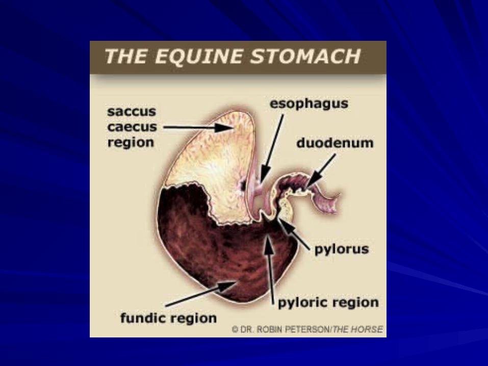
StomachAlmost half the mucosal surface is lined with squamous instead of glandular epithelium
Glandular mucosa is divided into: – Fundic (5.4) and pyloric (2.6) regions
Fundic mucosa contain– Parietal cells that secrete HCl (substrate?)– Zymogen cells which secret pepsin (substrate?)
Pyloric Region – Secretes the polypeptide hormone gastrin in to the blood
plasma

Stomach
What triggers the secretion of gastrin?– A meal
What triggers the cease of gastrin secretion?
– Distention of stomach wall
Most prolonged gastrin secretion occurs – When horses eat hay freely

Stomach
Some fermentation takes place– Primarily yields lactic acid– Occurs in esophageal, fundic, and saccus caecus regions– All lined by squamous cells– As digesta approaches pylorus pH falls
↑ HCl → ↑Pepsin → ↓ fermentation
Protein digestion is slight, why?– Small stomach– Short dwell time


Gastric Ulceration
• Two General Types:• Squamous Ulceration• Glandular Ulceration
• One research study indicated:• TB’s in training = 80%• TB’s off one month = 52%
• Clinical signs generally include colic and pronounced teeth grinding
• In general, ↑ grain = ↑ ulceration

Squamous Ulceration
The dorsal region is covered by squamous epithelium and ulcers occur here as a direct result of extended exposure to acid secretions. Many equine stomach ulcers occur in the area near to the esophagus. In foals the developing cell lining is thinner than in adults, making foals especially prone to gastric ulceration

Glandular UlcerationThe ventral region is covered in glandular epithelium and ulcers occur here when the protective mucus layer is compromised e.g. due a side-effect of certain medications, enabling acid erosion of the stomach wall.

Small Intestine– Divided into three parts:
Duodenum
Jejunum
Ilieum
– How long?50 - 70 feet long
3 – 4 “ diameter
– May hold up to 10 to 12 gallons Some food passes In as little as 15 minutes
Most takes 10 hrs

Gastro-intestinal Tract


Small Intestine
Pancreatic juices secreted due to: – Food in stomach– Mediated
Vagal nerve fibers in S.I.
Presence of HCl in duodenum– Stimulates secretin production in blood
Secretin – Controls secretions in duodenum– Also increase pancreatic juice secretion by 4 to 5
times– Creates buffering effect

Small Intestine
Other pancreatic secretions present include:– Trypsin (protease)– Lipase
Bile– Secreted by liver– Stimulated by HCl in duodenum– Stored in gall bladder?– Fat digestion
Bicarbonate content ↑ towards ileum– Buffer to large intestinal VFA’s

Small Intestine
– What is digested in the S.I.?Fat
Protein
50 -70% soluble CHO’s
Vitamins and Minerals also absorbed
Liquids pass through rapidly– Reach cecum 2 to 8 hrs post ingestion– 5 hours later, liquid reaches colon

Small Intestine
What leaves the small intestine?– Fibrous Feed residues– Undigested feed starch– Protein– Microorganisms– Intestinal Secretions– Cell debris

Large Intestine
What occurs in the LI?– Fermentation of digesta by microorganisms
produces:VFA’s
Lactate
– Slow in comparison to the digestion ofStarch and protein
– What is located at distal end of illeum that allows fermentation?
Cecum

Cecum
3 to 4‘ in length
Holds 7 to 8 gallons
Contains bacteria
What is digested?– Large amounts of fiber– ~ ½ of the soluble CHO’s ingested
Absorption can also occur
Bacterial protein – Produced, digested, and absorbed

Ascending Colon
10 to 12 ‘ long
Diameter of 8 to 10”
Holds 14 to 16 gallons
Four Portions:– Right ventral colon– Sternal flexure or left ventral colon– Pelvic Flexure to the left dorsal colon– Diaphragmatic flexure to the right dorsal colon
(connects to small colon)

Ascending Colon
Passage of particulate matter and liquids slow– 36 to 48 hrs
What is digested in the Colon?
Nearly all – CF– Cellulose– Over 50% soluble CHO’s – Passes through S.I. into cecum

Cecum and Colon
Digestion depends almost entirely on:– Constituent bacteria and ciliate protozoa– Walls contain only mucous-secreting glands– No digestive enzymes