Embed Size (px)
Citation preview
449
C H A P T E R
S e c t i o n 5
Parasitic Diseases
54
Laboratory Diagnosis of Parasitic DiseasesEllis C. Greiner
Some helminth and protozoan parasites are easily diagnosed because the diagnostic stages passed in the feces are readily detected and identified. Other parasites require more effort to identify because the eggs are not distinguishable; larval fecal cultures are needed to determine the species or groups of nema-todes present, and some require special procedures to enhance diagnosis. This chapter assists the equine clinician in sample selection and interpretation of results to facilitate rapid and accurate diagnosis of helminth and protozoan parasitic diseases of horses.
Helminth Diagnosis
Some helminth parasites of horses have complex life cycles, and immature stages may go on circuitous journeys through viscera before arriving at the site where the adult worms will develop and produce offspring. Sometimes, these worms do not follow the correct route and migrate to the central nervous system (CNS), where they may cause severe damage. These conditions are usually confirmed at necropsy, when the parasites may be recovered and identified.
When larval stages of parasites migrate through tissues, normal parasite diagnosis by fecal examination is not possible because the mature adults have not developed and have not begun to produce and release the eggs or larvae. This interval is referred to as the prepatent period.
Diagnosis of helminth infection based on observation of eggs or larvae is facilitated by examination of fresh fecal samples. Some parasites are common in horses at any age, whereas others are restricted to the foals because the adult horses mount a sufficient immune response to prevent the adult worms from developing.
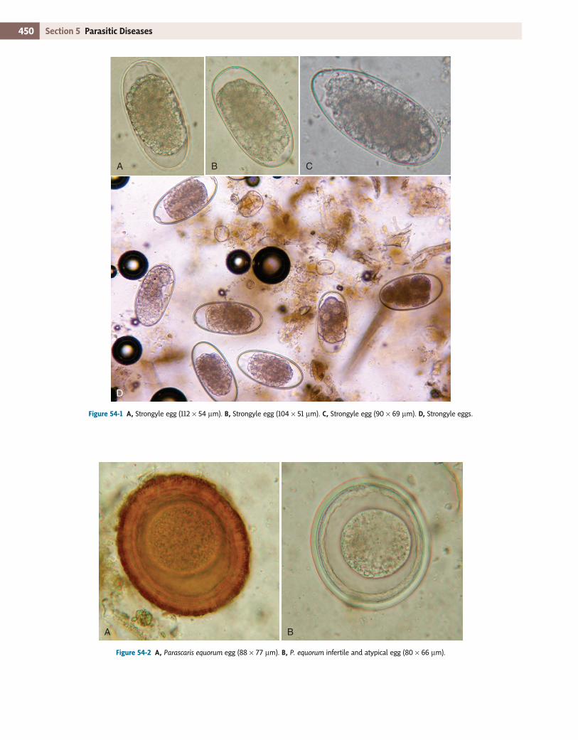
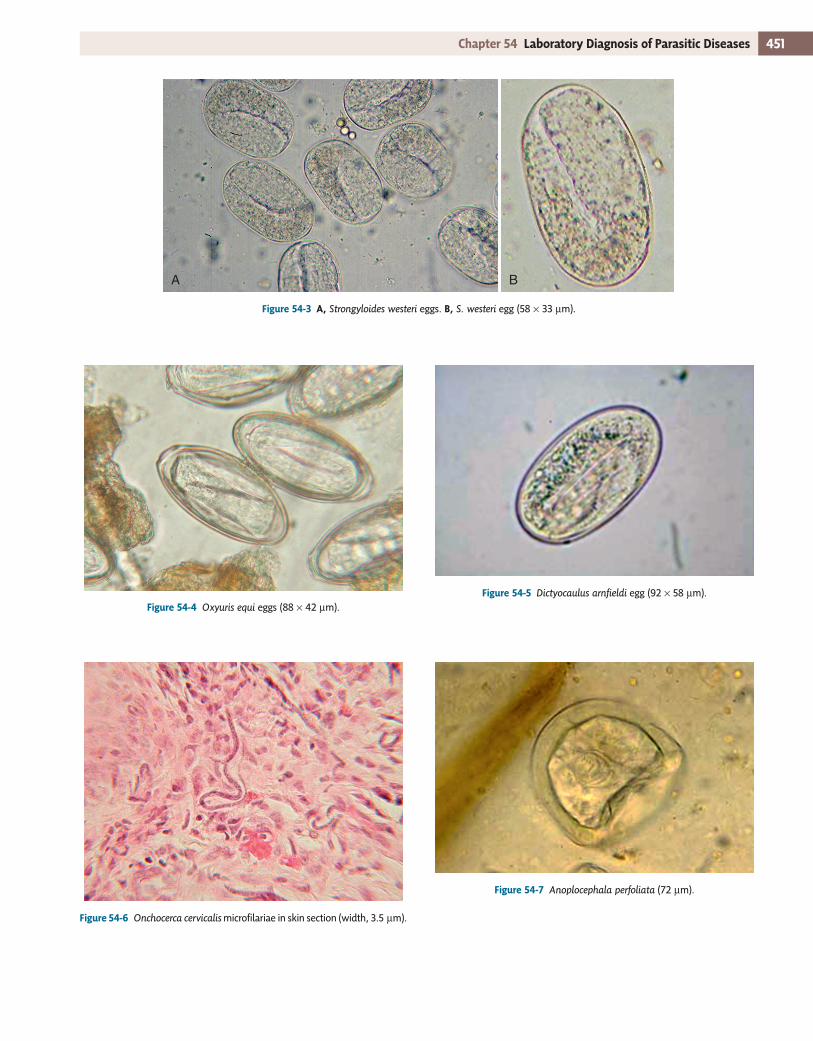
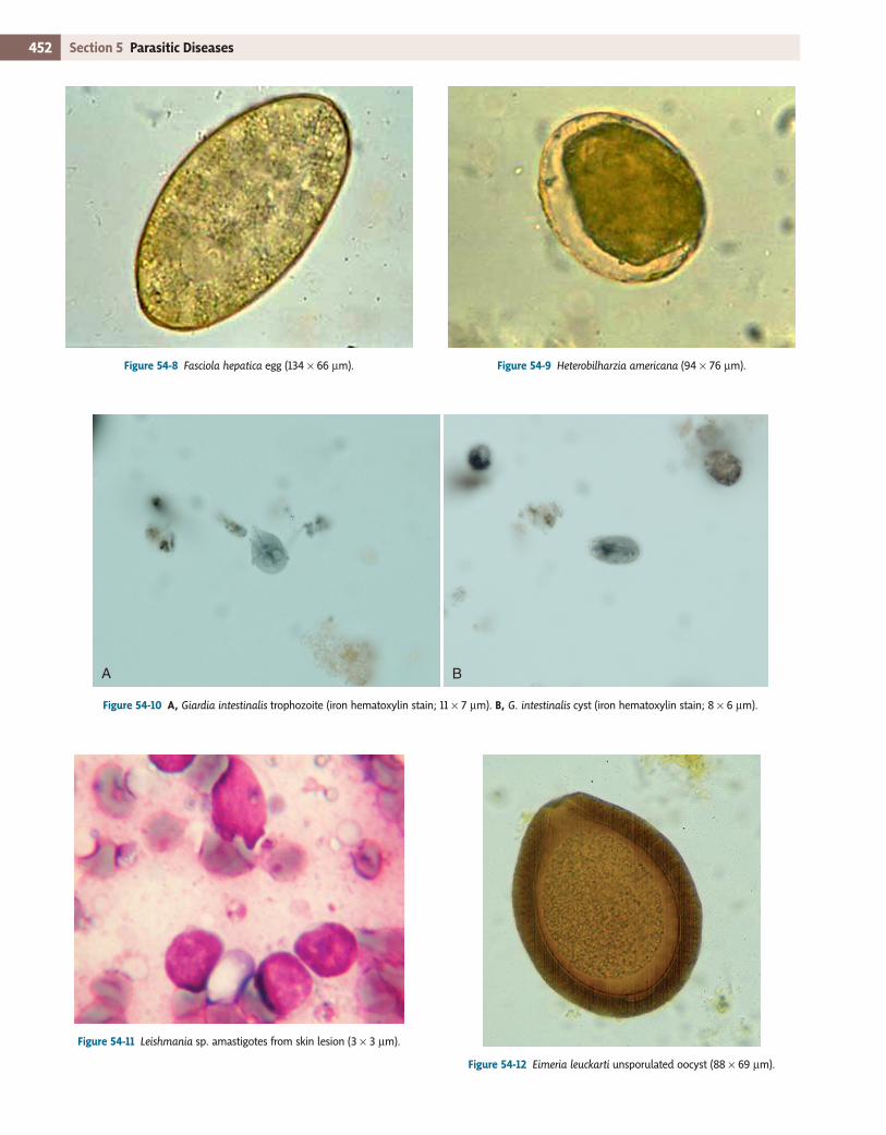
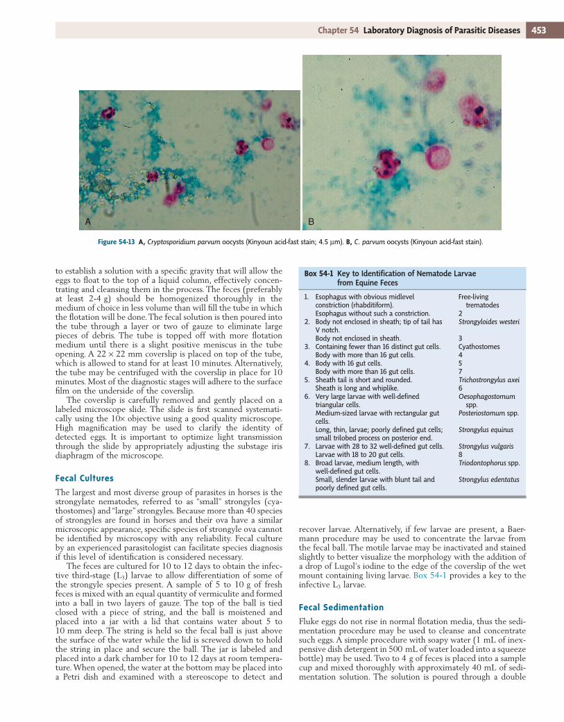
A large variety of nematode parasites infect horses. Most reside in the gastrointestinal (GI) tract and are detected by fecal examination; examples include the strongyles (Fig. 54-1), asca-rids (Fig. 54-2), threadworms (Fig. 54-3), pinworms (Fig. 54-4), lungworms (Fig.54-5), and stomach worms. Some nematodes reside in solid tissues and do not pass any stages in the feces; these include the filarial worms, Onchocerca cervicalis, O. reticu-lata, and Parafilaria multipapillosa, which are detected by dis-covery of microfilariae, which are motile embryos (Fig. 54-6). Some worms cause severe problems but do not produce any stages that leave the host, such as Halicephalobus deletrix, which is usually diagnosed by histopathology at necropsy. Summer sores are caused by larval stomach worms which produce no eggs or larvae and are species of Draschia and Habronema.
The diversity of flatworms is rather depauperate compared with the nematodes. Three species of tapeworms develop as adults in the intestines (Anoplocephala perfoliata [Fig. 54-7], A. magna and Paranoplocephala mamillana), and the larval cyst of
one tapeworm (hydatid cyst of Echinococcus) rarely develops in the liver. Two flukes, Fasciola hepatica in the liver (Fig. 54-8) and Heterobilharzia americana in mesenteric veins (Fig. 54-9), rarely infect horses because the horse is not the normal host for either. These flukes develop to adults that shed eggs in the feces, which could be detected by fecal sedimentation.
Protozoan Diagnosis
Very few protozoal organisms infect the GI tract of horses, and some are believed to be beneficial as the ciliate fauna of the large bowel. Others are pathogenic or potentially pathogenic, including flagellates, such as Giardia (Fig. 54-10) and possibly Leishmania (Fig. 54-11), and coccidians, such as one species of Eimeria (Fig. 54-12) and Cryptosporidium (Fig. 54-13).
Laboratory Procedures
Procedures that are used to diagnose parasitic infections in horses include gross fecal examination, fecal flotation, fecal culture, fecal sedimentation, direct smear, Baermann procedure, cellophane (Scotch) tape test, McMaster’s counts, stained fecal smears, impression smears, skin biopsy examination, and blood smears. It is highly desirable to have a compound microscope calibrated with an ocular micrometer to facilitate precise mea-surement of ova or other parasite structures.
Gross Fecal Examination
Before altering the feces for diagnostic purposes, examination without magnification for consistency, evidence of blood, and the macroscopic presence of worms should be performed. If the horse is impacted or there is delayed movement of ingesta, parasite ova may be more developed than normal. Conversely, if there is diarrhea, ova may not be as developed as expected. Blood might indicate high numbers of strongyles migrating within the intestinal mucosa, with resultant petechial hemor-rhages and frank hemorrhage.
Fecal Flotation
Fecal flotation techniques use a variety of solutions, including sodium nitrate, zinc sulfate, sucrose, and sodium chloride, to identify parasite ova or larvae. The author prefers sodium nitrate as a fecal flotation solution because it will concentrate most nematode eggs and larvae, tapeworm eggs, flagellate cysts, and coccidian oocysts, as well as parasitic mites consumed when the host is trying to alleviate mite-associated pruritus. The goal is
450 Section 5 Parasitic Diseases
Figure 54-1 A, Strongyle egg (112 × 54 µm). B, Strongyle egg (104 × 51 µm). C, Strongyle egg (90 × 69 µm). D, Strongyle eggs.
A B C
D
Figure 54-2 A, Parascaris equorum egg (88 × 77 µm). B, P. equorum infertile and atypical egg (80 × 66 µm).
A B
451Chapter 54 Laboratory Diagnosis of Parasitic Diseases
Figure 54-3 A, Strongyloides westeri eggs. B, S. westeri egg (58 × 33 µm).
A B
Figure 54-4 Oxyuris equi eggs (88 × 42 µm). Figure 54-5 Dictyocaulus arnfieldi egg (92 × 58 µm).
Figure 54-6 Onchocerca cervicalis microfilariae in skin section (width, 3.5 µm).
Figure 54-7 Anoplocephala perfoliata (72 µm).
452 Section 5 Parasitic Diseases
Figure 54-8 Fasciola hepatica egg (134 × 66 µm). Figure 54-9 Heterobilharzia americana (94 × 76 µm).
Figure 54-10 A, Giardia intestinalis trophozoite (iron hematoxylin stain; 11 × 7 µm). B, G. intestinalis cyst (iron hematoxylin stain; 8 × 6 µm).
A B
Figure 54-11 Leishmania sp. amastigotes from skin lesion (3 × 3 µm).
Figure 54-12 Eimeria leuckarti unsporulated oocyst (88 × 69 µm).
453Chapter 54 Laboratory Diagnosis of Parasitic Diseases
to establish a solution with a specific gravity that will allow the eggs to float to the top of a liquid column, effectively concen-trating and cleansing them in the process. The feces (preferably at least 2-4 g) should be homogenized thoroughly in the medium of choice in less volume than will fill the tube in which the flotation will be done. The fecal solution is then poured into the tube through a layer or two of gauze to eliminate large pieces of debris. The tube is topped off with more flotation medium until there is a slight positive meniscus in the tube opening. A 22 × 22 mm coverslip is placed on top of the tube, which is allowed to stand for at least 10 minutes. Alternatively, the tube may be centrifuged with the coverslip in place for 10 minutes. Most of the diagnostic stages will adhere to the surface film on the underside of the coverslip.
The coverslip is carefully removed and gently placed on a labeled microscope slide. The slide is first scanned systemati-cally using the 10× objective using a good quality microscope. High magnification may be used to clarify the identity of detected eggs. It is important to optimize light transmission through the slide by appropriately adjusting the substage iris diaphragm of the microscope.
Fecal Cultures
The largest and most diverse group of parasites in horses is the strongylate nematodes, referred to as “small” strongyles (cya-thostomes) and “large” strongyles. Because more than 40 species of strongyles are found in horses and their ova have a similar microscopic appearance, specific species of strongyle ova cannot be identified by microscopy with any reliability. Fecal culture by an experienced parasitologist can facilitate species diagnosis if this level of identification is considered necessary.
The feces are cultured for 10 to 12 days to obtain the infec-tive third-stage (L3) larvae to allow differentiation of some of the strongyle species present. A sample of 5 to 10 g of fresh feces is mixed with an equal quantity of vermiculite and formed into a ball in two layers of gauze. The top of the ball is tied closed with a piece of string, and the ball is moistened and placed into a jar with a lid that contains water about 5 to 10 mm deep. The string is held so the fecal ball is just above the surface of the water while the lid is screwed down to hold the string in place and secure the ball. The jar is labeled and placed into a dark chamber for 10 to 12 days at room tempera-ture. When opened, the water at the bottom may be placed into a Petri dish and examined with a stereoscope to detect and
Figure 54-13 A, Cryptosporidium parvum oocysts (Kinyoun acid-fast stain; 4.5 µm). B, C. parvum oocysts (Kinyoun acid-fast stain).
A B
recover larvae. Alternatively, if few larvae are present, a Baer-mann procedure may be used to concentrate the larvae from the fecal ball. The motile larvae may be inactivated and stained slightly to better visualize the morphology with the addition of a drop of Lugol’s iodine to the edge of the coverslip of the wet mount containing living larvae. Box 54-1 provides a key to the infective L3 larvae.
Fecal Sedimentation
Fluke eggs do not rise in normal flotation media, thus the sedi-mentation procedure may be used to cleanse and concentrate such eggs. A simple procedure with soapy water (1 mL of inex-pensive dish detergent in 500 mL of water loaded into a squeeze bottle) may be used. Two to 4 g of feces is placed into a sample cup and mixed thoroughly with approximately 40 mL of sedi-mentation solution. The solution is poured through a double
Box 54-1 KeytoIdentificationofNematodeLarvaefromEquineFeces
1. Esophagus with obvious midlevel constriction (rhabditiform).
Free-living trematodes
Esophagus without such a constriction. 22. Body not enclosed in sheath; tip of tail has
V notch.Strongyloides westeri
Body not enclosed in sheath. 33. Containing fewer than 16 distinct gut cells. Cyathostomes
Body with more than 16 gut cells. 44. Body with 16 gut cells. 5
Body with more than 16 gut cells. 75. Sheath tail is short and rounded. Trichostrongylus axei
Sheath is long and whiplike. 66. Very large larvae with well-defined
triangular cells.Oesophagostomum
spp.Medium-sized larvae with rectangular gut cells.
Posteriostomum spp.
Long, thin, larvae; poorly defined gut cells; small trilobed process on posterior end.
Strongylus equinus
7. Larvae with 28 to 32 well-defined gut cells. Strongylus vulgarisLarvae with 18 to 20 gut cells. 8
8. Broad larvae, medium length, with well-defined gut cells.
Triodontophorus spp.
Small, slender larvae with blunt tail and poorly defined gut cells.
Strongylus edentatus
454 Section 5 Parasitic Diseases
Cellophane (Scotch) Tape Test
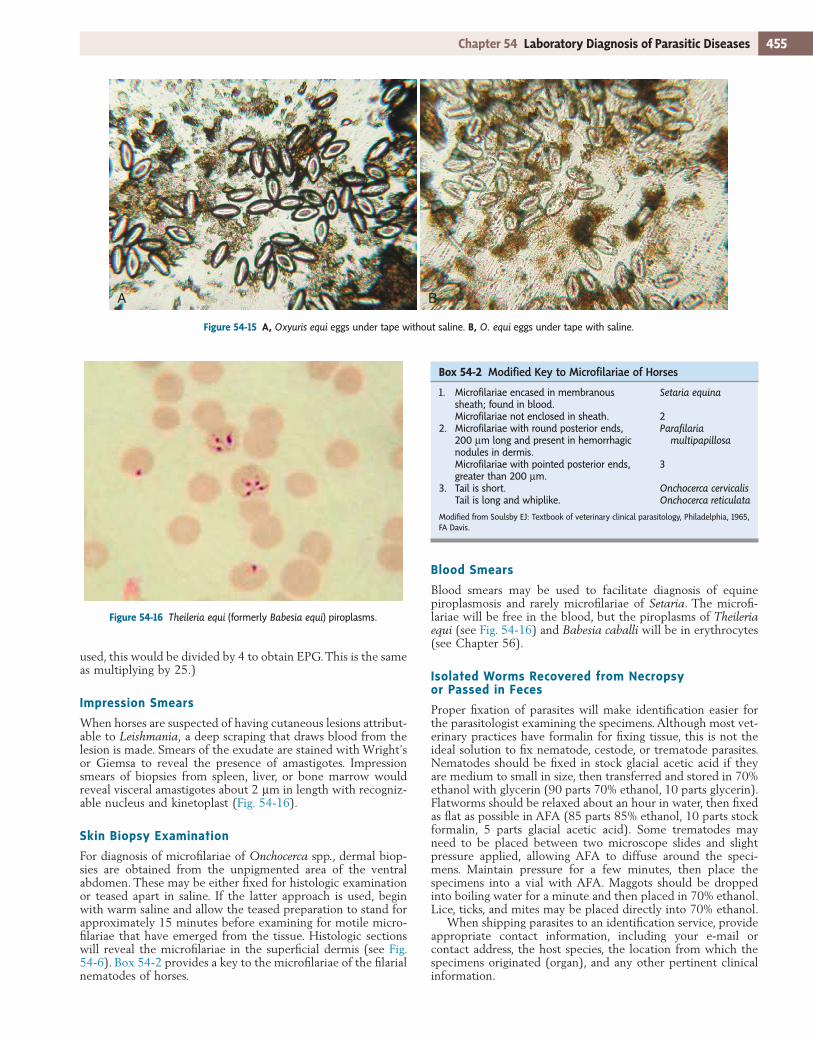
The cellophane (Scotch) tape test is the preferred test for detec-tion of pinworm (Oxyuris equi) ova (see Fig. 54-4). These ova adhere to the perineum and are rarely observed in routine fecal flotation or sedimentation samples. A length of clear cellophane tape is wrapped around three or four fingers with the sticky side out, and the tape is pressed against the perineum, then placed onto a glass slide. Saline or water may be placed over the tape and a long coverslip added to facilitate visualization of the ova (Fig. 54-15).
McMaster’s Procedure
The McMaster’s procedure is used to estimate the level of fecal contamination with parasite ova. Because it is a dilution tech-nique, it may produce a false-negative result if low numbers of ova are present in a sample. Therefore it is best used in combi-nation with an initial standard flotation. The McMaster’s tech-nique is often used to determine whether an anthelmintic treatment was effective. Pretreatment results are compared to results from samples collected 10 to 14 days after therapy.
A 4-g sample of fresh feces is added to a 120-mL screw-cap sample cup with sufficient sodium nitrate solution (fecal flota-tion medium) to bring the total volume to 30 mL and is mixed thoroughly. The solution is poured through one layer of gauze into another cup. After thorough mixing, the solution is used to fill the McMaster chamber. After standing for 10 minutes, the slide is placed on a compound microscope with the 10× objective in place. The width of each of the six lanes of the McMaster chamber is equal to the diameter of the field of view. The slide is systematically scanned to count the total number of each type of egg present on both sides of the chamber. If the counts between sides vary more than 20%, it is considered evidence of insufficient mixing of the sample, and the proce-dure should be repeated. The total count by parasite type present is multiplied by 25 to determine the eggs per gram (EPG) of feces. (NOTE: The volume of fluid and the weight of feces used can vary and are based on the following: the volume under each grid is 0.15 mL, thus the volume under both grids is 0.3 mL. This is 1
100 the volume used [30 mL], thus the number of eggs would be multiplied by 100; if 4 g of feces were
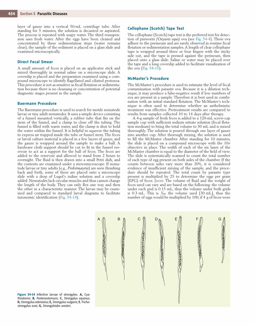
Figure 54-14 Infective larvae of strongyles. A, Cya-thostome; B, Posteriostomum; C, Strongylus equinus; D, Strongylus edentatus; E, Strongylus vulgaris; F, Tricho-strongylus axei; G, Strongyloides westeri.
A B C D E F G
layer of gauze into a vertical 50-mL centrifuge tube. After standing for 5 minutes, the solution is decanted or aspirated. The process is repeated with soapy water. The third resuspen-sion uses fresh water. After the eggs have been cleaned and concentrated by these sedimentation steps (water remains clear), the sample of the sediment is placed on a glass slide and examined microscopically.
Direct Fecal Smear
A small amount of feces is placed on an applicator stick and mixed thoroughly in normal saline on a microscope slide. A coverslip is placed and the preparation examined using a com-pound microscope to identify flagellated and ciliated protozoa. This procedure is not as sensitive as fecal flotation or sedimenta-tion because there is no cleansing or concentration of potential diagnostic stages present in the sample.
Baermann Procedure
The Baermann procedure is used to search for motile nematode larvae or tiny adult nematodes. It uses a simple device consisting of a funnel mounted vertically, a rubber tube that fits on the stem of the funnel, and a clamp to close off the tubing. The funnel is filled with warm water, and the clamp is shut to hold the water within the funnel. It is helpful to squeeze the tubing to express air trapped inside the tube or funnel stem. The feces or larval culture material is placed in a few layers of gauze, and the gauze is wrapped around the sample to make a ball. A hardware cloth support should be cut to fit in the funnel res-ervoir to act as a support for the ball of feces. The feces are added to the reservoir and allowed to stand from 2 hours to overnight. The fluid is then drawn into a small Petri dish, and the contents are examined under a stereomicroscope. If nema-tode larvae or tiny adults (e.g., Probstmayria) are seen thrashing back and forth, some of these are placed onto a microscope slide with a drop of Lugol’s iodine solution and a coverslip added. Nematodes lack circular muscles and thus cannot change the length of the body. They can only flex one way and then the other in a characteristic manner. The larvae may be exam-ined and compared to standard larval diagrams to facilitate taxonomic identification (Fig. 54-14).
455Chapter 54 Laboratory Diagnosis of Parasitic Diseases
Blood Smears
Blood smears may be used to facilitate diagnosis of equine piroplasmosis and rarely microfilariae of Setaria. The microfi-lariae will be free in the blood, but the piroplasms of Theileria equi (see Fig. 54-16) and Babesia caballi will be in erythrocytes (see Chapter 56).
Isolated Worms Recovered from Necropsy or Passed in Feces
Proper fixation of parasites will make identification easier for the parasitologist examining the specimens. Although most vet-erinary practices have formalin for fixing tissue, this is not the ideal solution to fix nematode, cestode, or trematode parasites. Nematodes should be fixed in stock glacial acetic acid if they are medium to small in size, then transferred and stored in 70% ethanol with glycerin (90 parts 70% ethanol, 10 parts glycerin). Flatworms should be relaxed about an hour in water, then fixed as flat as possible in AFA (85 parts 85% ethanol, 10 parts stock formalin, 5 parts glacial acetic acid). Some trematodes may need to be placed between two microscope slides and slight pressure applied, allowing AFA to diffuse around the speci-mens. Maintain pressure for a few minutes, then place the specimens into a vial with AFA. Maggots should be dropped into boiling water for a minute and then placed in 70% ethanol. Lice, ticks, and mites may be placed directly into 70% ethanol.
When shipping parasites to an identification service, provide appropriate contact information, including your e-mail or contact address, the host species, the location from which the specimens originated (organ), and any other pertinent clinical information.
Figure 54-15 A, Oxyuris equi eggs under tape without saline. B, O. equi eggs under tape with saline.
A B
Figure 54-16 Theileria equi (formerly Babesia equi) piroplasms.
Box 54-2 ModifiedKeytoMicrofilariaeofHorses
1. Microfilariae encased in membranous sheath; found in blood.
Setaria equina
Microfilariae not enclosed in sheath. 22. Microfilariae with round posterior ends,
200 µm long and present in hemorrhagic nodules in dermis.
Parafilaria multipapillosa
Microfilariae with pointed posterior ends, greater than 200 µm.
3
3. Tail is short. Onchocerca cervicalisTail is long and whiplike. Onchocerca reticulata
Modified from Soulsby EJ: Textbook of veterinary clinical parasitology, Philadelphia, 1965, FA Davis.
used, this would be divided by 4 to obtain EPG. This is the same as multiplying by 25.)
Impression Smears
When horses are suspected of having cutaneous lesions attribut-able to Leishmania, a deep scraping that draws blood from the lesion is made. Smears of the exudate are stained with Wright’s or Giemsa to reveal the presence of amastigotes. Impression smears of biopsies from spleen, liver, or bone marrow would reveal visceral amastigotes about 2 µm in length with recogniz-able nucleus and kinetoplast (Fig. 54-16).
Skin Biopsy Examination
For diagnosis of microfilariae of Onchocerca spp., dermal biop-sies are obtained from the unpigmented area of the ventral abdomen. These may be either fixed for histologic examination or teased apart in saline. If the latter approach is used, begin with warm saline and allow the teased preparation to stand for approximately 15 minutes before examining for motile micro-filariae that have emerged from the tissue. Histologic sections will reveal the microfilariae in the superficial dermis (see Fig. 54-6). Box 54-2 provides a key to the microfilariae of the filarial nematodes of horses.

Suggested ReadingsBowman DD: Georgi’s parasitology for veterinarians, Philadelphia, 2009,
Saunders.Jacobs DE: A color atlas of equine parasites, Philadelphia, 1986, Lea & Febiger.Lichtenfels JR: Helminths of domestic equids, Proc Helm Soc Wash 42, Special
Issue 92, 1975.Manual of veterinary parasitological laboratory techniques, Technical Bulletin 18,
London, 1979, Ministry of Agriculture, Fisheries and Food.Soulsby EJ: Textbook of veterinary clinical parasitology, vol 1, Helminths, Phila-
delphia, 1965, FA Davis.
The adult helminths passed in the feces and large enough to see with the naked eye include Anoplocephala magna (see Fig. 58-1, B), Anoplocephala perfoliata (see Fig. 58-1, A), Parascaris (see Fig. 57-6), Oxyuris equi (see Fig. 57-10), and nematodes observed on the sleeves of gloves used in rectal palpations include species of Strongylus (see Fig. 57-4) and cyathostomes (see Fig. 57-3).