Embed Size (px)
Citation preview
J Clin Pathol 1986;39:435-439
Epithelial membrane antigen and cytokeratinexpression by meningiomas: an immunohistologicalstudyJM THEAKER, KC GATTER, MM ESIRI, KA FLEMING
From the Nuffield Department ofPathology, University ofOxford, John Radeliffe Hospital, Oxford
SUMMARY Thirteen meningiomas of varying light microscopic features were studied immuno-histologically using a panel of monoclonal antibodies directed against epithelial, mesenchymal, andneural components. All 13 meningiomas showed expression of epithelial membrane antigen,vimentin, and S100 protein, as did normal meninges. Five of the 13 meningiomas also showed focalexpression of cytokeratins, with double labelling showing expression of cytokeratins and vimentinby different cells. The cytokeratin expression was especially noticeable in cells surrounding thehyaline bodies of meningiomas. These results provide further evidence that meningiomas havefeatures of both mesenchymal and epithelial tissues.
Meningiomas are primary tumours thought to arisefrom the arachnoidal cap cell of the meninges.' As themeninges are believed to develop from the connectivetissue surrounding the neural tube, meningiomas areconsidered to be mesenchymal tumours.' Meningio-mas, however, show considerable heterogeneity onlight microscopy' 2 and, indeed, can have epithelialfeatures such as large numbers of desmosomes.3 Fur-thermore, occasional tumours contain periodic acidSchiff (PAS) positive hyaline bodies, which, ultra-structurally, can resemble the intracytoplasmiclumina of some adenocarcinomas, and which Kepeshas suggested reflects secretory differentiation bymeningioma cells.4
In this study a series of meningiomas of varyingmorphological patterns was examined with a panel ofmonoclonal antibodies, which recognises a variety ofantigens of epithelial, mesenchymal, and neural type.The aim of this study was to clarify the origin ofmeningiomas, their relation to other cells and tissues,and to establish whether a panel of monoclonal anti-bodies might be useful in their differential diagnosis.
Material and methods
Thirteen cases of meningioma were recovered fromthe files of the neuropathology department, TheRadcliffe Infirmary, Oxford, or the histopathologydepartment, John Radcliffe Hospital, Oxford. All hadbeen routinely fixed in formalin and embedded inparaffin wax.
Table 1 gives details of the monoclonal antibodiesAccepted for publication 18 December 1985
used in this study. Peroxidase conjugated antibodieswere obtained from Dakopatts.
IMMUNOENZYMATIC TECHNIQUESSingle labelled sections were stained by a three stageimmunoperoxidase technique, as described pre-viously.7 To visualise two antigens simultaneously(double labelling) slides were first stained by theimmunoperoxidase technique for one antigen andthen by the APAAP immunoalkaline phosphatasesystem for the other antigen.8 9
Results
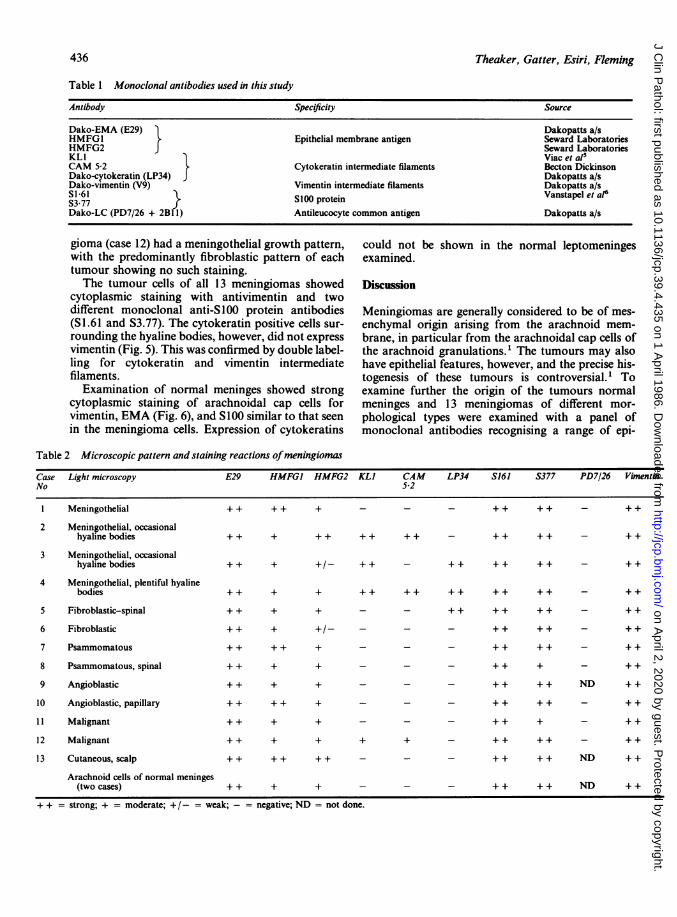
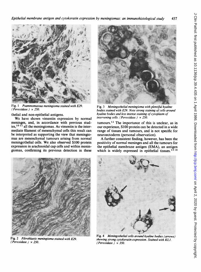
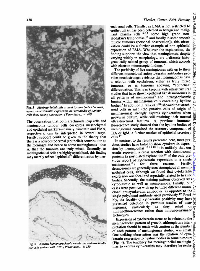
Table 2 summarises the results obtained. The moststriking feature was that the tumour cells of all 13 ofthe meningiomas, regardless of morphology, werepositive for epithelial membrane antigen (EMA),shown most consistently and strongly by antibodyE29. The staining was both cytoplasmic and mem-brane bound (Figs. 1, 2, and 3). The psammoma bod-ies seen in many of the tumours were, however,always negative, although the hyaline bodies in cases3 and 4 did stain.
Five of the meningiomas were also positivelylabelled by at least one of the three anticytokeratinantibodies. This staining was restricted mainly to thecytoplasm of a minority of the tumour cells and wasparticularly noticeable in the cells surrounding thePAS positive hyaline bodies seen in cases 2, 3, and 4(Fig. 4). The hyaline material itself was unstained.The foci showing cytokeratin expression both in thefibroblastic tumour (case 5) and the malignant menin-
435
on April 2, 2020 by guest. P
rotected by copyright.http://jcp.bm
j.com/
J Clin P
athol: first published as 10.1136/jcp.39.4.435 on 1 April 1986. D
ownloaded from
Theaker, Gatter, Esiri, Fleming
Table I Monoclonal antibodies used in this study
Antibody Specificity Source
Dako-EMA (E29) Dakopatts a/sHMFGI Epithelial membrane antigen Seward LaboratoriesHMFG2 J Seward LaboratoriesKLI Viac et al'CAM 5-2 3 Cytokeratin intermediate filaments Becton DickinsonDako-cytokeratin (LP34) Dakopatts a/sDako-vimentin (V9) Vimentin intermediate filaments Dakopatts a/sSI 61 i} SlOO protein Vanstapel et al6
Dako-LC (PD7/26 + 2B 1) Antileucocyte common antigen Dakopatts a/s
gioma (case 12) had a meningothelial growth pattern,with the predominantly fibroblastic pattern of eachtumour showing no such staining.The tumour cells of all 13 meningiomas showed
cytoplasmic staining with antivimentin and twodifferent monoclonal anti-S100 protein antibodies(S1.61 and S3.77). The cytokeratin positive cells sur-rounding the hyaline bodies, however, did not express
vimentin (Fig. 5). This was confirmed by double label-ling for cytokeratin and vimentin intermediatefilaments.
Examination of normal meninges showed strongcytoplasmic staining of arachnoidal cap cells forvimentin, EMA (Fig. 6), and S100 similar to that seen
in the meningioma cells. Expression of cytokeratins
could not be
examined.
shown in the normal leptomeninges
Discussion
Meningiomas are generally considered to be of mes-enchymal origin arising from the arachnoid mem-brane, in particular from the arachnoidal cap cells ofthe arachnoid granulations.' The tumours may alsohave epithelial features, however, and the precise his-togenesis of these tumours is controversial.' Toexamine further the origin of the tumours normalmeninges and 13 meningiomas of different mor-phological types were examined with a panel ofmonoclonal antibodies recognising a range of epi-
Table 2 Microscopic pattern and staining reactions ofmeningiomas
Case Light microscopy E29 HMFGI HMFG2 KLI CAM LP34 S161 S377 PD7/26 VimentinNo 52
1 Meningothelial ++ ++ + - - - ++ ++ - ++
2 Meningothelial, occasionalhyalinebodies ++ + ++ ++ ++ - ++ ++ - ++
3 Meningothelial, occasionalhyalinebodies ++ + +/- ++ - ++ ++ ++ - ++
4 Meningothelial, plentiful hyalinebodies + + + + ++ ++ ++ ++ ++ - ++
5 Fibroblastic-spinal + + + + - - + + + + + + - + +
6 Fibroblastic + + + +/- - - - + + + + - + +
7 Psammomatous + + + + + - - - + + + + - ++
8 Psammomatous, spinal + + + + - - - ++ + - + +
9 Angioblastic + + + + - - - + + + + ND + +
10 Angioblastic, papillary + + + + + - - - + + + + - ++
11 Malignant + + + + - - - + + + - ++
12 Malignant + + + + + + - + + + + - ++
13 Cutaneous, scalp + + + + + + - - - + + + + ND + +
Arachnoid cells of normal meninges(two cases) + + + + - - - + + + + ND + +
+ + = strong; + = moderate; +/- = weak; - = negative; ND = not done.
436
on April 2, 2020 by guest. P
rotected by copyright.http://jcp.bm
j.com/
J Clin P
athol: first published as 10.1136/jcp.39.4.435 on 1 April 1986. D
ownloaded from
Epithelial membrane antigen and cytokeratin expression by meningiomas: an immunohistological study 437
D,}v
TsXt.. X.-
9,%*4,,.'I: . j
a 'w7-l -A~ 4-F -p-N -W 20Fig. 1 Psammomatous meningioma stained with E29.(Peroxidase.) x 250.
thelial and non-epithelial antigens.We have shown vimentin expression by normal
meninges, and, in accordance with previous stud-ies,10 11 all the meningiomas. As vimentin is the inter-mediate filament of mesenchymal cells this result canbe interpreted as supporting the view that meningio-mas are mesenchymal tumours arising from normalmeningothelial cells. We also observed S1OO proteinexpression in arachnoidal cap cells and within menin-giomas, confirming its previous detection in these
Fig. 2 Fibroblastic meningioma stained with E29.(Peroxidase.) x 250.
Fig. 3 Meningothelial meningioma with plentiful hyalinebodies stained with E29. Note strong staining ofcells aroundhyaline bodies and less intense staining ofcytoplasm ofintervening cells. (Peroxidase.) x 250.
tumours. 2 The importance of this is unclear, as inour experience, SI 00 protein can be detected in a widerange of tissues and tumours, and is not specific forneuroectoderm (personal observation).A further consistent finding, however, has been the
positivity of normal meninges and all the tumours forthe epithelial membrane antigen (EMA), an antigenwhich is widely expressed in epithelial tissues.13 14
Fig. 4 Meningothelial cells around hyaline bodies (arrows)soigstrong cytokeratin expression. Stained with KUI.
(Peroxidase.) x 350.
-,
.'v:t
to
IN
on April 2, 2020 by guest. P
rotected by copyright.http://jcp.bm
j.com/
J Clin P
athol: first published as 10.1136/jcp.39.4.435 on 1 April 1986. D
ownloaded from
438
Fig. 5 Meningothelial cells around hyaline bodies (arrows)do not show vimentin expression, but remainder oftumourcells show strong expression. (Peroxidase.) x 400.
The observation that both arachnoidal cap cells andmeningioma tumour cells coexpress mesenchymaland epithelial markers-namely, vimentin and EMA,respectively, can be interpreted in several ways.
Firstly, support could be given to the theory thatthere is a neuroectodermal (epithelial) contribution tothe meninges and hence to some meningiomas-thatis, that the tumours are truly mixed. Secondly, as
meningothelial cells are highly specialised, this findingmay merely reflect "epithelial" differentiation by mes-
..,,*.....< .t
*f _*1.. :. .
§F £ _ g
e ,M":! __
... st_
h: 5 s*' o __.t M . :e.
W oiS F_
Fig. 6 Normal human arachnoid membrane and arachnidatcap cells stained with E29. (Peroxidase.) x 150.
Theaker, Gatter, Esiri, Fleming
enchymal cells. Thirdly, as EMA is not restricted toepithelium (it has been detected in benign and malig-nant plasma cells,14 15 some high grade non-Hodgkin's lymphomas,15 and focally in some smoothmuscle tumours (personal observation)), this obser-vation could be a further example of non-epithelialexpression of EMA. Whatever the explanation, thefinding supports the view that meningiomas, despitevarying widely in morphology, are a discrete histo-genetically related group of tumours, which accordswith electron microscopic findings.3The positivity of five meningiomas with up to three
different monoclonal anticytokeratin antibodies pro-vides much stronger evidence that meningiomas havea relation with epithelium, either as truly mixedtumours, or as tumours showing "epithelial"differentiation. This is in keeping with ultrastructuralstudies that have shown epithelial like desmosomes inall patterns of menigiomas3 and intracytoplasmiclumina within meningioma cells containing hyalinebodies.4 In addition, Frank et all6 showed that arach-noid cells in man (the putative cells of origin ofmeningiomas) strongly express cytokeratins whengrown in culture, while still retaining their normalultrastructural features. A previous immuno-fluorescence study showed that the hyaline bodies ofmeningiomas contained the secretory component ofIgA or IgM, a further marker of epithelial secretorycells. 7
In contrast to the results presented here, most pre-vious studies have failed to show cytokeratin expres-sion by meningiomas.'0 1 18 It is unlikely that our
results represent a cross reaction with desmosomalproteins (a postulated explanation'0 of the only pre-vious report of cytokeratin expression in a singlemeningioma19) for three reasons. Firstly,desmosomes are generally seen throughout all menin-gothelial cells, although we found that cytokeratinexpression was focal and especially related to hyalinebodies. Secondly, the staining pattern observed was
cytoplasmic as well as membranous. Finally, our
cases were positive with up to three different mono-clonal anticytokeratin antibodies, as opposed to thesingle polyclonal antibody used previously.'9 Possi-bly, the focality of cytokeratin positivity may haveprevented detection in previous studies of men-
ingiomas, particularly as they relied on
immunofluorescence rather than immunoenzymatictechniques.Expression of cytokeratin seems to be related to the
meningothelial pattern of growth, although this inter-pretation should be made with caution as the numberof each pattern of meningiomas studied was small.One striking observation was the relation of cyto-keratin expression to hyaline bodies in some tumours(Fig. 4). The tendency for meningothelial meningio-mas to express cytokeratins may therefore be expla-
on April 2, 2020 by guest. P
rotected by copyright.http://jcp.bm
j.com/
J Clin P
athol: first published as 10.1136/jcp.39.4.435 on 1 April 1986. D
ownloaded from
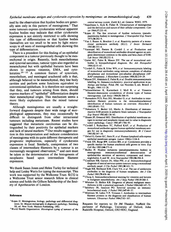
Epithelial membrane antigen and cytokeratin expression by meningiomas: an immunohistological studyined by the observation that hyaline bodies are gener-ally seen only in this pattern of meningioma.1 Thatsome tumours express cytokeratins without apparenthyaline bodies may indicate that either cytokeratinexpression is not entirely restricted to cells showingthis type of differentiation or that hyaline bodiesthemselves are too small to be seen by light micro-scopy in all nests of meningothelial cells showing thistype of differentiation.
There is a precedent for this finding of an epithelialcomponent in a tumour previously thought to be mes-enchymal in origin. Recently, both mesotheliomasand synovial sarcomas, tumour types also regarded asmesenchymal but with epithelial features have beenshown to express vimentin, EMA, and cyto-keratins.20 -22 A common feature of synovium,mesothelium, and meningeal arachnoid cells is that,although believed to be mesenchymal, they line bodycavities and perform some functions equivalent toconventional epithelium. It is therefore not surprisingthat they, and tumours arising from them, shouldexpress markers of "epithelial" differentiation despitetheir mesenchymal origin. This mechanism seems amore likely explanation than the mixed tumourtheory.Although meningiomas are usually a straight-
forward diagnosis despite their wide range of mor-phological appearances, they may occasionally bedifficult to distinguish from other intracranialtumours including metastases. Recent studies havesuggested that metastatic carcinomas can be dis-tinguished by their positivity for epithelial markersand lack of neural markers.18 Our results suggest cau-tion in this interpretation and indicate considerationofmeningioma with its quite different therapeutic andprognostic implications, especially if cytokeratinexpression is focal. Similarly, coexpression of twoclasses of intermediate filaments by a tumour is anincreasingly recognised observation,23 and care mustbe taken in the determination of the histogenesis ofneoplasms based upon intermediate filamentexpression.
We thank Susan Jones and Helen Turley for technicalhelp and Lesley Watts for typing the manuscript. Thiswork was supported by the Wellcome Trust. KCG isa Wellcome Trust senior research fellow in clinicalscience and holds the Gillson Scholarship of the Soci-ety of Apothecaries of London.
References
1 Kepes JJ. Meningiomas, biology, pathology and differential diag-nosis. In: Masson monographs in diagnostic pathology. SteinbergSS, ed. New York: Masson Publishing, 1982.
'World Health Organization. Histological typing of tumours of the
central nervous system. Zulch KJ, ed. Geneva: WHO, 1979.3Napolitano L, Kyle R, Fisher R. Ultrastructure of meningiomas
and the derivation and nature of their cellular components. Can-cer 1964;17:233-41.
'Kepes JJ. The fine structure of hyaline inclusions (pseudo-psammoma bodies) in meningiomas. J Neuropathol Exp Neurol1975;36:282-9.
5Viac J, Reano A, Brochier J, et al. Reactivity pattern of a mono-clonal antikeratin antibody (KL 1). J Invest Dermatol1983;81:351-4.
6Vanstapel MJ, Peeters B, Cordell J, et al. Production andidentification of monoclonal antibodies directed against an anti-genic determinant common to the alpha and beta chain of SIOO.Lab Invest 1985;52:232-8.
7Gatter KC, Falini B, Mason DY. The use of monoclonal anti-bodies in histopathological diagnosis. Rec Adv Histopathol1984;12:35-67.
8Cordell JL, Falini B, Erber WN, et al. Immunoenzymatic labelingof monoclonal antibodies using immune complexes of alkalinephosphatase and monoclonal anti-alkaline phosphatase (AP-AAP complexes). J Histochem Cytochem 1984;32:219-29.
'Mason DY, Abdulaziz Z, Falini B, et al. Double immunoenzymaticlabelling. In: Immunocytochemistry: practical applications in pa-thology and biology. Polak JM, van Noorden S, eds. Bristol: JWright, 1984:113-28.
'0Schwechheimer K, Kartenbeck J, Moll R, et al. Vimentinfilament-desmosome cytoskeleton of diverse types of humanmeningiomas. Lab Invest 1984;51:584-91.
l Ramaekers FCS, Puts JJG, Moesker 0, et al. Antibodies to inter-mediate filament proteins in the immunohistochemicalidentification of human tumours: an overview. Histochem J1983;15:691-71 3.
12Nakamura Y, Becker LE, Marks A. Distribution of immuno-reactive SIOO protein in pediatric brain tumours. J NeuropatholExp Neurol 1983;42:136-45.
13Sloane JP, Ormerod MG. Distribution of epithelial membrane an-tigen in normal and neoplastic tissues and its value in diagnostictumor pathology. Cancer 1981;47:1786-95.
14Cordell J, Richardson TC, Pulford KAF, et al. Production ofmonoclonal antibodies against human epithelial membrane anti-gen for use in diagnostic immunocytochemistry. Br J Cancer1985;52:347-54.
5Delsol G, Gatter KC, Stein H, et al. Human lymphoid cells expressepithelial membrane antigen. Lancet 1984;ii: 1 124-8.
16 Frank EH, Burge BW, Liwnicz BH, et al. Cytokeratin provides aspecific marker for human arachnoid cells grown in vitro. ExpCell Res 1983;146:371-6.
Budka H. Hyaline inclusions (pseudopsammoma bodies) inmeningiomas: immunocytochemical demonstration ofepithelial-like secretion of secretory component and immu-noglobulins A and M. Acta Neuropathol 1982;56:294-8.
18Coakham HB, Garson JA, Allan PM, et al. Immunohistologicaldiagnosis of central nervous system tumours using a monoclonalantibody panel. J Clin Pathol 1985;38:165-73.
Nagle RB, McDaniel KM, Clark VA, et al. The use of antikeratinantibodies in the diagnosis of human neoplasms. Am J ClinPathol 1983;79:458-66.
20Churg A. Immunohistochemical staining for vimentin and keratinin malignant mesothelioma. Am J Surg Pathol 1985;9:360-5.
21Whitaker D, Shilkin KB. Diagnosis of pleural malignant meso-thelioma in life: a practical approach. J Pathol 1984;143:147-75.
Salisbury JR, Isaacson PG. Synovial sarcoma: an immuno-histochemical study. J Pathol 1985;147:49-57.
23 Miettinen M, Lehto V-P, Virtanen I. Antibodies to intermediatefilament proteins in the diagnosis and classification of humantumours. Ultrastruct Pathol 1984;7:83-107.
Requests for reprints to: Dr JM Theaker, Nuffield De-partment of Pathology, University of Oxford, JohnRadcliffe Hospital, Oxford, OX3 9DU, England.
439
on April 2, 2020 by guest. P
rotected by copyright.http://jcp.bm
j.com/
J Clin P
athol: first published as 10.1136/jcp.39.4.435 on 1 April 1986. D
ownloaded from