Embed Size (px)
Citation preview
Research Paper 277
Enzyme-containing liposomes can endogenously produce membrane-constituting lipids Roger W ick and Pier Luigi Luisi
Badqround: ‘Giant vesicles’ are l iposomes that have diameters of several micrometers. It is possible to microinject biochemicals into a single vesicle and follow the progress of a chemical reaction in real t ime by light microscopy. We have previously used this technique to inject phospholipase A, into giant vesicles; the vesicles disappeared as their components were hydrolyzed. Here we investigate whether the lipid components of a vesicle can be synthesized inside it.
Results: Giant vesicles composed of 1 -palmitoyl-2-oleoyl-sn-glycerol-3- phosphocholine (POPC) and palmitoyl-CoA were prepared in a solution containing sn-glycerol-3-phosphate. Microinjection of the enzyme sn-glycerol-3- phosphate-acyltransferase into the vesicle catalyzes the in situ production of the lipid membrane precursor 1 -palmitoyl-sn-glycerol-3-phosphate, which remains incorporated in the membrane. The altered membrane chemistry causes shrinkage of the vesicle and formation of smaller l iposomes on the inner surface at the site of injection. Similar transformations were seen when the enzyme was added to the outside of the vesicle.
Address: ETH-Zentrum, lnstitut fib Polymere, Universitatstrasse 6, CH-8092 Zurich, Switzerland.
Correspondence: Pier Luigi Luisi e-mail: [email protected]
Key words: giant l iposomes, giant vesicles, microinjection, minimal cell model, sn-glycerol-3- phosphate-acyltransferase
Received: 12 Feb 1996 Revisions requested: 26 Feb 1996 Revisions received: 25 Mar 1996 Accepted: 26 Mar 1996
Chemistry 81 Biology April 1996, 3:277-265
0 Current Biology Ltd ISSN 1074-5521
Conclusions: We have used the first step of the ‘salvage pathway’ for synthesis of POPC to demonstrate that it is possible to localize the synthesis of a lipid membrane precursor inside a giant vesicle. In the future it may be possible to combine the necessary enzymes and substrates to carry out the reactions for a complete metabolic pathway within a l iposome.
Introduction Liposomes are probably the prebiotic precursors of cells [l-5]; they are the simplest spherical structures in which an aqueous microenvironment is enclosed by a lipid bilayer membrane. Recently, a variety of chemical reactions have been carried out inside liposomes [6-Z%]. These studies have helped to determine the effects of a restricted compartment on biochemical reactions, including problems of trafficking, local concentration effects and the influence of concentration gradients across the surrounding membrane. The spherical microenvironment inside the liposome mimics the intracellular compartment, and it may be possible to construct a ‘synthetic cell’ in which the coupled reac- tions of a self-sustaining metabolic cycle are entrapped in a liposome.
In the past, entrapped reactions have been carried out in submicroscopic liposomes, which have diameters ranging from 50 to 400 nm and can only be observed by electron microscopy. These studies can be extended by using so- called ‘giant vesicles’ (or ‘giant l iposomes’), which reach the dimensions of microns and can be observed using normal light microscopy. Several recent papers have described the behaviour of giant vesicles, in particular the shape transformations that they can undergo [29-361.
We have recently developed a micromanipulation technique that allows the microinjection of biochemicals into a single giant vesicle [36]. In this way, it is possible to carry out a localized chemical reaction in an individual l iposome and follow its progress in real time by light microscopy. We have used this technique previously to microinject phospholipase A, (PLA,) into giant vesicles composed of 1-palmitoyl-Z-oleoyl-sn-glycerol-3-phospho- choline (POPC) [36]. Hydrolysis of POPC into the corresponding lysolecithin and oleic acid causes com- plex transformations in the shape of the liposome. Microinjection of PLA, onto the surface of the giant vesicle, or into the external medium a few microns away from the bilayer, also induces changes in the structure of the giant vesicle.
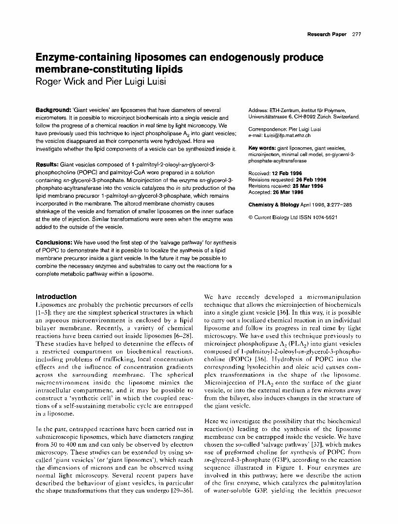
Here we investigate the possibility that the biochemical reaction(s) leading to the synthesis of the liposome membrane can be entrapped inside the vesicle. We have chosen the so-called ‘salvage pathway’ [37], which makes use of preformed choline for synthesis of POPC from sn-glycerol-3-phosphate (G3P), according to the reaction sequence illustrated in Figure 1. Four enzymes are involved in this pathway; here we describe the action of the first enzyme, which catalyzes the palmitoylation of water-soluble G3P, yielding the lecithin precursor
278 Chemistry 8 Biology 1996, Vol3 No 4
Figure 1
1 / 0-CoA
1 -Acyl-sn-glycerol- 3-phosphate- acyltransferase K
$ G CoA
f f:
H>C-0-C-C,5H3, I
H&Z,,-C-0-CH I ::
H&-O--P-OH
H2O
Phosphatidicacid- phosphohydrolase
H2POd-
i I: H&-0-C-C,5H3,
I H&T-C-0-CH
H&-OH
1 fl CDP-choline
1 ,P-Diacylglycerol- phosphocholine- transferase K
+\- CMP
I: f H2y-0-C-C,5H3, I I
H&Z,,-C-0-CH 0 CH:,
1 -Palmitoyl-2-oleoyl-sn-gl cerol-3-phosphocholine SOPC)
Pathway of POPC biosynthesis in four enzymatic reaction steps starting from G3P, acyl-CoA and CDP-choline 1371. The box highlights the reaction carried out inside giant vesicles in this study.
1-palmitoyl-sn-glycerol-3-phosphate (lP-G3P). This product is incorporated in the POPC membrane.
Results and discussion Formation of giant vesicles We prepared giant vesicles, according to a modification of the electroformation method of Angelova and Dimitrov [38,39], as described in detail elsewhere [36]. The vesicles were composed of POPC and had diameters of lo-60 p,m. We previously developed a microinjection technique, based on the micromanipulation of biological cells [40,41], to inject substances into the interior of giant vesicles ([36], see Materials and methods).
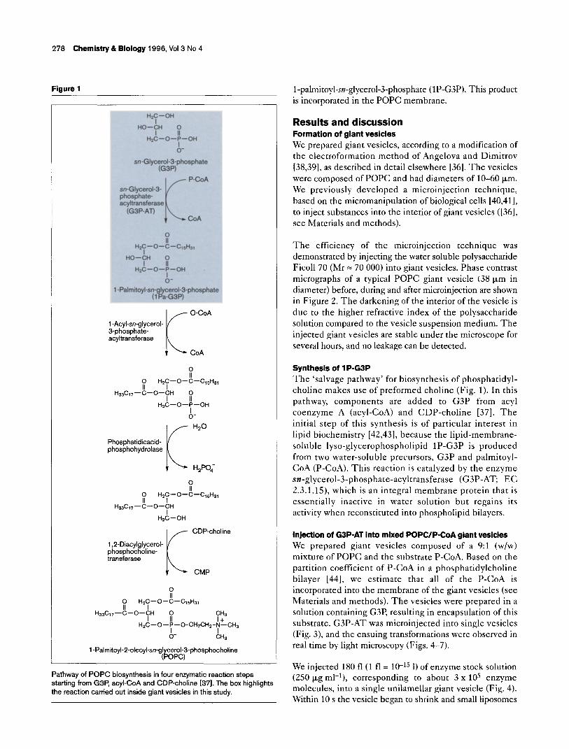
The efficiency of the microinjection technique was demonstrated by injecting the water soluble polysaccharide Ficoll 70 (Mr = 70 000) into giant vesicles. Phase contrast micrographs of a typical POPC giant vesicle (38 pm in diameter) before, during and after microinjection are shown in Figure 2. The darkening of the interior of the vesicle is due to the higher refractive index of the polysaccharide solution compared to the vesicle suspension medium. The injected giant vesicles are stable under the microscope for several hours, and no leakage can be detected.
Synthesis of 1 P-GSP The ‘salvage pathway’ for biosynthesis of phosphatidyl- choline makes use of preformed choline (Fig. 1). In this pathway, components are added to G3P from acyl coenzyme A (acyl-CoA) and CDP-choline [37]. The initial step of this synthesis is of particular interest in lipid biochemistry [42,43], because the lipid-membrane- soluble lyso-glycerophospholipid lP-G3P is produced from two water-soluble precursors, G3P and palmitoyl- CoA (P-CoA). This reaction is catalyzed by the enzyme sn-glycerol-3-phosphate-acyltransferase (G3P-AT; EC 2.3.1.15), which is an integral membrane protein that is essentially inactive in water solution but regains its activity when reconstituted into phospholipid bilayers.
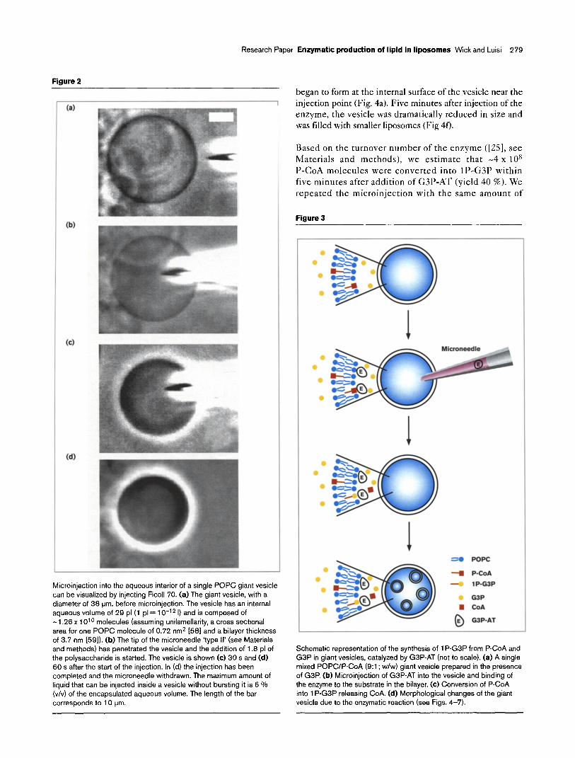
Injection of GBP-AT into mixed POPCYP-CoA giant vesides We prepared giant vesicles composed of a 9:l (w/w) mixture of POPC and the substrate P-CoA. Based on the partition coefficient of P-CoA in a phosphatidylcholine bilayer [44], we estimate that all of the P-CoA is incorporated into the membrane of the giant vesicles (see Materials and methods). The vesicles were prepared in a solution containing G3P, resulting in encapsulation of this substrate. G3P-AT was microinjected into single vesicles (Fig. 3), and the ensuing transformations were observed in real time by light microscopy (Figs. 4-7).
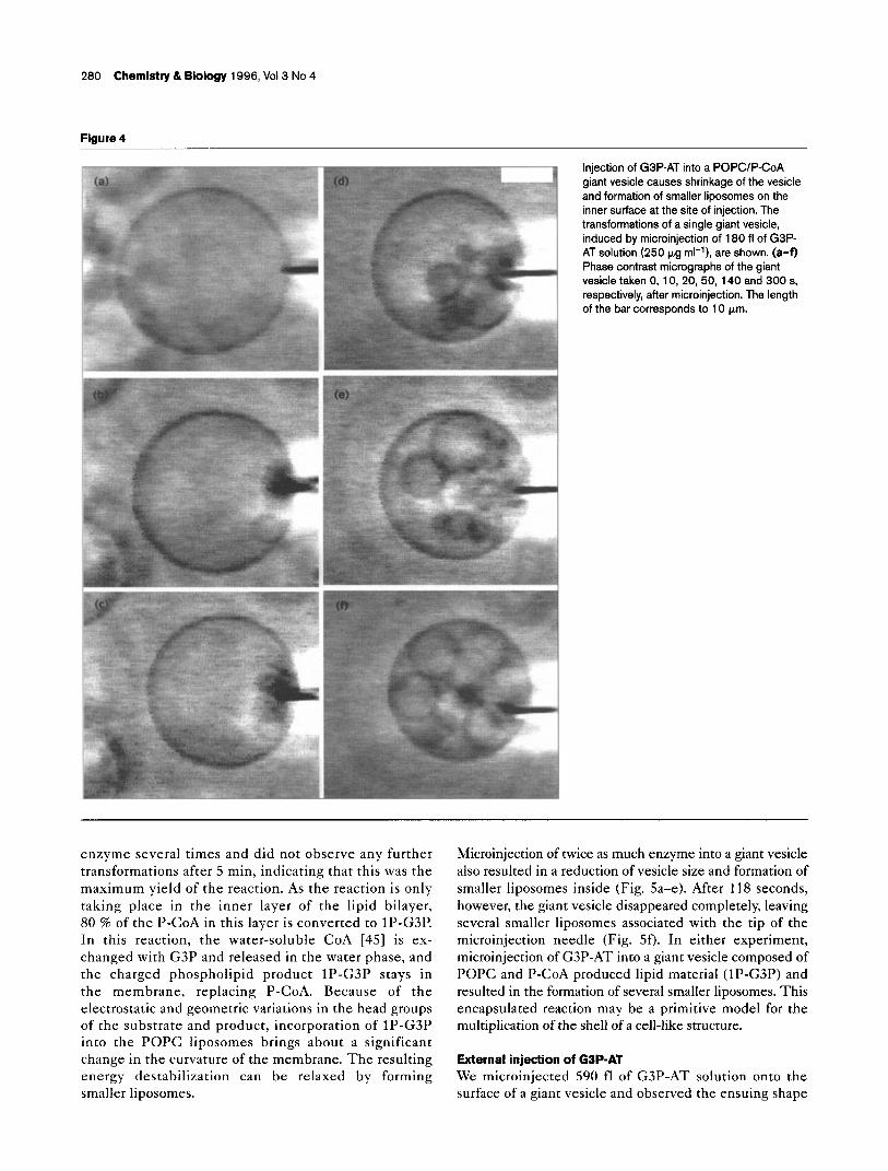
We injected 180 fl(1 fl = lo-l5 1) of enzyme stock solution (250 p,g ml-r), corresponding to about 3 x lo5 enzyme molecules, into a single unilamellar giant vesicle (Fig. 4). Within 10 s the vesicle began to shrink and small l iposomes
Research Paper Enzymatic production of lipid in liposomes Wick and Luisi 279
Figure 2
(a)
(c)
Microinjection into the aqueous interior of a single POPC giant vesicle can be visualized by injecting Ficoll 70. (a) The giant vesicle, with a diameter of 38 urn, before microinjection. The vesicle has an internal aqueous volume of 29 pl(1 pl = 10-l 2 I) and is composed of -1.26x 1 O’O molecules (assuming unilamellarity, a cross sectional area for one POPC molecule of 0.72 nm* [58] and a bilayer thickness of 3.7 nm [59]). (b) The tip of the microneedle ‘type II’ (see Materials and methods) has penetrated the vesicle and the addition of 1.8 pl of the polysaccharide is started. The vesicle is shown (c) 30 s and (d) 60 s after the start of the injection. In (d) the injection has been completed and the microneedle withdrawn. The maximum amount of liquid that can be injected inside a vesicle without bursting it is 6 Vo (v/v) of the encapsulated aqueous volume. The length of the bar corresponds to 10 urn.
began to form at the internal surface of the vesicle near the injection point (Fig. 4a). Five minutes after injection of the enzyme, the vesicle was dramatically reduced in size and was filled with smaller liposomes (Fig 4f).
Based on the turnover number of the enzyme ([25], see Materials and methods), we estimate that -4 x lo* P-CoA molecules were converted into lP-G3P within five minutes after addition of G3P-AT (yield 40 %). We repeated the microinjection with the same amount of
Figure 3
so POPC
-m P-COA
- lP-G3P
G3P ; CoA
0 GBP-AT
Schematic representation of the synthesis of 1 P-G3P from P-CoA and G3P in giant vesicles, catalyzed by GSP-AT (not to scale). (a) A single mixed POPCIP-CoA (9:l; w/w) giant vesicle prepared in the presence of G3P. (b) Microinjection of G3P-AT into the vesicle and binding of the enzyme to the substrate in the bilayer. (c) Conversion of P-CoA into 1 P-G3P releasing CoA. (d) Morphological changes of the giant vesicle due to the enzymatic reaction (see Figs. 4-7).
280 Chemistry & Biology 1996, Vol3 No 4
Figure 4
Injection of G3P-AT into a POPCIP-CoA giant vesicle causes shrinkage of the vesicle and formation of smaller l iposomes on the inner surface at the site of injection. The transformations of a single giant vesicle, induced by microinjection of 180 fl of G3P- AT solution (250 pg ml-l), are shown. (a-f) Phase contrast micrographs of the giant vesicle taken 0, 10, 20, 50, 140 and 300 s, respectively, after microinjection. The length of the bar corresponds to 10 pm.
enzyme several times and did not observe any further transformations after 5 min, indicating that this was the maximum yield of the reaction. As the reaction is only taking place in the inner layer of the lipid bilayer, 80 % of the P-CoA in this layer is converted to lP-G3P. In this reaction, the water-soluble CoA [4.5] is ex- changed with G3P and released in the water phase, and the charged phospholipid product lP-G3P stays in the membrane, replacing P-CoA. Because of the electrostatic and geometric variations in the head groups of the substrate and product, incorporation of lP-G3P into the POPC liposomes brings about a significant change in the curvature of the membrane. The resulting energy destabilization can be relaxed by forming smaller liposomes.
Microinjection of twice as much enzyme into a giant vesicle also resulted in a reduction of vesicle size and formation of smaller l iposomes inside (Fig. Sa-e). After 118 seconds, however, the giant vesicle disappeared completely, leaving several smaller l iposomes associated with the tip of the microinjection needle (Fig. 5). In either experiment, microinjection of G3P-AT into a giant vesicle composed of POPC and P-CoA produced lipid material (lP-G3P) and resulted in the formation of several smaller liposomes. This encapsulated reaction may be a primitive model for the multiplication of the shell of a cell-like structure.
External injection of GBP-AT We microinjected 590 fl of G3P-AT solution onto the surface of a giant vesicle and observed the ensuing shape
Research Paper Enzymatic production of lipid in l iposomes Wick and Luisi 281
Figure 5
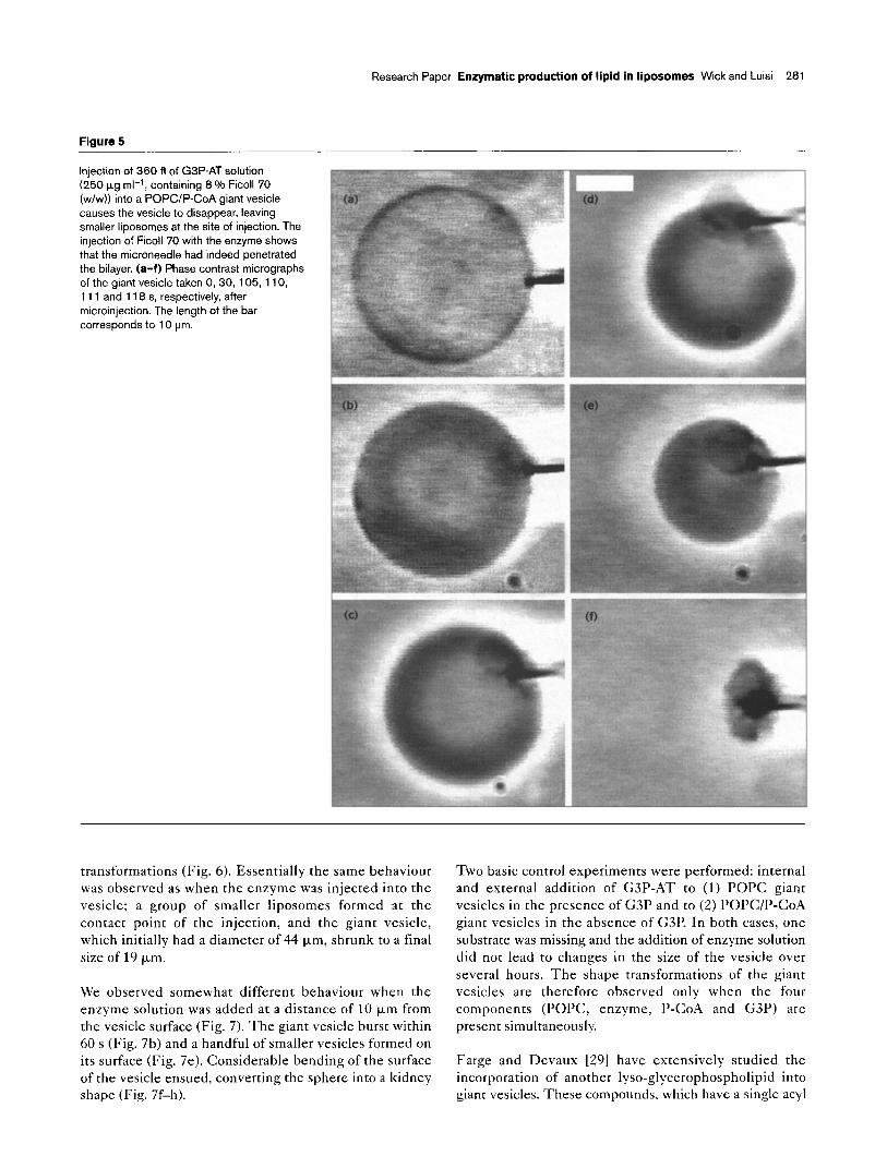
Injection of 360 fl of G3P-AT solution (250 pg ml-‘, containing 8 % Ficoll 70 (w/w)) into a POPCIP-CoA giant vesicle causes the vesicle to disappear, leaving smaller l iposomes at the site of injection. The injection of Ficoll 70 with the enzyme shows that the microneedle had indeed penetrated the bilayer. (a-f) Phase contrast micrographs of the giant vesicle taken 0, 30, 105, 110, 111 and 118 s, respectively, after microinjection. The length of the bar corresponds to 10 urn.
transformations (Fig. 6). Essentially the same behaviour was observed as when the enzyme was injected into the vesicle; a group of smaller l iposomes formed at the contact point of the injection, and the giant vesicle, which initially had a diameter of 44 pm, shrunk to a final size of 19 p,m.
We observed somewhat different behaviour when the enzyme solution was added at a distance of 10 pm from the vesicle surface (Fig. 7). The giant vesicle burst within 60 s (Fig. 7b) and a handful of smaller vesicles formed on its surface (Fig. 7e). Considerable bending of the surface of the vesicle ensued, converting the sphere into a kidney shape (Fig. 7f-h).
Two basic control experiments were performed: internal and external addition of G3P-AT to (1) POPC giant vesicles in the presence of G3P and to (2) POPC/P-CoA giant vesicles in the absence of G3P. In both cases, one substrate was missing and the addition of enzyme solution did not lead to changes in the size of the vesicle over several hours. The shape transformations of the giant vesicles are therefore observed only when the four components (POPC, enzyme, P-CoA and G3P) are present simultaneously.
Farge and Devaux [29] have extensively studied the incorporation of another lyso-glycerophospholipid into giant vesicles. These compounds, which have a single acyl
282 Chemistry 8 Biology 1996, Vol3 No 4
Figure 6
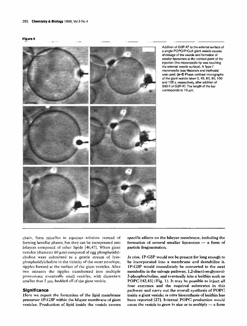
Addition of G3P-AT to the external surface of a single POPCIP-CoA giant vesicle causes shrinkage of the vesicle and formation of smaller l iposomes at the contact point of the injection (the microneedle tip was touching the external vesicle surface). A ‘type I’ microneedle (see Materials and methods) was used. (a-f) Phase contrast micrographs of the giant vesicle taken 0, 45, 80, 90, 100 and 105 s, respectively, after addition of 590 fl of GSP-AT The length of the bar corresponds to 10 pm.
chain, form micelles in aqueous solution instead of forming lamellar phases, but they can be incorporated into bilayers composed of other lipids [46,47]. When giant vesicles (diameter 60 p,m) composed of egg phosphatidyl- choline were submitted to a gentle stream of lyso- phosphatidylcholine in the vicinity of the outer envelope, ripples formed at the surface of the giant vesicles. After two minutes the ripples transformed into multiple protrusions; eventually small vesicles, with diameters smaller than 5 urn, budded off of the giant vesicle.
Significance Here we report the formation of the lipid membrane precursor lP-G3P within the bilayer membrane of giant vesicles. Production of lipid inside the vesicle causes
specific effects on the bilayer membrane, including the formation of several smaller l iposomes - a form of particle fragmentation.
In vivo, lP-G3P would not be present for long enough to be incorporated into a membrane and destabilize it. lP-G3P would immediately be converted to the next metabolite in the salvage pathway, 1,2-diacyl-sn-glycerol- 3-phosphocholine, and eventually into a lecithin such as POPC 142,431 (Fig. 1). It may be possible to inject all four enzymes and the required substrates in this pathway and carry out the overall synthesis of POPC inside a giant vesicle; in vitro biosynthesis of lecithin has been reported [27]. Internal POPC production would cause the vesicle to grow in size or to multiply - a form
Research Paper Enzymatic production of lipid in liposomes Wick and Luisi 283
Figure 7
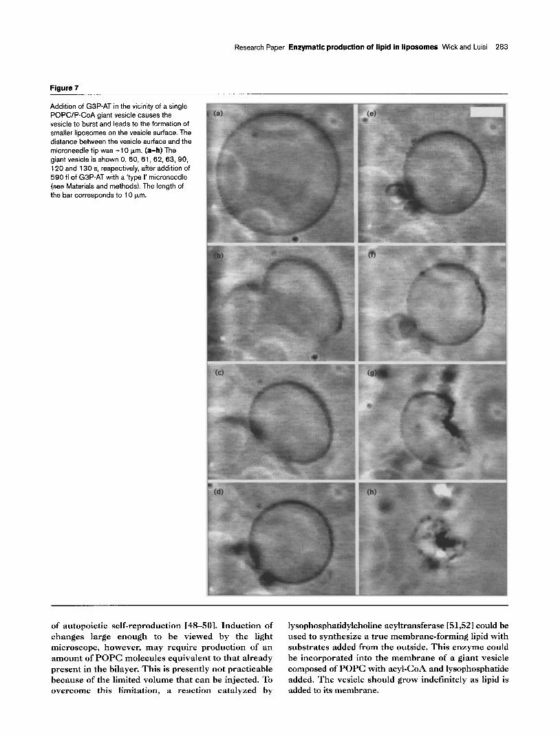
Addition of G3P-AT in the vicinity of a single POPCIP-CoA giant vesicle causes the vesicle to burst and leads to the formation of smaller liposomes on the vesicle surface. The distance between the vesicle surface and the microneedle tip was -10 km. (a-h) The giant vesicle is shown 0, 60, 61, 62, 63, 90, 120 and 130 s, respectively, after addition of 590 fl of G3P-AT with a ‘type I’ microneedle (see Materials and methods). The length of the bar corresponds to 10 pm.
of autopoietic self-reproduction 148-501. Induction of lysophosphatidylcholine acyltransferase 151,521 could be changes large enough to be viewed by the light used to synthesize a true membrane-forming lipid with microscope, however, may require production of an substrates added from the outside. This enzyme could amount of POPC molecules equivalent to that already be incorporated into the membrane of a giant vesicle present in the bilayer. This is presently not practicable composed of POPC with acyl-CoA and lysophosphatide because of the limited volume that can be injected. To added. The vesicle should grow indefinitely as lipid is overcome this limitation, a reaction catalyzed by added to its membrane.
284 Chemistry & Biology 1996, Vol3 No 4
We are presently working on solving the technical limit- ations to carrying out biosynthetic reactions inside a l iposome. An ultimate goal of this research is to construct a minimal cell model in which a l iposome contains all of the components for a self-sustaining metabolic cycle and perhaps even the protein biosynthetic apparatus to produce the enzymes for that cycle.
Materials and methods Isolation and purification of the enzyme G3P-AT The enzyme GBP-AT was isolated from Escherichia co/i VL3/plB3-4, which harbours a hybrid plasmid containing the plsB gene encoding G3P-AT and overproduces the enzyme 153,541. The purification procedure was based on a modification of previously described protocols 155,561 and included a chromatographic separation on a Matrex Gel Green A (Amicon, USA) and on hydroxylapatite. Details of the procedure are given elsewhere 1251. The purified protein was analyzed by SDS polyacrylamide gel electrophoresis (1 O-l 5 % Phast gel on a PhastSystem (Pharmacia, Sweden)). A single band was stained with Coomassie blue, corresponding to a relative molecular mass of 83 000. The GSP-AT had a specific activity of 47.3 nmol mg-’ min-’ when measured using a 3 m M soybean phospholipid vesicular solution in 30 m M Tris (containing 2 m M G3P, 25 pM P-CoA, 4 m M Ca 2+, 70 m M NaCI, 1 mg ml-’ bovine serum albumin, 7.5 m M 8-mercaptoethanol and <O.l % (w/v) Triton X-100) at pH 7.4 and 30 ‘C [251. To determine the kinetic parameters of the G3P-AT-catalyzed reaction, we measured the specific activity as a function of P-CoA concentration (l-l 0 FM) in excess G3P (2.2 mM) using a 3.6 m M soybean phospholipid vesicular solution in 30 m M Tris (containing 2 m M G3P, 25 p,M P- CoA, 4 m M Ca2+, 70 m M NaCI, 1 mg ml-’ bovine serum albumin, 7.5 m M 8-mercaptoethanol and <O.l % (w/v) Triton X-l 00) at pH 7.4 and 30 “C. The enzyme concentration was 25 kg ml-‘. The apparent values for K, = 12 P M and V,,, = 75 nmol mg-’ min-’ were calculated from a Lineweaver-Burk plot.
Micromanipulation of giant vesicles Microinjection was carried out as previously described (36). An 8 ‘J/o solution of the water soluble polysaccharide Ficoll 70 (from Pharmacia, Sweden) was injected into a single POPC giant vesicle (prepared in 1 m M Tris, 1 m M Ca2+, pH 7.4) to prove that the injection was indeed into the interior aqueous volume of the vesicle.
Transformations of single giant vesicles were studied after addition of G3P-AT. Each experiment (Figs 4-7) was carried out at room temperature, and a concentrated GSP-AT solution (250 p,g ml-’ G3P-AT in 30 m M Tris pH 7.4, 0.1 % (w/v) Triton X-100) was added into, onto or near a particular mixed POPCIP-CoA (9:l; w/w) giant vesicle (prepared in 1 m M Tris, 1 m M G3P, 1 m M Ca2+, pH 7.4). P-CoA forms micelles in water when present at a concentration above the ‘critical micellar concentration’ (cmc), which is in the low m M range [571. The P-CoA molecules are located mainly in the membrane of the phospholipid vesicles. Requero et al. [44] determined an egg- phosphatidylcholine bilayerjwater partition coefficient of 3.8 x 1 03, by adding [14ClP-CoA and separating the l iposomes by centrifugation. On the basis of this figure, which indicates a very high affinity of P-CoA for the lipid phase, we estimate that all of the P-CoA used in formation of the giant vesicles is bound to the vesicle membrane. Our own studies revealed that there exists a critical ratio of POPC:P-CoA of about 4:1, at which no more mixed giant vesicles but only mixed micelles are formed in solution. We therefore prepared mixed POPCIP-CoA giant vesicles starting from a POPC/P-CoA lipid f i lm (9:l; w/w). This experimental setup insured a quantitative incorporation of the activated fatty acid in the membrane. The G3P, a water-soluble molecule, was located in the internal water pool of the vesicles and in the outer bulk, as it was added together with the buffer solution during the swelling of the lipid deposit.
Two types of microneedles were prepared from borosilicate tubes as previously described (361. The ‘type I’ microneedle was used when reagents were added externally to the vesicles; the ‘type II’ microneedle was used for injection into the interior of the vesicles
The characteristics of the two microneedles used were: microneedle ‘type I’, inner diameter = 135 & 5 nm, outer diameter = 245 & 10 nm, injection volume = 590 + 10 fl (2000 hPa, 1 s); microneedle ‘type II’, inner diameter = 100 + 5 nm, outer diameter = 205 f 6 nm, injection volume = 180 + 10 fl (2000 hPa, 1 s).
Acknowledgements We are grateful to Robert M. Bell for kindly providing us with Eschen’chia co/i VL3/plB3-4 and to Heinrich Riiger and Eveline Blochliger for the isolation and purification of G3P-AT. We also thank Antonio Lazcano and Peter Walde for critically reading our manuscript.
References 1.
2.
3.
4.
5.
6.
7.
8.
9.
10.
11.
12.
13.
14.
15.
16.
1 7.
18.
19.
Day, W. (1984). Genesis of Planet farfh. Yale University Press, New Haven. Deamer, D.W. (1986). Role of amphiphil ic compounds in the evolution of membrane structure on the early earth. Origins Life Evol. Biosphere 17,3-25. Deamer. D.W. & Or& J. (1980). Role of lioids in orebiotic structures. Biosysf~ms 12, 167-l 75. Lazcano. A., Fox, G.E. & Or6. J. (1992). Life before DNA: the oriain and evolution of’early archean cells. In. The Evolution of Mefabolyc function. (Mortlock, RR, ed.), pp. 237-295, CRC Press Inc., Boca Raton, FL. Morowitz, H.J., Heinz, B. & Deamer, D.W. (1988). The chemical logic of a min imum protocell. Origins Life Evol. Biosphere 18, 281-287. Ambartsumian, T.G., Adamian, S.Y., Petrosian, L.S. & Simonian, A.L. (1992). Incorporation of water-soluble enzymes glucose oxidase and urate oxidase into phosphatidylcholine l iposomes. B/o/. Mem. 5, 1878-l 887. Annesini, M.C., Di Giulio, A., Di Marzio, L., Finazzi-Agrb, A. & Mossa, G. (1992). Diffusion and catalysis processes in unilamellar l iposomes. J. Liposome Res. 2,455-467. Annesini, M.C., Di Giorgio, L., Di Marzio, L., Finazzi-Agrb, A., Serafino, A.L. & Mossa, G. (1994). Carbon dioxide hydration with l iposomes entrapping carbonic anhydrase. J. Liposome Res. 3, 639-648. Annesini, M.C., Di Marzio, L., Finazzi-Agrb, A., Serafino, A.L. & Mossa, G. (1994). Interaction of cationic phospholipid vesicles with carbonic anhydrase. Bioch. Mol. Biol. Inf. 32, 6j-94: Burrier, R.E. & Brecher, P (1984). Effect of surface composition on triolein hydrolysis in phospholipid vesicles and microemulsions by a purified acid lipase. Biochemistry 23, 5366-5371. Chakrabarti, A.C., Breaker, R.R., Joyce, G.F. & Deamer, D.W. (1994). Production of RNA by a polvmerase protein encapsulated within phospholipid vesicles. J.‘M&. Evol. 39, 555-559: Cho. Y.. Ko, T.-S.. Cha. S-H. & Sok. D.-E. (1995). Properties of acetylcholinesterase reconstituted in l iposomes of a d’ifferent charge. Neurochem. Res. 20,681-687. Holm, C., Fredrikson, G., Sundler, R. & Belfrage, P (1990). lncorooration of hormone-sensitive lioase into ohosohatidvlcholine vesicles. Lipids 25, 254-259. . Luisi. P.L.. Walde. P. & Oberholzer. T. (1994). Enzvmatic RNA _ synthesis’in self-reproducing vesicles: an approach to the construction of a minimal synthetic cell. Ber. Bunsenges. Phys. Chem. 98, 1160-l 165. McCurley, M.F. & Glazier, S.A. (1995). Optical control of enzymatic conversion of sucrose to glucose by bacteriorhodopsin incorporated into self-assembled phosphatidylcholine vesicles. Analytica Chimica Acta311, 211-215. Menger, FM.. & Johnston, D.E., Jr (1991). Specific enzyme-induced decapsulation. J. Am. Chem. Sot. 113,5467-5468. Mosmuller, E.W.J., Franssen, M.C.R. & Engbersen, J.F.J. (1993). Lipase activity in vesicular systems: characterization of Candida cylindracea lipase and its activity in polymerizable dialkylammonium surfactant vesicles. Biofechnol. Bioeng. 42, 196-204. Mossa, G., Annesini, MC., Di Giulio, A., Dini, L. & Finazzi-Agrb, A. (1969). Lipsomes as bioreactors: transport phenomena in proteoliposomes. In Biological and Synthetic Membranes. (Butterfield, D. A., ed.), pp. 227-236, Alan R. Liss, New York. Oberholzer, T., Wick, R., Luisi, PL. & Biebricher, C.K. (1995).
Research Paper Enzymatic production of ilpid in l iposomes Wick and Luisi 285
20.
21.
22.
Enzymatic RNA replication in self-reproducing vesicles: an approach to a minimal cell. Biochem. Biophys. Res. Commun. 207, 250-257. Oberholzer, T., Albrizio, M. & Luisi, PL. (1995). Polymerase chain reaction in l iposomes. Chemistry & Biofogy 2, 677-882. Ohno, K., Sohda, K., Kosaka, A.-& KitanoyH. (1995). Galactose- containing amphiphiles prepared with a lipophilic radical initiator: association processes between l iposomes triggered by enzymatic reaction. Bioconjugare Chem. 6, 361-368. Orlando, A.R., Arcovito, C., Palombo, A., Serafino, A.L. & Mossa, G. (I 993). Enzymatic kinetic change of ascorbate oxidase loaded into l iposomes induced by microwave field exposure. J. Liposome Res. 3, 717-724.
23.
24.
25.
26.
Orlando, A.R., Mossa, G. & D’lnzeo, G. (1993). Effect of microwave radiation on the permeability of carbonic anhydrase loaded unilamellar l iposomes. 8ioe/ectromagnerics 15, 303-313. Rosenberg, ME, Jones, M.N. & Yadgama, PM. (1991). A l iposomal enzyme electrode for measuring glucose. Biochim. Biophys. Acta 1115, 157-165. Riioer, H. (1995). Enzymatische Synthese von 1 -Acyl-sn-Glycerin-3- phosphat in Phospholipid-Vesikeln-als Teil eines Mobelles fir chemische Autopoiese. PhD thesis, Diss. ETH Nr. 11135. Ziirich. Sada, E., Katoh,‘S., Terashima, M., Shiraga, H. & Miura, Y. (1990). Stability and reaction characteristics of reverse-phase evaporation vesicles (REVS) as enzyme containers. Biotechnof. Bioeng. 36, 665-671.
27.
28.
29.
30.
31.
Schmidli, PK., Schurtenberger, P. & Luisi, P.L. (1991). Liposome- mediated enzymatic synthesis of phosphatidylcholine as an approach to self-replicating l iposomes. J. Am. Chem. Sot. 113, 8127-8130. Walde, P., Goto, A., Monnard, P-A., Wessicken, M. & Luisi, P.L. (1994). Oparin’s reactions revisited: enzymatic synthesis of poly(adenylic acid) in micelles and self-reproducing vesicles. J. Am. Chem.Soc. 116,7541-7547. Farge, E. & Devaux, F!F. (I 992). Shape changes of giant l iposomes induced by an asymmetric transmembrane distribution of phospholipids. Biophys. J. 61, 347-357. Kss. J. & Sackmann, E. (1991). Shape transitions and shape stability of giant phospholipid vesicles in pure water induced by area-to-volume changes. Biophys. J. 60,825-844. Menger, FM. & Gabrielson, K.D. (I 995). Die cytomimetische organische Chemie - ein erster Bericht. Anger Chemie 107, 2260-2278.
32. Menaer. FM. & Balachander, N.J. (1992). Chemicallv-induced I
33.
34.
35.
36.
37.
38.
39.
aggregation, budding, and fusion in giant vesicles: direct observation bv liaht microscopy. J. Am. Chem. Sot. 114.5862-5863. Menger, F.M. & dabrielson, K. (I 994). Chemically-induced birthing and foraging in vesicle systems. J. Am. Chem. Sot. 116, 1567-l 568. Sackmann, E., Duwe, H.-P & Engelhardt, H. (1986). Membrane bending elasticity and its role for shape fluctuations and shape transformations of cells and vesicles. Faraday Discuss. Chem. Sot. 81~281-290. Wrck, R., Walde, P & Luisi, P.L. (1995). Light microscopic investigations of the autocatalytic self-reproduction of giant vesicles. J. Am. Chem. Sot. 117,1435-l 436. Wick, R., Angelova, M.I., Walde, P & Luisi, PL. (1996). Microinjection into Giant vesicles and light microscopy investigation of enzyme mediated vesicle transfo;mations. Chemistry &-Biology 3, 105-l 11. Lehninger, A.L., Nelson, D.L. & Cox MM. (1993). Principles of Biochemistry. Worth Publishers, New York Anoelova. M.I. & Dimitrov, D.S. (1986). Liposome electroformation. Faraday Discuss. Chem. Sot. 81,303-31 I. Dimitrov, D.S. & Angelova, M.I. (1988). Lipid swelling and l iposome formation mediated by electric fields. Bioelecfrochem. Bioenerg. 19, 323-336.
40. Celis, J.E. (I 984). Microinjection of somatic cells with micropipettes: comparison with other transfer techniques. Biochem. J. 223, 281-291.
41.
42.
43.
44.
Schnorf, M., Potrykus, I. & Neuhaus, G. (1994). Microinjection technique: routine system for characterization of microcapillaries by bubble pressure measurement. Exp. Cell Res. 210, 260-287. Cronan, J.E., Jr (1978). Molecular biology of bacterial membrane lipids. Annu. Rev. Biochem. 47, 163-I 89. Rae& C.R.H. (1978). Enzymology, genetics and regulation of membrane phospholipid synthesis in Escherichia co/i. Microbiof. Rev. 42,814-659. _ Requero, M.A., Got%, FM. & Alonso, A. (1995). The membrane- perturbing properties of palmitoyl-coenzyme A and palmitoylcsrnitine. A comparative study. Biochemistry 34, 10400-I 0405.
45.
46.
47.
48.
49.
50.
51.
52.
53.
54.
55.
56.
57.
58.
59.
Windholz, M. (1983). The Merck index (Tenth Edition), Merck & Co., Inc., Rahway, NJ. Bangham, A.D. & Horne, R.W. (1964). Negative staining of phospholipids and their structural modification by surface-active agents as observed in the electron microscope. J. Mol. Biof. 8, 660-668. lain, M.K. (I 983). Fantasy and fun with l iposomes. In Liposome Letters. (Bangham, A.D, ed.), pp. 73-83, Academic Press, London. Varela, F.J., Maturana, H.R. & Uribe, R. (I 974). Autopoiesis: the organization of living systems, its characterizaton and a model. Biosystems 5, 187-I 96. Maturana, H.R. & Varela, F.J. (1980). Autopoiesis and Cognition: The Realization of the Living. D. Reidel Publishing Co., Boston, MA. Fleischaker, G.R. (1990). Origins of life: an operational definition. Origins Life Evol. Biosphere 20, 127-I 37. Deamer, D.W. & Boatman, D.E. (1980). An enzymatically driven membrane reconstitution from solubilized components. J. Cell. Biof. 84,48l-467. Gaviano, V.C. & Deamer, D.W. (I 982). Purification of acyl CoA:l -acyl- sn-glycero-3-phosphorylcholine acyltransferase. J. Bioenerg. Biomembr. 14,513-526. Liahtner. V.A.. et a/.. & Modrich. P (1980). Membrane ohosoholipid synthesis in Esche&hia co/i. J. Biol. Chem. 255,941 j-9420. ’ Larson. T.J.. Liahtner. V.A.. Green. P.R.. Modrich. P & Bell. R.M. (1980). Membrane phospholipid synthesis in Escherichia co/i. J. Biof. Chem. 255,9421-9428. Green, P.R., Merrill, A.H. & Bell, R.M. (I 981). Membrane phospholipid synthesis in fscherichia co/i. J. Biol. Chem. 256, 11151-l 1159. Scheideler, M.A. & Bell, R.M. (I 986). Efficiency of reconstitution of the membrane-associated sn-glycerol-3-phosphate acyltransferase of Escherichia co/i. J. Biol. Chem. 261, 10990-l 0995. Requero, M.A., Gofii, F.M. & Alonso, A. (1993). The critical micellar concentrations of fatty acyl coenzyme A and fatty acyl camitines. J. Colloid. Interface Sci. 161, 343-346. Cornell, B.A., Middlehurst, J. & Separovic, F. (1980). The molecular packing and stability within highly curved phospholipid bilayers. Biochim. Biophys. Acta 598,405-410. Tahara, Y. & Fujiyoshi, Y. (1994). A new method to measure bilayer thickness: cryo-electron microscopy of frozen hydrated l iposomes and image simulation. Micron 25, 141-i 49.