Embed Size (px)
Citation preview
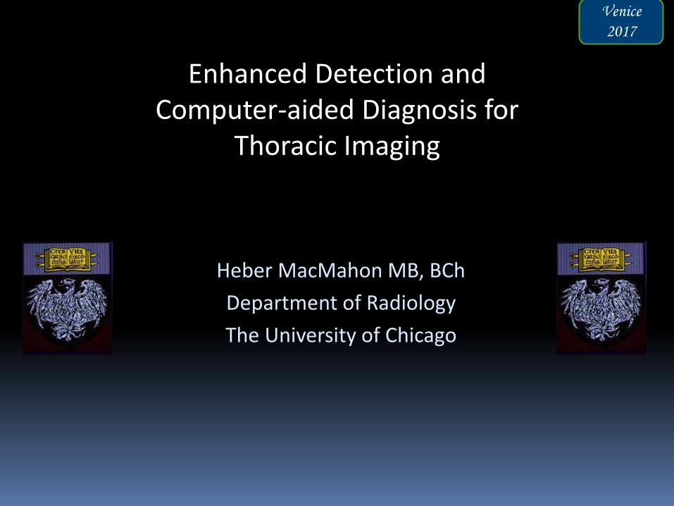
Heber MacMahon MB, BCh
Department of Radiology
The University of Chicago
Enhanced Detection and Computer-aided Diagnosis for
Thoracic Imaging
Venice
2017

Disclosures
Consultant for Riverain Medical
Minor stockholder in Hologic, Inc.
License and royalty fees from University of
Chicago (UCTech)
Research support from Philips Healthcare
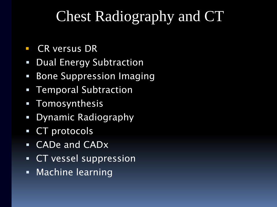
CR versus DR
Dual Energy Subtraction
Bone Suppression Imaging
Temporal Subtraction
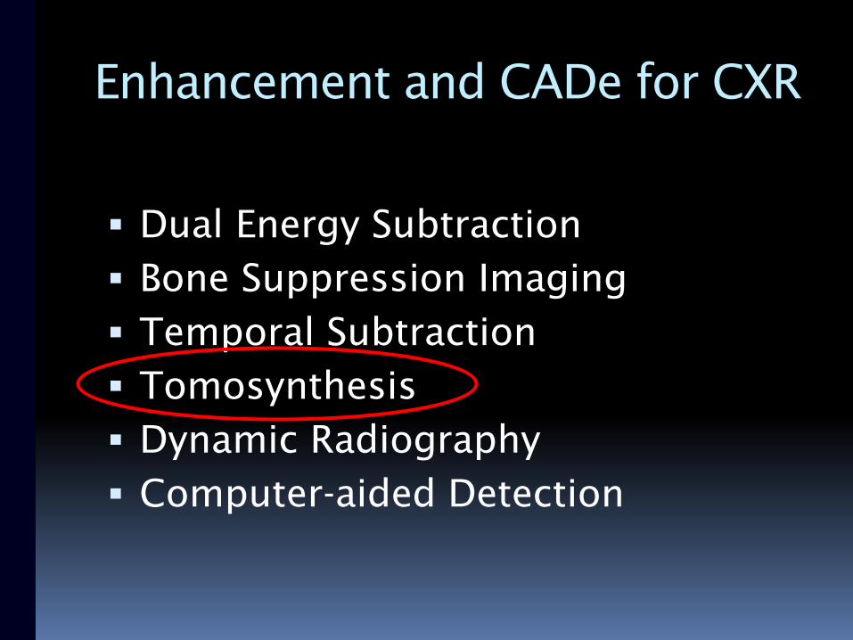
Tomosynthesis
Dynamic Radiography
CT protocols
CADe and CADx
CT vessel suppression
Machine learning
Chest Radiography and CT
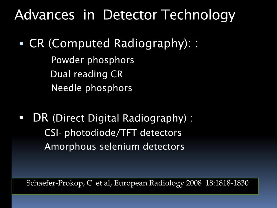
Advances in Detector Technology
CR (Computed Radiography): :
Powder phosphors
Dual reading CR
Needle phosphors
DR (Direct Digital Radiography) :
CSI- photodiode/TFT detectors
Amorphous selenium detectors
Schaefer-Prokop, C et al, European Radiology 2008 18:1818-1830
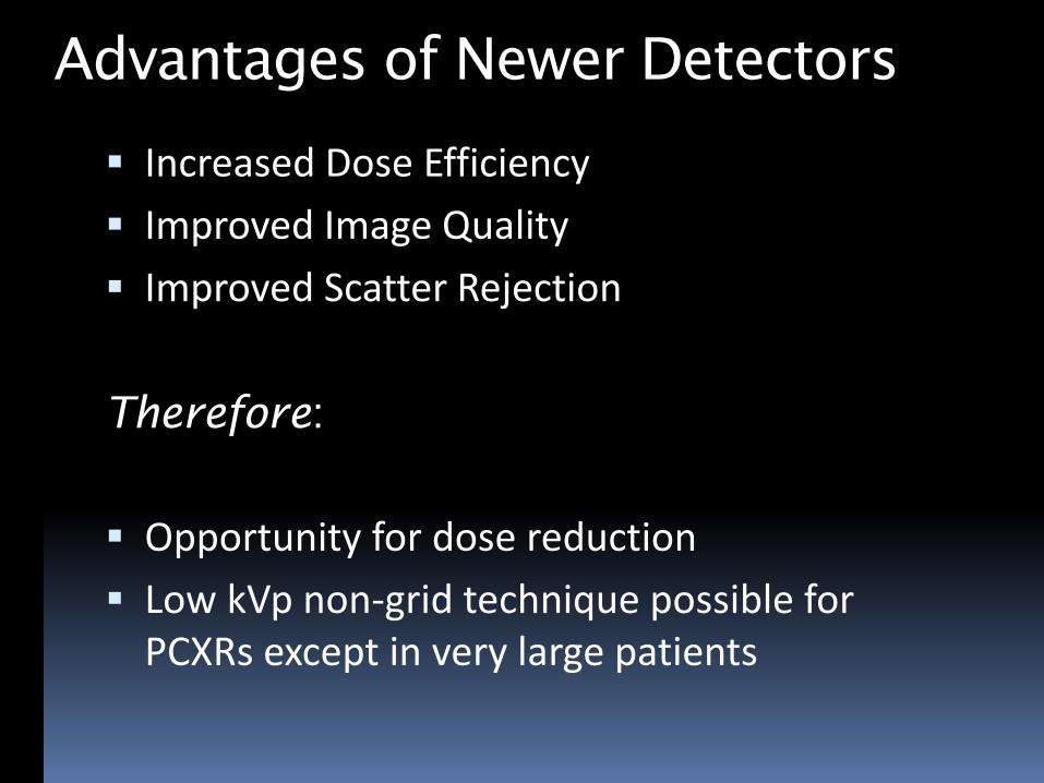
Advantages of Newer Detectors
Increased Dose Efficiency
Improved Image Quality
Improved Scatter Rejection
Therefore:
Opportunity for dose reduction
Low kVp non-grid technique possible for PCXRs except in very large patients
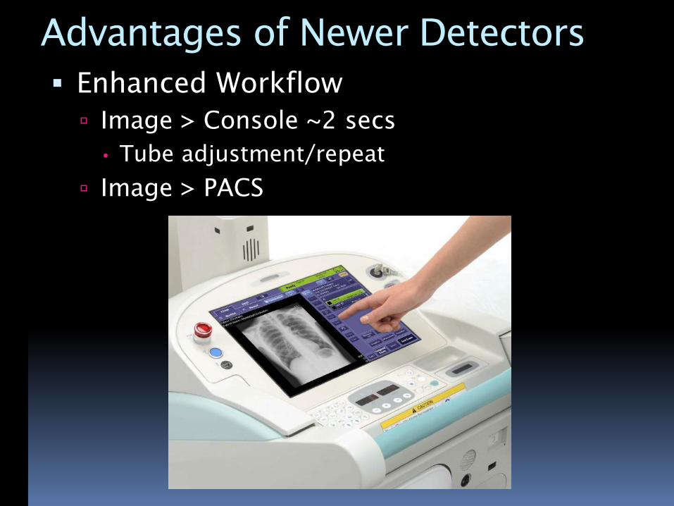
Advantages of Newer Detectors
Enhanced Workflow
Image > Console ~2 secs
• Tube adjustment/repeat
Image > PACS
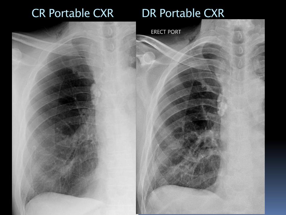
CR Portable CXR DR Portable CXR
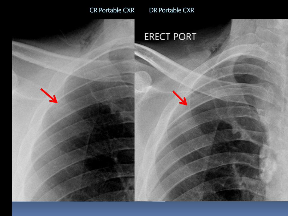
CR Portable CXR DR Portable CXR
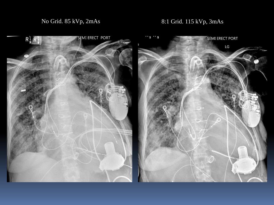
No Grid. 85 kVp, 2mAs 8:1 Grid. 115 kVp, 3mAs
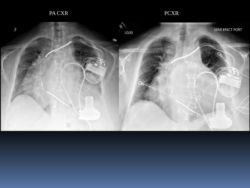
PA CXR PCXR
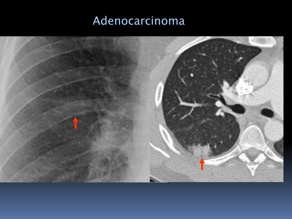
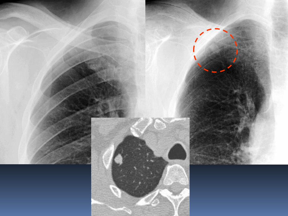
Adenocarcinoma
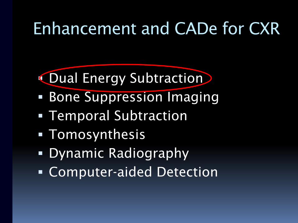
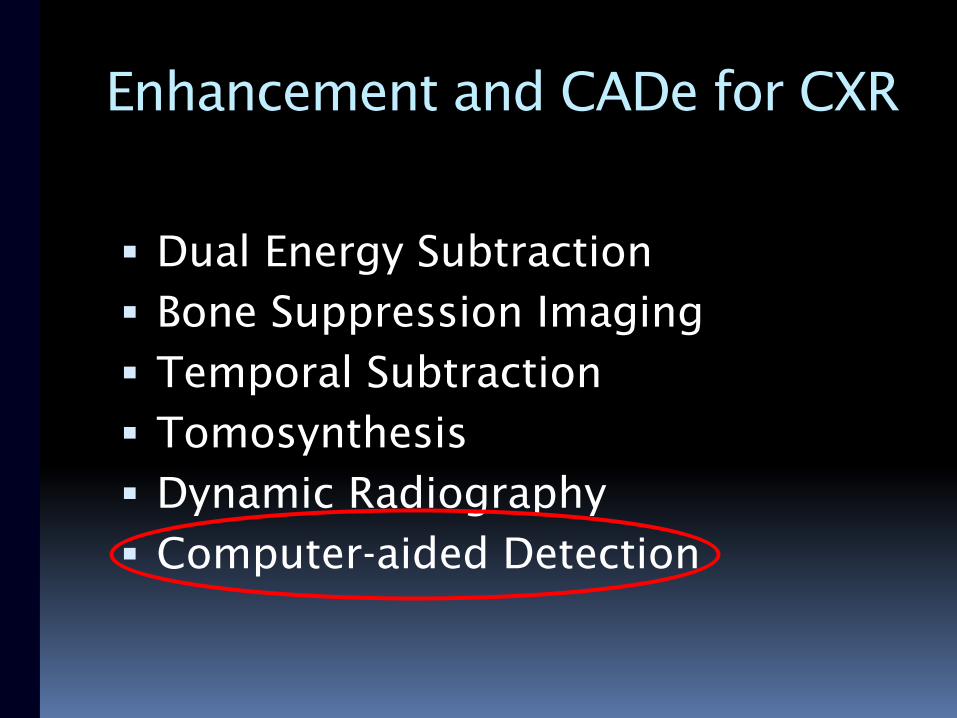
Enhancement and CADe for CXR
Dual Energy Subtraction
Bone Suppression Imaging
Temporal Subtraction
Tomosynthesis
Dynamic Radiography
Computer-aided Detection
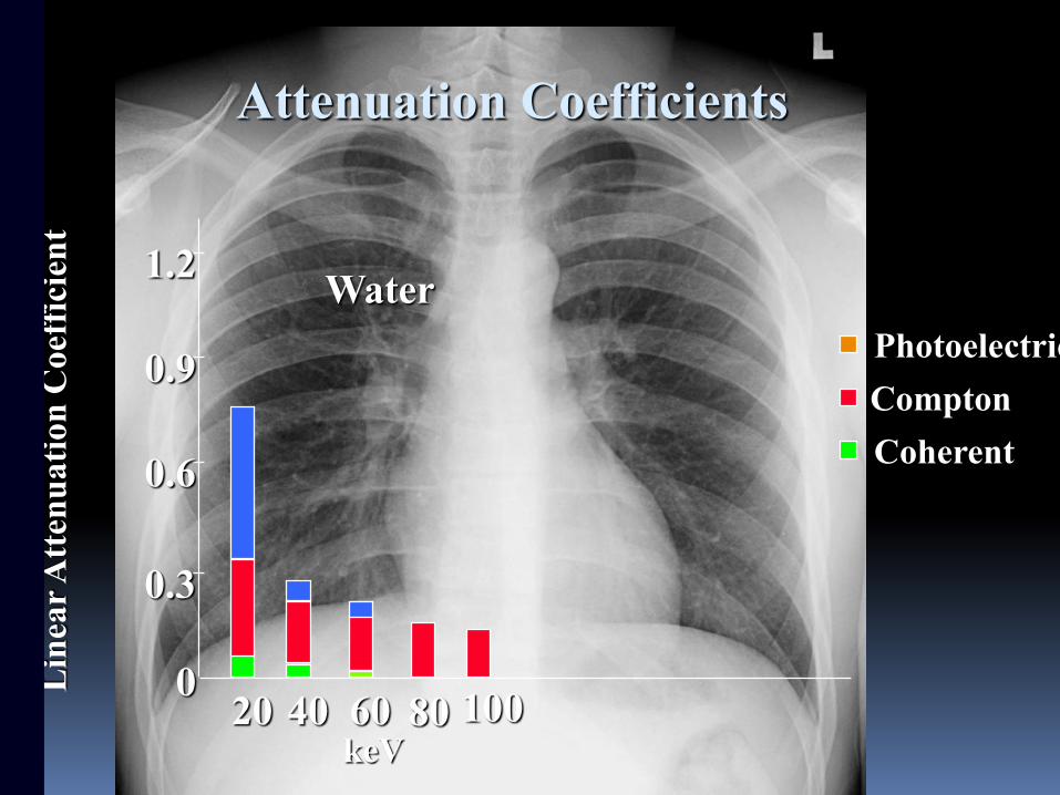
0
0.3
0.6
0.9
1.2
20 40 60 80 100
Water
Lin
ear
Att
enu
ati
on
Coef
fici
ent
Coherent
Compton
Photoelectric
keV
Attenuation Coefficients
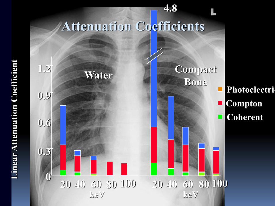
4.8
0
0.3
0.6
0.9
1.2
20 40 60 80 100 20 40 60 80 100
WaterCompact
Bone
Lin
ear
Att
enu
ati
on
Coef
fici
ent
Coherent
Compton
Photoelectric
keV keV
Attenuation Coefficients
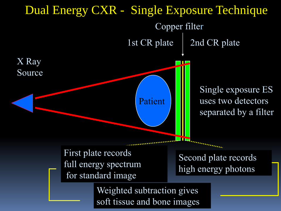
Single exposure ES
uses two detectors
separated by a filter
1st CR plate 2nd CR plate
Copper filter
X Ray
Source
Patient
Dual Energy CXR - Single Exposure Technique
Weighted subtraction gives
soft tissue and bone images
First plate records
full energy spectrum
for standard image
Second plate records
high energy photons
1st CR plate
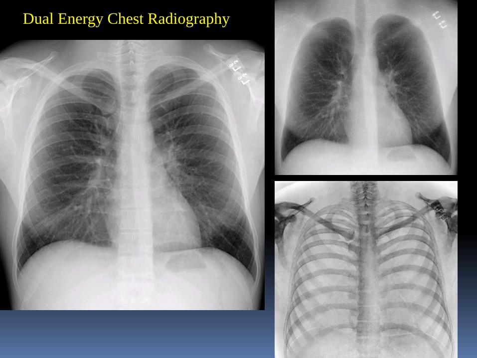
Dual Energy Chest Radiography
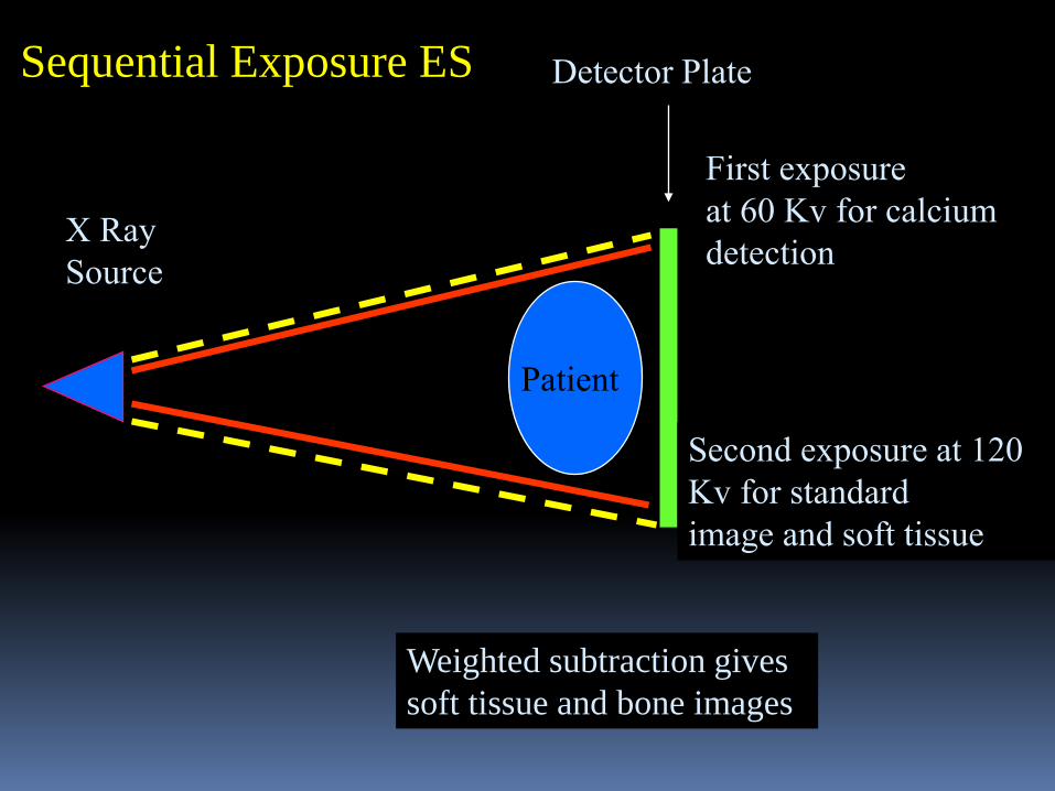
Detector Plate
X Ray
Source
Sequential Exposure ES
Second exposure at 120
Kv for standard
image and soft tissue
First exposure
at 60 Kv for calcium
detection
Weighted subtraction gives
soft tissue and bone images
Patient
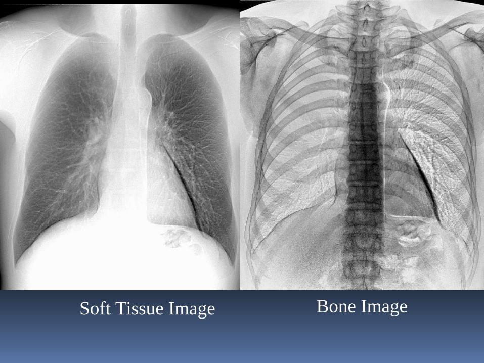
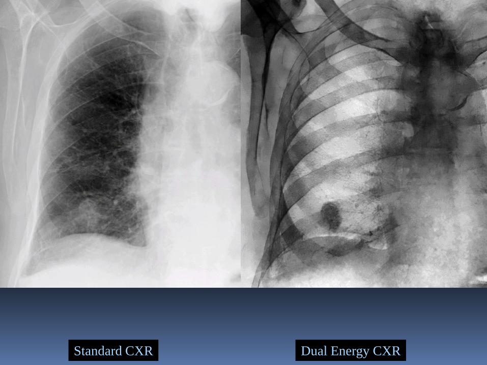
Soft Tissue Image Bone Image
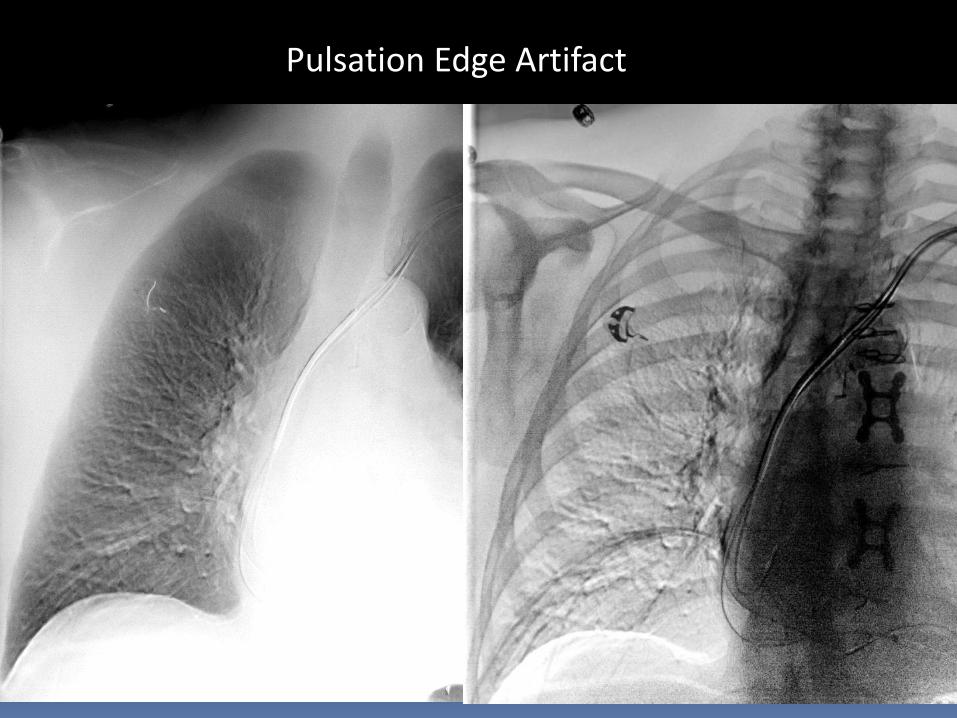
Pulsation Edge Artifact
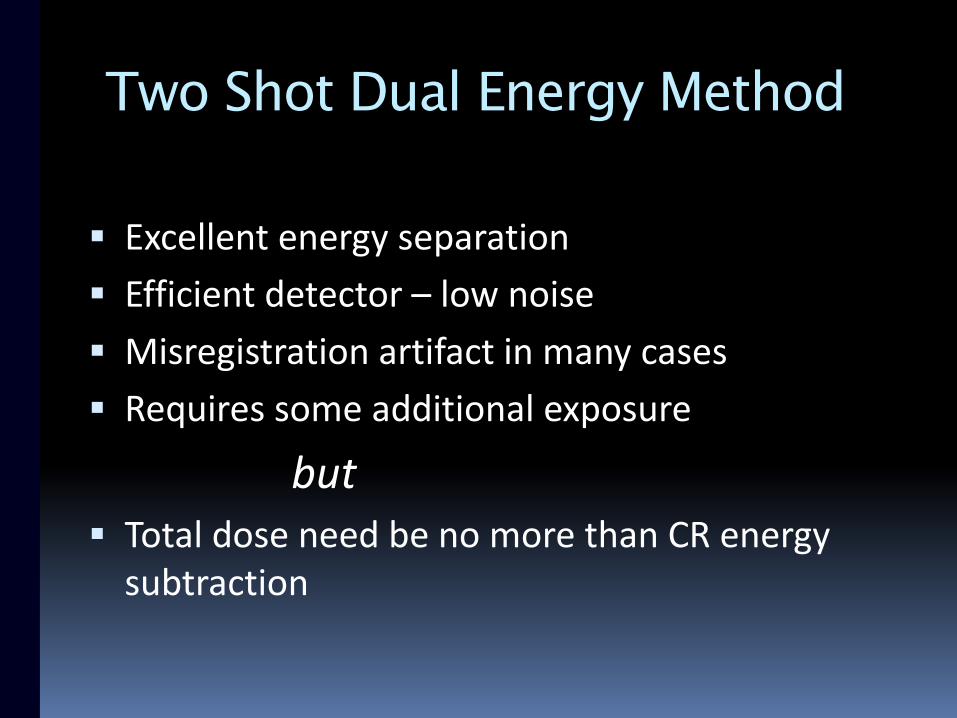
Two Shot Dual Energy Method
Excellent energy separation
Efficient detector – low noise
Misregistration artifact in many cases
Requires some additional exposure
but Total dose need be no more than CR energy
subtraction
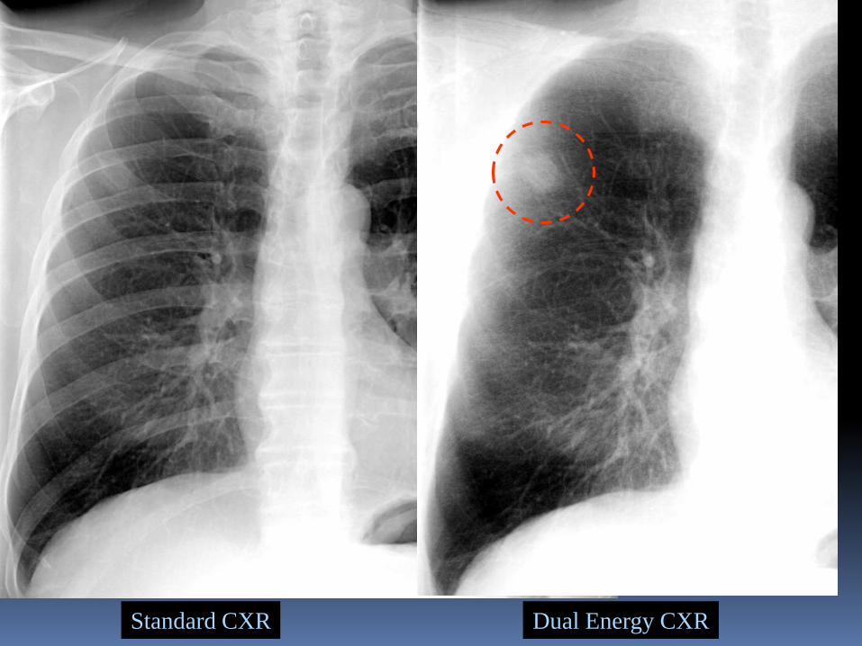
Standard CXR Dual Energy CXR
Dual Energy CXRStandard CXR
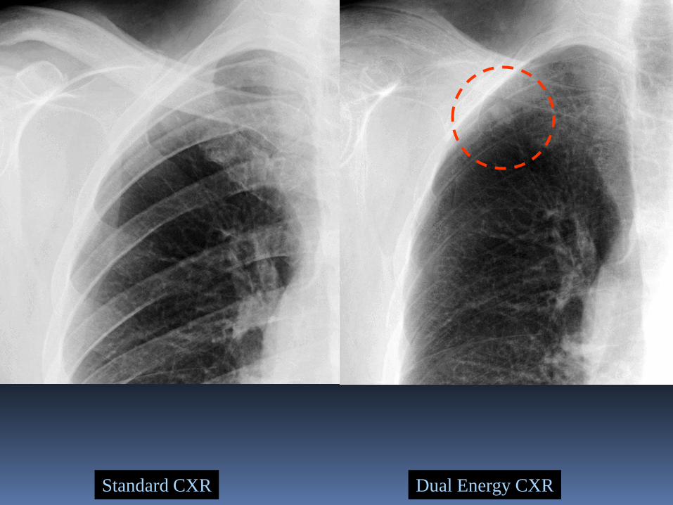
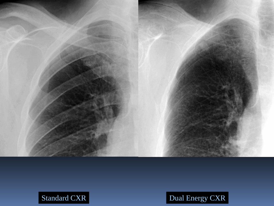
Standard CXR Dual Energy CXR
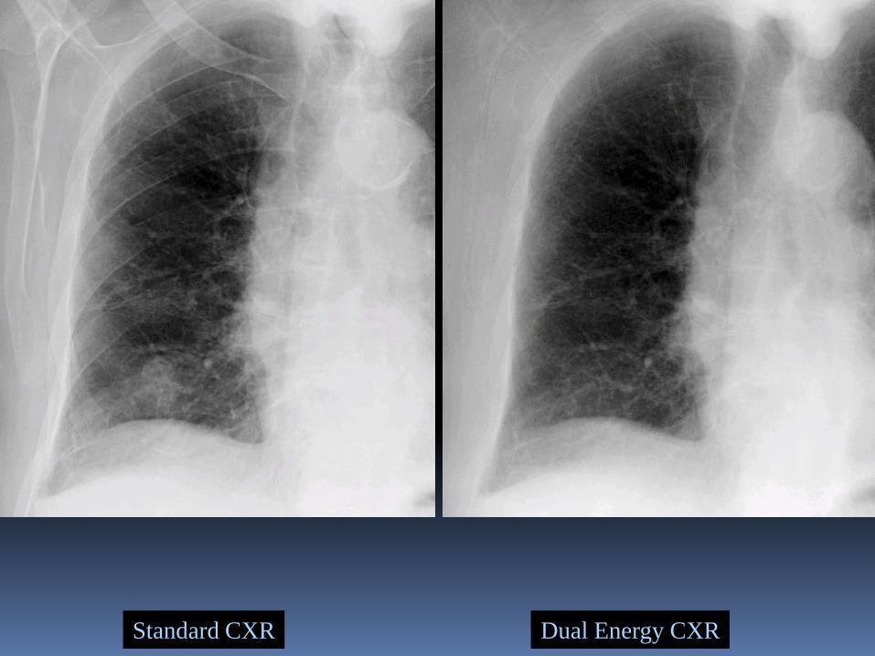
Standard CXR Dual Energy CXR
Standard CXR Dual Energy CXR
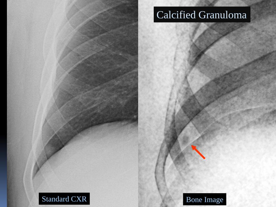
Calcified Granuloma
Standard CXR Bone Image
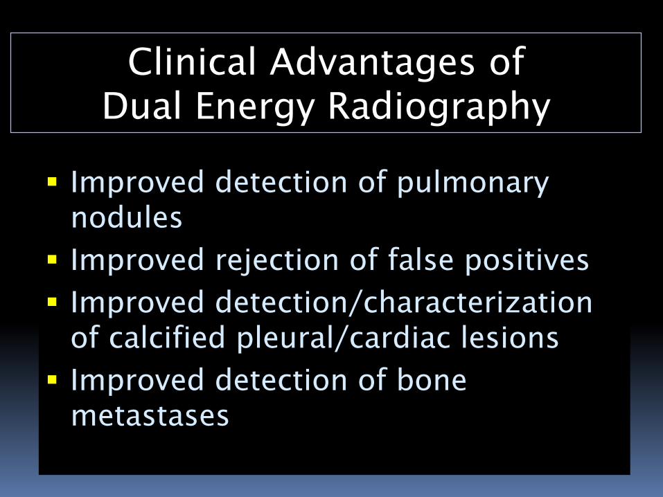
Clinical Advantages of
Dual Energy Radiography
Improved detection of pulmonary
nodules
Improved rejection of false positives
Improved detection/characterization
of calcified pleural/cardiac lesions
Improved detection of bone
metastases
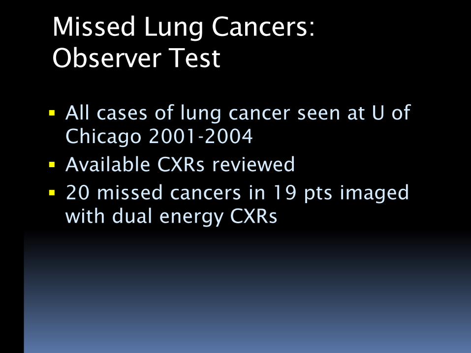
Missed Lung Cancers:
Observer Test
All cases of lung cancer seen at U of
Chicago 2001-2004
Available CXRs reviewed
20 missed cancers in 19 pts imaged
with dual energy CXRs
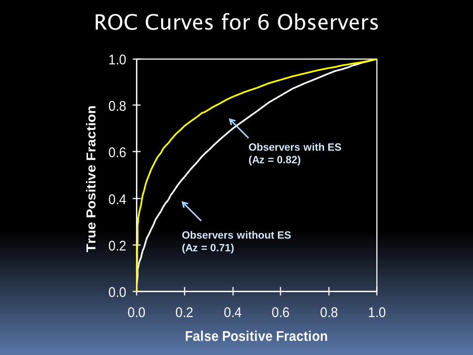
ROC Curves for 6 Observers
0.0
0.2
0.4
0.6
0.8
1.0
0.0 0.2 0.4 0.6 0.8 1.0
False Positive Fraction
Tru
e P
os
itiv
e F
rac
tio
n
Observers with ES
(Az = 0.82)
Observers without ES
(Az = 0.71)
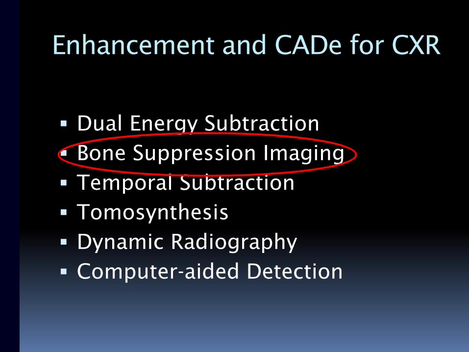
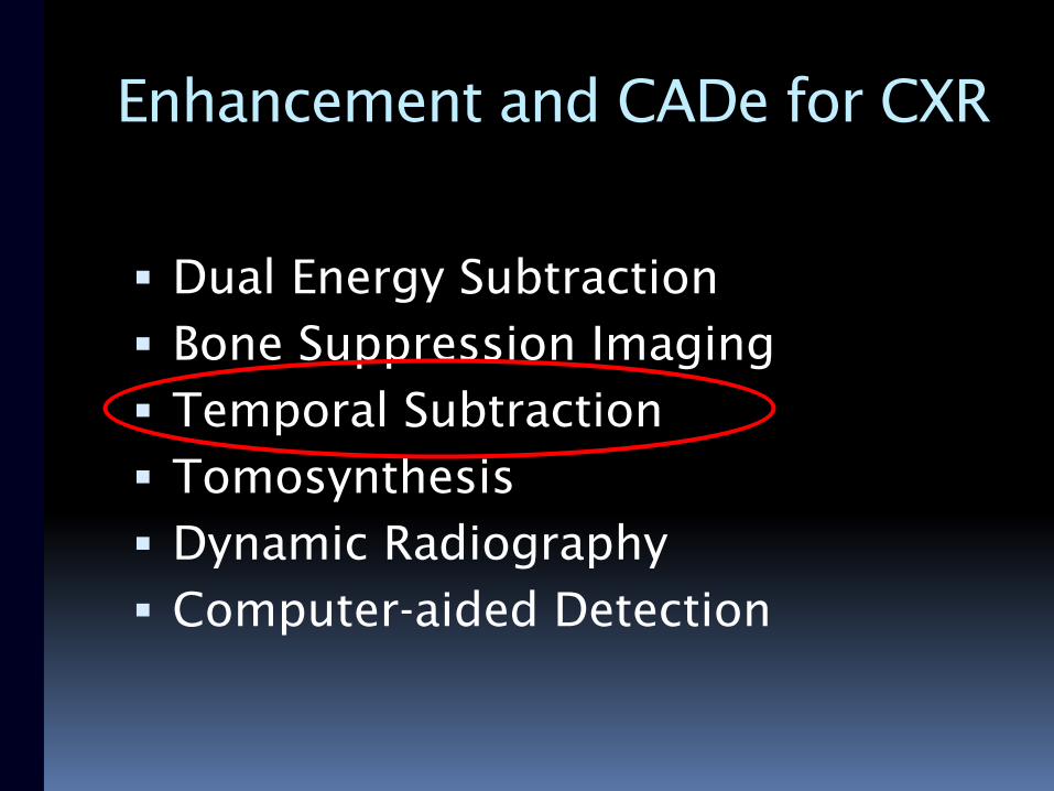
Enhancement and CADe for CXR
Dual Energy Subtraction
Bone Suppression Imaging
Temporal Subtraction
Tomosynthesis
Dynamic Radiography
Computer-aided Detection
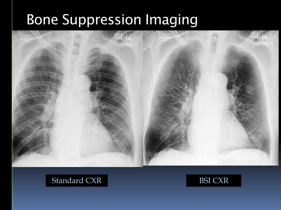
BSI CXR
Bone Suppression Imaging
Standard CXR
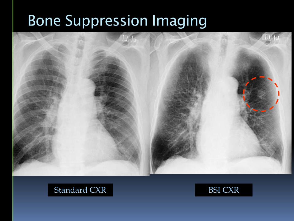
BSI CXR
Bone Suppression Imaging
Standard CXR
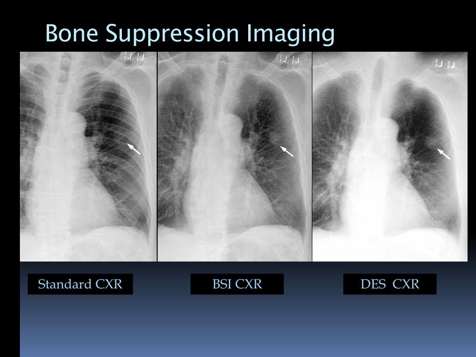
BSI CXR DES CXR
Bone Suppression Imaging
Standard CXR
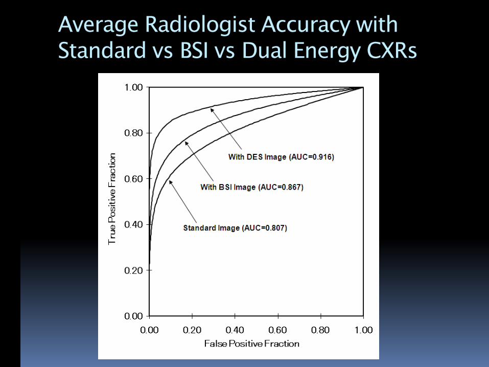
Average Radiologist Accuracy with
Standard vs BSI vs Dual Energy CXRs
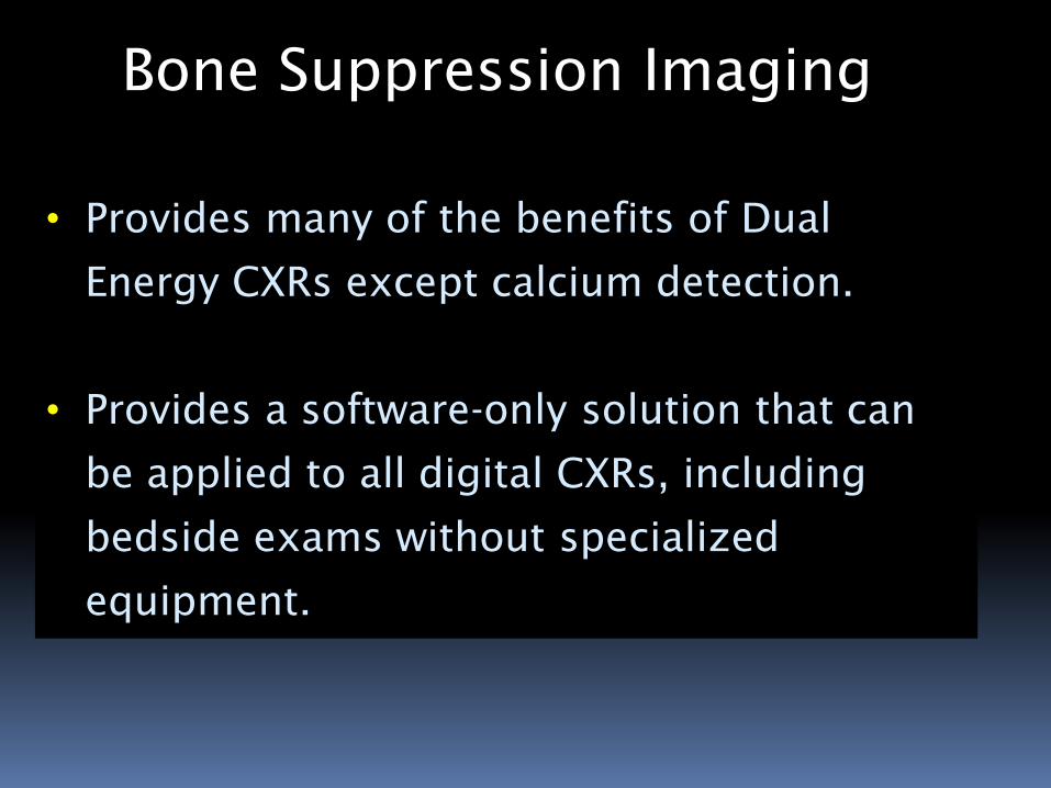
• Provides many of the benefits of Dual
Energy CXRs except calcium detection.
• Provides a software-only solution that can
be applied to all digital CXRs, including
bedside exams without specialized
equipment.
Bone Suppression Imaging
Bone Suppression Imaging
Enhancement and CADe for CXR
Dual Energy Subtraction
Bone Suppression Imaging
Temporal Subtraction
Tomosynthesis
Dynamic Radiography
Computer-aided Detection
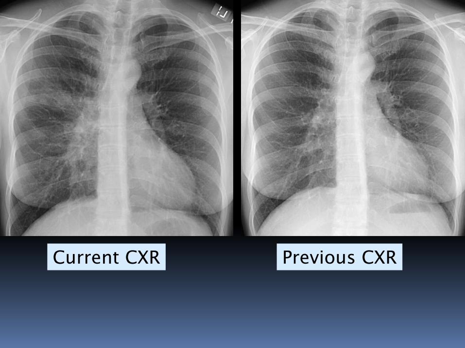
Current CXR Previous CXR
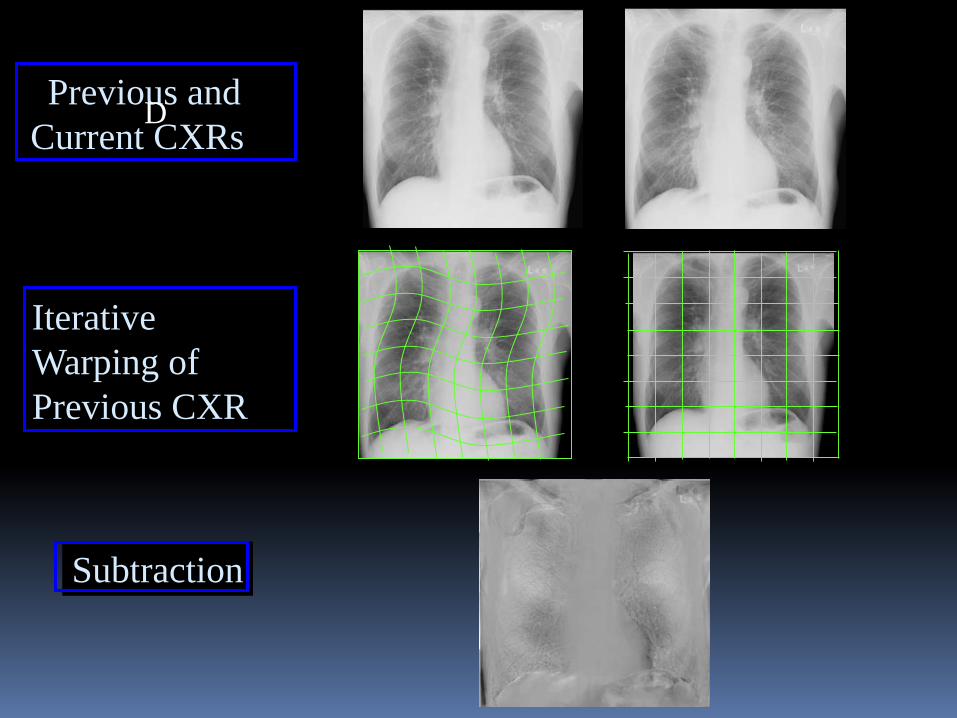
Previous and
Current CXRs
Iterative
Warping of
Previous CXR
Subtraction
D
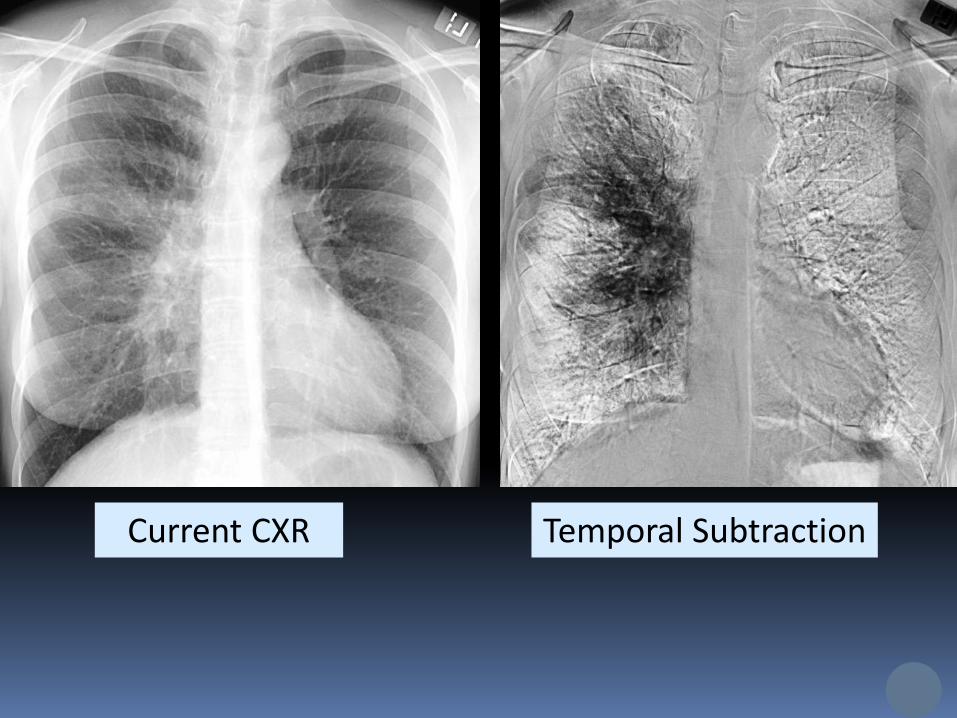
Temporal SubtractionCurrent CXR
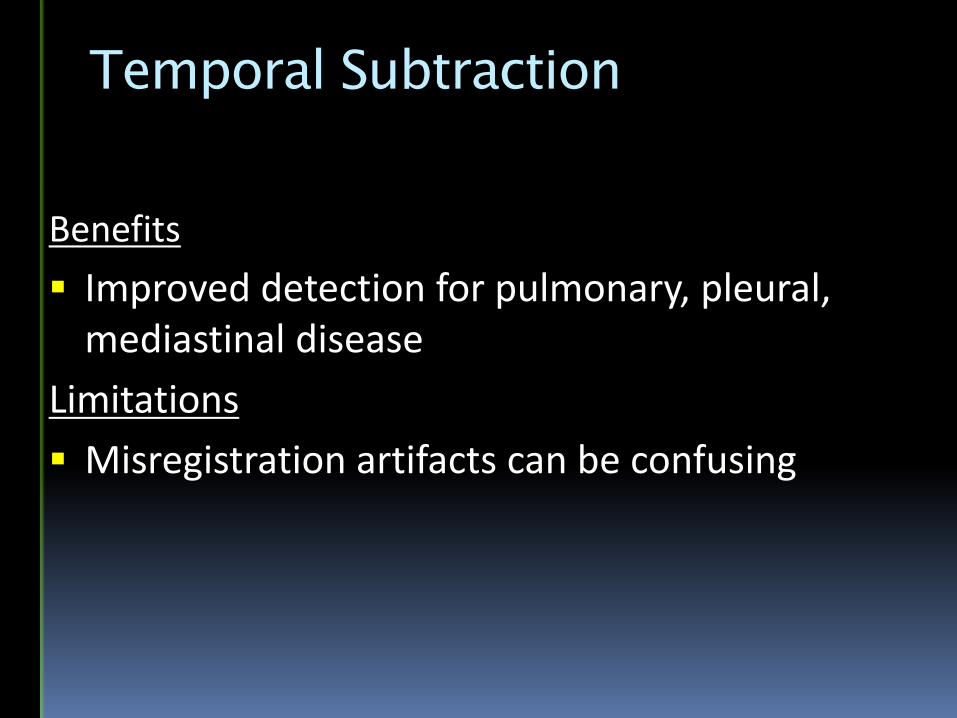
Temporal Subtraction
Benefits
Improved detection for pulmonary, pleural, mediastinal disease
Limitations
Misregistration artifacts can be confusing
Enhancement and CADe for CXR
Dual Energy Subtraction
Bone Suppression Imaging
Temporal Subtraction
Tomosynthesis
Dynamic Radiography
Computer-aided Detection
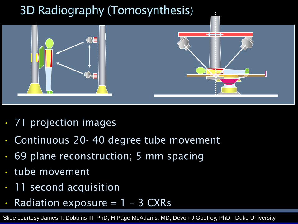
3D Radiography (Tomosynthesis)
• 71 projection images
• Continuous 20- 40 degree tube movement
• 69 plane reconstruction; 5 mm spacing
• tube movement
• 11 second acquisition
• Radiation exposure = 1 – 3 CXRs
Slide courtesy James T. Dobbins III, PhD, H Page McAdams, MD, Devon J Godfrey, PhD; Duke University
CXR Tomosynthesis
CXR Tomosynthesis
CXR Tomosynthesis
CXR Tomosynthesis
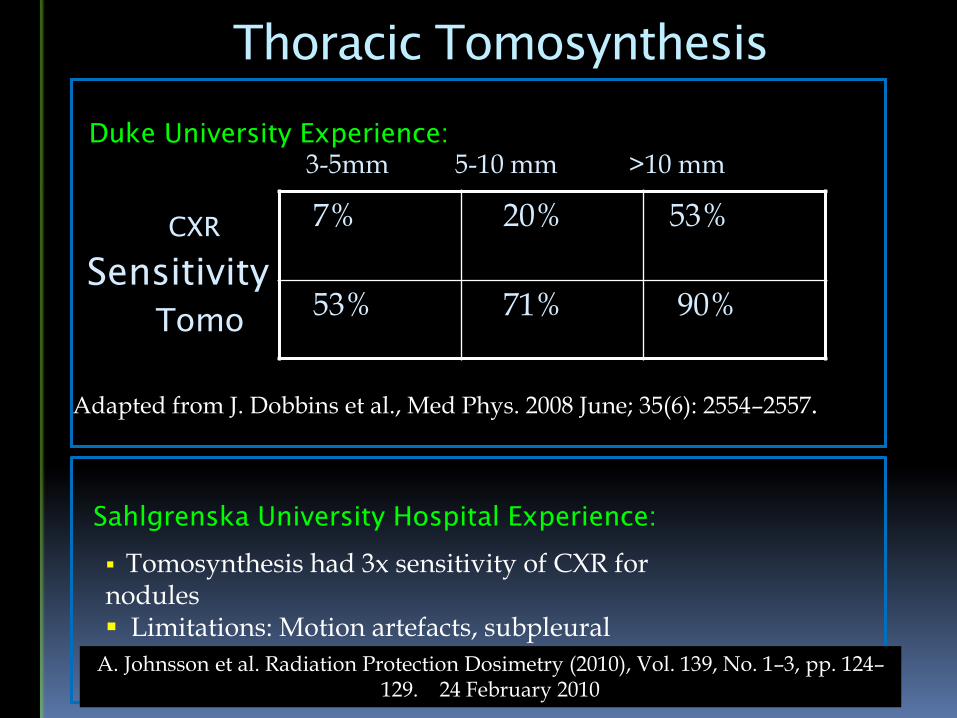
Thoracic Tomosynthesis
7% 20% 53%
53% 71% 90%
3-5mm 5-10 mm >10 mm
CXR
Tomo
Adapted from J. Dobbins et al., Med Phys. 2008 June; 35(6): 2554–2557.
Sensitivity
Duke University Experience:
Sahlgrenska University Hospital Experience:
Tomosynthesis had 3x sensitivity of CXR for nodules Limitations: Motion artefacts, subpleuralnodulesA. Johnsson et al. Radiation Protection Dosimetry (2010), Vol. 139, No. 1–3, pp. 124–
129. 24 February 2010
Enhancement and CADe for CXR
Dual Energy Subtraction
Bone Suppression Imaging
Temporal Subtraction
Dynamic Radiography
Tomosynthesis
Computer-aided Detection
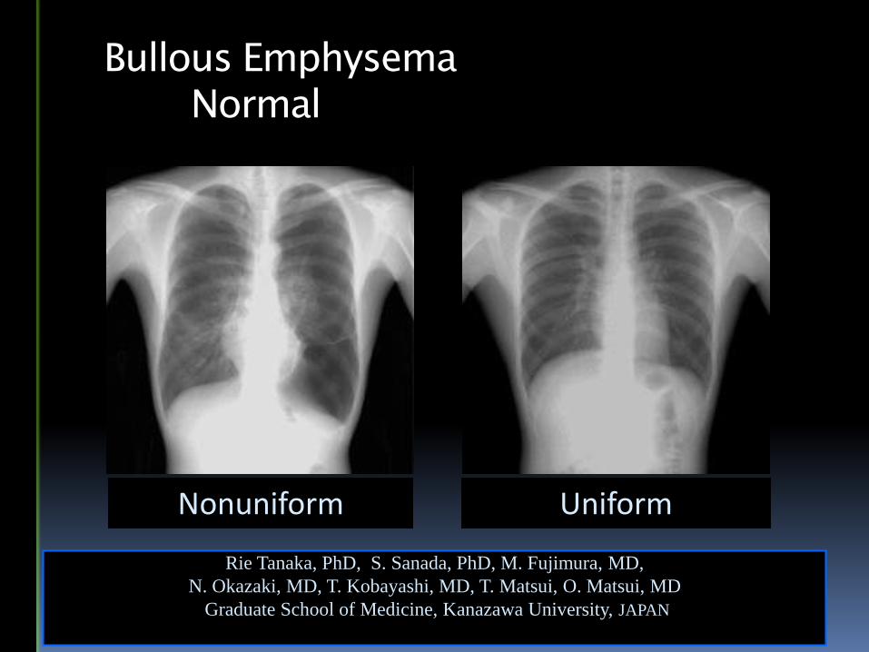
UniformNonuniform
Rie Tanaka, PhD, S. Sanada, PhD, M. Fujimura, MD,
N. Okazaki, MD, T. Kobayashi, MD, T. Matsui, O. Matsui, MD
Graduate School of Medicine, Kanazawa University, JAPAN
Bullous Emphysema
Normal
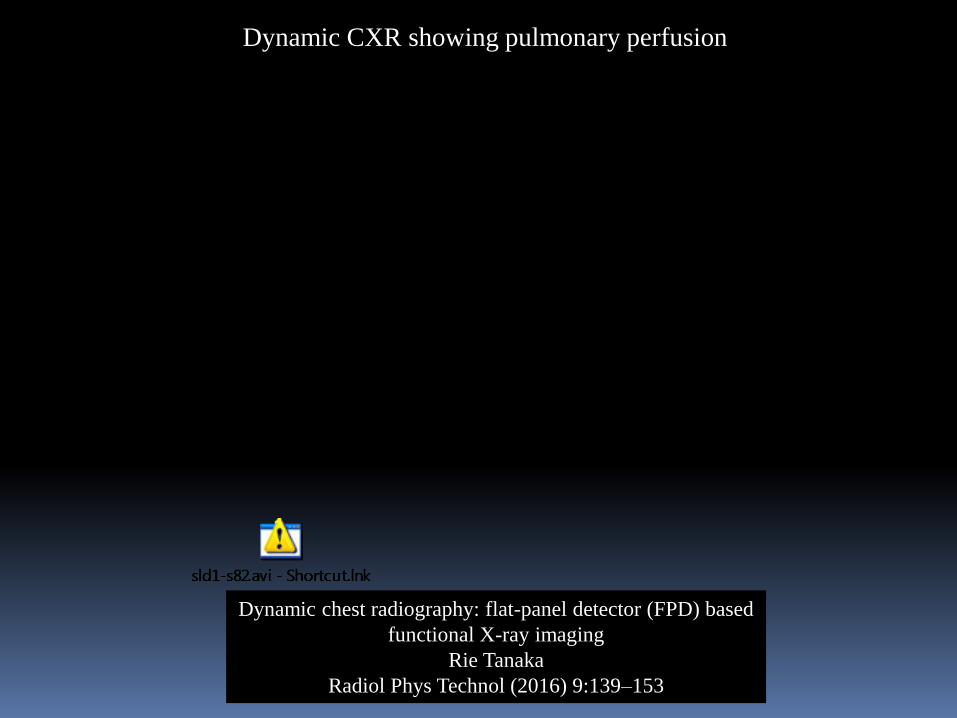
Dynamic chest radiography: flat-panel detector (FPD) based
functional X-ray imaging
Rie Tanaka
Radiol Phys Technol (2016) 9:139–153
Dynamic CXR showing pulmonary perfusion
Enhancement and CADe for CXR
Dual Energy Subtraction
Bone Suppression Imaging
Temporal Subtraction
Tomosynthesis
Dynamic Radiography
Computer-aided Detection
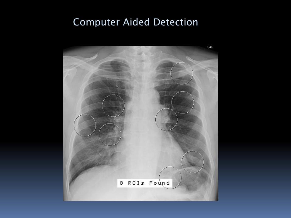
Computer Aided Detection
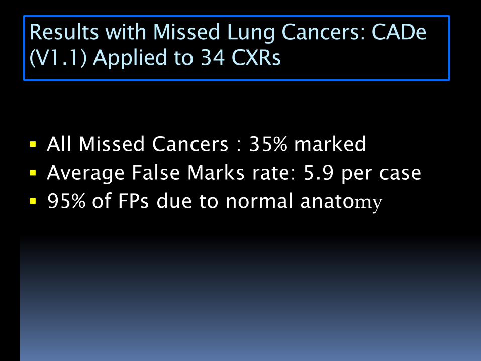
Results with Missed Lung Cancers: CADe
(V1.1) Applied to 34 CXRs
All Missed Cancers : 35% marked
Average False Marks rate: 5.9 per case
95% of FPs due to normal anatomy
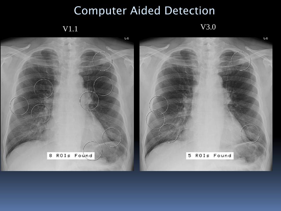
V1.1 V3.0
Computer Aided Detection
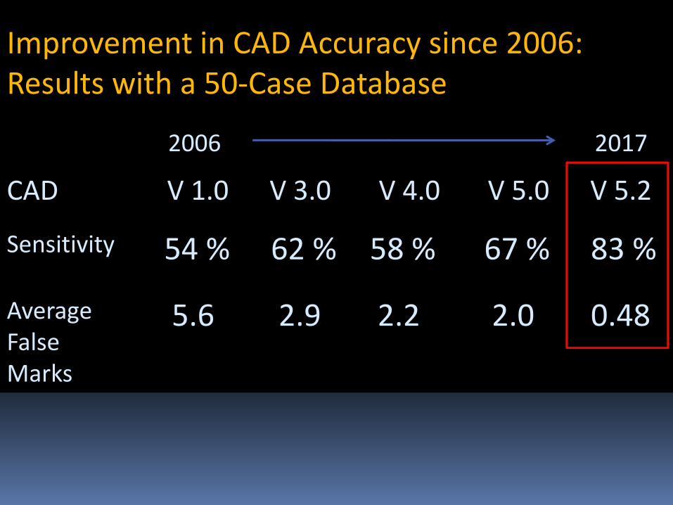
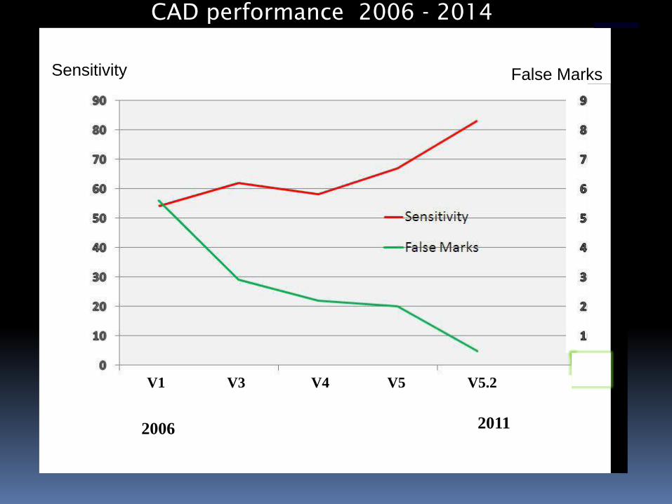
Sensitivity 54 % 62 % 58 % 67 % 83 %
Average False Marks
5.6 2.9 2.2 2.0 0.48
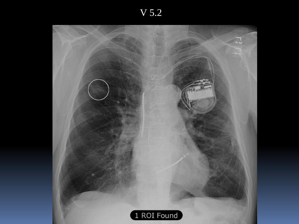
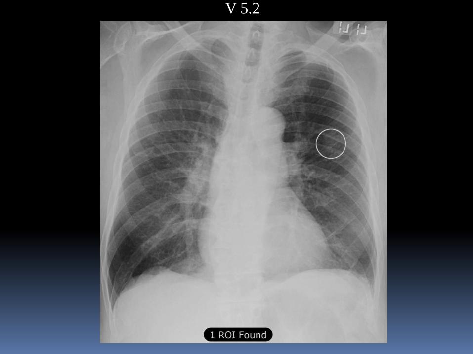
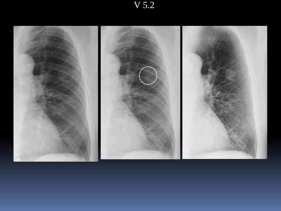
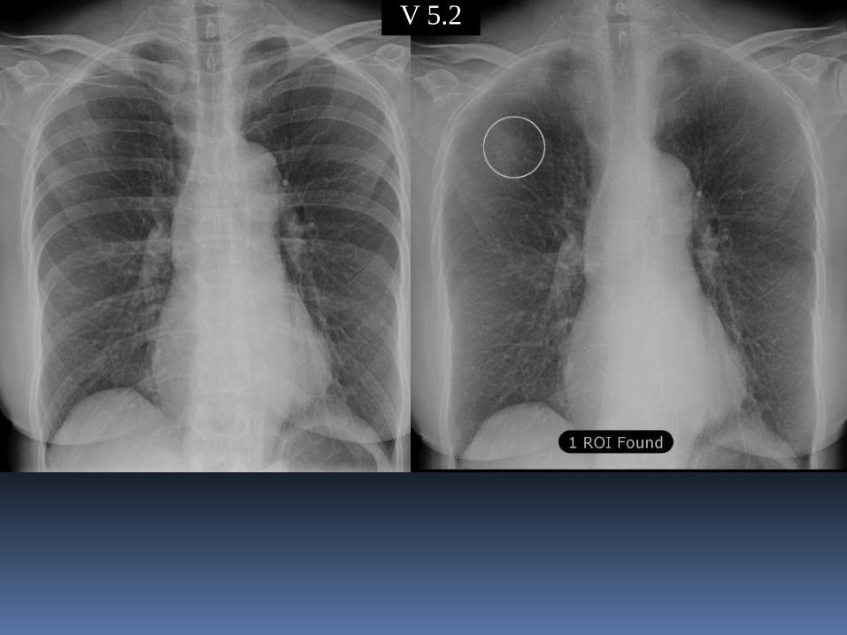
CAD V 1.0 V 3.0 V 4.0 V 5.0 V 5.2
Improvement in CAD Accuracy since 2006:Results with a 50-Case Database
20062006 2017
)Sensitivity False Marks(
Progress in CXR CAD
CAD performance 2006 - 2014
V1 V3 V4 V5 V5.2
2006 2011
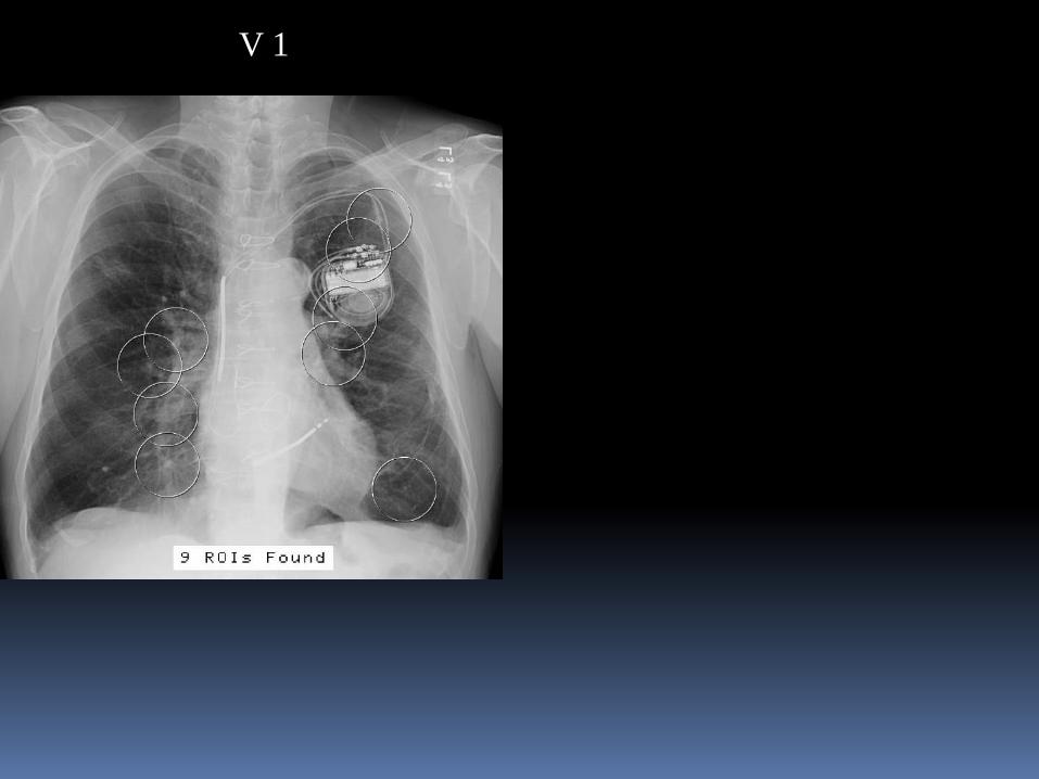
V 1 V 3V 1
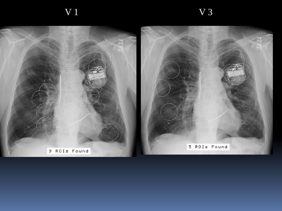
V 1 V 3
V 5.2
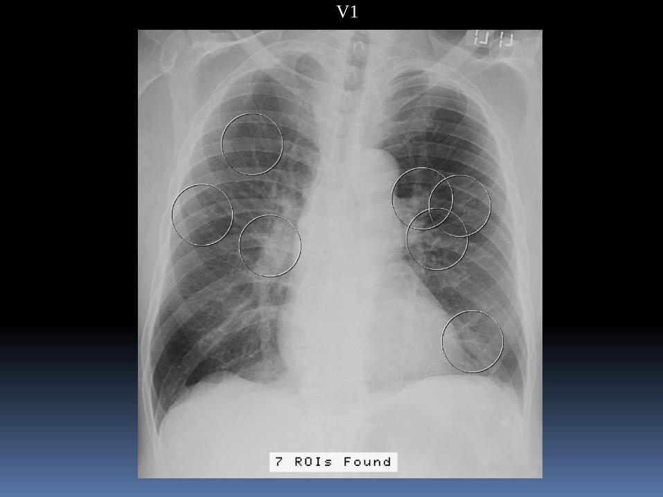
V1
V 5.2
V 5.2V 5.2
V 5.2
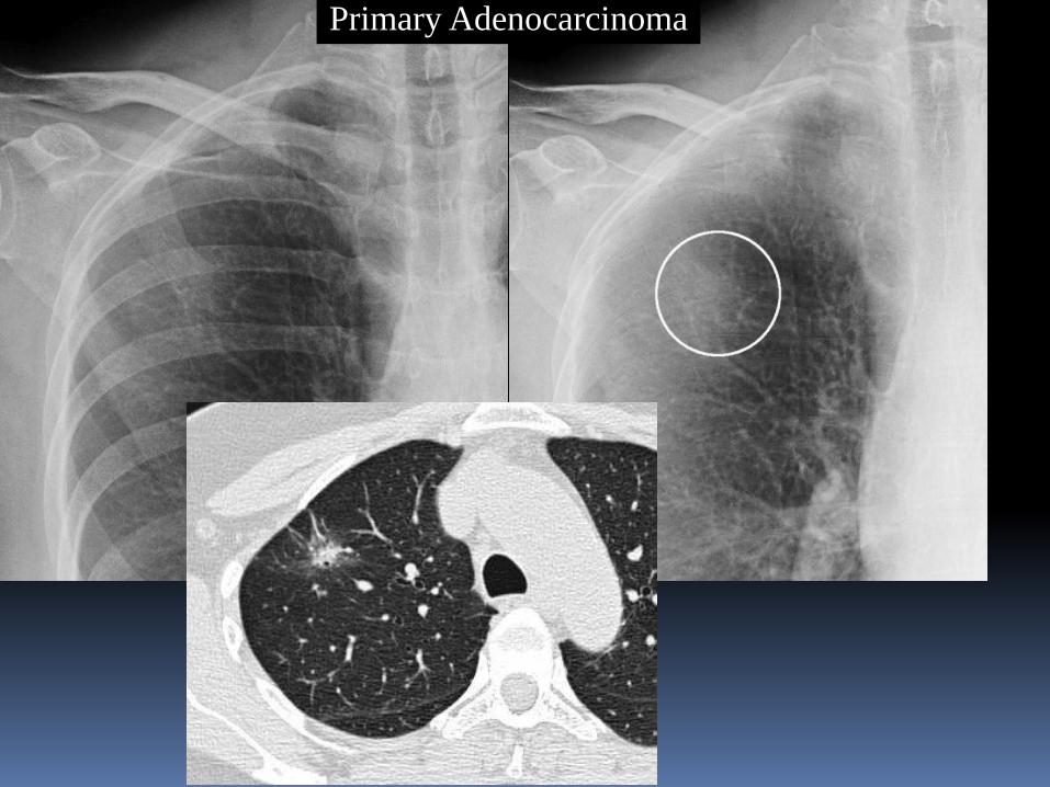
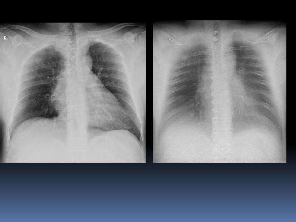
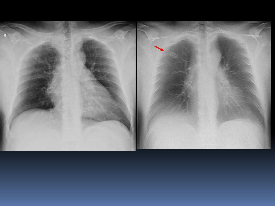
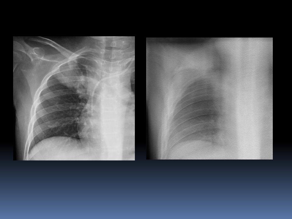
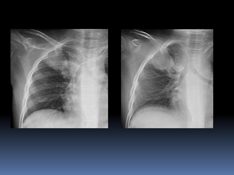
Primary Adenocarcinoma
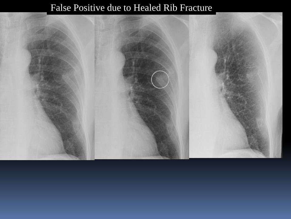
False Positive due to Healed Rib Fracture
Recent Improvements in CXR CAD
Increased sensitivity
Greatly reduced false positive rate
Nearly 50% of false positives are now due to focal benign opacities
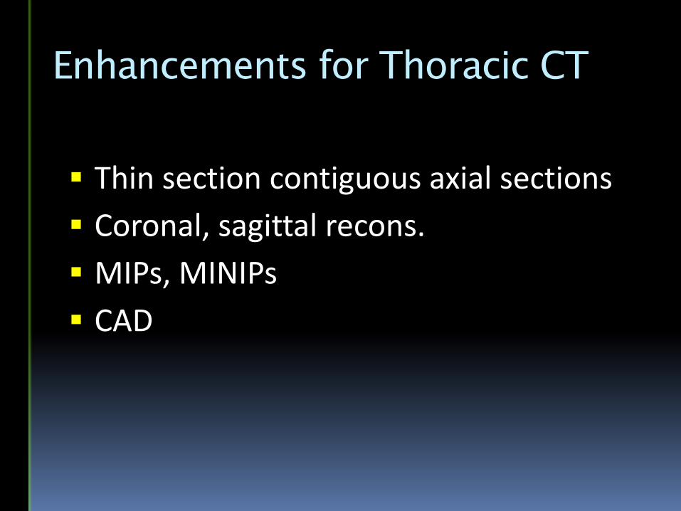
Enhancements for Thoracic CT
Thin section contiguous axial sections
Coronal, sagittal recons.
MIPs, MINIPs
CAD
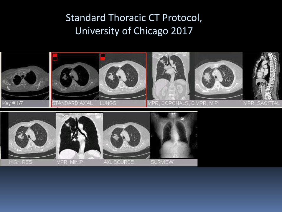
Standard Thoracic CT Protocol, University of Chicago 2017

3mm
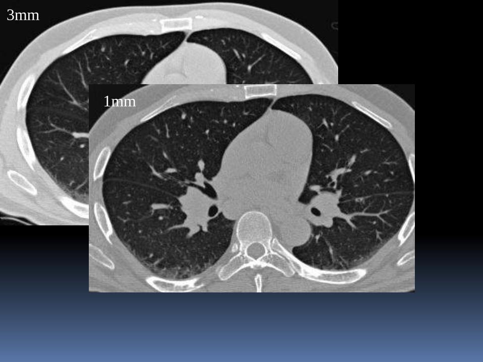
3mm
1mm
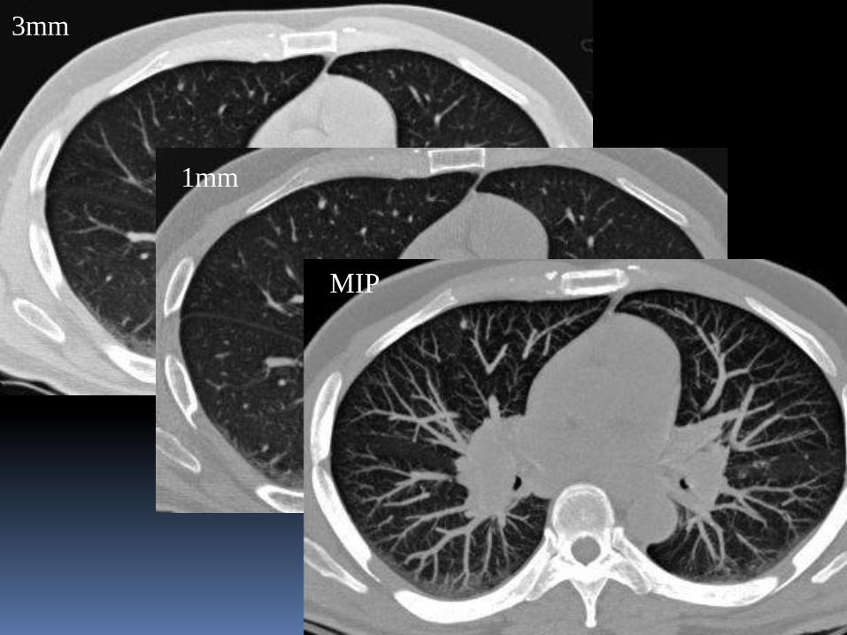
3mm
1mm
MIP
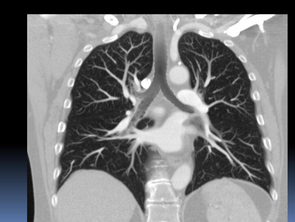
Coronal MIP
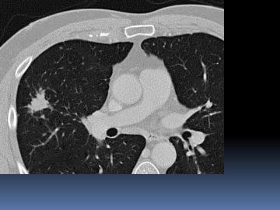
3mm slab
3mm slab
MIP
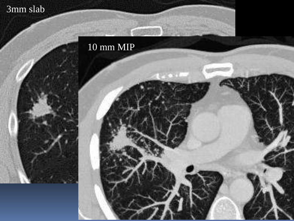
10 mm MIP
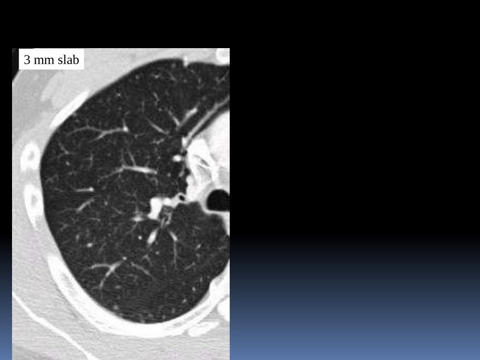
3 mm slab
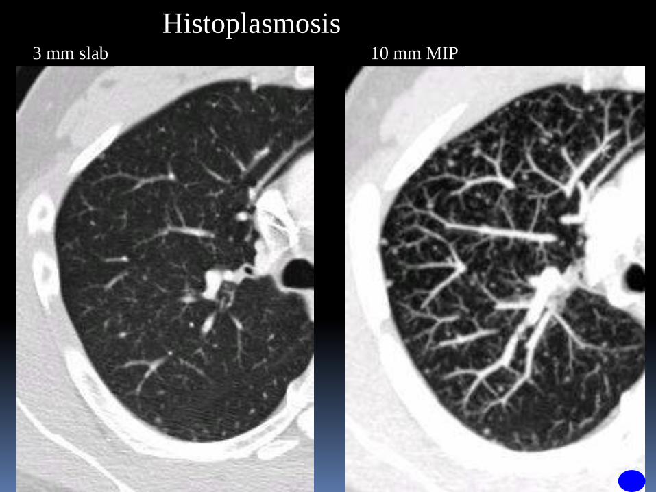
Histoplasmosis3 mm slab 10 mm MIP
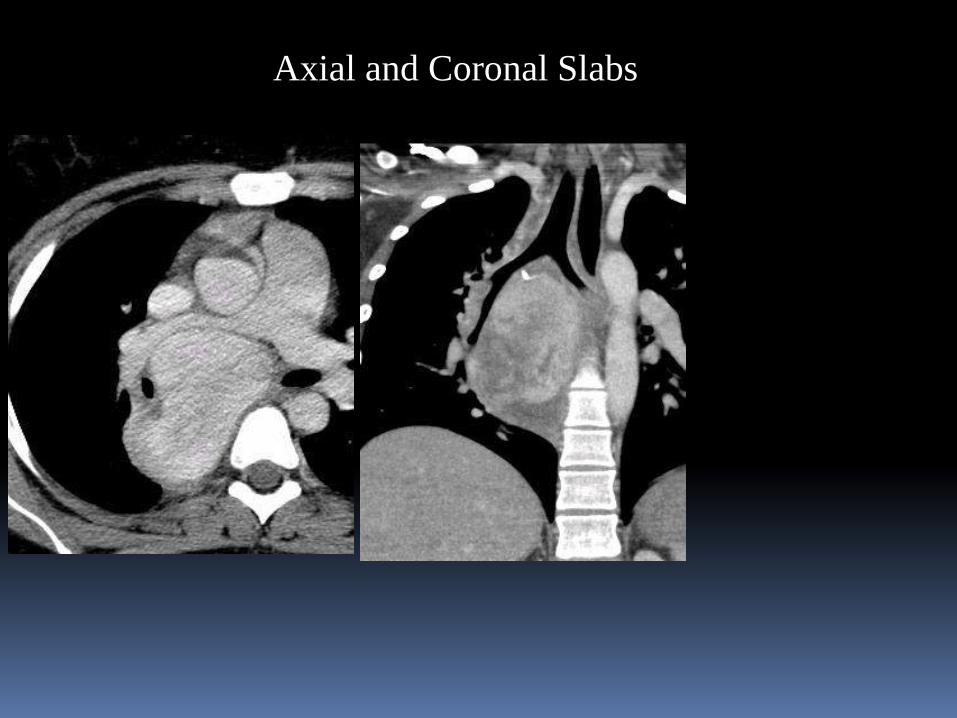
Axial and Coronal Slabs
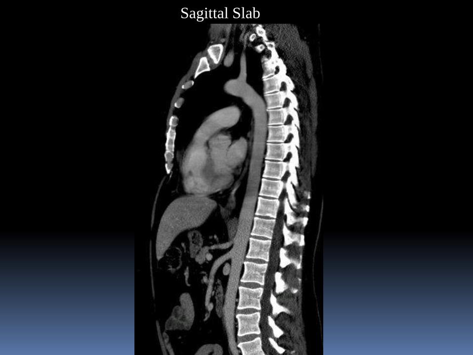
Sagittal Slab
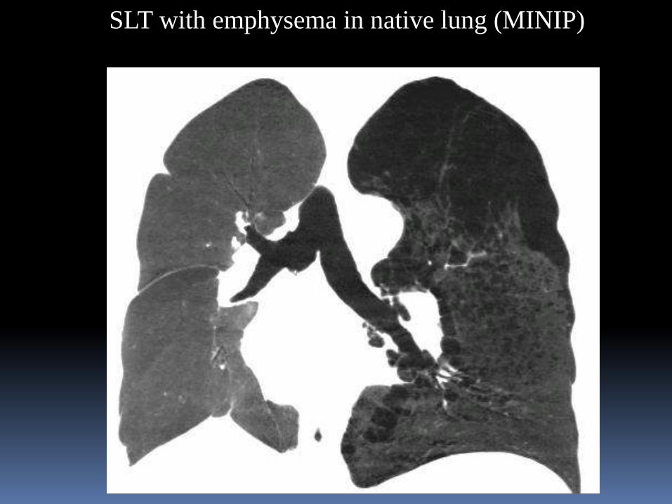
SLT with emphysema in native lung (MINIP)
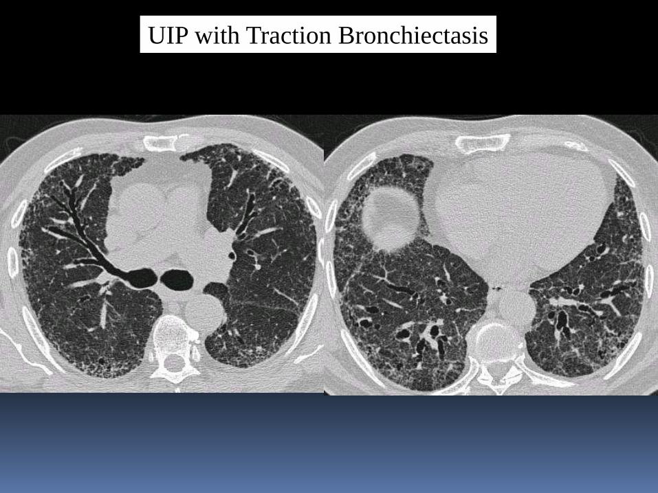
UIP with Traction Bronchiectasis
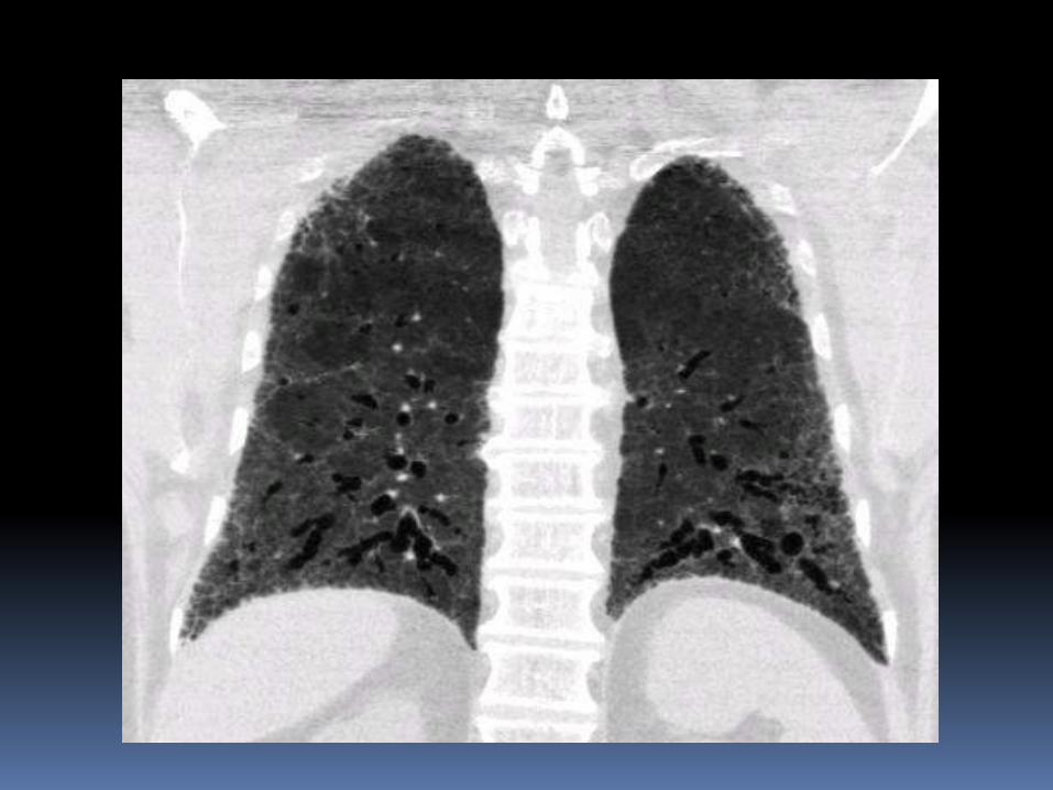
MINIP Image
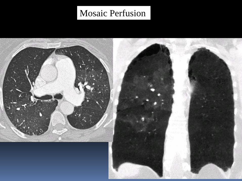
Mosaic Perfusion
Slab MINIP
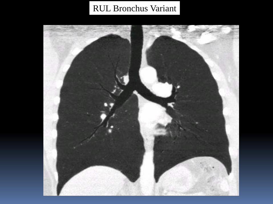
RUL Bronchus Variant
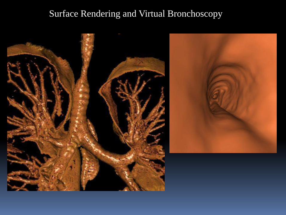
Surface Rendering and Virtual Bronchoscopy
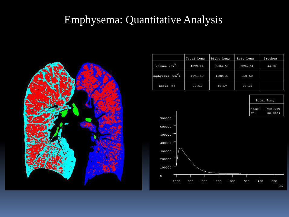
Emphysema: Quantitative Analysis
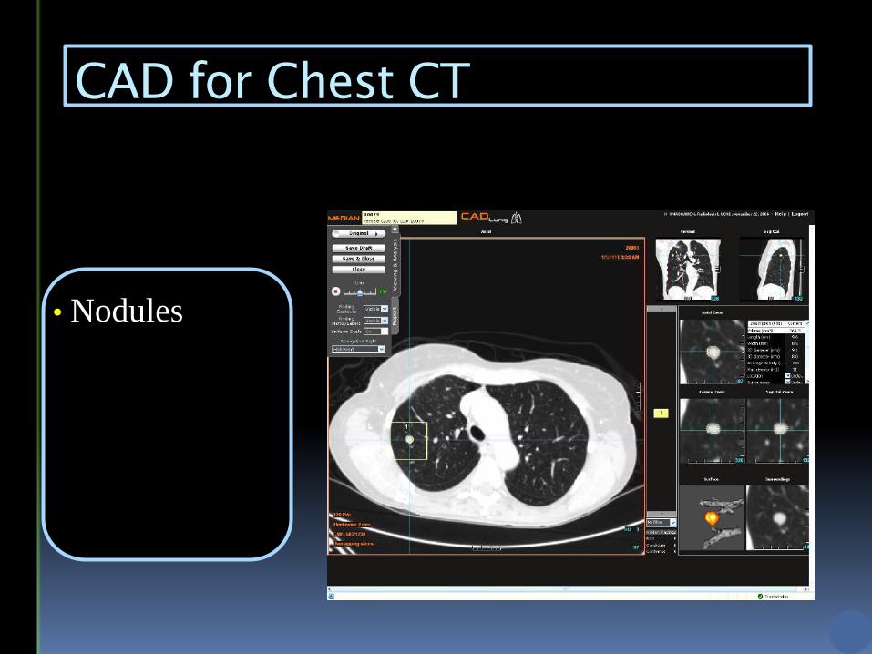
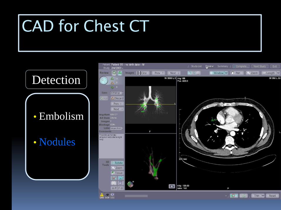
CAD for Chest CT
• Nodules
-detect
-measure
-compare
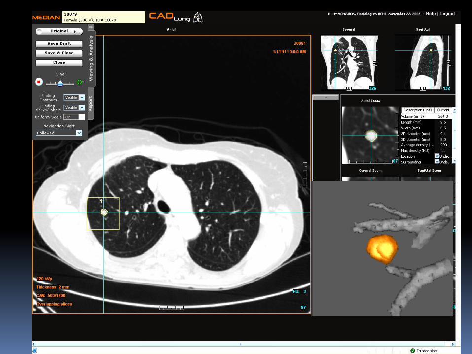
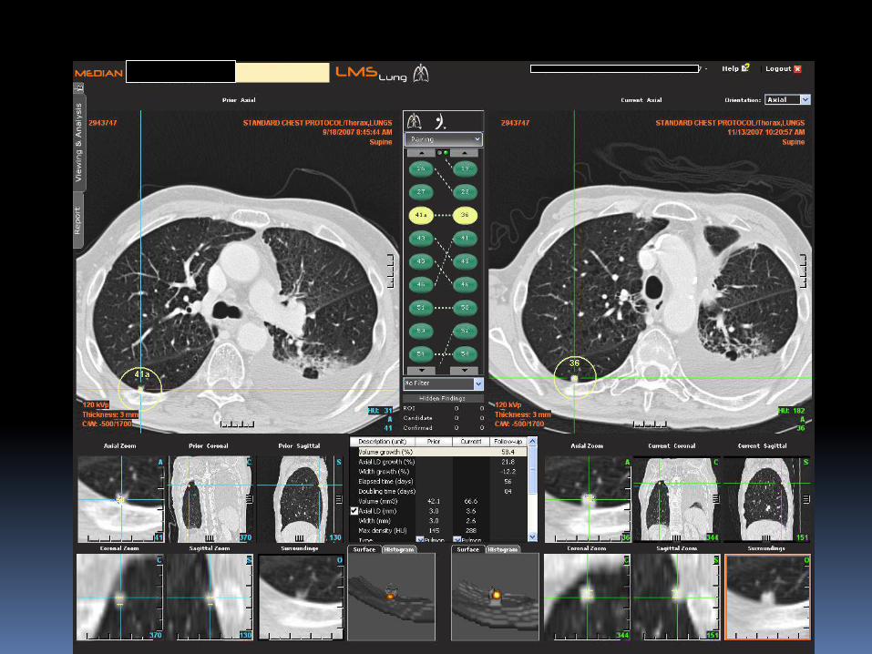
Nodule Tracking and Comparison
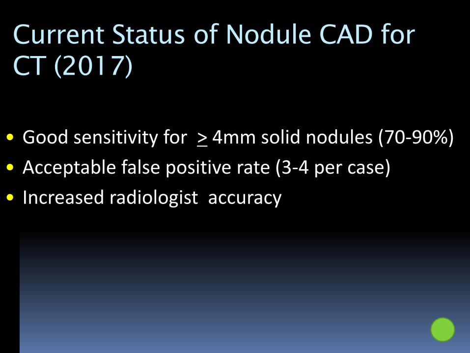
Current Status of Nodule CAD for
CT (2017)
• Good sensitivity for > 4mm solid nodules (70-90%)
• Acceptable false positive rate (3-4 per case)
• Increased radiologist accuracy
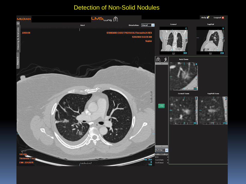
Detection of Non-Solid Nodules
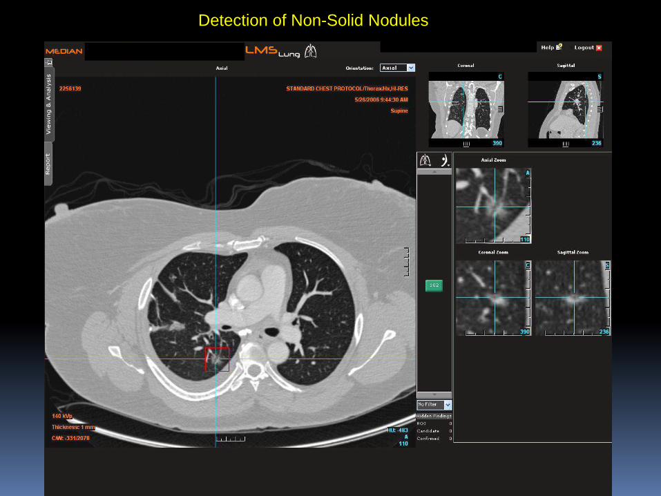
Detection of Non-Solid Nodules
CAD for Chest CT
Detection
• Nodules
• Embolism
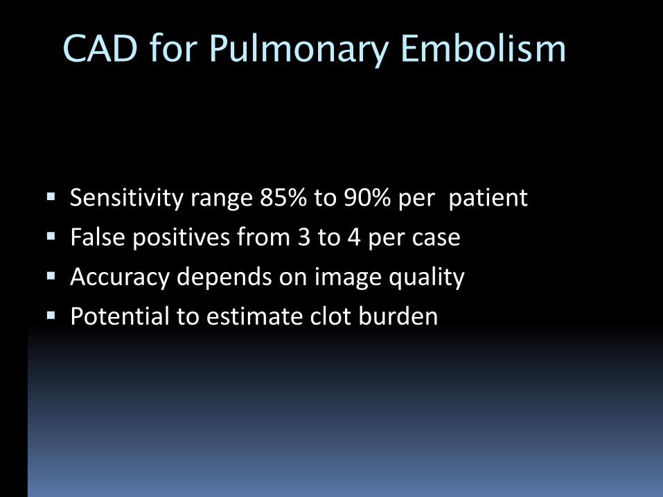
CAD for Pulmonary Embolism
Sensitivity range 85% to 90% per patient
False positives from 3 to 4 per case
Accuracy depends on image quality
Potential to estimate clot burden
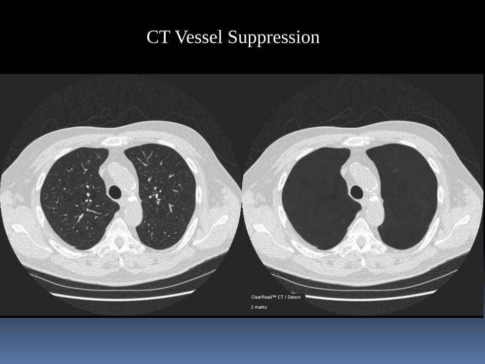
CT Vessel Suppression
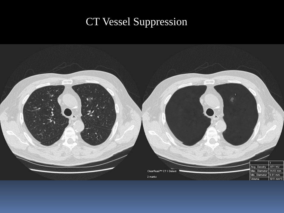
CT Vessel Suppression
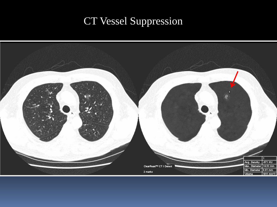
CT Vessel Suppression
Future Directions
Big Data
Convolutional Neural Networks
Machine Learning
Cognitive computing

Uber Self Driving Car
“In theory, there’s no difference between theory and practice.But in practice, there is”.
Yogi Berra
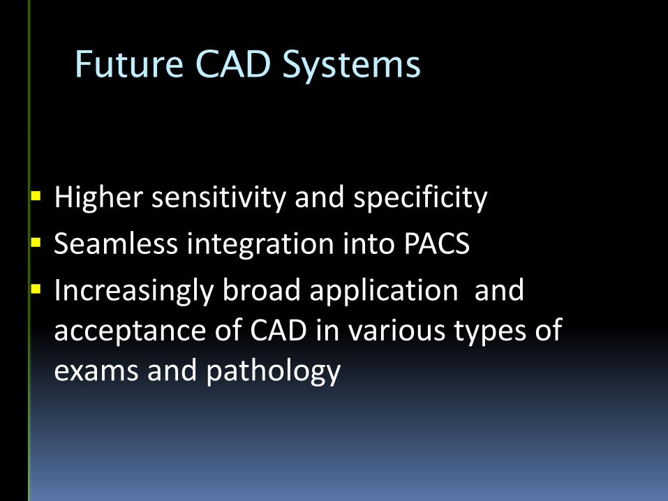
Future CAD Systems
Higher sensitivity and specificity
Seamless integration into PACS
Increasingly broad application and acceptance of CAD in various types of exams and pathology
Conclusions
Enhanced Radiography can improve diagnostic accuracy without reducing productivity
Venice
2017


![Automatic Computer Aided Diagnosis of Breast Cancer in ...€¦ · Automatic Computer Aided Diagnosis of Breast Cancer in Dynamic Contrast Enhanced Magnetic ... [38]. Research into](https://img.dokumen.tips/doc/110x75/5fed569fc69d0e2eda06e93e/automatic-computer-aided-diagnosis-of-breast-cancer-in-automatic-computer-aided.jpg)