Embed Size (px)
Citation preview
Full Terms & Conditions of access and use can be found athttp://www.tandfonline.com/action/journalInformation?journalCode=gpom20
Download by: [Universite Laval] Date: 10 May 2016, At: 01:59
International Journal of Polymeric Materials andPolymeric Biomaterials
ISSN: 0091-4037 (Print) 1563-535X (Online) Journal homepage: http://www.tandfonline.com/loi/gpom20
Albumin-Based Biomaterial for Lungs TissueEngineering Applications
Hammed T. Aiyelabegan, Sadaf S. Z. Zaidi, Songwe Fanuel, Ali Eatemadi,Malihe T.K. Ebadi & Esmaeil Sadroddiny
To cite this article: Hammed T. Aiyelabegan, Sadaf S. Z. Zaidi, Songwe Fanuel, Ali Eatemadi,Malihe T.K. Ebadi & Esmaeil Sadroddiny (2016): Albumin-Based Biomaterial for LungsTissue Engineering Applications, International Journal of Polymeric Materials and PolymericBiomaterials, DOI: 10.1080/00914037.2016.1180610
To link to this article: http://dx.doi.org/10.1080/00914037.2016.1180610
Accepted author version posted online: 09May 2016.
Submit your article to this journal
View related articles
View Crossmark data
1
Albumin-Based Biomaterial for Lungs Tissue Engineering Applications
Hammed T. Aiyelabegan1, 2
, Sadaf S. Z. Zaidi1, 2
, Songwe Fanuel1,2
, Ali Eatemadi1,
Malihe T.K. Ebadi1, Esmaeil Sadroddiny
1
1Department of Medical Biotechnology, School of Advanced Technologies in Medicine,
Tehran University of Medical Sciences, Tehran, Iran. 2Tehran University of Medical Sciences International Campus, Tehran, Iran.
Corresponding author: Dr. Esmaeil Sadroddiny No 88 Italia Street, P.O. Box.
1417755469. Department of Medical Biotechnology, School of Advanced Technologies
in Medicine, Tehran University of Medical Sciences, Tehran, Iran. Email:
Abstract
The role of albumin-based biomaterials in Tissue engineering (TE) cannot be over-
emphasized. In this paper, we reviewd the role of albumin in lungs scaffold grafting
which promotes cell seeding. Albumin grafted on decellularized lungs scaffold is
presented as a great support material for cell-tissue interaction, for ease in attachment,
growth and differentiation when seeded with different types of cells. Albumin scaffold
fabrication from different sources is a promising approach that may facilitate medical
treatments from bench-to-bed, although, the role of this scaffold in lungs surfactant
proteins regeneration and binding need to be fully elucidated.
Keyword; Albumin, Scaffold, Tissue Engineering, Regenerative Medicine, Lungs and
Biomaterial.
LIST OF ABBREVIATION
3D Three Dimension
Ad-MSc Adipose tissue-derived mesenchymal stem cells
ADSCs Adipose derived stem cells
Dow
nloa
ded
by [
Uni
vers
ite L
aval
] at
01:
59 1
0 M
ay 2
016
2
ASA Autologous serum-derived albumin
BALF Bronchoalveolar lavage fluid
BSA Bovine Serum Albumin
CC Clara cells
CPG Calcium phosphate glass
CRD Carbohydrate recognition domain
DNA Deoxy ribonucleic acid
ECM Extracellular matrix
ECs Endothelial Cells
EPS Exogenous pulmonary surfactant
HA Hydroxyapatite
HSA Human Serum Albumin
MSCs Messenchymal stem cell
OECs Olfactory ensheathing cells
PCL Polycaprolactone
PLGA Poly (lactic-coglycolic acid)
PLLA Poly (L-lactide)
PSA Porcine serum albumin
SCI Spinal Cord Injury
SP Surfactant protein
SPARC Secreted proteins acid and rich in cysteine
TE Tissue engineering
UV Ultra violet
Dow
nloa
ded
by [
Uni
vers
ite L
aval
] at
01:
59 1
0 M
ay 2
016
3
VSCs Smooth vascular muscle cells
1.0 INTRODUCTION
Langer & Vacanti [1] introduced biomaterial-based TE. It was defined as an
interdisciplinary field employing engineering and life sciences principles to support
biological substitutes which restore, maintain, or improve the function of a tissue or a
whole organ [1], [2]. Most researchers have outlined six components for TE, namely,
cells, cell-cell interaction, scaffolds, growth factors, bioreactors and stimulation
(mechanical etc.).
Cellularizing decellularized organs like lungs, represents another method of obtaining a
whole transplantable, and functional organs [3]–[6]. An intersection between
biotechnology and biomedical engineering named TE, was established to support and
promote denovo synthesis of scaffolds, so also as to repair defective damaged tissues
according to the patient’s regeneration potential. In recent therapy, it is necessary to
provide cells with a surrounding that will regulates and improve their proliferation and
differentiation for regeneration of tissue i.e. monitoring cellular growth and
differentiation in TE constructs. Biomaterial scaffold is capable of providing in addition
to physical support, the chemical and biological clues needed in forming functional tissue
[7]–[9]. Biomaterial technology is essential in the creation of this local cell environment,
and various synthetic and natural materials such as polymers, ceramics, conjugated
metals, or their composites, have been studied and utilized in different manners [8], [10].
Protein derived biomaterials like albumin, vitronectin, fibronectin, laminin, collagen,
elastin, casein, zein, also provides suitable enviroment for the growth of cells [7], [8].
Dow
nloa
ded
by [
Uni
vers
ite L
aval
] at
01:
59 1
0 M
ay 2
016
4
Transplantation and biomaterial engineering were recently utilized for disease healing,
with the aftermath of the former, being cancer and graft rejection [8]. The unique
characteristics like mechanical properties, high availability, low cost, easy design and
synthesis of polymeric materials have made them a good substrates [9], [12] for organ
scaffold synthesis and support. However, only a few polymers provide the
biocompatibility [13] and affinity with other matrix proteins and growth factor in vitro
and in vivo [3], [13], [14].
In other to initiate lungs regeneration process and to simultaneously solve the problem of
graft rejection, biomaterial polymers research has been focused on, as biopolymers are
biodegradable, biocompatible and could be replaced by human tissue produced by the
cells surrounding the material [15].
In addition to being biodegradable, proteins scaffolds are cheap and can be produced in
large scale [16], [17]. For these reasons, great effort has been made in the utilization of
proteins as a biomaterial scaffold for TE application [18]–[21]. However, the lack of
mechanical strength, high rate of biodegradation, risk of auto-immune rejection, some
properties, like wettability, adhesion, and surface composition are insufficient for many
applications [22]–[24].
Fabrication is also important when synthesizing a protein scaffold, the protein chains
functional groups, for instance, must be cross-linked either within themselves or with the
Dow
nloa
ded
by [
Uni
vers
ite L
aval
] at
01:
59 1
0 M
ay 2
016
5
help of chemically cross-linking agents like glutaraldehyde, formaldehyde [25] or
grafting them on decellularized lungs tissue, and simultaneously maintaining their
structure, which in-turn reserve their functionality. Self-crosslinking have been said to be
better since there won’t be an incorporation of toxic chemicals into the scaffold by the
use of the reagents, which in-turn may interfere with the process of lungs tissue
regeneration.
Albumin; the most abundant human serum protein, have been said to influence the
attachment of cells to various scaffold material in a similar and better fashion as collagen
and fibronectin after some treatments. They can serve as an interface between cells and
scaffold, thereby mediating the integration of these two components. In this review,
milestone of albumin based TE biomaterials was addressed, and ways they have assisted
in cellular adhesion to scaffold in bone, cardiac, and neural TE. Their potential role in
grafting decellularized lungs tissue before recellularization with various kinds of cells
was discussed, and possible challenges in Lungs TE was pointed.
2.0 ALBUMIN-BASED BIOMATERIALS
Found in blood serum (human, horse, pig, sheep etc), egg white, milk, and many other
plants and animal tissues, with a slight variation in amino acid composition. Previous
data have supported the notion that different sources of albumin are relevant substrate for
TE application (Table 1). The most common albumin types used in TE scaffold include,
Human serum albumin (HSA), Bovine Serum Albumin (BSA), Ovalbumin, and Porcine
Serum Albumin (PSA).
Dow
nloa
ded
by [
Uni
vers
ite L
aval
] at
01:
59 1
0 M
ay 2
016
6
2.1 Types
2.1.1 Hsa
Structure And Properties Of HSA
HSA protein was observed as a component of human blood and to be the most abundant
blood protein (35 - 50 g/L) as early as the 20th
century. From then on, it has been
extensively studied and applied in several aspects of biomedical science. Its structural
information was obtained based on low angle X-ray scattering, hydrodynamics as well as
in silico prediction studies. The structures of HSA purified from blood and that obtained
by recombinant DNA technology are basically identical. However, despite its well-
characterized molecular structure (Fig. 1), it has been slow to find its use as scaffolding
material in TE.
According to Carter and Ho [26], X-ray crystallographic studies , HSA is a heart-shaped
molecule (globular), coded for by a gene located on chromosome 4 and has a high alpha-
helical content (~67%), with a molecular mass of 66.5 kDa, rich in disulphide bridges (17
in total) and has one free cysteine residue at position 34 [26]. It is made of 585 amino
acid residues that make up three repeating helical peptide domains (labeled I–III) and
each of these domains is divided into two sub-domains (Fig. 1). Hydrodynamic studies
have revealed that HSA has a high affinity to a very wide range of materials, including
metals such as copper and zinc, fatty acids, amino acids as well as a vast number of
metabolites. Of late, a number of structures of HSA have been published which helped
shed light on the structural features of the HSA and how the protein binds a number of
ligands in some biomedical applications.
Dow
nloa
ded
by [
Uni
vers
ite L
aval
] at
01:
59 1
0 M
ay 2
016
7
Considering normal physiological conditions, about 10-15 grams of HSA are produced in
the hepatocytes daily, with none or very low intracellular storage. HSA can be
polymerized by cross-linking [22] and other fabrication methods, its synthesis is
stimulated by hormones, such as insulin, cortisol and growth hormone, and inhibited by
pro-inflammatory substances, including interleukin-6 and tumor necrosis factor-α [27],
[28]. Albumin scaffold have been said to display a very similar result when compared
with collagen based tissue scaffold, both of them have high water binding characteristics
and excellent resilience capacity, exhibiting approximately the same tensile strengths and
possessing extremely high porosities (97%). In addition, they have good cell binding
properties because they are peptide based biomaterial and this has been helpful for
cellular growth, thus, in this regard, they have been examined [29].
Function
HSA binds and carries lots of hydrophobic molecules like endogenous molecules (i.e.,
cholesterol, bilirubin, fatty acids, thyroxina) or exogenous substances (i.e., toxins and
drugs), gas like NO with consequent solubilization, transport, metabolism and
detoxification [30], [31], maintaining the reducing power, osmotic pressure and pH of the
mammalian blood [32], [33]. The HSA molecule also contribute to the stabilization of the
endothelial layer and for the maintenance of the normal capillary permeability probably
by reducing oxidative damage and modulating inflammation [34].
Dow
nloa
ded
by [
Uni
vers
ite L
aval
] at
01:
59 1
0 M
ay 2
016
8
HSA has been established for hepatitis B virus and hepatocyte binding which in turn
partake in hepatitis pathology [35]. It is capable of binding proteins like membrane
associated gp60 (albondin), drugs, as well as secreted proteins acid and rich in cysteine
(SPARC). The receptors of albondin localized on tumor vessels endothelial cells allows
for transcytosis of albumin via continuous endothelium whereas overexpressed SPARC
leads to the buildup of interstitium tumor albumin. HSA has been a suitable gene therapy
agent since it prevent undesired reaction with serum that often follow intravenous
injection of transfection complexes. Recombinant HSA (Recombumin) in addition has
been synthesized and their tolerability, safety, pharmacodynamics and pharmacokinetics
to the native HSA have been presented [36], [37].
2.1.2 Bsa
Obtained from cows blood, BSA is one of the component in tissue culture medium,
albumin-based scaffold will remain a good substrate since it plays a structural support
part in cell and TE [29], [30]. BSA is a globular protein (66kDa), containing 583 amino
acid residues linked together on a single chain with a known sequence [38]. The 3D
(three- dimensional) conformation of BSA comprises of 3 homologous domains (I, II, III)
which is specific for fatty acids and metals. Each domain represent the product of 2
subdomains, which are chiefly helical and extensively cross linked by sulfide bridges
[38].
2.1.3 Ovalbumin
Dow
nloa
ded
by [
Uni
vers
ite L
aval
] at
01:
59 1
0 M
ay 2
016

9
Ovalbumin is one of the first isolated proteins, which is the major protein in avian egg-
white (60-65% of total egg-white protein) [39]. The structure and function of ovalbumin
reveal that this protein belongs to the seprin superfamily [40]. It is basically a
glycoprotein comprising of 386 amino acids [41] and a carbohydrate side chain linked
covalently to amide N of Asn293. Ovalbumin does not show any protease inhibitory
activity despite sequence identity of 30% with antitrypsin and other inhibitor proteins of
serpin family [39].
2.2 Albumin Scaffold
A scaffold is a support, matrix or delivery vehicle for promoting the binding and
migration of cells or bioactive molecules used for restoring, repairing or regenerating
tissues [42]. Scaffold provides 3D cell culture template upon which activation of seeded
cells is initiated for full tissue regeneration and controlled degradation of the scaffold,
after the tissue has been fully grown. Tissue engineered scaffold plays a role in
mimicking functions of native ECM partially.
Pore structure and size should also be considered when synthesizing a scaffold for TE,
wider and well linked pores gives rise to a good mass transfer and in-turn increase cell
viability. Pore size between the range of 100 to 150mm is always essential for tissue
growth [43], this size also shows a longitudinal larger pore size when compared to cross
sectional size of a novel scaffold.
Dow
nloa
ded
by [
Uni
vers
ite L
aval
] at
01:
59 1
0 M
ay 2
016
10
However, when synthesizing an albumin scaffold (Fig. 2), the type of method employed
should be taken into account as well. A number of process have been established for the
fabrication of different types of albumin scaffold which have been employed in bone,
cardiac and neural tissue engineering, these include but not limited to chemical/enzymatic
cross-linking method, freeze-drying method, templating and leaching method, solution
evaporation method and 3-D printing method [54]–[59]. Albumin scaffold synthesized by
cross-linking (Table 1) has a good wettability [44], [45], aids craniofacial regeneration
[46]–[49], when combine with collagenI promotes osteogenesis differentiation [50],
possesses a very porous structure, resilience, moderate mechanical strength, good
compatibility and support long period of osteogenic differentiation of MSCs [29].
On the other hand, albumin scaffold synthesized through heat aggregation (Table 1) has
been said to possess mechanical strength at pH8.5, and those at isoelectric albumin pH4.8
showed lowest biodegradation rate and those at pH12 gives the highest biodegradation
rate [51]. Using another method; electrospinning (Table 1) albumin to form a scaffold
was reported to produce a scaffold that is nontoxic, biodegradable, and supported
endothelial and muscle cell adhesion [52] in vivo, the functional group present on the
electron fiber encourage facile protein conjugation, and this can contribute to the growth
of tissue [53] like lungs by enhancing physiological processes. Generally for proteins and
specifically albumin, cross-linking, freeze-drying, heat aggregation and electrospinning
methods, have proved to be efficient in the fabrication of their scaffold.
2.3 Albumin Scaffold Protein Degradation
Dow
nloa
ded
by [
Uni
vers
ite L
aval
] at
01:
59 1
0 M
ay 2
016

11
The principle behind an ideal scaffold and how it works involve, the migration of cells
throughout the entire part of the scaffold, after which the scaffold is being degraded by
specific enzyme, this must be a long time process to maintain the shape of the scaffold
after which the tissue has been fully developed. Increasing protein instability and
solubility with time in vivo and in vitro, due to enzymatic action are important in
characterization of proteins. Long-term scaffold integrity and mechanical stability are
crucial for cells for enough period of time and stiffness to create their tissue-specific
matrix. Therefore, adjusting factors like physical properties of scaffold, protein type, and
other factors (inhibitors or promoters to proteases, the cross-linking between molecules of
protein, processing conditions, and the biocompatibility of scaffolds) could control
protein scaffold degradation [60].
It is difficult to sterilize collagen without altering its structure [61], rapid degradation of
collagen based matrices remain an issue, since they are degraded and dissolved by
cellular activities leading to an unstable architecture when they are being seeded with cell
in vivo [29]. Cross-linked albumin may follow a different degradation pattern when
compared to other polymers like collagen and fibrin. Collagenase degrade collagen,
plasmin degrade fibrin, but specific protease responsible for serum albumin or
polymerized albumin degradation is rear [29] and this has made it a better scaffold than
its counterpart. Moreover, biodegradation of albumin to its building blocks, that is, amino
acids, may provide nutrition to cells in the microenvironment [53].
3.0 SURFACE CHEMISTRY OF LUNGS
Dow
nloa
ded
by [
Uni
vers
ite L
aval
] at
01:
59 1
0 M
ay 2
016
12
Heterogeneous epithelium links the lungs with its external environment. The proximal
conducting airways are lined with a pseudostratified epithelium which is continuously
replaced by distal airways cuboidal cells and by a very thin epithelial lining covering the
aveoli lungs surface. Goblet, Clara cells, ciliated, and basal cells, are all present along the
airways [62]–[64]. The lungs epithelium and alveolar primary function is to provide thin
surface for the exchange of gases. The pulmonary epithelium also preserve the gaseous
exchange capacity, providing a barrier that shield the host from the environment by
separating inhaled foreign agents, and it controls the movement of solutes and water,
contributing to the maintenance of lung fluid balance. The lung epithelium also plays an
active role in the metabolism of endogenous mediators and xenobiotic agents, and is
capable of regeneration, allowing normal cell turnover and restoration of airway and
alveolar functions after lung injury. Beyond this, the lung epithelium produces complex
secretions, among which is the mucus blanket, a surface-active agent (surfactant), as well
as several proteins important for host defense [65]–[68]. Among these proteins are the
16-kD Clara cell secretory protein (CC16, CC10), three surfactant-associated proteins
(surfactant protein [SP]-A, SP-B, and SP-D) and mucin associated antigens, as
recognized by monoclonal antibodies (KL-6, 17-B1, 17-Q2).
Pulmonary surfactant is a complex material shielding the alveolar surface of the lung. It
comprises chiefly of structurally heterogeneous phospholipids. Pulmonary surfactant
reduces the surface tension at the air–liquid interface of the alveolus, thereby inhibiting
alveolar disintegration on expiration. Four surfactant-specific proteins, possessing
different functional and structural properties, have been studied. They were named SP-A
Dow
nloa
ded
by [
Uni
vers
ite L
aval
] at
01:
59 1
0 M
ay 2
016
13
[69]–[71], SP-B [72], [73], SP-C [74], [75], and SP-D [76]–[78] according to how they
are discover. Organic solvents extraction of the lipid-rich pellet recovered after
ultracentrifugation of bronchoalveolar lavage fluid (BALF) helps in the separation of this
protein into two groups: lower molecular-weight hydrophobic SP-B and SP-C, and higher
molecular-weight hydrophilic SP-A and SP-D [79], [80].
The interaction between a collagenous tail with a globular head possessing a
carbohydrate recognition domain (CRD) is a functional and structural properties of an
ancient family of proteins present in prokaryotes: the collectins or lectins (collagenous
lectins) [81]–[85]. CC16, or CC10 is a homodimer having 70 aminoacid subunits in
antiparallel orientation linked by two disulfide bonds [86]. Molecular mass of clara
protein determined by mass spectrometry is 15,840, justifying CC16 abbreviation to
designate the protein. However, because of an irregular electrophoretic movement, the
protein molecular size of both 10 kD and 7.8 kD on gel electrophoresis, under reducing
and non-reducing state, abbreviated as CC10 [87].
Albumin has been reported to cause disaggregation in EPS (exogenous pulmonary
surfactant) on interaction, without a loss in its surface activity, serum protein also caused
inactivation and disaggregation of EPS, although, it has been argued that serum
component different from albumin has been responsive for this inactivation [88].
4.0 APPLICATION OF ALBUMIN IN TISSUE ENGINEERING
4.1 Bone Tissue Engineering
Dow
nloa
ded
by [
Uni
vers
ite L
aval
] at
01:
59 1
0 M
ay 2
016
14
Bone defects arising from disease or trauma often lead to loss of function, and their
successful repair is challenging in reconstructive surgery [89]. Bone TE using appropriate
scaffold with MSCs provides an alternative means [50]. Appropriate scaffold is important
for osteogenic differentiation and cell growth in bone TE. A highly porous material is
essential for an ideal scaffold, it must also have a good osteointegrative and
biocompatible properties. Various porous scaffolds like protein, hydroxyapatite,
tricalcium phosphate, and polymethylmethacrylate have been developed [90], [91].
Albumin scaffold has been successfully verified and applied in bone tissue engineering
[44], [45], [47], [48], [50], [92], [93], using various fabrication method as depicted in
(Table 1), they have high seeding efficiency, biocompatible, non-immunogenic, cheap
and possesses controlled degradation.
4.2 Cardiac Tissue Engineering
Albumin fiber scaffolds with mechanical properties related to cardiac tissue has been
successfully fabricated. This fibers serves as scaffolds for engineering functional cardiac
tissues [53], and proves to be superior in function when compared to PCL fibers. The
ability of this cardiac scaffold to bind serum proteins promotes cell adherence, the
functional groups on albumin scaffold promotes facile protein conjugation, enhancing
physiological processes and improving tissue growth. However, serum protein binding,
the release of therapeutic biomolecules that will improve tissue function, and ability of
the engineered patches to improve heart functions after infarction is yet to be explored
fully [53].
4.3 Neural Tissue Engineering
Dow
nloa
ded
by [
Uni
vers
ite L
aval
] at
01:
59 1
0 M
ay 2
016
15
Spinal cord injury (SCI) is an overwhelming situation that usually produces partial or
complete motor and sensory loss below the injury level [94]. Biodegradable polymer
scaffolds provides structural support to connect the injury site, and also guide axon
regeneration [95], [96].
Progress in SCI research, biomaterials and cell culture techniques foresee future
treatments of patients with SCI or some other nerve injuries [94]. Human serum-derived
spongy scaffold is capable of improving motor function reconstruction in SCI rats, and
provide future therapy for studies devised to examine the potential and safety of this
novel albumin scaffold [94].
4.4 Prospective Role Of Albumin In Lungs Tissue Regeneration
The challenges in cultivating complex 3D functional lung tissues ex vivo will be in
reiterating the normal dynamic integrated network of fundamental cells, function and
orientation of its ECM, perfusion-ventilation relationships, and immune response, which
are all required for perfect lungs function [97].
Inoculation of different cell lines through the airway or vascular routes into small, cut out
segments of the decellularized lungs showed lung scaffolds to fully support initial
engraftment and proliferation of each cell type for just a month. On the other hand, even
though cells bind, they couldn’t survive for more than a week in emphysematous lungs.
However, cell engraftment and growth on solubilized ECM homogenates of
decellularized normal, and emphysematous lungs tissue coated onto the tissue culture
Dow
nloa
ded
by [
Uni
vers
ite L
aval
] at
01:
59 1
0 M
ay 2
016
16
plates was similar but not defective, this suggested that the 3-D decellularized
emphysematous scaffolds is probably deficient in fundamental ECM architecture to assist
cell growth [98].
There have been several reports about the use of albumin as surface grafting material
(Table 1), grafting it on the surface of HA/CPG for periodontal intrabony defects, causes
a resorption and regeneration of cementum [92]. Seeding bone graft with cells, or
grafting them with collagen or fibronectin is not sufficient enough for regeneration,
whereas albumin-coated bone chips is capable of the mediation of tissue regeneration
[93], the active functional group present in the albumin might have been responsible for
this rapid regeneration process [99], [100]. In a similar way, isolated albumin can be
grafted on lungs before seeding with a stem cell of interest, since the lungs surface
marker will cooperate with the albumin scaffold.
5.0 CONCLUSION AND FUTURE DIRECTION
Since there is a shortage of donor organ world-wide, the regeneration of human-scale
lung scaffolds brings the goal of organ regeneration one step closer to clinical
application. The field must now tackle the larger challenges of recellularization and
restoring organ function to ultimately create transplantable organs for clinical use [101].
Albumin scaffold is an autogenic biomaterial which is at ones disposal in an unlimited
quantities, since they are easily synthesize, biodegradable, biocompatible with different
cells and scaffold materials, and non-immunogenic. Albumin-based biomaterial
application has been established in Bone, Cardiac and Neural TE, this biomaterial can as
Dow
nloa
ded
by [
Uni
vers
ite L
aval
] at
01:
59 1
0 M
ay 2
016
17
well be grafted on decellularized lungs before recellularization process. Thus we propose
the supportive role of this biomaterial for a robust cell engraftment, proliferation and
differentiation on the lungs. This will be possible considering their interaction with lungs
surface protein. Further research will be needed to clarify the serum content other than
albumin responsible for ESP inactivation.
REFERENCES
[1] R. Langer and J. P. Vacanti, “Tissue Engineering,” Science (80-. )., vol. 260, pp.
920–926, 1993.
[2] U. Meyer, T. Meyer, J. Handschel, and H.-P. Wiesmann, Fundamentals of Tissue
Engineering and Regenerative Medicine, vol. 7, no. 1. 2009.
[3] H. C. Ott, T. S. Matthiesen, S.-K. Goh, L. D. Black, S. M. Kren, T. I. Netoff, and
D. A. Taylor, “Perfusion-decellularized matrix: using nature’s platform to engineer a
bioartificial heart.,” Nat. Med., vol. 14, pp. 213–221, 2008.
[4] H. C. Ott, B. Clippinger, C. Conrad, C. Schuetz, I. Pomerantseva, L. Ikonomou,
D. Kotton, and J. P. Vacanti, “Regeneration and orthotopic transplantation of a
bioartificial lung.,” Nat. Med., vol. 16, pp. 927–933, 2010.
[5] B. E. Uygun, A. Soto-Gutierrez, H. Yagi, M.-L. Izamis, M. A. Guzzardi, C.
Shulman, J. Milwid, N. Kobayashi, A. Tilles, F. Berthiaume, M. Hertl, Y. Nahmias, M.
L. Yarmush, and K. Uygun, “Organ reengineering through development of a
transplantable recellularized liver graft using decellularized liver matrix.,” Nat. Med., vol.
16, pp. 814–820, 2010.
Dow
nloa
ded
by [
Uni
vers
ite L
aval
] at
01:
59 1
0 M
ay 2
016
18
[6] G. Orlando, A. C. Farney, S. S. Iskandar, S.-H. Mirmalek-Sani, D. C. Sullivan, E.
Moran, T. AbouShwareb, D. C. Paolo, K. J. Wood, R. J. Stratta, A. Atala, J. J. Yoo, and
S. Soker, “Production and Implantation of Renal Extracellular Matrix Scaffolds From
Porcine Kidneys as a Platform for Renal Bioengineering Investigations,” Annals of
Surgery, vol. 256. pp. 363–370, 2012.
[7] J. Leor, Y. Amsalem, and S. Cohen, “Cells, scaffolds, and molecules for
myocardial tissue engineering,” Pharmacology and Therapeutics, vol. 105. pp. 151–163,
2005.
[8] Y. Tabata, “Biomaterial technology for tissue engineering applications.,” J. R.
Soc. Interface, vol. 6 Suppl 3, pp. S311–S324, 2009.
[9] Q. Shen, P. Shi, M. Gao, X. Yu, Y. Liu, L. Luo, and Y. Zhu, “Progress on
materials and scaffold fabrications applied to esophageal tissue engineering,” Materials
Science and Engineering C, vol. 33. pp. 1860–1866, 2013.
[10] L. S. Nair and C. T. Laurencin, “Polymers as biomaterials for tissue engineering
and controlled drug delivery,” Advances in Biochemical Engineering/Biotechnology, vol.
102. pp. 47–90, 2006.
[11] B. D. Ratner and S. J. Bryant, “Biomaterials: where we have been and where we
are going.,” Annu. Rev. Biomed. Eng., vol. 6, pp. 41–75, 2004.
[12] C. Oehr, “Plasma surface modification of polymers for biomedical use,” in
Nuclear Instruments and Methods in Physics Research, Section B: Beam Interactions
with Materials and Atoms, 2003, vol. 208, pp. 40–47.
Dow
nloa
ded
by [
Uni
vers
ite L
aval
] at
01:
59 1
0 M
ay 2
016

19
[13] H. McKellop, F. W. Shen, B. Lu, P. Campbell, and R. Salovey, “Development of
an extremely wear-resistant ultra high molecular weight polyethylene for total hip
replacements.,” J. Orthop. Res., vol. 17, pp. 157–167, 1999.
[14] D. Huh, B. D. Matthews, A. Mammoto, M. Montoya-Zavala, H. Y. Hsin, and D.
E. Ingber, “Reconstituting organ-level lung functions on a chip.,” Science, vol. 328, pp.
1662–1668, 2010.
[15] R. Gauvin and A. Khademhosseini, “Microscale technologies and modular
approaches for tissue engineering: Moving toward the fabrication of complex functional
structures,” ACS Nano, vol. 5. pp. 4258–4264, 2011.
[16] X. Wang, H. J. Kim, C. Wong, C. Vepari, A. Matsumoto, and D. L. Kaplan,
“Fibrous proteins and tissue engineering,” Mater. Today, vol. 9, no. 12, pp. 44–53, 2006.
[17] E. Hannachi Imen, M. Nakamura, M. Mie, and E. Kobatake, “Construction of
multifunctional proteins for tissue engineering: epidermal growth factor with collagen
binding and cell adhesive activities.,” J. Biotechnol., vol. 139, no. 1, pp. 19–25, 2009.
[18] F. Quaglia, “Bioinspired tissue engineering: The great promise of protein delivery
technologies,” Int. J. Pharm., vol. 364, no. 2, pp. 281–297, 2008.
[19] X. Wang, E. Wenk, X. Hu, G. R. Castro, L. Meinel, X. Wang, C. Li, H. Merkle,
and D. L. Kaplan, “Silk coatings on PLGA and alginate microspheres for protein
delivery.,” Biomaterials, vol. 28, no. 28, pp. 4161–9, 2007.
[20] K. Kottke-Marchant, J. M. Anderson, Y. Umemura, and R. E. Marchant, “Effect
of albumin coating on the in vitro blood compatibility of Dacron arterial prostheses.,”
Biomaterials, vol. 10, no. 3, pp. 147–155, 1989.
Dow
nloa
ded
by [
Uni
vers
ite L
aval
] at
01:
59 1
0 M
ay 2
016
20
[21] A. Maltais, G. E. Remondetto, and M. Subirade, “Soy protein cold-set hydrogels
as controlled delivery devices for nutraceutical compounds,” Food Hydrocoll., vol. 23,
no. 7, pp. 1647–1653, 2009.
[22] E. T. Kang and Y. Zhang, “Surface modification of fluoropolymers via molecular
design,” Adv. Mater., vol. 12, pp. 1481–1494, 2000.
[23] Y. S. Lin, S. S. Wang, T. W. Chung, Y. H. Wang, S. H. Chiou, J. J. Hsu, N. K.
Chou, T. H. Hsieh, and S. H. Chu, “Growth of endothelial cells on different
concentrations of Gly- Arg-Gly-Asp photochemically grafted in polyethylene glycol
modified polyurethane,” Artif. Organs, vol. 25, pp. 617–621 ST – Growth of endothelial
cells on diffe, 2001.
[24] V. Švorčík, V. Hnatowicz, P. Stopka, L. Bačáková, J. Heitz, R. Öchsner, and H.
Ryssel, “Amino acids grafting of Ar+ ions modified PE,” Radiat. Phys. Chem., vol. 60,
pp. 89–93, 2001.
[25] M. Geiger, R. H. Li, and W. Friess, “Collagen sponges for bone regeneration with
rhBMP-2,” Advanced Drug Delivery Reviews, vol. 55. pp. 1613–1629, 2003.
[26] D. C. Carter and J. X. Ho, “Structure of serum albumins,” Adv. Protein Chem.,
vol. 45, pp. 153–203, 1994.
[27] T. W. Evans, “Review article: albumin as a drug--biological effects of albumin
unrelated to oncotic pressure.,” Aliment. Pharmacol. Ther., vol. 16 Suppl 5, pp. 6–11,
2002.
[28] J. P. Nicholson, M. R. Wolmarans, and G. R. Park, “The role of albumin in
critical illness.,” Br. J. Anaesth., vol. 85, pp. 599–610, 2000.
Dow
nloa
ded
by [
Uni
vers
ite L
aval
] at
01:
59 1
0 M
ay 2
016
21
[29] P.-S. Li, I. -Liang Lee, W.-L. Yu, J.-S. Sun, W.-N. Jane, and H.-H. Shen, “A
novel albumin-based tissue scaffold for autogenic tissue engineering applications.,” Sci.
Rep., vol. 4, p. 5600, 2014.
[30] G. Fanali, A. Di Masi, V. Trezza, M. Marino, M. Fasano, and P. Ascenzi,
“Human serum albumin: From bench to bedside,” Mol. Aspects Med., vol. 33, pp. 209–
290, 2012.
[31] R. Garcia-Martinez, P. Caraceni, M. Bernardi, P. Gines, V. Arroyo, and R. Jalan,
“Albumin: Pathophysiologic basis of its role in the treatment of cirrhosis and its
complications,” Hepatology, vol. 58. pp. 1836–1846, 2013.
[32] K. Oettl and R. E. Stauber, “Physiological and pathological changes in the redox
state of human serum albumin critically influence its binding properties.,” Br. J.
Pharmacol., vol. 151, pp. 580–590, 2007.
[33] S. Sugio, A. Kashima, S. Mochizuki, M. Noda, and K. Kobayashi, “Crystal
structure of human serum albumin at 2.5 A resolution.,” Protein Eng., vol. 12, pp. 439–
446, 1999.
[34] T.-A. Chen, Y.-C. Tsao, A. Chen, G.-H. Lo, C.-K. Lin, H.-C. Yu, L.-C. Cheng,
P.-I. Hsu, and W.-L. Tsai, “Effect of intravenous albumin on endotoxin removal,
cytokines, and nitric oxide production in patients with cirrhosis and spontaneous bacterial
peritonitis.,” Scand. J. Gastroenterol., vol. 44, pp. 619–625, 2009.
[35] S. N. Thung, D. F. Wang, T. M. Fasy, A. Hood, and M. A. Gerber, “Hepatitis B
surface antigen binds to human serum albumin cross-linked by transglutaminase.,”
Hepatology, vol. 9, pp. 726–730, 1989.
Dow
nloa
ded
by [
Uni
vers
ite L
aval
] at
01:
59 1
0 M
ay 2
016

22
[36] D. Bosse, M. Praus, P. Kiessling, L. Nyman, C. Andresen, J. Waters, and F.
Schindel, “Phase I comparability of recombinant human albumin and human serum
albumin.,” 2005.
[37] B. Elsadek and F. Kratz, “Impact of albumin on drug delivery - New applications
on the horizon,” Journal of Controlled Release, vol. 157, no. 1. pp. 4–28, 2012.
[38] T. Peters, “Serum albumin.,” Adv. Protein Chem., vol. 37, pp. 161–245, 1985.
[39] J. A. Huntington and P. E. Stein, “Structure and properties of ovalbumin,” J.
Chromatogr. B Biomed. Sci. Appl., vol. 756, no. 1–2, pp. 189–198, 2001.
[40] L. T. Hunt and M. O. Dayhoff, “A surprising new protein superfamily containing
ovalbumin, antithrombin-III, and alpha 1-proteinase inhibitor.,” Biochem. Biophys. Res.
Commun., vol. 95, no. 31, pp. 864–871, 1980.
[41] L. McReynolds, B. W. O’Malley, A. D. Nisbet, J. E. Fothergill, D. Givol, S.
Fields, M. Robertson, and G. G. Brownlee, “Sequence of chicken ovalbumin mRNA.,”
Nature, vol. 273, no. 5665, pp. 723–8, 1978.
[42] G. Chen, T. Ushida, and T. Tateishi, “Scaffold design for tissue engineering,”
Macromolecular Bioscience, vol. 2. pp. 67–77, 2002.
[43] J. Zeltinger, J. K. Sherwood, D. A. Graham, R. Müeller, and L. G. Griffith,
“Effect of Pore Size and Void Fraction on Cellular Adhesion, Proliferation, and Matrix
Deposition,” Tissue Engineering, vol. 7, no. 5. pp. 557–572, 2001.
[44] H. Yamazoe and T. Tanabe, “Preparation of water-insoluble albumin film
possessing nonadherent surface for cells and ligand binding ability,” J. Biomed. Mater.
Res. - Part A, vol. 86, pp. 228–234, 2008.
Dow
nloa
ded
by [
Uni
vers
ite L
aval
] at
01:
59 1
0 M
ay 2
016
23
[45] H. Yamazoe, T. Uemura, and T. Tanabe, “Facile cell patterning on an albumin-
coated surface,” Langmuir, vol. 24, pp. 8402–8404, 2008.
[46] L. Gallego, L. Junquera, A. Meana, E. García, and V. García, “Three-dimensional
culture of mandibular human osteoblasts on a novel albumin scaffold: growth,
proliferation, and differentiation potential in vitro.,” Int. J. Oral Maxillofac. Implants,
vol. 25, no. 4, pp. 699–705, 2009.
[47] L. Gallego, L. Junquera, A. Meana, M. Alvarez-Viejo, and M. Fresno, “Ectopic
bone formation from mandibular osteoblasts cultured in a novel human serum-derived
albumin scaffold.,” J. Biomater. Appl., vol. 25, pp. 367–381, 2010.
[48] L. Gallego, L. Junquera, E. García, V. García, M. Alvarez-Viejo, S. Costilla, M.
F. Fresno, and A. Meana, “Repair of rat mandibular bone defects by alveolar osteoblasts
in a novel plasma-derived albumin scaffold.,” Tissue Eng. Part A, vol. 16, pp. 1179–
1187, 2010.
[49] O. K. D. Yoon, B. Kang, Y. Kim, S. H. Lee, D. Rhew, W. H. Kim, “Effect of
serum-derived albumin scaffold and canine adipose tissue-derived 5 mesenchymal stem
cells on osteogenesis in canine segmental bone defect model,” J Vet. Sc., 2015.
[50] B. Kang, Y. Kim, S. H. Lee, W. H. Kim, and H. Woo, “Collagen I gel promotes
homogenous osteogenic differentiation of adipose tissue-derived mesenchymal stem cells
in serum-derived albumin scaffold,” vol. 24, no. 10, 2013.
[51] R. Rohanizadeh and N. Kokabi, “Heat denatured/aggregated albumin-based
biomaterial: Effects of preparation parameters on biodegradability and mechanical
properties,” J. Mater. Sci. Mater. Med., vol. 20, pp. 2413–2418, 2009.
Dow
nloa
ded
by [
Uni
vers
ite L
aval
] at
01:
59 1
0 M
ay 2
016
24
[52] N. Nseir, O. Regev, T. Kaully, J. Blumenthal, S. Levenberg, and E. Zussman,
“Biodegradable scaffold fabricated of electrospun albumin fibers: mechanical and
biological characterization,” Tissue Eng. Part C Methods, vol. 19, no. 4, p.
120810100455006, 2012.
[53] S. Fleischer, A. Shapira, O. Regev, N. Nseir, E. Zussman, and T. Dvir, “Albumin
Fiber Scaffolds for Engineering Functional Cardiac Tissues,” vol. 111, no. 6, pp. 1246–
1257, 2014.
[54] S. Fuchs, X. Jiang, H. Schmidt, E. Dohle, S. Ghanaati, C. Orth, A. Hofmann, A.
Motta, C. Migliaresi, and C. J. Kirkpatrick, “Dynamic processes involved in the pre-
vascularization of silk fibroin constructs for bone regeneration using outgrowth
endothelial cells,” Biomaterials, vol. 30, no. 7, pp. 1329–1338, 2009.
[55] T. Osathanon, M. L. Linnes, R. M. Rajachar, B. D. Ratner, M. J. Somerman, and
C. M. Giachelli, “Microporous nanofibrous fibrin-based scaffolds for bone tissue
engineering,” Biomaterials, vol. 29, no. 30, pp. 4091–4099, 2008.
[56] S. Roche, M. C. Ronzière, D. Herbage, and a M. Freyria, “Native and DPPA
cross-linked collagen sponges seeded with fetal bovine epiphyseal chondrocytes used for
cartilage tissue engineering.,” Biomaterials, vol. 22, no. 1, pp. 9–18, 2001.
[57] J. J. Song, S. S. Kim, Z. Liu, J. C. Madsen, D. J. Mathisen, J. P. Vacanti, and H.
C. Ott, “Enhanced in vivo function of bioartificial lungs in rats,” Ann. Thorac. Surg., vol.
92, pp. 998–1006, 2011.
[58] H. Liu and T. J. Webster, “Nanomedicine for implants: A review of studies and
necessary experimental tools,” Biomaterials, vol. 28, no. 2. pp. 354–369, 2007.
Dow
nloa
ded
by [
Uni
vers
ite L
aval
] at
01:
59 1
0 M
ay 2
016
25
[59] C. Z. Liu, Z. D. Xia, Z. W. Han, P. a. Hulley, J. T. Triffitt, and J. T. Czernuszka,
“Novel 3D collagen scaffolds fabricated by indirect printing technique for tissue
engineering,” J. Biomed. Mater. Res. - Part B Appl. Biomater., vol. 85, no. 2, pp. 519–
528, 2008.
[60] H.-J. Wang, L. Di, Q.-S. Ren, and J.-Y. Wang, “Applications and Degradation of
Proteins Used as Tissue Engineering Materials,” Materials (Basel)., vol. 2, no. 2, pp.
613–635, 2009.
[61] R. Parenteau-Bareil, R. Gauvin, and F. Berthod, “Collagen-based biomaterials for
tissue engineering applications,” Materials (Basel)., vol. 3, no. 3, pp. 1863–1887, 2010.
[62] and E. B. W. Breeze, R. G., “The cells of the pulmonary airways,” Am. Rev.
Respir. Dis., pp. 116:705–777, 1977.
[63] and K. H. A. Staub, N. C., “The structure of the lungs relative to their principal
functions,” J. F. Murray J. A. Nadel, Ed. Textb. Respir. Med. WB Saunders,
Philadelphia, vol. 12–36., 1988.
[64] and C. C. F. Richardson, J. B., “Morphology of the airways,” J. A. Nadel, Ed.
Physiol. Pharmacol. Airways. Dekker, New York, vol. 1–30., 1980.
[65] and S. I. R. Robbins, R. A., “Biology of airway epithelial cells,” Lung Sci.
Found. Lippincott–Raven, Philadelphia., pp. 445–457., 1997.
[66] S. Shak, “Mucins and lung secretions,” R. G. Crystal, J. B. West, E. R. Weibel, P.
J. Barnes, Ed. Lung Sci. Found. Lippincott–Raven, Philadelphia, pp. 479–486, 1996.
[67] S. Hawgood, “Surfactant: composition, structure, and metabolism,” R. G. Crystal,
J. B. West, E. R. Weibel, P. J. Barnes, Ed. Lung Sci. Found. Lippincott–Raven,
Philadelphia, pp. 557–571., 1996.
Dow
nloa
ded
by [
Uni
vers
ite L
aval
] at
01:
59 1
0 M
ay 2
016
26
[68] and E. D. C. Lubman, R. L., K. J. Kim, “Alveolar epithelial barrier properties,”
R. G. Crystal, J. B. West, E. R. Weibel, P. J. Barnes, Ed. Lung Sci. Found. Lippincott–
Raven, Philadelphia, pp. 585–602, 1996.
[69] and J. A. C. King, R. J., D. J. Klass, E. G. Gikas, “Isolation of apoproteins from
canine surface active material,” Am. J. Physiol, vol. 224, pp. 788–795, 1973.
[70] R. T. White, D. Damm, J. Miller, K. Spratt, J. Schilling, S. Hawgood, B. Benson,
and B. Cordell, “Isolation and characterization of the human pulmonary surfactant
apoprotein gene.,” Nature, vol. 317, no. 6035, pp. 361–363, 1985.
[71] J. Floros, R. Steinbrink, K. Jacobs, D. Phelps, R. Kriz, M. Recny, L. Sultzman, S.
Jones, H. W. Taeusch, and H. A. Frank, “Isolation and characterization of cDNA clones
for the 35-kDa pulmonary surfactant-associated protein.,” J. Biol. Chem., vol. 262, no.
22, pp. 12036–12043, 1987.
[72] S. W. Glasser, T. R. Korfhagen, T. Weaver, T. Pilot-Matias, J. L. Fox, and J. A.
Whitsett, “cDNA and deduced amino acid sequence of human pulmonary surfactant-
associated proteolipid SPL(Phe),” Proc. Natl. Acad. Sci. U. S. A., vol. 84, no. 12, pp.
4007–4011, 1987.
[73] S. Hawgood, B. J. Benson, J. Schilling, D. Damm, J. a Clements, and R. T. White,
“Nucleotide and amino acid sequences of pulmonary surfactant protein SP 18 and
evidence for cooperation between SP 18 and SP 28-36 in surfactant lipid adsorption.,”
Proc. Natl. Acad. Sci. U. S. A., vol. 84, no. 1, pp. 66–70, 1987.
[74] J. H. Fisher, J. M. Shannon, T. Hofmann, and R. J. Mason, “Nucleotide and
deduced amino acid sequence of the hydrophobic surfactant protein SP-C from rat:
Dow
nloa
ded
by [
Uni
vers
ite L
aval
] at
01:
59 1
0 M
ay 2
016
27
expression in alveolar type II cells and homology with SP-C from other species.,”
Biochim. Biophys. Acta, vol. 995, no. 3, pp. 225–30, 1989.
[75] R. G. Warr, S. Hawgood, D. I. Buckley, T. M. Crisp, J. Schilling, B. J. Benson, P.
L. Ballard, J. a Clements, and R. T. White, “Low molecular weight human pulmonary
surfactant protein (SP5): isolation, characterization, and cDNA and amino acid
sequences.,” Proc. Natl. Acad. Sci. U. S. A., vol. 84, no. 22, pp. 7915–9, 1987.
[76] A. Persson, D. Chang, K. Rust, M. Moxley, W. Longmore, and E. Crouch,
“Purification and biochemical characterization of CP4 (SP-D), a collagenous surfactant-
associated protein.,” Biochemistry, vol. 28, no. 15, pp. 6361–6367, 1989.
[77] A. Persson, K. Rust, D. Chang, M. Moxley, W. Longmore, and E. Crouch, “CP4:
a pneumocyte-derived collagenous surfactant-associated protein. Evidence for
heterogeneity of collagenous surfactant proteins.,” Biochemistry, vol. 27, no. 23, pp.
8576–8584, 1988.
[78] J. Lu, a C. Willis, and K. B. Reid, “Purification, characterization and cDNA
cloning of human lung surfactant protein D.,” Biochem. J., vol. 284 ( Pt 3, pp. 795–802,
1992.
[79] J. Johansson and T. Curstedt, “Molecular structures and interactions of pulmonary
surfactant components.,” Eur. J. Biochem., vol. 244, no. 3, pp. 675–693, 1997.
[80] C. Grathwohl, G. E. Newman, P. J. R. Phizackerley, and M.-H. Town, “Structural
studies on lamellated osmiophilic bodies isolated from pig lung. 31P NMR results and
water content,” Biochim. Biophys. Acta - Biomembr., vol. 552, no. 3, pp. 509–518, 1979.
[81] H. P. Haagsman, S. Hawgood, T. Sargeant, D. Buckley, R. T. White, K.
Drickamer, and B. J. Benson, “The major lung surfactant protein, SP 28-36, is a calcium-
Dow
nloa
ded
by [
Uni
vers
ite L
aval
] at
01:
59 1
0 M
ay 2
016
28
dependent, carbohydrate-binding protein.,” J. Biol. Chem., vol. 262, no. 29, pp. 13877–
80, 1987.
[82] J. S. Haurum, S. Thiel, H. P. Haagsman, S. B. Laursen, B. Larsen, and J. C.
Jensenius, “Studies on the carbohydrate-binding characteristics of human pulmonary
surfactant-associated protein A and comparison with two other collectins: mannan-
binding protein and conglutinin.,” Biochem. J., vol. 293 ( Pt 3, pp. 873–8, 1993.
[83] K. Drickamer, M. S. Dordal, and L. Reynolds, “Mannose-binding proteins
isolated from rat liver contain carbohydrate-recognition domains linked to collagenous
tails. Complete primary structures and homology with pulmonary surfactant apoprotein,”
J. Biol. Chem., vol. 261, no. 15, pp. 6878–6887, 1986.
[84] J. Lu, “Collectins: collectors of microorganisms for the innate immune system,”
Bioessays, vol. 19, no. 6, pp. 509–518, 1997.
[85] S. Thiel and K. B. Reid, “Structures and functions associated with the group of
mammalian lectins containing collagen-like sequences.,” FEBS Lett., vol. 250, no. 1, pp.
78–84, 1989.
[86] M. L. Nordlund, O. Andersson, R. Ahlgren, J. Schilling, M. Gillner, J. A.
Gustafsson, and J. Lund, “Cloning, structure, and expression of a rat binding protein for
polychlorinated biphenyls. Homology to the hormonally regulated progesterone-binding
protein uteroglobin,” J Biol Chem, vol. 265, no. 21, pp. 12690–12693, 1990.
[87] A. Bernard, H. Roels, R. Lauwerys, R. Witters, C. Gielens, A. Soumillion, J. Van
Damme, and M. De Ley, “Human urinary protein 1: evidence for identity with the Clara
cell protein and occurrence in respiratory tract and urogenital secretions,” Clin. Chim.
Acta., vol. 207, no. 3, pp. 239–249, 1992.
Dow
nloa
ded
by [
Uni
vers
ite L
aval
] at
01:
59 1
0 M
ay 2
016

29
[88] M. Martínez Sarrasague, A. Cimato, E. Rubin de Celis, and G. Facorro,
“Influence of serum protein and albumin addition on the structure and activity of an
exogenous pulmonary surfactant.,” Respir. Physiol. Neurobiol., vol. 175, no. 3, pp. 316–
21, 2011.
[89] R. Cancedda, P. Giannoni, and M. Mastrogiacomo, “A tissue engineering
approach to bone repair in large animal models and in clinical practice,” Biomaterials,
vol. 28, no. 29, pp. 4240–4250, 2007.
[90] A. J. Salgado, O. P. Coutinho, and R. L. Reis, “Bone tissue engineering: state of
the art and future trends.,” Macromol. Biosci., vol. 4, no. 8, pp. 743–65, 2004.
[91] D. W. Hutmacher, “Scaffolds in tissue engineering bone and cartilage.,”
Biomaterials, vol. 21, no. 24, pp. 2529–2543, 2000.
[92] C. Kim, Y. Lee, Y. Um, U. Jung, G. Chae, and C. Kim, “The effects of
hydroxyapatite / calcium phosphate glass scaffold and its surface modification with
bovine serum albumin on 1- wall intrabony defects of beagle dogs/: A preliminary stud ...
The effects of hydroxyapatite / calcium phosphate glass scaffold a,” no. October 2015,
2009.
[93] M. Weszl, G. Skaliczki, A. Cselenyák, L. Kiss, T. Major, K. Schandl, E. Bognár,
G. Stadler, A. Peterbauer, L. Csönge, and Z. Lacza, “Freeze-dried human serum albumin
improves the adherence and proliferation of mesenchymal stem cells on mineralized
human bone allografts,” J. Orthop. Res., no. March, pp. 489–496, 2012.
[94] A. Ferrero-Gutierrez, Y. Menendez-Menendez, M. Alvarez-Viejo, A. Meana, and
J. Otero, “New serum-derived albumin scaffold seeded with adipose-derived stem cells
Dow
nloa
ded
by [
Uni
vers
ite L
aval
] at
01:
59 1
0 M
ay 2
016
30
and olfactory ensheathing cells used to treat spinal cord injured rats.,” Histol.
Histopathol., vol. 28, no. 1, pp. 89–100, 2013.
[95] H. E. Olson, G. E. Rooney, L. Gross, J. J. Nesbitt, K. E. Galvin, A. Knight, B.
Chen, M. J. Yaszemski, and A. J. Windebank, “Neural stem cell- and Schwann cell-
loaded biodegradable polymer scaffolds support axonal regeneration in the transected
spinal cord.,” Tissue Eng. Part A, vol. 15, no. 7, pp. 1797–1805, 2009.
[96] K. S. Straley, C. W. P. Foo, and S. C. Heilshorn, “Biomaterial design strategies
for the treatment of spinal cord injuries,” J. Neurotrauma, vol. 27, no. 1, pp. 1–19, 2010.
[97] E. R. Weibel, “It takes more than cells to make a good lung,” Am. J. Respir. Crit.
Care Med., vol. 187, no. 4, pp. 342–346, 2013.
[98] D. E. Wagner, N. R. Bonenfant, C. S. Parsons, D. Sokocevic, E. M. Brooks, Z. D.
Borg, M. J. Lathrop, J. D. Wallis, A. B. Daly, Y. W. Lam, B. Deng, M. J. DeSarno, T.
Ashikaga, R. Loi, and D. J. Weiss, “Comparative decellularization and recellularization
of normal versus emphysematous human lungs,” Biomaterials, vol. 35, pp. 3281–3297,
2014.
[99] Z. Kolská, A. Řezníčková, M. Nagyová, N. Slepičková Kasálková, P. Sajdl, P.
Slepička, and V. Švorčík, “Plasma activated polymers grafted with cysteamine improving
surfaces cytocompatibility,” Polym. Degrad. Stab., vol. 101, pp. 1–9, 2014.
[100] N. Slepi, P. Slepi, P. Hoda, and Š. Václav, “Grafting of bovine serum albumin
proteins on plasma-modified polymers for potential application in tissue engineering,”
pp. 1–7, 2014.
[101] S. E. Gilpin, J. P. Guyette, G. Gonzalez, X. Ren, J. M. Asara, D. J. Mathisen, J. P.
Vacanti, and H. C. Ott, “Perfusion decellularization of human and porcine lungs:
Dow
nloa
ded
by [
Uni
vers
ite L
aval
] at
01:
59 1
0 M
ay 2
016
31
Bringing the matrix to clinical scale,” Journal of Heart and Lung Transplantation, vol.
33. pp. 298–308, 2014.
[102] K. M. Woo, V. J. Chen, and P. X. Ma, “Nano-fibrous scaffolding architecture
selectively enhances protein adsorption contributing to cell attachment.,” J. Biomed.
Mater. Res. A, vol. 67, pp. 531–537, 2003.
Dow
nloa
ded
by [
Uni
vers
ite L
aval
] at
01:
59 1
0 M
ay 2
016
32
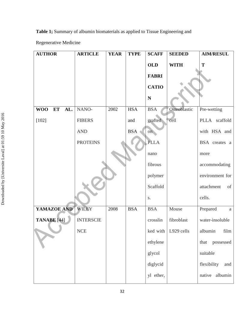
Table 1; Summary of albumin biomaterials as applied to Tissue Engineering and
Regenerative Medicine
AUTHOR ARTICLE YEAR TYPE SCAFF
OLD
FABRI
CATIO
N
SEEDED
WITH
AIM/RESUL
T
WOO ET AL.
[102]
NANO-
FIBERS
AND
PROTEINS
2002 HSA
and
BSA
BSA
grafted
on
PLLA
nano
fibrous
polymer
Scaffold
s.
Osteoblastic
cell
Pre-wetting
PLLA scaffold
with HSA and
BSA creates a
more
accommodating
environment for
attachment of
cells.
YAMAZOE AND
TANABE [44]
WILEY
INTERSCIE
NCE
2008 BSA BSA
crosslin
ked with
ethylene
glycol
diglycid
yl ether,
Mouse
fibroblast
L929 cells
Prepared a
water-insoluble
albumin film
that possessed
suitable
flexibility and
native albumin
Dow
nloa
ded
by [
Uni
vers
ite L
aval
] at
01:
59 1
0 M
ay 2
016
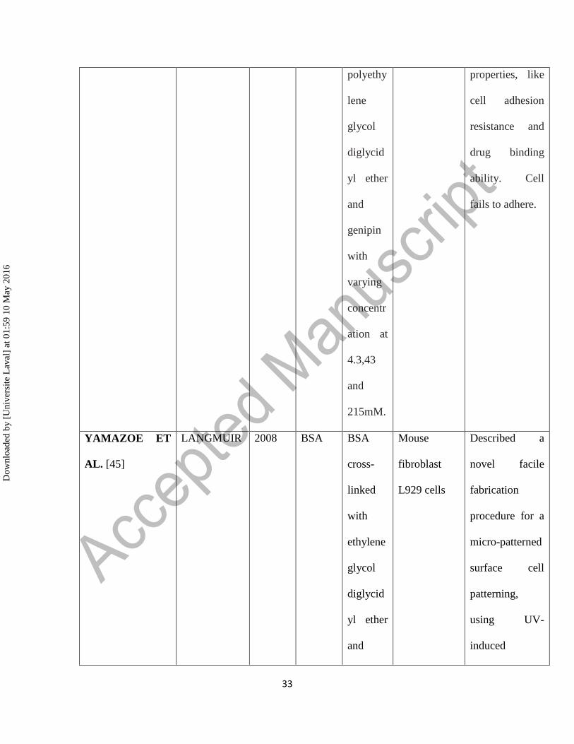
33
polyethy
lene
glycol
diglycid
yl ether
and
genipin
with
varying
concentr
ation at
4.3,43
and
215mM.
properties, like
cell adhesion
resistance and
drug binding
ability. Cell
fails to adhere.
YAMAZOE ET
AL. [45]
LANGMUIR 2008 BSA BSA
cross-
linked
with
ethylene
glycol
diglycid
yl ether
and
Mouse
fibroblast
L929 cells
Described a
novel facile
fabrication
procedure for a
micro-patterned
surface cell
patterning,
using UV-
induced
Dow
nloa
ded
by [
Uni
vers
ite L
aval
] at
01:
59 1
0 M
ay 2
016
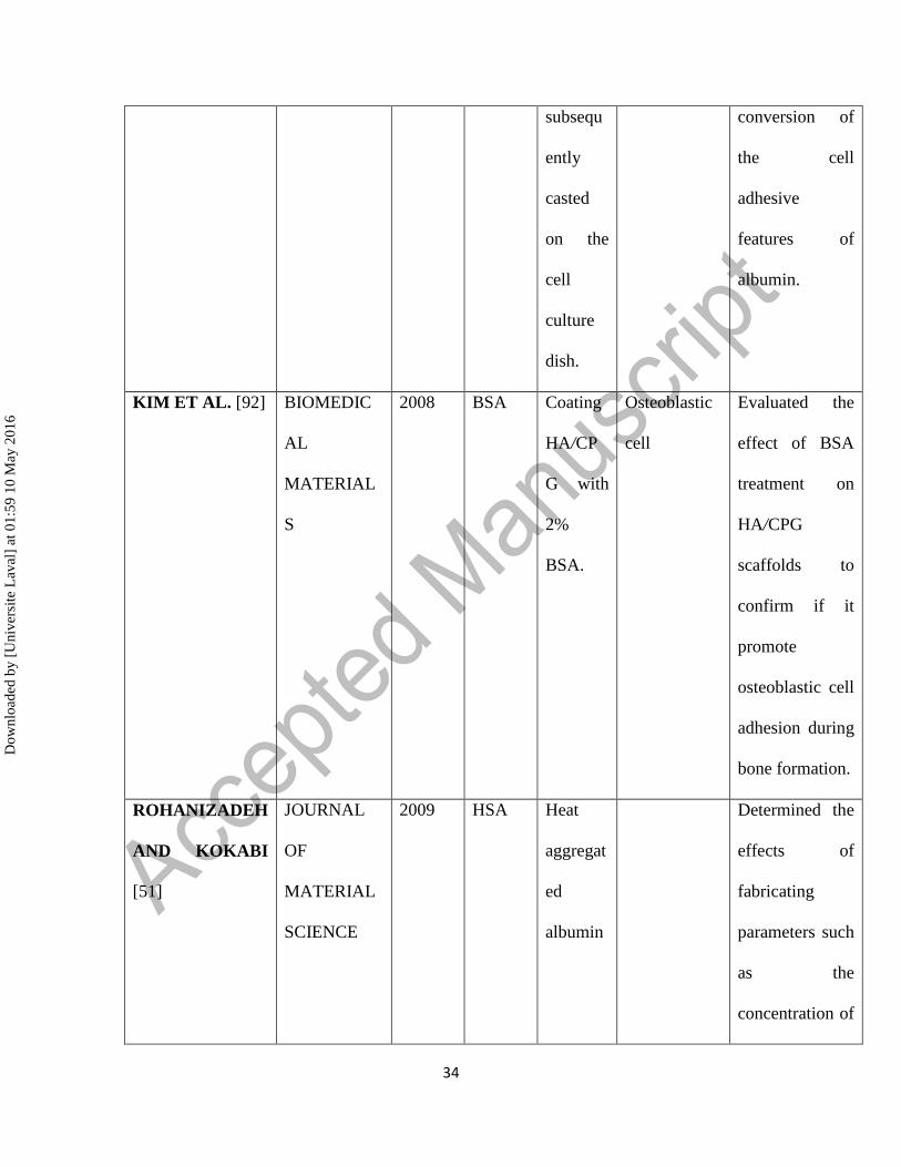
34
subsequ
ently
casted
on the
cell
culture
dish.
conversion of
the cell
adhesive
features of
albumin.
KIM ET AL. [92] BIOMEDIC
AL
MATERIAL
S
2008 BSA Coating
HA/CP
G with
2%
BSA.
Osteoblastic
cell
Evaluated the
effect of BSA
treatment on
HA/CPG
scaffolds to
confirm if it
promote
osteoblastic cell
adhesion during
bone formation.
ROHANIZADEH
AND KOKABI
[51]
JOURNAL
OF
MATERIAL
SCIENCE
2009 HSA Heat
aggregat
ed
albumin
Determined the
effects of
fabricating
parameters such
as the
concentration of
Dow
nloa
ded
by [
Uni
vers
ite L
aval
] at
01:
59 1
0 M
ay 2
016
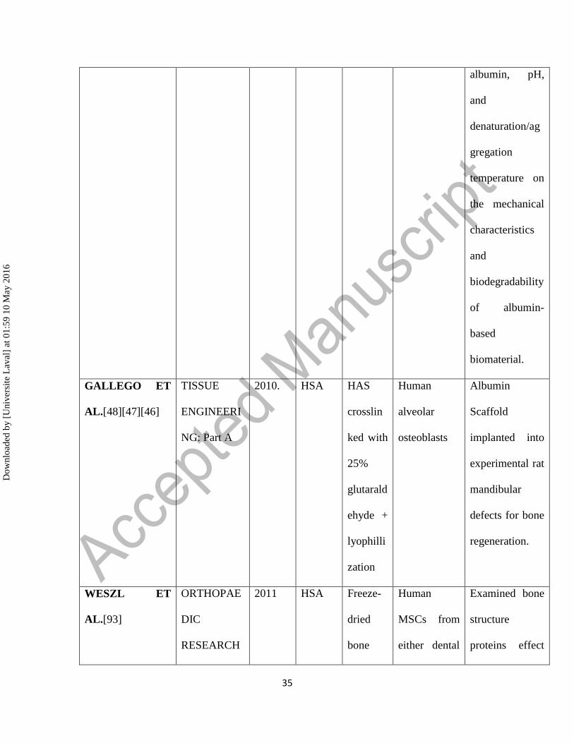
35
albumin, pH,
and
denaturation/ag
gregation
temperature on
the mechanical
characteristics
and
biodegradability
of albumin-
based
biomaterial.
GALLEGO ET
AL.[48][47][46]
TISSUE
ENGINEERI
NG: Part A
2010. HSA HAS
crosslin
ked with
25%
glutarald
ehyde +
lyophilli
zation
Human
alveolar
osteoblasts
Albumin
Scaffold
implanted into
experimental rat
mandibular
defects for bone
regeneration.
WESZL ET
AL.[93]
ORTHOPAE
DIC
RESEARCH
2011 HSA Freeze-
dried
bone
Human
MSCs from
either dental
Examined bone
structure
proteins effect
Dow
nloa
ded
by [
Uni
vers
ite L
aval
] at
01:
59 1
0 M
ay 2
016

36
graft pulp/bone
marrow.
or serum
components on
bone graft and
freeze dried
allograft
colonization by
MSCs.
FERRERO-
GUTIERREZ ET
AL. [94]
HISTOLOG
Y AND
HISTOPATH
OLOGY
2013 Serum-
derived
albumi
n
Crosslin
ked with
25%
glutarald
ehyde +
lyophilli
zation
Adipose
derived stem
cells
(ADSCs) and
olfactory
ensheathing
cells (OECs)
Verified the
regeneration of
axon, and
recovery of
locomotor in
rats induced
with spinal cord
injury and
treated with a
novel serum-
derived albumin
scaffold seeded
with (ADSCs)
and (OECs).
Their findings
Dow
nloa
ded
by [
Uni
vers
ite L
aval
] at
01:
59 1
0 M
ay 2
016
37
pointed to the
feasibility of
albumin
scaffold as a
being potent for
use in the
studies of spinal
cord injury
repair.
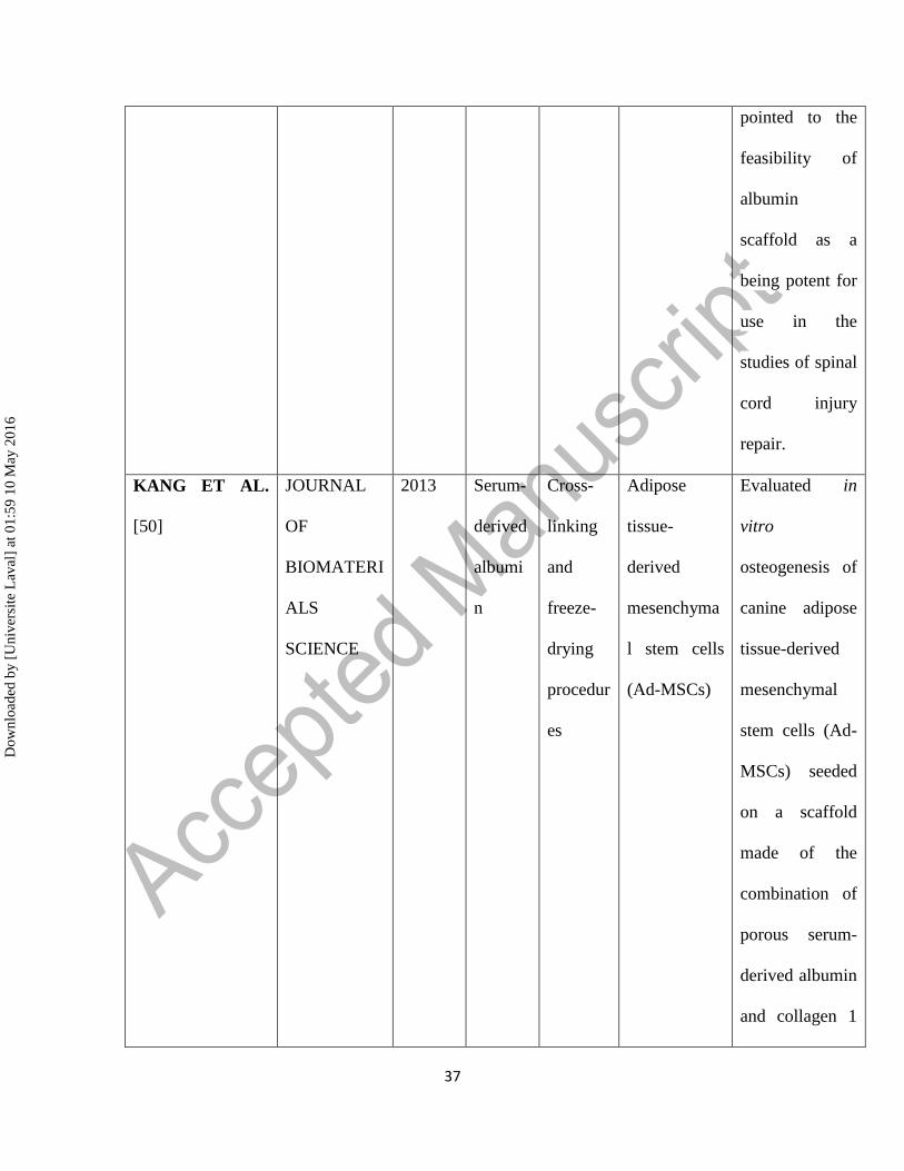
KANG ET AL.
[50]
JOURNAL
OF
BIOMATERI
ALS
SCIENCE
2013 Serum-
derived
albumi
n
Cross-
linking
and
freeze-
drying
procedur
es
Adipose
tissue-
derived
mesenchyma
l stem cells
(Ad-MSCs)
Evaluated in
vitro
osteogenesis of
canine adipose
tissue-derived
mesenchymal
stem cells (Ad-
MSCs) seeded
on a scaffold
made of the
combination of
porous serum-
derived albumin
and collagen 1
Dow
nloa
ded
by [
Uni
vers
ite L
aval
] at
01:
59 1
0 M
ay 2
016

38
gel.
NSEIR ET AL.
[52]
TISSUE
ENGINEERI
NG: Part C
2013 Serum
Albumi
n
Electros
pinning
and salt-
leached
techniqu
e
fibroblasts,
muscle cells,
and
endothelial
cells (ECs) in
vitro
Explored the
mechanical and
biological
features of
electrospun
scaffolds that
solely consist of
albumin
fibers, and
compared them
with those of
scaffolds made
of
polycaprolacton
e (PCL) and
poly (L-lactide)
/poly(lactic-
coglycolic
acid)
(PLLA/PLGA).
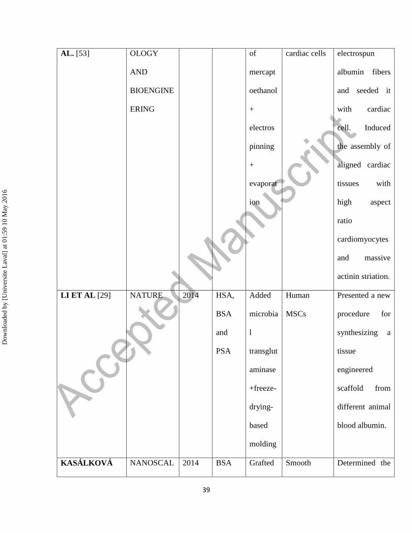
FLEISCHER ET BIOTECHN 2014 BSA Addition Rat neonatal Fabricated
Dow
nloa
ded
by [
Uni
vers
ite L
aval
] at
01:
59 1
0 M
ay 2
016
39
AL. [53] OLOGY
AND
BIOENGINE
ERING
of
mercapt
oethanol
+
electros
pinning
+
evaporat
ion
cardiac cells electrospun
albumin fibers
and seeded it
with cardiac
cell. Induced
the assembly of
aligned cardiac
tissues with
high aspect
ratio
cardiomyocytes
and massive
actinin striation.
LI ET AL [29] NATURE 2014 HSA,
BSA
and
PSA
Added
microbia
l
transglut
aminase
+freeze-
drying-
based
molding
Human
MSCs
Presented a new
procedure for
synthesizing a
tissue
engineered
scaffold from
different animal
blood albumin.
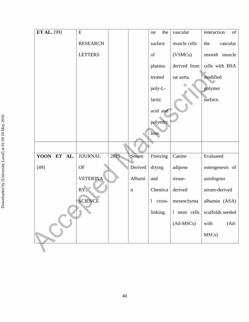
KASÁLKOVÁ NANOSCAL 2014 BSA Grafted Smooth Determined the
Dow
nloa
ded
by [
Uni
vers
ite L
aval
] at
01:
59 1
0 M
ay 2
016
40
ET AL. [99] E
RESEARCH
LETTERS
on the
surface
of
plasma-
treated
poly-L-
lactic
acid and
polyethy
lene.
vascular
muscle cells
(VSMCs)
derived from
rat aorta.
interaction of
the vascular
smooth muscle
cells with BSA
modified
polymer
surface.
YOON ET AL.
[49]
JOURNAL
OF
VETERINA
RY
SCIENCE
2015 Serum
Derived
Albumi
n
Freezing
drying
and
Chemica
l cross-
linking.
Canine
adipose
tissue-
derived
mesenchyma
l stem cells
(Ad-MSCs)
Evaluated
osteogenesis of
autologous
serum-derived
albumin (ASA)
scaffolds seeded
with (Ad-
MSCs)
Dow
nloa
ded
by [
Uni
vers
ite L
aval
] at
01:
59 1
0 M
ay 2
016
41
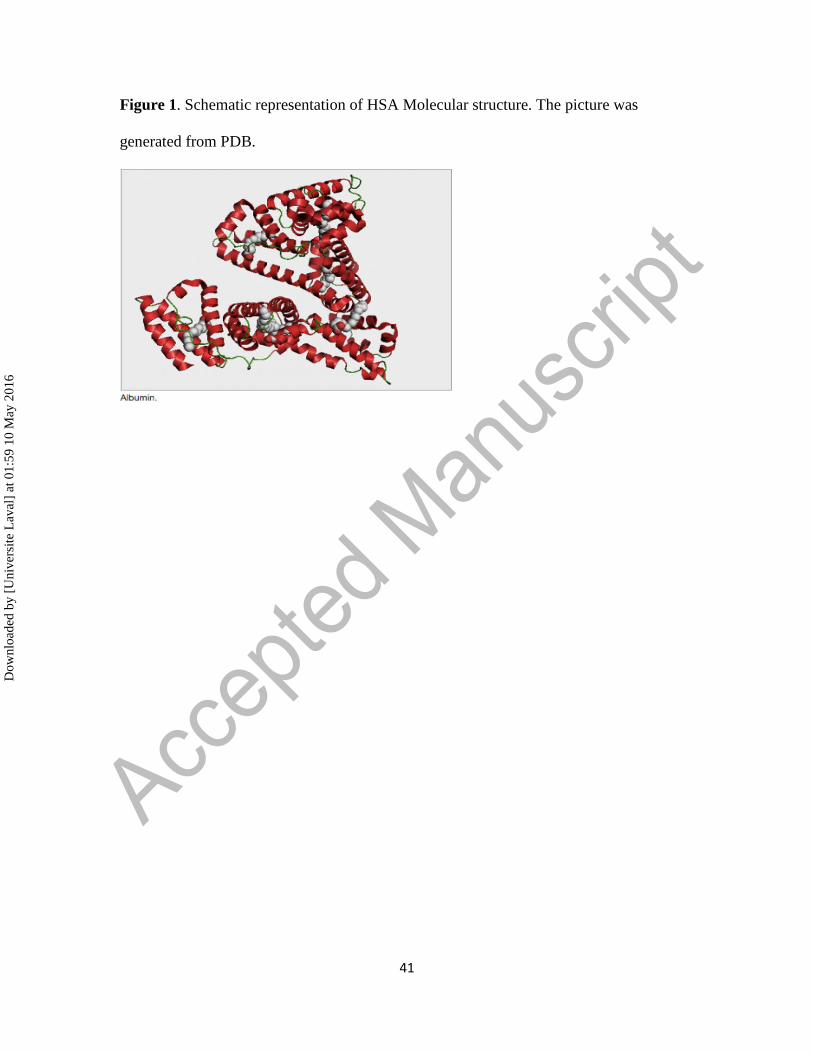
Figure 1. Schematic representation of HSA Molecular structure. The picture was
generated from PDB.
Dow
nloa
ded
by [
Uni
vers
ite L
aval
] at
01:
59 1
0 M
ay 2
016
42
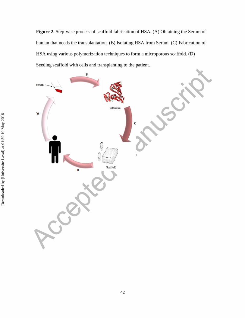
Figure 2. Step-wise process of scaffold fabrication of HSA. (A) Obtaining the Serum of
human that needs the transplantation. (B) Isolating HSA from Serum. (C) Fabrication of
HSA using various polymerization techniques to form a microporous scaffold. (D)
Seeding scaffold with cells and transplanting to the patient.
Dow
nloa
ded
by [
Uni
vers
ite L
aval
] at
01:
59 1
0 M
ay 2
016