Embed Size (px)
Citation preview
Lesson 3 Biology
© 2013 Region 4 Education Service Center STAAR™ Achievement Series for Science
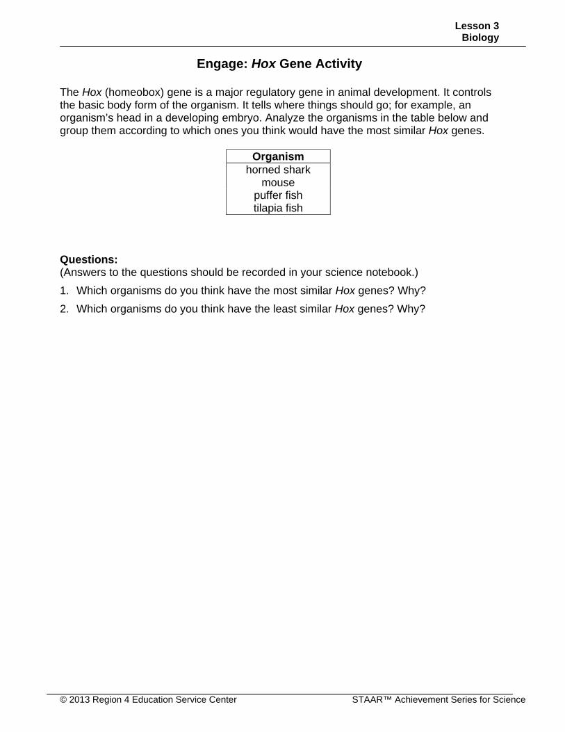
Engage: Hox Gene Activity
The Hox (homeobox) gene is a major regulatory gene in animal development. It controls the basic body form of the organism. It tells where things should go; for example, an organism’s head in a developing embryo. Analyze the organisms in the table below and group them according to which ones you think would have the most similar Hox genes.
Organism horned shark
mouse puffer fish tilapia fish
Questions: (Answers to the questions should be recorded in your science notebook.)
1. Which organisms do you think have the most similar Hox genes? Why?
2. Which organisms do you think have the least similar Hox genes? Why?
Lesson 3 Biology
STAAR™ Achievement Series for Science © 2013 Region 4 Education Service Center
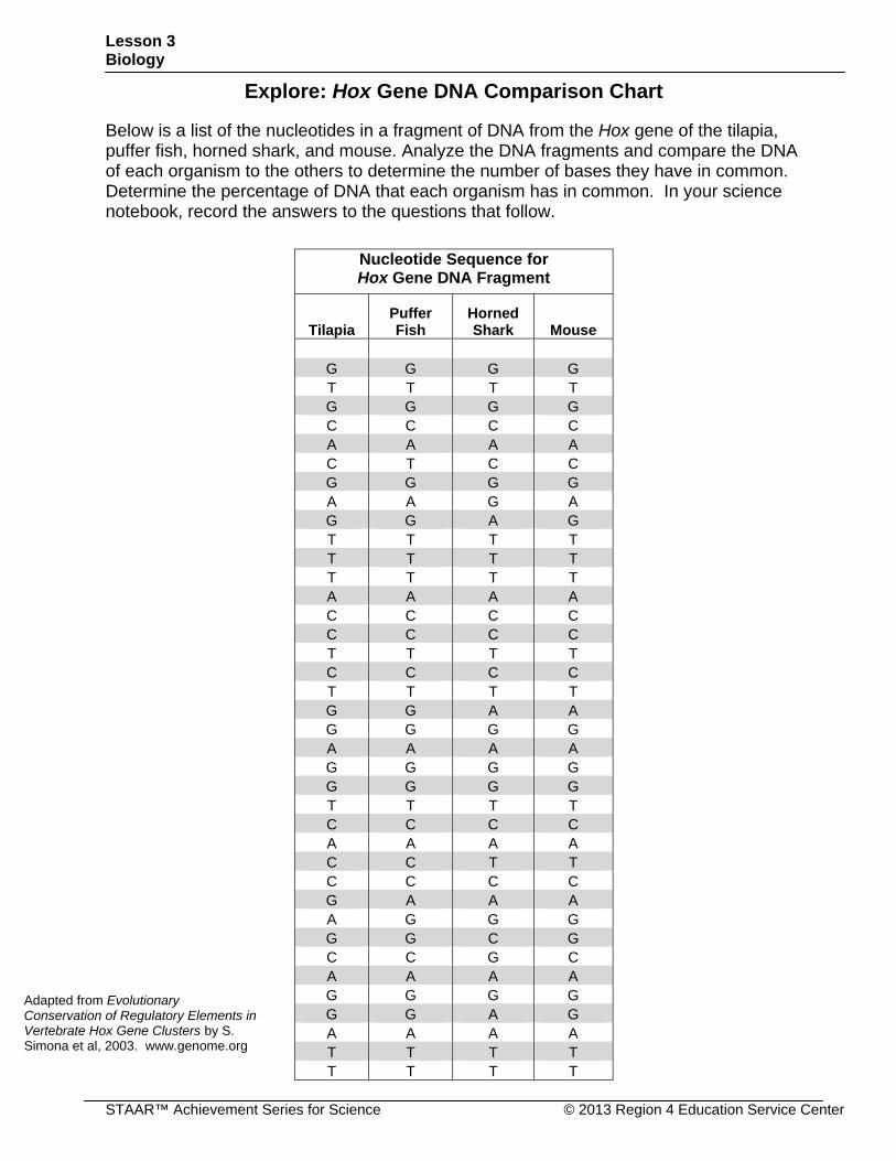
Explore: Hox Gene DNA Comparison Chart
Below is a list of the nucleotides in a fragment of DNA from the Hox gene of the tilapia, puffer fish, horned shark, and mouse. Analyze the DNA fragments and compare the DNA of each organism to the others to determine the number of bases they have in common. Determine the percentage of DNA that each organism has in common. In your science notebook, record the answers to the questions that follow.
Nucleotide Sequence for Hox Gene DNA Fragment
Tilapia Puffer Fish
Horned Shark Mouse
G G G G T T T T G G G G C C C C A A A A C T C C G G G G A A G A G G A G T T T T T T T T T T T T A A A A C C C C C C C C T T T T C C C C T T T T G G A A G G G G A A A A G G G G G G G G T T T T C C C C A A A A C C T T C C C C G A A A A G G G G G C G C C G C A A A A G G G G G G A G A A A A T T T T T T T T
Adapted from Evolutionary Conservation of Regulatory Elements in Vertebrate Hox Gene Clusters by S. Simona et al, 2003. www.genome.org
Lesson 3 Biology
© 2013 Region 4 Education Service Center STAAR™ Achievement Series for Science
Questions
1. Which organisms share the greatest number of bases in the Hox gene fragment with the tilapia?
2. Which two organisms shared the least number and percentage of the DNA fragment?
3. Analyzing all the organisms’ DNA bases for the Hox gene fragment, what can you conclude about the gene?
Lesson 3 Biology
STAAR™ Achievement Series for Science © 2013 Region 4 Education Service Center
Explain: DNA
The Structure and Function of DNA DNA, deoxyribonucleic acid, is a complex biological macromolecule, which is known as a nucleic acid. Nucleic acid is made up of smaller subunits called nucleotides that store information in the form of a code inside the nucleus of cells. The nucleotides themselves are made up of hydrogen, oxygen, carbon, nitrogen, and phosphorous and create three groups: the phosphate group, a simple sugar, and a nitrogen base. Each nucleotide is created by a sugar covalently bonded to a phosphate group and a nitrogen base. DNA is the foundation for an organism’s appearance and behavior and contains nearly all of the information needed to form enzymes and structural proteins. When cell division occurs, the genetic code is passed on from one generation of cells to the next. In the DNA molecule, the phosphate groups, simple sugars, and nitrogen bases form a long strand. This does not exist as an individual molecule. The nucleotides combine to create two long strands in the form of a ladder. The ladder twists, forming a double helix.
There are four nitrogen bases: adenine (A), guanine (G), cytosine (C), and thymine (T). Adenine and guanine are both double-ring bases called purines, and cytosine and thymine are smaller single-ring bases called pyrimidines. In the double helix, the base from one strand bonds to the base on the second strand with a hydrogen bond, forming base pairs. Adenine can only form a bond with thymine, forming two hydrogen bonds. Guanine and cytosine can only bond together, forming three hydrogen bonds.
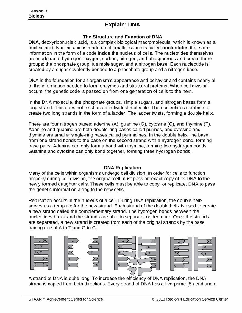
DNA Replication Many of the cells within organisms undergo cell division. In order for cells to function properly during cell division, the original cell must pass an exact copy of its DNA to the newly formed daughter cells. These cells must be able to copy, or replicate, DNA to pass the genetic information along to the new cells. Replication occurs in the nucleus of a cell. During DNA replication, the double helix serves as a template for the new strand. Each strand of the double helix is used to create a new strand called the complementary strand. The hydrogen bonds between the nucleotides break and the strands are able to separate, or denature. Once the strands are separated, a new strand is created from each of the original strands by the base pairing rule of A to T and G to C.
A strand of DNA is quite long. To increase the efficiency of DNA replication, the DNA strand is copied from both directions. Every strand of DNA has a five-prime (5’) end and a
Lesson 3 Biology
© 2013 Region 4 Education Service Center STAAR™ Achievement Series for Science
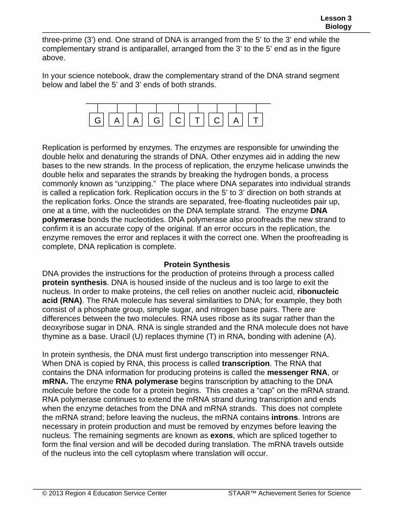
three-prime (3’) end. One strand of DNA is arranged from the 5’ to the 3’ end while the complementary strand is antiparallel, arranged from the 3’ to the 5’ end as in the figure above. In your science notebook, draw the complementary strand of the DNA strand segment below and label the 5’ and 3’ ends of both strands. Replication is performed by enzymes. The enzymes are responsible for unwinding the double helix and denaturing the strands of DNA. Other enzymes aid in adding the new bases to the new strands. In the process of replication, the enzyme helicase unwinds the double helix and separates the strands by breaking the hydrogen bonds, a process commonly known as “unzipping.” The place where DNA separates into individual strands is called a replication fork. Replication occurs in the 5’ to 3’ direction on both strands at the replication forks. Once the strands are separated, free-floating nucleotides pair up, one at a time, with the nucleotides on the DNA template strand. The enzyme DNA polymerase bonds the nucleotides. DNA polymerase also proofreads the new strand to confirm it is an accurate copy of the original. If an error occurs in the replication, the enzyme removes the error and replaces it with the correct one. When the proofreading is complete, DNA replication is complete.
Protein Synthesis
DNA provides the instructions for the production of proteins through a process called protein synthesis. DNA is housed inside of the nucleus and is too large to exit the nucleus. In order to make proteins, the cell relies on another nucleic acid, ribonucleic acid (RNA). The RNA molecule has several similarities to DNA; for example, they both consist of a phosphate group, simple sugar, and nitrogen base pairs. There are differences between the two molecules. RNA uses ribose as its sugar rather than the deoxyribose sugar in DNA. RNA is single stranded and the RNA molecule does not have thymine as a base. Uracil (U) replaces thymine (T) in RNA, bonding with adenine (A). In protein synthesis, the DNA must first undergo transcription into messenger RNA. When DNA is copied by RNA, this process is called transcription. The RNA that contains the DNA information for producing proteins is called the messenger RNA, or mRNA. The enzyme RNA polymerase begins transcription by attaching to the DNA molecule before the code for a protein begins. This creates a “cap” on the mRNA strand. RNA polymerase continues to extend the mRNA strand during transcription and ends when the enzyme detaches from the DNA and mRNA strands. This does not complete the mRNA strand; before leaving the nucleus, the mRNA contains introns. Introns are necessary in protein production and must be removed by enzymes before leaving the nucleus. The remaining segments are known as exons, which are spliced together to form the final version and will be decoded during translation. The mRNA travels outside of the nucleus into the cell cytoplasm where translation will occur.
CGG A A TC AT
Lesson 3 Biology
STAAR™ Achievement Series for Science © 2013 Region 4 Education Service Center
In your science notebook, use the DNA strand below to create the mRNA strand.
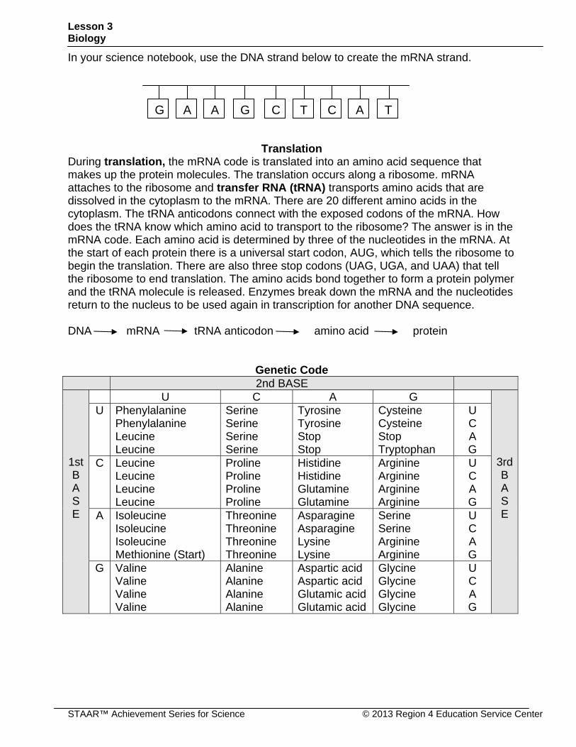
Translation During translation, the mRNA code is translated into an amino acid sequence that makes up the protein molecules. The translation occurs along a ribosome. mRNA attaches to the ribosome and transfer RNA (tRNA) transports amino acids that are dissolved in the cytoplasm to the mRNA. There are 20 different amino acids in the cytoplasm. The tRNA anticodons connect with the exposed codons of the mRNA. How does the tRNA know which amino acid to transport to the ribosome? The answer is in the mRNA code. Each amino acid is determined by three of the nucleotides in the mRNA. At the start of each protein there is a universal start codon, AUG, which tells the ribosome to begin the translation. There are also three stop codons (UAG, UGA, and UAA) that tell the ribosome to end translation. The amino acids bond together to form a protein polymer and the tRNA molecule is released. Enzymes break down the mRNA and the nucleotides return to the nucleus to be used again in transcription for another DNA sequence. DNA mRNA tRNA anticodon amino acid protein
Genetic Code 2nd BASE
1st B A S E
U C A G
3rdB A S E
U Phenylalanine Phenylalanine Leucine Leucine
Serine Serine Serine Serine
Tyrosine Tyrosine Stop Stop
Cysteine Cysteine Stop Tryptophan
U C A G
C Leucine Leucine Leucine Leucine
Proline Proline Proline Proline
Histidine Histidine Glutamine Glutamine
Arginine Arginine Arginine Arginine
U C A G
A Isoleucine Isoleucine Isoleucine Methionine (Start)
Threonine Threonine Threonine Threonine
Asparagine Asparagine Lysine Lysine
Serine Serine Arginine Arginine
U C A G
G Valine Valine Valine Valine
Alanine Alanine Alanine Alanine
Aspartic acid Aspartic acid Glutamic acid Glutamic acid
Glycine Glycine Glycine Glycine
U C A G
CGG A A TC AT
Lesson 3 Biology
© 2013 Region 4 Education Service Center STAAR™ Achievement Series for Science
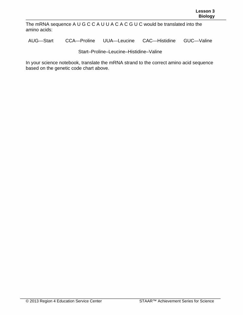
The mRNA sequence A U G C C A U U A C A C G U C would be translated into the amino acids: AUG—Start CCA—Proline UUA—Leucine CAC—Histidine GUC—Valine
Start–Proline–Leucine–Histidine–Valine
In your science notebook, translate the mRNA strand to the correct amino acid sequence based on the genetic code chart above.
Lesson 3 Biology
STAAR™ Achievement Series for Science © 2013 Region 4 Education Service Center
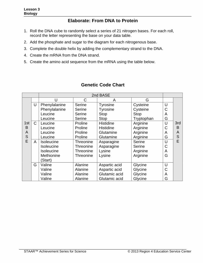
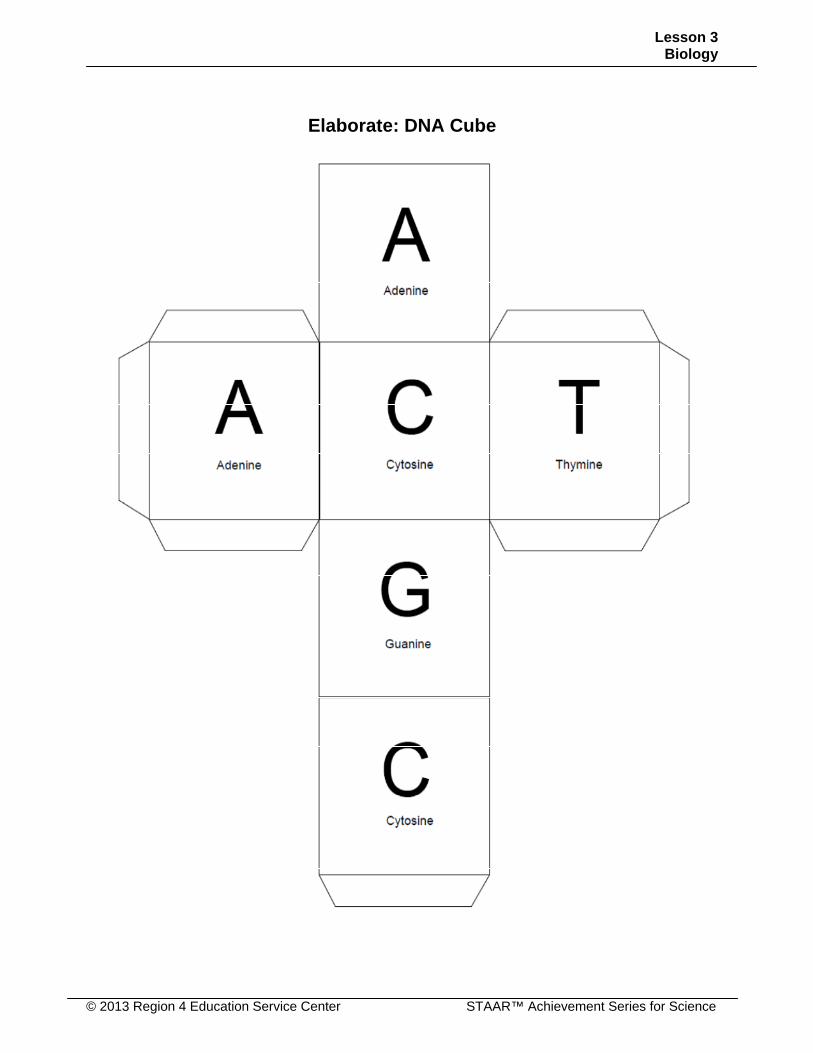
Elaborate: From DNA to Protein 1. Roll the DNA cube to randomly select a series of 21 nitrogen bases. For each roll,
record the letter representing the base on your data table.
2. Add the phosphate and sugar to the diagram for each nitrogenous base.
3. Complete the double helix by adding the complementary strand to the DNA.
4. Create the mRNA from the DNA strand.
5. Create the amino acid sequence from the mRNA using the table below.
Genetic Code Chart
2nd BASE
1st B A S E
U C A G
3rdB A S E
U Phenylalanine Phenylalanine Leucine Leucine
Serine Serine Serine Serine
Tyrosine Tyrosine Stop Stop
Cysteine Cysteine Stop Tryptophan
U C A G
C Leucine Leucine Leucine Leucine
Proline Proline Proline Proline
Histidine Histidine Glutamine Glutamine
Arginine Arginine Arginine Arginine
U C A G
A Isoleucine Isoleucine Isoleucine Methionine (Start)
Threonine Threonine Threonine Threonine
Asparagine Asparagine Lysine Lysine
Serine Serine Arginine Arginine
U C A G
G Valine Valine Valine Valine
Alanine Alanine Alanine Alanine
Aspartic acid Aspartic acid Glutamic acid Glutamic acid
Glycine Glycine Glycine Glycine
U C A G
Lesson 3 Biology
© 2013 Region 4 Education Service Center STAAR™ Achievement Series for Science
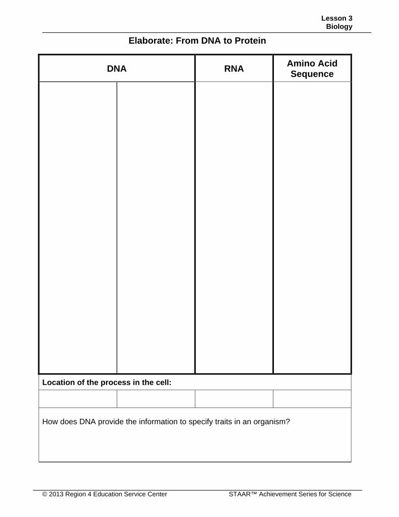
Elaborate: From DNA to Protein
DNA RNA Amino Acid Sequence
Location of the process in the cell:
How does DNA provide the information to specify traits in an organism?
Lesson 3 Biology
© 2013 Region 4 Education Service Center STAAR™ Achievement Series for Science
Elaborate: DNA Cube
Lesson 3 Biology
STAAR™ Achievement Series for Science © 2013 Region 4 Education Service Center
Name: _____________________________________________ Date: __________
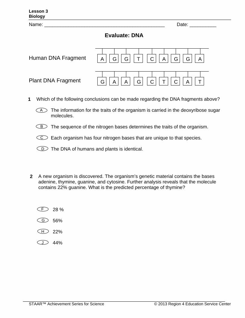
Evaluate: DNA
Human DNA Fragment
Plant DNA Fragment
2 A new organism is discovered. The organism’s genetic material contains the bases adenine, thymine, guanine, and cytosine. Further analysis reveals that the molecule contains 22% guanine. What is the predicted percentage of thymine?
28 % 56% 22% 44%
G
F
H
J
CTA G G AG G A
CGG A A TC A T
Which of the following conclusions can be made regarding the DNA fragments above?
The information for the traits of the organism is carried in the deoxyribose sugar molecules.
The sequence of the nitrogen bases determines the traits of the organism. Each organism has four nitrogen bases that are unique to that species. The DNA of humans and plants is identical.
1
B
A
C
D
Lesson 3 Biology
© 2013 Region 4 Education Service Center STAAR™ Achievement Series for Science
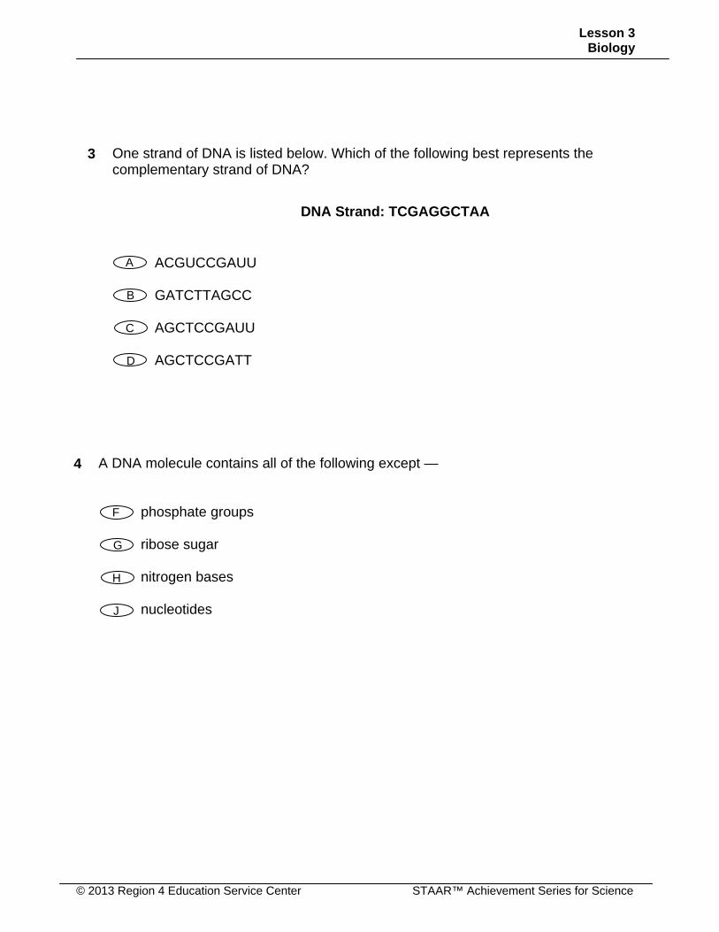
4 A DNA molecule contains all of the following except —
phosphate groups ribose sugar nitrogen bases nucleotides
G
F
H
J
3 One strand of DNA is listed below. Which of the following best represents the complementary strand of DNA?
DNA Strand: TCGAGGCTAA
ACGUCCGAUU GATCTTAGCC AGCTCCGAUU AGCTCCGATT
B
A
C
D
Lesson 3 Biology
STAAR™ Achievement Series for Science © 2013 Region 4 Education Service Center
2nd BASE
1st B A S E
U C A G
3rd
B A S E
U Phenylalanine Phenylalanine Leucine Leucine
Serine Serine Serine Serine
Tyrosine Tyrosine Stop Stop
Cysteine Cysteine Stop Tryptophan
U C A G
C Leucine Leucine Leucine Leucine
Proline Proline Proline Proline
Histidine Histidine Glutamine Glutamine
Arginine Arginine Arginine Arginine
U C A G
A Isoleucine Isoleucine Isoleucine Methionine (Start)
Threonine Threonine Threonine Threonine
Asparagine Asparagine Lysine Lysine
Serine Serine Arginine Arginine
U C A G
G Valine Valine Valine Valine
Alanine Alanine Alanine Alanine
Aspartic acid Aspartic acid Glutamic acid Glutamic acid
Glycine Glycine Glycine Glycine
U C A G
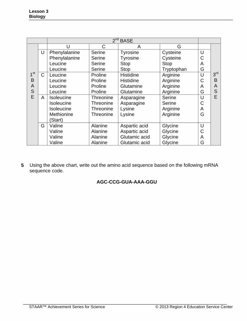
5 Using the above chart, write out the amino acid sequence based on the following mRNA sequence code.
AGC-CCG-GUA-AAA-GGU