Embed Size (px)
Citation preview
Diagnostic and Therapeutic Endoscopy, 1997, Vol. 3, pp. 161-170
Reprints available dinctly from the publisherPhotocopying permitted by license only
(C) 1997 OPA (Overseas Publishers Association)Amsterdam B.V. Published in The Netherlands
by Harwood Academic Publishers
Printed in Singapore
Endoscopic Nasobiliary Drainage in the Management ofAcute Cholangitis: An Experience in 143 Patients
M.K. GOENKA,*,t D.K. BHASIN, R. KOCHHAR, B. NAGI,U. RUNGTA, K. DAS and K. SINGH
Sections of Radiology and Clinical Gastroenterology, Department of Gastroenterology,Postgraduate Institute of Medical Education and Research, Chandigar 160012, India
(Received 8 May 1996; Revised 20 June 1996; In final form 8 July 1996)
Acute cholangitis is associated with a high mortality and morbidity and often requiresdrainage of the obstructed biliary system. The purpose of this study was to evaluate theusefulness and safety ofendoscopic nasobiliary drainage in the treatment and preventionof acute cholangitis due to diverse etiology. During a 32-month period, 143 patients(67 males, 76 females) with age range of 15 to 84 years underwent urgent fluoroscopyguided endoscopic nasobiliary drainage using a 7 Fr catheter either to treat acutecholangitis not responding to antibiotics (groupA,n 116) or to prevent its developmentfollowing endoscopic retrograde cholangiography performed in an obstructed biliarysystem (group B, n 27). Underlying etiology included bile duct stones (92), malignantbiliary obstruction (34), choledochal cyst (4), chronic pancreatitis (4), ruptured hydatidcyst (3), portal hypertensive cholangiopathy (3) and liver abscess (3). Endoscopicnasobiliary drainage was performed successfully in 129 patients (90.2%). Cholangitisimproved within I to 3 days (in group A) or did not develop (in Group B) in 125 patients(96.7%) with successful endoscopic nasobiliary drainage.Two patients however requiredadditional drainage by percutaneous transhepatic route, while two died inspite ofeffectiveendoscopic drainage. Of the 14 patients (9.8%) with failed endoscopic drainage, 9 weremanaged by surgical decompression or percutaneous transhepatic drainage, 3 died ofsepticemia. Endoscopic nasobiliary drainage is a safe and effective method to treatpatients with acute cholangitis as well as to prevent its development followingcholangiography performed in an obstructed biliary system.
Keywords: Bile ducts stenosis, bile ducts prosthesis, cholangitis, endoscopic sphincterotomy
*Corresponding Author: Tel: 0091-33-3341193 Fax: 0091-33-2428098.Present address: Eko Endoscopy Centre, 54 JLN Road, Calcutta 700 071, India.
161
162 M.K. GOENKA et al.
INTRODUCTION
Acute cholangitis often complicates obstructed biliarysystem due to choledocholithiasis, biliary stricture andless commonly due to malignancy ofpancreaticobiliaryregion. Risk of acute cholangitis is increased if
radiographic contrast material is injected into an
obstructed biliary system[l]. While all such patientsrequire antibiotic treatment, a significant proportiondo need drainage of the obstructed system[l]. Thedrainage was conventionally done by surgery[2-4],but over the last decade, radiological[5-7] andendoscopic techniques[1,8-14] for drainage have beenintroduced. Endoscopic drainage can be achieved byendoscopic sphincterotomy, biliary stenting or
nasobiliary catheter drainage[1,8-14]. While a numberof studies have shown endotherapy of cholangitis to bea safe and useful technique, these are mostly based ona limited number of patients[8,10-13], and most haveincluded only patients with choledocholithiasis[9-14].We report our experience of 143 patients treated byendoscopic nasobiliary drainage to treat acute
cholangitis or to prevent its development in patientswith obstructed biliary system due to a diverseunderlying etiology.
MATERIAL AND METHODS
Between May, 1993 and December, 1995, 143patients underwent endoscopic nasobiliary drainage(ENBD) either to treat acute cholangitis (Group A,therapeutic group, n 116) or to prevent the riskof cholangitis following endoscopic retrogradecholangiopancreatography (ERCP) performed in anobstructed biliary system with failure to relieve theobstruction endoscopically (Group B, prophylacticgroup, n 27).
All patients underwent clinical evaluation,laboratory investigations including liver functiontests, renal parameters and serum electrolytes. Anabdominal ultrasonogram was also performed in all
patients. Initial treatment of patients with featuresof cholangitis (Group A) included intravenous
antibiotic (ciprofloxacin or piperacillin), parenteralfluid and injections of vitamin K, while those without
cholangitis (Group B) were started on oral ciprofloxacin12-24 hours prior to ERCP. Four patients in Group Ahaving hyperkalemia due to renal failure secondary to
cholangitis and septicemia received dialysis prior to
ERCP. ERCP was performed in GroupA patients onlyafter a sub-optimum response to medical therapyinstituted for a period of 1 to 3 days.ERCP was performed under fluoroscopic
guidance using a side-viewing duodenoscope (JF-IT or JF-IT20, Olympus or FD34X, Ashai Optical)after intravenous diazepam (5-10 mg) and/or
hyoscine N-butyl bromide (20-60 mg). A smallamount of 60% meglumine iothalamate was usedfor diagnostic ERCP. Endoscopic sphincterotomywas performed in only 11 patients as a pre-requisitefor attempted stone extraction. A 0.035 inch guide-wire (Zebra, Microvasive, Boston Scientific, USA)was passed through the ERCP cannula and afterpositioning its tip proximal to the site of obstruction,the cannula was withdrawn. In patients with tightbiliary stricture, a 6 Fr biliary dilator (Wilson Cook,Winston-Salem) was passed over the guide wire.
Nasobiliary catheter (7 Fr, pig-tail) was threadedover the guide wire, passed through the biopsychannel of the endoscope and its tip positionedproximal to the site of obstruction or into the abscesscavity (in patients with liver abscess, hydatid cystand choledochal cyst). Guide wire was thenwithdrawn followed by the endoscope. Afterensuring a free flow of bile from the external endofENBD catheter, the catheter was rerouted throughthe nose by a rail-road technique using a nasogastrictube. A cholangiogram was repeated through theENBD catheter using 60% meglumine iothalamateto adequately visualise the bitiary system. ENBDcatheter, while in position, was irrigated once dailyusing 20 ml-60 ml of sterile normal saline.
Response to endoscopic drainage was monitoredin terms of clinical improvement as well as changesin liver function tests. Patients were offereddefinitive therapy for the underlying etiology, oncethe cholangitis settled and patient stabilised.
ENDOSCOPIC NASOBILIARY DRAINAGE IN THE MANAGEMENT OF ACUTE CHOLANGITIS 163
TABLE Etiology in patients undergoing endoscopic nasobiliary drainage
Etiology Group A Group Total
(Therapeutic) (Prophylactic) n 143n 116 n 27
Benign (n=109)Bile duct stones 83 9*** 92Choledochal cyst 4 4Chronic pancreatitis 3 4
with biliary strictureHydatid cyst with biliary 3 3
rupturePortal cholangiopathy 3 3Cholangitic abscess 3 3Malignant (n 34)Ampullary carcinoma 7 4 11Malignant biliary 8"* 10 18
structure*Pancreatic head cancer 2 3 5
*Includes gall bladder cancer with bile duct infiltration as well as cholangiocarcinoma**Two of these patients ahd cholangitis following occlusion of biliary stent***Failed stone extraction because of large stones (4), associated stricture (2), abnormalcoagulogram (1)
RESULTS
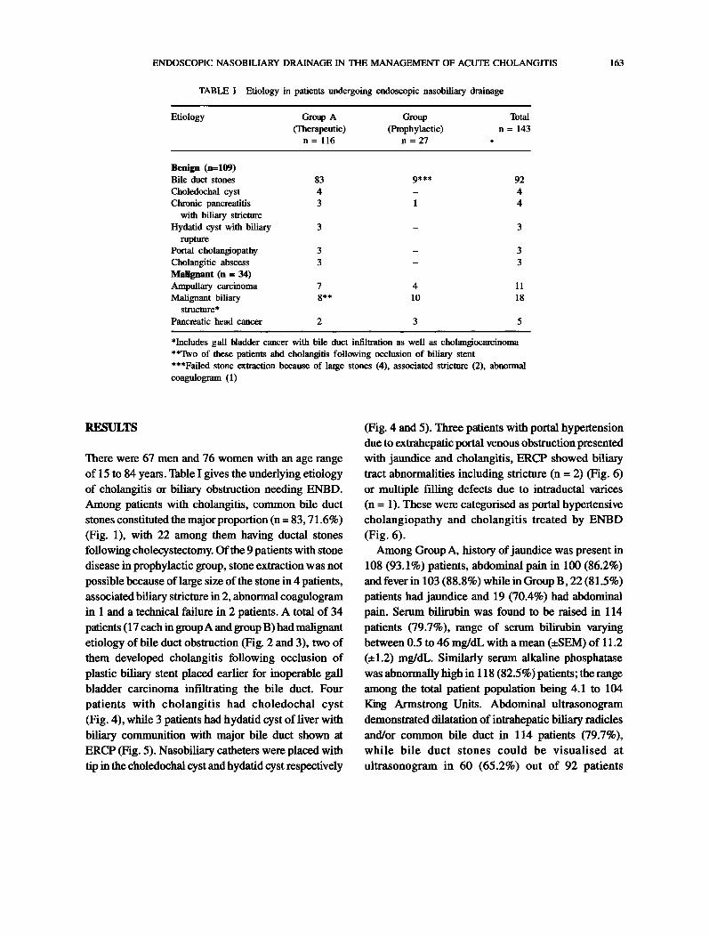
There were 67 men and 76 women with an age rangeof 15 to 84 years. Table I gives the underlying etiologyof cholangitis or biliary obstruction needing ENBD.Among patients with cholangitis, common bile ductstones constituted the major proportion (n 83, 71.6%)(Fig. 1), with 22 among them having ductal stones
following cholecystectomy. Ofthe 9 patients with stone
disease in prophylactic group, stone extraction was not
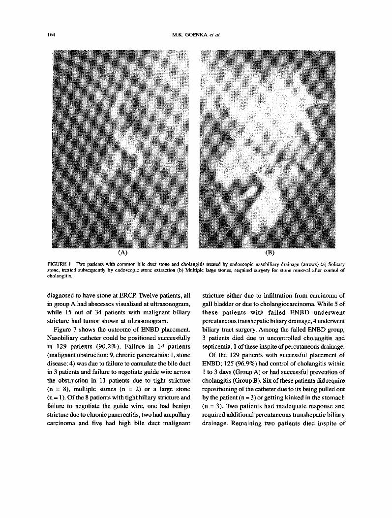
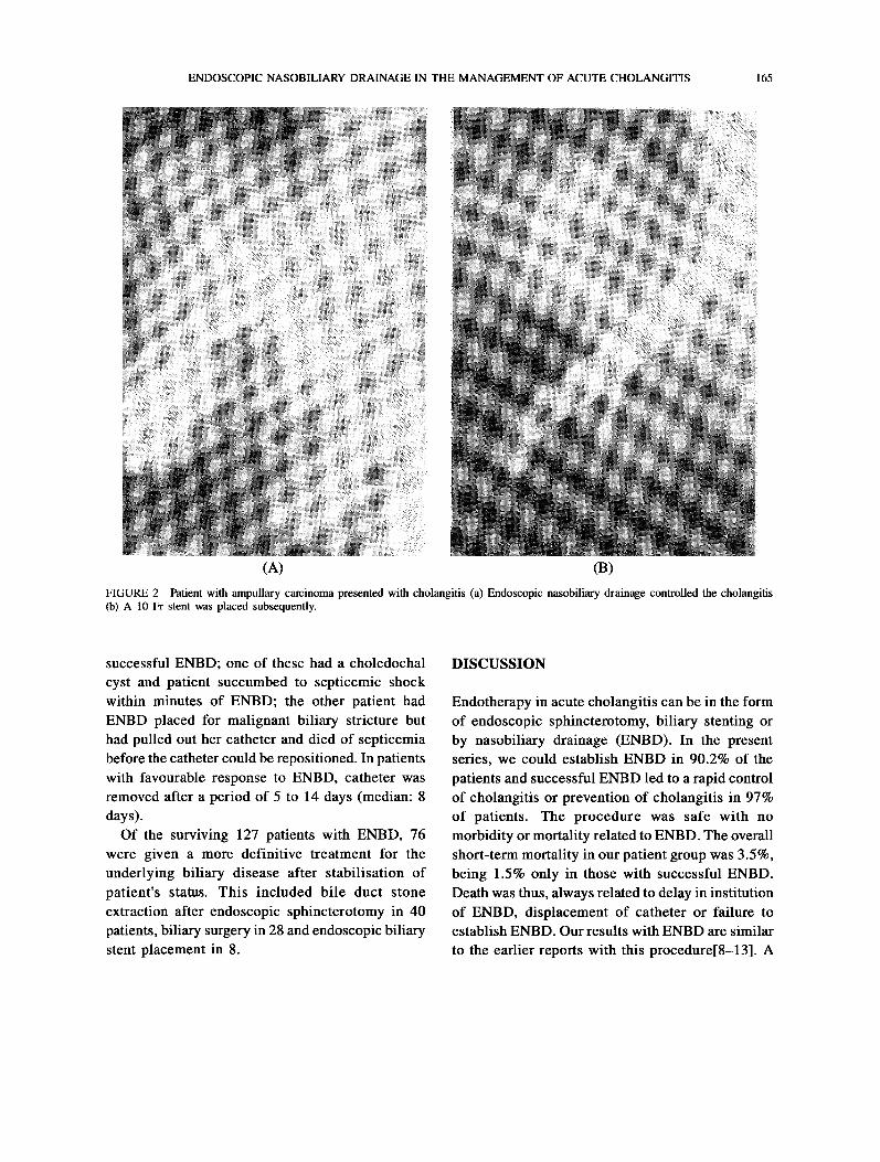
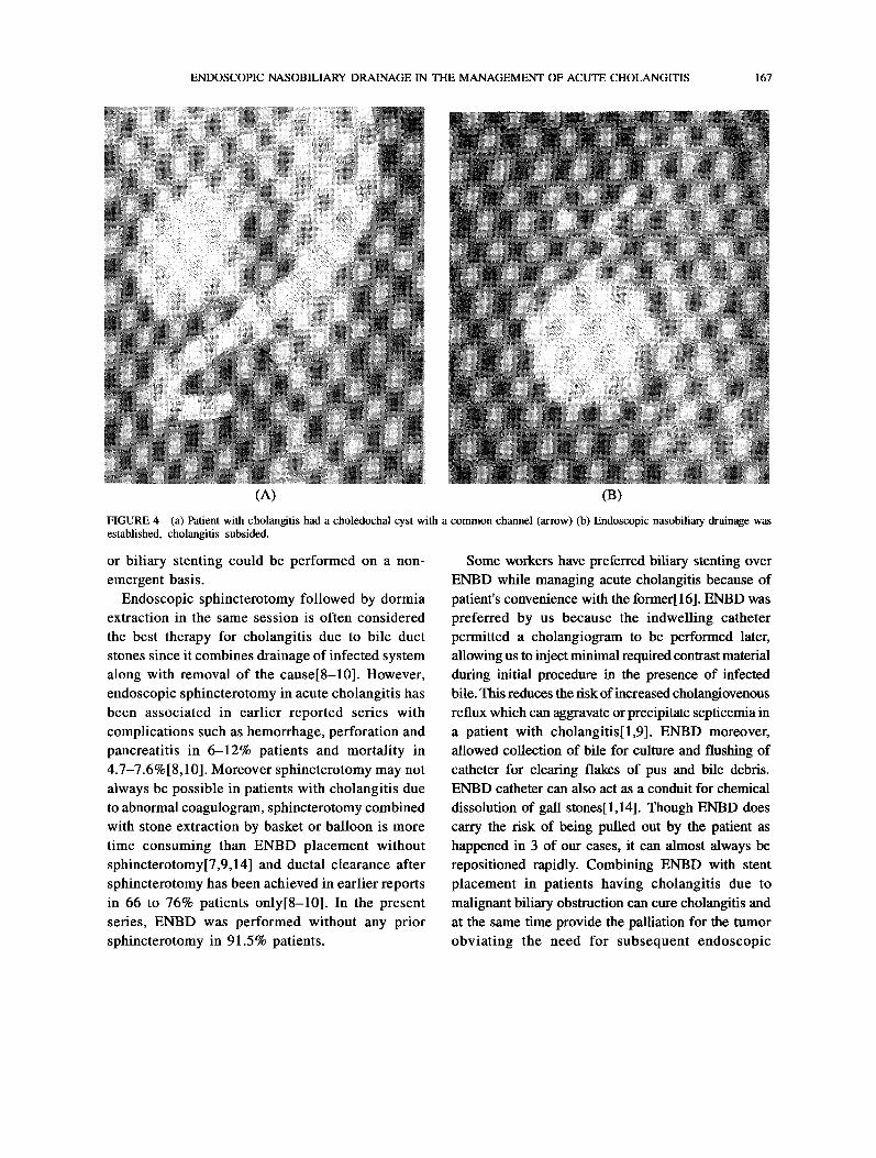
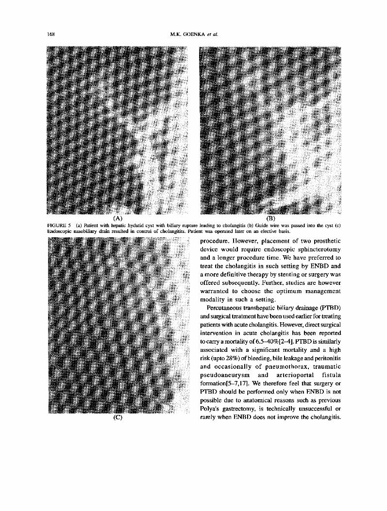
possible because of large size of the stone in 4 patients,associated biliary stricture in 2, abnormal coagulogramin 1 and a technical failure in 2 patients. A total of 34patients (17 each in groupA and group B) had malignantetiology of bile duct obstruction (Fig. 2 and 3), two ofthem developed cholangitis following occlusion ofplastic biliary stent placed earlier for inoperable gallbladder carcinoma infiltrating the bile duct. Fourpatients with cholangitis had choledochal cyst(Fig. 4), while 3 patients had hydatid cyst of liver withbiliary communition with major bile duct shown atERCP (Fig. 5). Nasobiliary catheters were placed with
tip in the choledochal cyst and hydatid cyst respectively
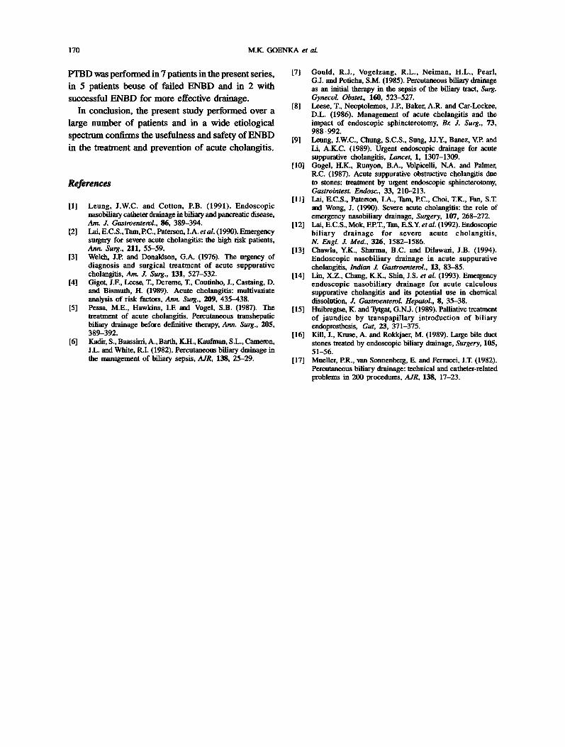
(Fig. 4 and 5). Three patients with portal hypertensiondue to extrahepatic portal venous obstruction presentedwith jaundice and cholangitis, ERCP showed biliarytract abnormalities including stricture (n 2) (Fig. 6)or multiple filling defects due to intraductal varices
(n 1). These were categorised as portal hypertensivecholangiopathy and cholangitis treated by ENBD(Fig. 6).Among Group A, history ofjaundice was present in
108 (93.1%) patients, abdominal pain in 100 (86.2%)and fever in 103 (88.8%) while in Group B, 22 (81.5%)patients had jaundice and 19 (70.4%) had abdominal
pain. Serum bilirubin was found to be raised in 114patients (79.7%), range of serum bilirubin varyingbetween 0.5 to 46 mg/dL with a mean (+/-SEM) of 11.2(+/-1.2) mg/dL. Similarly serum alkaline phosphatasewas abnormally high in 118 (82.5%) patients; the rangeamong the total patient population being 4.1 to 104King Armstrong Units. Abdominal ultrasonogramdemonstrated dilatation of intrahepatic biliary radiclesand/or common bile duct in 114 patients (79.7%),while bile duct stones could be visualised at
ultrasonogram in 60 (65.2%) out of 92 patients
164 M.K. GOENKA et al.
(A) (B)
FIGURE Two patients with common bile duct stone and cholangitis treated by endoscopic nasobiliary drainage (arrows) (a) Solitarystone, treated subsequently by endoscopic stone extraction (b) Multiple large stones, required surgery for stone removal after control ofcholangitis.
diagnosed to have stone at ERCE Twelve patients, allin group A had abscesses visualised at ultrasonogram,while 15 out of 34 patients with malignant biliarystricture had tumor shown at ultrasonogram.
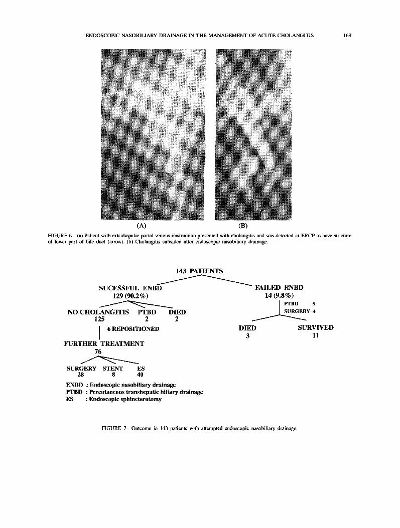
Figure 7 shows the outcome of ENBD placement.Nasobiliary catheter could be positioned successfullyin 129 patients (90.2%). Failure in 14 patients(malignant obstruction: 9, chronic pancreatitis: 1, stonedisease: 4) was due to failure to cannulate the bile ductin 3 patients and failure to negotiate guide wire across
the obstruction in 11 patients due to tight stricture
(n 8), multiple stones (n 2) or a large stone
(n 1). Of the 8 patients with tight biliary stricture andfailure to negotiate the guide wire, one had benignstricture due to chronic pancreatitis, two had ampullarycarcinoma and five had high bile duct malignant
stricture either due to infiltration from carcinoma ofgall bladder or due to cholangiocarcinoma. While 5 ofthese patients with failed ENBD underwentpercutaneous transhepatic biliary drainage, 4 underwentbiliary tract surgery. Among the failed ENBD group,3 patients died due to uncontrolled cholangitis andsepticemia, 1 of these inspite ofpercutaneous drainage.Of the 129 patients with successful placement of
ENBD; 125 (96.9%) had control of cholangitis within
1 to 3 days (Group A) or had successful prevention ofcholangitis (Group B). Six of these patients did requirerepositioning of the catheter due to its being pulled out
by the patient (n 3) or getting kinked in the stomach(n 3). Two patients had inadequate response andrequired additional percutaneous transhepatic biliarydrainage. Remaining two patients died inspite of
ENDOSCOPIC NASOBILIARY DRAINAGE IN THE MANAGEMENT OF ACUTE CHOLANGITIS 165
(A) (B)
FIGURE 2 Patient with ampullary carcinoma presented with cholangitis (a) Endoscopic nasobiliary drainage controlled the cholangitis(b) A 10 Fr stent was placed subsequently.
successful ENBD; one of these had a choledochalcyst and patient succumbed to septicemic shockwithin minutes of ENBD; the other patient hadENBD placed for malignant biliary stricture buthad pulled out her catheter and died of septicemiabefore the catheter could be repositioned. In patientswith favourable response to ENBD, catheter was
removed after a period of 5 to 14 days (median: 8days).Of the surviving 127 patients with ENBD, 76
were given a more definitive treatment for the
underlying biliary disease after stabilisation of
patient’s status. This included bile duct stone
extraction after endoscopic sphincterotomy in 40
patients, biliary surgery in 28 and endoscopic biliarystent placement in 8.
DISCUSSION
Endotherapy in acute cholangitis can be in the formof endoscopic sphincterotomy, biliary stenting or
by nasobiliary drainage (ENBD). In the presentseries, we could establish ENBD in 90.2% of the
patients and successful ENBD led to a rapid controlof cholangitis or prevention of cholangitis in 97%of patients. The procedure was safe with no
morbidity or mortality related to ENBD. The overallshort-term mortality in our patient group was 3.5%,being 1.5% only in those with successful ENBD.Death was thus, always related to delay in institutionof ENBD, displacement of catheter or failure to
establish ENBD. Our results with ENBD are similar
to the earlier reports with this procedure[8-13]. A
166 M.K. GOENKA et al.
(M (B)FIGURE 3 Patient with porta hepatitis block due to malignancy (a) Following ERCP, nasobiliary drain was established withoutsphincterotomy to prevent cholangitits (b) Subsequently sphincterotomy was performed followed by dilatation of porta block by biliarydilator (arrows) (c) A 10 Fr stent was placed.
slightly higher failure rate in the present study couldbe due to the fact that we have included patientswith diverse etiology including malignant bile ductobstruction. ENBD failure were mostly in malignantetiology group because of tightness of these
strictures, resulting in difficulty to negotiate the
guide wire across the stricture. Among the patientswith malignant bile duct strictures, those havinghigh bile duct strictures resulting from
cholangiocarcinoma or due to infiltration fromcarcinoma of gall bladder had a relatively higherfailure rate for guide wire negotiation and ENBDplacement, an experience similar to the earlier
reports[15]. While as expected choledocholithiasiswas the commonest benign cause for cholangitis,we unlike earlier series[9-14] also encountered othercauses such as chronic pancreatitis with biliarystricture, choledochal cyst, hydatid cyst with biliaryrupture and portal cholangiopathy. ENBD resultedin control of cholangitis and stabilised the patient,
(C) so that a more definite therapy in the form of surgery
ENDOSCOPIC NASOBILIARY DRAINAGE IN THE MANAGEMENT OF ACUTE CHOLANGITIS 167
(A) (B)
FIGURE 4 (a) Patient with cholangitis had a choledochal cyst with a common channel (arrow) (b) Endoscopic nasobiliary drainage wasestablished, cholangitis subsided.
or biliary stenting could be performed on a non-
emergent basis.
Endoscopic sphincterotomy followed by dormiaextraction in the same session is often consideredthe best therapy for cholangitis due to bile ductstones since it combines drainage of infected systemalong with removal of the cause[8-10]. However,endoscopic sphincterotomy in acute cholangitis hasbeen associated in earlier reported series with
complications such as hemorrhage, perforation andpancreatitis in 6-12% patients and mortality in
4.7-7.6%[8,10]. Moreover sphincterotomy may not
always be possible in patients with cholangitis dueto abnormal coagulogram, sphincterotomy combinedwith stone extraction by basket or balloon is moretime consuming than ENBD placement without
sphincterotomy[7,9,14] and ductal clearance aftersphincterotomy has been achieved in earlier reportsin 66 to 76% patients only[8-10]. In the presentseries, ENBD was performed without any priorsphincterotomy in 91.5% patients.
Some workers have preferred biliary stenting over
ENBD while managing acute cholangitis because ofpatient’s convenience with the former[ 16]. ENBD was
preferred by us because the indwelling catheter
permitted a cholangiogram to be performed later,allowing us to inject minimal required contrast material
during initial procedure in the presence of infected
bile. This reduces the risk ofincreased cholangiovenousreflux which can aggravate or precipitate septicemia in
a patient with cholangitis[1,9]. ENBD moreover,allowed collection of bile for culture and flushing ofcatheter for clearing flakes of pus and bile debris.ENBD catheter can also act as a conduit for chemical
dissolution of gall stones[I,14]. Though ENBD does
carry the risk of being pulled out by the patient as
happened in 3 of our cases, it can almost always be
repositioned rapidly. Combining ENBD with stent
placement in patients having cholangitis due to
malignant biliary obstruction can cure cholangitis andat the same time provide the palliation for the tumor
obviating the need for subsequent endoscopic
168 M.K. GOENKA et al.
(A) (B)FIGURE 5 (a) Patient with hepatic hydatid cyst with biliary rupture leading to cholangitis (b) Guide wire was passed into the cyst (c)Endoscopic nasobiliary drain resulted in control of cholangitits. Patient was operated later on an elective basis.
procedure. However, placement of two prostheticdevice would require endoscopic sphincterotomyand a longer procedure time. We have preferred to
treat the cholangitis in such setting by ENBD anda more definitive therapy by stenting or surgery wasoffered subsequently. Further, studies are howeverwarranted to choose the optimum managementmodality in such a setting.
Percutaneous transhepatic biliary drainage (PTBD)and surgical treatment have been used earlier for treatingpatients with acute cholangitis. However, direct surgicalintervention in acute cholangitis has been reportedto carry a mortality of6.5-40%[2-4]. PTBD is similarlyassociated with a significant mortality and a highrisk (upto 28%) ofbleeding, bile leakage and peritonitisand occasionally of pneumothorax, traumatic
pseudoaneurysm and arterioportal fistula
formation[5-7,17]. We therefore feel that surgery or
PTBD should be performed only when ENBD is not
possible due to anatomical reasons such as previousPolya’s gastrectomy, is technically unsuccessful or
(C) rarely when ENBD does not improve the cholangitis.
ENDOSCOPIC NASOBILIARY DRAINAGE IN THE MANAGEMENT OF ACUTE CHOLANGITIS 169
(A) (B)FIGURE 6 (a) Patient with extrahepatic portal venous obstruction presented with cholangitis and was detected at ERCP to have strictureof lower part of bile duct (arrow). (b) Cholangitis subsided after endoscopic nasobiliary drainage.
143 PATIENTS
SUCESSFUL ENBD129 (90.2%)
NO CHOLANGITIS PTBD DIED125 2 2
6 REPOSITIONED
FURTHER TREATMENT76
SURGERY STENT ES28 8 40
FAILED ENBD14(9.8%)
PTBD 5
SURGERY 4
DIED SURVIVED3 11
ENBD Endoscopic nasobiliary drainagePTBD Percutaneous transhepatic biliary drainageES Endoscopic sphincterotomy
FIGURE 7 Outcome in 143 patients with attempted endoscopic nasobiliary drainage.
170 M.K. GOENKA et al.
PTBD was performed in 7 patients in the present series,in 5 patients beuse of failed ENBD and in 2 with
successful ENBD for more effective drainage.In conclusion, the present study performed over a
large number of patients and in a wide etiologicalspectrum confirms the usefulness and safety ofENBDin the treatment and prevention of acute cholangitis.
References
[1] Leung, J.W.C. and Cotton, P.B. (1991). Endoscopicnasobiliary catheter drainage in biliary and pancreatic disease,Am. J. Gastroenterol., 86, 389-394.
[2] Lai, E.C.S., Tam, P.C., Paterson, I.A. et al. (1990). Emergencysurgery for severe acute cholangitis: the high risk patients,Ann. Surg., 211, 55-59.
[3] Welch, J.P. and Donaldson, G.A. (1976). The urgency ofdiagnosis and surgical treatment of acute suppurativecholangitis, Am. J. Surg., 131, 527-532.
[4] Gigot, J.F., Leese, T., Dereme, T., Coutinho, J., Castaing, D.and Bismuth, H. (1989). Acute cholangitis: multivariateanalysis of risk factors, Ann. Surg., 209, 435-438.
[5] Pessa, M.E., Hawkins, I.E and Vogel, S.B. (1987). Thetreatment of acute cholangitis. Percutaneous transhepaticbiliary drainage before definitive therapy, Ann. Surg., 205,389-392.
[6] Kadir, S., Baassirri, A., Barth, K.H., Kaufman, S.L., Cameron,J.L. and White, R.I. (1982). Percutaneous biliary drainage inthe management of biliary sepsis, AJR, 138, 25-29.
[7] Gould, R.J., Vogelzang, R.L., Neiman, H.L., Pearl,G.J. and Poticha, S.M. (1985). Percutaneous biliary drainageas an initial therapy in the sepsis of the biliary tract, Surg.Gynecol. Obstet, 160, 523-527.
[8] Leese, T., Neoptolemos, J.P., Baker, A.R. and Car-Lockee,D.L. (1986). Management of acute cholangitis and theimpact of endoscopic sphincterotomy, Br. J. Surg., 73,988-992.
[9] Leung, J.W.C., Chung, S.C.S., Sung, J.J.Y., Banez, V.P. andLi, A.K.C. (1989). Urgent endoscopic drainage for acutesuppurative cholangitis, Lancet, 1, 1307-1309.
[10] Gogel, H.K., Runyon, B.A., Volpicelli, N.A. and Palmer,R.C. (1987). Acute suppurative obstructive cholangitis dueto stones: treatment by urgent endoscopic sphincterotomy,Gastrointest. Endosc. 33, 210-213.
[11] Lai, E.C.S., Paterson, I.A., Tam, P.C., Choi, T.K., Fan, S.T.and Wong, J. (1990). Severe acute cholangitis: the role ofemergency nasobiliary drainage, Surgery, 107, 268-272.
12] Lai, E.C.S., Mok, EP.T., Tan, E.S.Y. et al. (1992). Endoscopicbiliary drainage for severe acute cholangitis,N. Engl. J. Med., 326, 1582-1586.
[13] Chawla, Y.K., Sharma, B.C. and Dilawari, J.B. (1994).Endoscopic nasobiliary drainage in acute suppurativecholangitis, Indian J. Gastroenterol., 13, 83-85.
[14] Lin, X.Z., Chang, K.K., Shin, J.S. et al. (1993). Emergencyendoscopic nasobiliary drainage for acute calculoussuppurative cholangitis and its potential use in chemicaldissolution, J. Gastroenterol. Hepatol., 8, 35-38.
15] Huibregtse, K. and Tytgat, G.N.J. (1989). Palliative treatmentof jaundice by transpapillary introduction of biliaryendoprosthesis, Gut, 23, 371-375.
[16] Kill, J., Kruse, A. and Rokkjaer, M. (1989). Large bile ductstones treated by endoscopic biliary drainage, Surgery, 105,51-56.
[17] Mueller, P.R., van Sonnenberg, E. and Fermcci, J.T. (1982).Percutaneous biliary drainage: technical and catheter-relatedproblems in 200 procedures, AJR, 138, 17-23.