Embed Size (px)
Citation preview
GE Port J Gastroenterol. 2015;22(4):137---142
www.elsevier.pt/ge
ORIGINAL ARTICLE
Endoscopic Mucosal Resection of Jejunal Polyps usingDouble-Balloon Enteroscopy
Paul Thomas Krönera, Aytekin Sancara, Lucia C. Fryb, Helmut Neumanna,c,1,Klaus Mönkemüllera,b,∗,1
a Basil I. Hirschowitz Endoscopic Center of Excellence, Division of Gastroenterology and Hepatology, University of Alabama atBirmingham, Birmingham, USAb Division of Gastroenterology, Hepatology and Infectious Diseases, Otto-von-Guericke University, Magdeburg, Germanyc Department of Medicine 1, Interdisciplinary Endoscopy, University of Erlangen-Nuremberg, Erlangen, Germany
Received 3 January 2015; accepted 23 April 2015Available online 2 July 2015
KEYWORDSDouble-BalloonEnteroscopy;Intestinal Mucosa;Intestinal Polyps;Jejunum
AbstractBackground: There are only two single case reports describing double-balloon enteroscopy(DBE)-assisted endoscopic mucosal resection (EMR) of the jejunum. The aim of this case serieswas to evaluate the feasibility and utility of DBE-assisted EMR in patients with familial andnon-familial jejunal polyps.Patients and methods: Observational, open-label, retrospective, single-arm case series in twohospitals.Results: Eight patients underwent DBE assisted jejunal EMR. Median age of patients was 42 years(range 24---62 years), male: female ratio 1.5:1. DBE was done through the antegrade (i.e. oral)route in all patients. Four patients had FAP; two had Peutz-Jeghers syndrome, one had a sporadicadenoma and one had a bleeding jejunal polyp, which on histological examination turned outto be lipoma. 3/8 underwent piece-meal EMR. No immediate adverse events occurred.Conclusions: This is the first case series presenting the technical details, feasibility and out-comes of EMR of the small bowel. EMR of the jejunum is feasible and safe during DBE.© 2015 Sociedade Portuguesa de Gastrenterologia. Published by ElsevierEspaña, S.L.U. This is an open access article under the CC BY-NC-ND license(http://creativecommons.org/licenses/by-nc-nd/4.0/).
∗ Corresponding author.E-mail address: [email protected] (K. Mönkemüller).
1 These authors contributed equally to this study.
http://dx.doi.org/10.1016/j.jpge.2015.04.0052341-4545/© 2015 Sociedade Portuguesa de Gastrenterologia. Published by Elsevier España, S.L.U. This is an open access article under theCC BY-NC-ND license (http://creativecommons.org/licenses/by-nc-nd/4.0/).
138 P.T. Kröner et al.
PALAVRAS-CHAVEEnteroscopia deDuplo Balão;Jejuno;Mucosa Intestinal;Pólipos Intestinais
Mucosectomia Endoscópica de Pólipos Jejunais por Enteroscopia de Duplo Balão
ResumoIntroducão: Existem apenas duas séries clínicas na literatura a descrever os resultados da muco-sectomia no jejuno por enteroscopia de duplo balão (DBE). O objetivo desta série de casos foiavaliar a exequibilidade e utilidade da mucosectomia por DBE em doentes com pólipos jejunaisfamiliares e não familiares.Métodos: Estudo observacional, retrospectivo, open-label, descrevendo uma série de casos emdois hospitais.Resultados: Oito doentes realizaram mucosectomia por DBE. A idade mediana foi 42 anos(âmbito 24---62 anos), razão homem:mulher 1,5:1. Foi realizada DBE por via anterógrada (oral)em todos os doentes. Quatro doentes tinham polipose adenomatosa familiar (PAF); dois tinhamsíndroma de Peutz-Jeghers, um tinha um adenoma esporádico e um tinha um pólipo jejunalsangrante, cuja avaliacão anatomopatológica revelou tratar-se de um lipoma. A mucosectomiafoi fragmentada em 3 dos 8 doentes. Não se verificou nenhum efeito adverso imediato.Conclusões: Este é o primeiro estudo que descreve os detalhes técnicos, exequibilidade e result-ados da mucosectomia no intestino delgado. A mucosectomia no jejuno por DBE é exequível esegura.© 2015 Sociedade Portuguesa de Gastrenterologia. Publicado por ElsevierEspaña, S.L.U. Este é um artigo Open Access sob a licença de CC BY-NC-ND(http://creativecommons.org/licenses/by-nc-nd/4.0/).
1. Introduction
Endoscopic mucosal resection (EMR) has become a well-accepted and practiced method for treating neoplasticand non-neoplastic lesions of the esophagus, stomach,duodenum and colon.1 Until recently, primary surgical orintraoperative endoscopic resection was the only avail-able means of treating polyps of the mid-small bowel.2
Since the advent of balloon-assisted enteroscopy (includingdouble-balloon, single balloon) endoscopic polypectomy hasbecome a viable option for treatment of small-bowel disor-ders. Indeed, at present balloon-assisted enteroscopy hasbecome the primary mode for the removal of small bowelpolyps. However, all yet published studies have only focusedon standard polypectomy techniques.3,4 Because the smallbowel has a thinner wall as compared to other luminal partsof the gastrointestinal (GI) tract, the experience using EMRis very limited. Indeed, there are currently only two casereports describing EMR of the jejunum.5,6 Therefore, theaim of this case series study was to report on the feasibilityand utility of double balloon enteroscopy assisted mucosalresection (EMR) in patients with familial and non-familialjejunal polyps.
2. Patients and methods
Forty-two patients with jejunal polyp(s) (familial adeno-matous polyposis syndrome (FAP), n = 17, Peutz-Jegherssyndrome (PJS), n = 12, sporadic adenomas, n = 7, nodularlymphoid hyperplasia, n = 3, lipomas, n = 3) undergoing DBE-assisted resection of their lesions at the MarienhospitalBottrop, University of Magdeburg Medical Center betweenDecember 2007 and December 2012 were included andtheir data recorded in a computerized database. For thiscase study we only included patients undergoing EMR.
EMR was defined as the resection of the entire mucosallayer and part of the submucosa using advanced endo-scopic resection techniques (i.e. mucosectomy). Patientsundergoing standard snare polypectomy were excluded.The patients provided written informed consent to undergoendoscopy with the double balloon enteroscopy system.Double balloon enteroscopy was performed using the Fuji-non enteroscope (Fujinon EN-450T5, Fujifilm, Saitama,Japan). The study was approved by the ethics committeeof the University of Magdeburg and conducted out in accor-dance with those of the Code of Ethics of the World MedicalAssociation (Declaration of Helsinki).
2.1. Mucosectomy technique
Before resection the mucosal Kudo pit pattern of thelesion was analyzed using high resolution white-light andchromoendoscopy methods (i.e. standard and virtual chro-moendoscopy) to better define their surface, borders andassist during resection (Fig. 1).7 The basis for a successfulmucosectomy is the creation of a ‘‘submucosal cushion’’,thus lifting the lesion from the submucosa. The submucosalcushion was created using epinephrine-saline and indigo-carmine solution (1:20,000 epinephrine:saline; 0.1 ml ofindigo-carmine 3% in 100 ml of saline). A maximum of 10 mlof this solution was used, as there are reports of bowelischemia induced by epinephrine.8 If more injection wasrequired to raise the polyp before or during the mucosec-tomy just normal saline was used. The submucosal cushionwas initiated by injecting 1---3 ml of epinephrine-saline solu-tion to the most distal part of the lesion. This maneuverplaces the polyp ‘‘en-face’’ to the endoscopist. Then oneor both lateral sides were injected, allowing for a homoge-nous lifting (Fig. 2). If no lifting was observed mucosectomywas aborted. In addition, no mucosectomy was attempted
Endoscopic mucosal resection of jejunal polyps 139
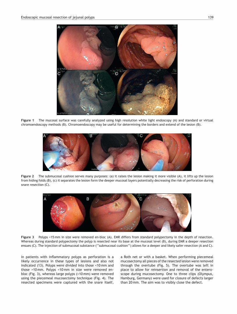
Figure 1 The mucosal surface was carefully analyzed using high resolution white light endoscopy (A) and standard or virtualchromoendoscopy methods (B). Chromoendoscopy may be useful for determining the borders and extend of the lesion (B).
Figure 2 The submucosal cushion serves many purposes: (a) it raises the lesion making it more visible (A), it lifts up the lesionfrom hiding folds (B), (c) it separates the lesion form the deeper mucosal layers potentially decreasing the risk of perforation duringsnare resection (C).
Figure 3 Polyps <15 mm in size were removed en-bloc (A). EMR differs from standard polypectomy in the depth of resection.Whereas during standard polypectomy the polyp is resected near its base at the mucosal level (B), during EMR a deeper resectionensues (C). The injection of submucosal substance (‘‘submucosal cushion’’) allows for a deeper and likely safer resection (A and C).
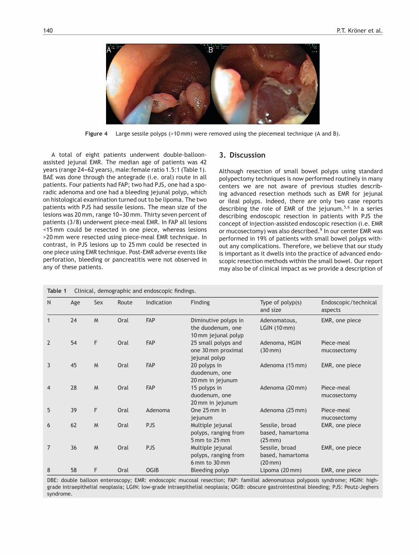
in patients with inflammatory polyps as perforation is alikely occurrence in these types of lesions and also notindicated (13). Polyps were divided into those <10 mm andthose >10 mm. Polyps <10 mm in size were removed en-bloc (Fig. 3), whereas large polyps (>10 mm) were removedusing the piecemeal mucosectomy technique (Fig. 4). Theresected specimens were captured with the snare itself,
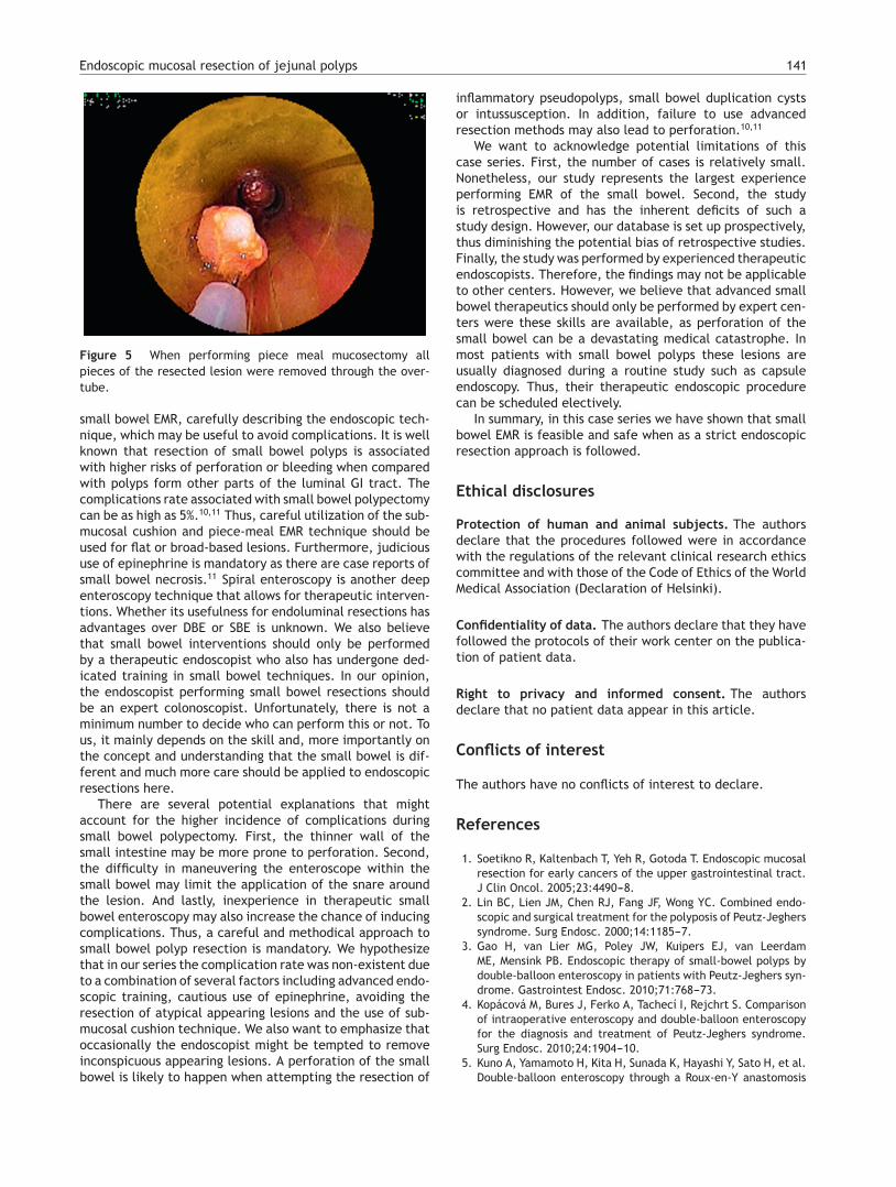
a Roth net or with a basket. When performing piecemealmucosectomy all pieces of the resected lesion were removedthrough the overtube (Fig. 5). The overtube was left inplace to allow for reinsertion and removal of the entero-scope during mucosectomy. One to three clips (Olympus,Hamburg, Germany) were used for closure of defects largerthan 20 mm. The aim was to visibly close the defect.
140 P.T. Kröner et al.
Figure 4 Large sessile polyps (>10 mm) were removed using the piecemeal technique (A and B).
A total of eight patients underwent double-balloon-assisted jejunal EMR. The median age of patients was 42years (range 24---62 years), male:female ratio 1.5:1 (Table 1).BAE was done through the antegrade (i.e. oral) route in allpatients. Four patients had FAP; two had PJS, one had a spo-radic adenoma and one had a bleeding jejunal polyp, whichon histological examination turned out to be lipoma. The twopatients with PJS had sessile lesions. The mean size of thelesions was 20 mm, range 10---30 mm. Thirty seven percent ofpatients (3/8) underwent piece-meal EMR. In FAP all lesions<15 mm could be resected in one piece, whereas lesions>20 mm were resected using piece-meal EMR technique. Incontrast, in PJS lesions up to 25 mm could be resected inone piece using EMR technique. Post-EMR adverse events likeperforation, bleeding or pancreatitis were not observed inany of these patients.
3. Discussion
Although resection of small bowel polyps using standardpolypectomy techniques is now performed routinely in manycenters we are not aware of previous studies describ-ing advanced resection methods such as EMR for jejunalor ileal polyps. Indeed, there are only two case reportsdescribing the role of EMR of the jejunum.5,6 In a seriesdescribing endoscopic resection in patients with PJS theconcept of injection-assisted endoscopic resection (i.e. EMRor mucosectomy) was also described.9 In our center EMR wasperformed in 19% of patients with small bowel polyps with-out any complications. Therefore, we believe that our studyis important as it dwells into the practice of advanced endo-scopic resection methods within the small bowel. Our reportmay also be of clinical impact as we provide a description of
Table 1 Clinical, demographic and endoscopic findings.
N Age Sex Route Indication Finding Type of polyp(s)and size
Endoscopic/technicalaspects
1 24 M Oral FAP Diminutive polyps inthe duodenum, one10 mm jejunal polyp
Adenomatous,LGIN (10 mm)
EMR, one piece
2 54 F Oral FAP 25 small polyps andone 30 mm proximaljejunal polyp
Adenoma, HGIN(30 mm)
Piece-mealmucosectomy
3 45 M Oral FAP 20 polyps induodenum, one20 mm in jejunum
Adenoma (15 mm) EMR, one piece
4 28 M Oral FAP 15 polyps induodenum, one20 mm in jejunum
Adenoma (20 mm) Piece-mealmucosectomy
5 39 F Oral Adenoma One 25 mm injejunum
Adenoma (25 mm) Piece-mealmucosectomy
6 62 M Oral PJS Multiple jejunalpolyps, ranging from5 mm to 25 mm
Sessile, broadbased, hamartoma(25 mm)
EMR, one piece
7 36 M Oral PJS Multiple jejunalpolyps, ranging from6 mm to 30 mm
Sessile, broadbased, hamartoma(20 mm)
EMR, one piece
8 58 F Oral OGIB Bleeding polyp Lipoma (20 mm) EMR, one piece
DBE: double balloon enteroscopy; EMR: endoscopic mucosal resection; FAP: familial adenomatous polyposis syndrome; HGIN: high-grade intraepithelial neoplasia; LGIN: low-grade intraepithelial neoplasia; OGIB: obscure gastrointestinal bleeding; PJS: Peutz-Jegherssyndrome.
Endoscopic mucosal resection of jejunal polyps 141
Figure 5 When performing piece meal mucosectomy allpieces of the resected lesion were removed through the over-tube.
small bowel EMR, carefully describing the endoscopic tech-nique, which may be useful to avoid complications. It is wellknown that resection of small bowel polyps is associatedwith higher risks of perforation or bleeding when comparedwith polyps form other parts of the luminal GI tract. Thecomplications rate associated with small bowel polypectomycan be as high as 5%.10,11 Thus, careful utilization of the sub-mucosal cushion and piece-meal EMR technique should beused for flat or broad-based lesions. Furthermore, judicioususe of epinephrine is mandatory as there are case reports ofsmall bowel necrosis.11 Spiral enteroscopy is another deepenteroscopy technique that allows for therapeutic interven-tions. Whether its usefulness for endoluminal resections hasadvantages over DBE or SBE is unknown. We also believethat small bowel interventions should only be performedby a therapeutic endoscopist who also has undergone ded-icated training in small bowel techniques. In our opinion,the endoscopist performing small bowel resections shouldbe an expert colonoscopist. Unfortunately, there is not aminimum number to decide who can perform this or not. Tous, it mainly depends on the skill and, more importantly onthe concept and understanding that the small bowel is dif-ferent and much more care should be applied to endoscopicresections here.
There are several potential explanations that mightaccount for the higher incidence of complications duringsmall bowel polypectomy. First, the thinner wall of thesmall intestine may be more prone to perforation. Second,the difficulty in maneuvering the enteroscope within thesmall bowel may limit the application of the snare aroundthe lesion. And lastly, inexperience in therapeutic smallbowel enteroscopy may also increase the chance of inducingcomplications. Thus, a careful and methodical approach tosmall bowel polyp resection is mandatory. We hypothesizethat in our series the complication rate was non-existent dueto a combination of several factors including advanced endo-scopic training, cautious use of epinephrine, avoiding theresection of atypical appearing lesions and the use of sub-mucosal cushion technique. We also want to emphasize thatoccasionally the endoscopist might be tempted to removeinconspicuous appearing lesions. A perforation of the smallbowel is likely to happen when attempting the resection of
inflammatory pseudopolyps, small bowel duplication cystsor intussusception. In addition, failure to use advancedresection methods may also lead to perforation.10,11
We want to acknowledge potential limitations of thiscase series. First, the number of cases is relatively small.Nonetheless, our study represents the largest experienceperforming EMR of the small bowel. Second, the studyis retrospective and has the inherent deficits of such astudy design. However, our database is set up prospectively,thus diminishing the potential bias of retrospective studies.Finally, the study was performed by experienced therapeuticendoscopists. Therefore, the findings may not be applicableto other centers. However, we believe that advanced smallbowel therapeutics should only be performed by expert cen-ters were these skills are available, as perforation of thesmall bowel can be a devastating medical catastrophe. Inmost patients with small bowel polyps these lesions areusually diagnosed during a routine study such as capsuleendoscopy. Thus, their therapeutic endoscopic procedurecan be scheduled electively.
In summary, in this case series we have shown that smallbowel EMR is feasible and safe when as a strict endoscopicresection approach is followed.
Ethical disclosures
Protection of human and animal subjects. The authorsdeclare that the procedures followed were in accordancewith the regulations of the relevant clinical research ethicscommittee and with those of the Code of Ethics of the WorldMedical Association (Declaration of Helsinki).
Confidentiality of data. The authors declare that they havefollowed the protocols of their work center on the publica-tion of patient data.
Right to privacy and informed consent. The authorsdeclare that no patient data appear in this article.
Conflicts of interest
The authors have no conflicts of interest to declare.
References
1. Soetikno R, Kaltenbach T, Yeh R, Gotoda T. Endoscopic mucosalresection for early cancers of the upper gastrointestinal tract.J Clin Oncol. 2005;23:4490---8.
2. Lin BC, Lien JM, Chen RJ, Fang JF, Wong YC. Combined endo-scopic and surgical treatment for the polyposis of Peutz-Jegherssyndrome. Surg Endosc. 2000;14:1185---7.
3. Gao H, van Lier MG, Poley JW, Kuipers EJ, van LeerdamME, Mensink PB. Endoscopic therapy of small-bowel polyps bydouble-balloon enteroscopy in patients with Peutz-Jeghers syn-drome. Gastrointest Endosc. 2010;71:768---73.
4. Kopácová M, Bures J, Ferko A, Tachecí I, Rejchrt S. Comparisonof intraoperative enteroscopy and double-balloon enteroscopyfor the diagnosis and treatment of Peutz-Jeghers syndrome.Surg Endosc. 2010;24:1904---10.
5. Kuno A, Yamamoto H, Kita H, Sunada K, Hayashi Y, Sato H, et al.Double-balloon enteroscopy through a Roux-en-Y anastomosis
142 P.T. Kröner et al.
for EMR of a jejunal adenoma in the afferent duodenal limb.Gastrointest Endosc. 2004;60:1032---4.
6. Suzuki H, Yamada A, Watabe H, Kobayashi Y, Hirata Y, YamajiY, et al. Successful treatment of early-stage jejunum adenocar-cinoma by endoscopic mucosal resection using double-balloonendoscopy: a case report. Diagn Ther Endosc. 2012:521960.
7. Mönkemüller K, Fry LC, Ebert M, Bellutti M, Venerito M, Knip-pig C, et al. Feasibility of double-balloon enteroscopy-assistedchromoendoscopy of the small bowel in patients with familialadenomatous polyposis. Endoscopy. 2007;39:52---7.
8. Yen HH, Chen YY, Su WW, Soon MS. Intestinal necrosis as acomplication of epinephrine injection therapy during double-balloon enteroscopy. Endoscopy. 2006;38:542.
9. Serrano M, Mão-de-Ferro S, Pinho R, Marcos-Pinto R, FigueiredoP, Ferreira S, Claro I, Mascarenhas-Saraiva M, Dias-Pereira A.Double-balloon enteroscopy in the management of patients withPeutz-Jeghers syndrome: a retrospective cohort multicenterstudy. Rev Esp Enferm Dig. 2013;105:594---9.
10. Spahn TW, Kampmann W, Eilers M. Small-bowel perfora-tion after endoscopic resection of a Peutz-Jeghers polypin an infant using double-balloon enteroscopy. Endoscopy.2007;39(S1):E217.
11. Möschler O, May A, Müller MK, Ell C, German DBE StudyGroup. Complications in and performance of double-balloonenteroscopy (DBE): results from a large prospective DBEdatabase in Germany. Endoscopy. 2011;43:484---9.