Embed Size (px)
Citation preview
4/10/2014
1
Endoscopic Assisted resection for congenital Midline Nasal Mass
Ahmed Aly Ibrahim
A.prof ORL Department
Alexandria University
Emad. A Magdy
prof ORL Department
Alexandria University
Haytham Morsi,MD Mohammad Fawzy,MD
Embryology
• During formation of skull base and nose,mesenchymal structures are formed from severalcenters which will eventually fuse and ossify.
• Before their fusion, there are recognized spaceswhich are important in the development ofcongenital midline nasal masses– Fonticulus frontalis
– Prenasal space
– Foramen cecum
4/10/2014
2
Embyrology
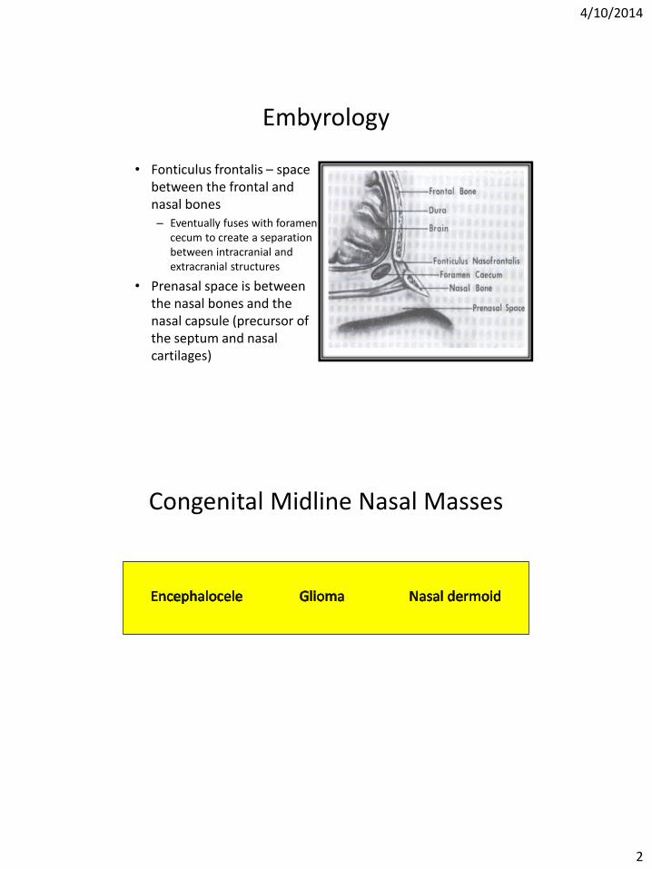
• Fonticulus frontalis – space between the frontal and nasal bones– Eventually fuses with foramen
cecum to create a separation between intracranial and extracranial structures
• Prenasal space is between the nasal bones and the nasal capsule (precursor of the septum and nasal cartilages)
Congenital Midline Nasal Masses
4/10/2014
3
Encephalocele
• Extracranial herniations of cranial contents through a defect in the skull.
• May include meninges only (meningocele), or both brain and meninges (meningoencephalocele).
• 40% have other associated anomalies.
Encephalocele
• Divided into three categories:– Occipital 75%– Sincipital 15%– Basal 10%
• Sincipital encephaloceles:– A bony defect between the frontal and ethmoid bones anterior
to the crista galli– Nasofrontal, nasoethmoidal, nasoorbital.
• Basal encephaloceles:– A bony defect between the cribriform plate and the superior
orbital or posterior clinoid fissure, presenting as an intranasal mass.
– Trans-ethmoidal, Spheno-ethmoidal, Trans-sphenoidal, spheno-orbital.
4/10/2014
4
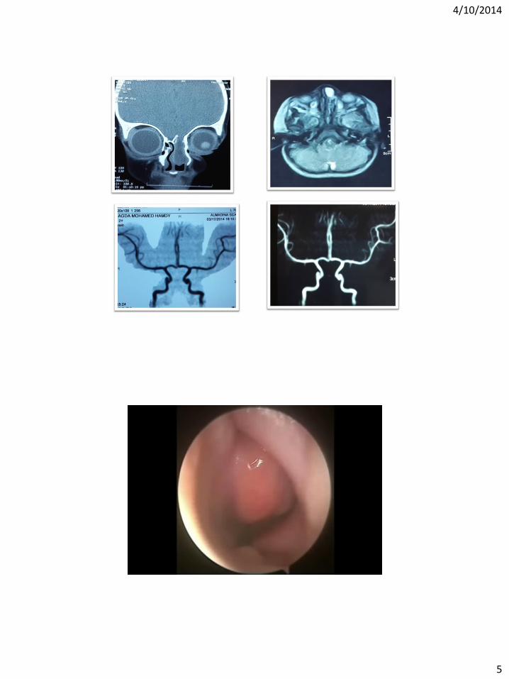
EncephaloceleImaging
• The diagnosis is radiologically confirmed by CT and/orMRI
• CT evaluation should include high-resolution which help delineate the infant’s cartilaginous skull base.
• MRI provides complementary information regarding the fluid and soft tissue characteristics of the mass and is valuable in identifying an intracranial connection. It is also useful in helping to differentiate a meningocelefrom a meningoencephalocele.
EncephaloceleTreatment
• Early intervention in the first few months of life:
– minimize the risk of meningitis and cosmetic deformities.
• Small lesions with minimal skull base defects may be managed endoscopically. Larger lesions require craniotomy / combined approach.
4/10/2014
5
4/10/2014
6
Glioma
• Gliomas are unencapsulated collections of glial cells situated outside the CNS.
• Possible theories of development include: (1) sequestration of glial tissue of the olfactory bulb entrapped during
cribriform plate fusion(2) ectopic neural tissue cells(3) pinched encephalocele(4) inappropriate closure of the anterior neuropore (fonticulus
frontalis), with failure of mesoderm to enter the region, resulting in inadequate bone formation.
• 15–20% of nasal gliomas have a fibrous stalk connection to the intracranial space.
Glioma
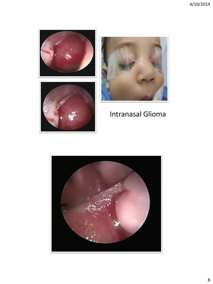
• Types:– extranasal (60%) – intranasal (30%)– combined (10%)
• Firm, noncompressible purple or gray mass.• Site:
– Extranasal: glabella or nasomaxillary suture– Intranasal: lateral nasal wall near MT or nasal septum
sometimes difficult to differentiate between gliomas andencephaloceles; however, the presence of ependymal tissue isconsistent with an encephalocele.
4/10/2014
7
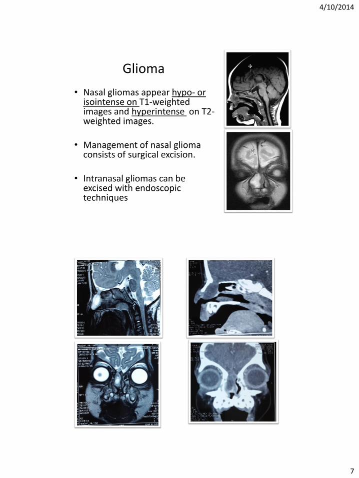
Glioma
• Nasal gliomas appear hypo- or isointense on T1-weighted images and hyperintense on T2-weighted images.
• Management of nasal gliomaconsists of surgical excision.
• Intranasal gliomas can be excised with endoscopic techniques
4/10/2014
8
Intranasal Glioma
4/10/2014
9
Nasal Dermoid
• Most common congenital nasal abnormality.
• 1–3% of all dermoids and 10–12% of head and neck dermoids.
• Congenital dermoids contain only ectodermal and mesodermalembryonic elements. Mesodermal elements, which include hair follicles, sebaceous glands, and sweat glands are found in the wall of the cyst and thus differentiate these masses from simple epidermoid cysts
• Teratomas, contain all three embryonal germ layers.
Nasal Dermoid
• Aetiology of these lesions is controversial.
• Prenasal space theory, is based on the abnormal development ofthe fonticulus frontalis.
• The retracting dura may drag the surface epithelium inward,causing formation of a sinus tract.
• In some patients, the sinus tract extends into the intracranial cavityor prenasal space.
• the dermal sinus or cyst may persist anywhere from the foramencecum to the nasal tip.
4/10/2014
10
Nasal Dermoids
• Manifest as a simple cyst, a cyst with a sinus tract, or a sinus tract alone. may be intermittent discharge or infection. Protruding hair is seen in a minority of patients, but is pathognomic.
• Firm, lobulated, noncompressible midline mass over the nasal dorsum +/- sinus opening. negative Furstenberg test and do not transilluminate.
• Intracranial extension 4–45%.
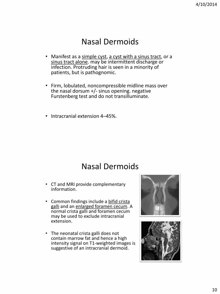
Nasal Dermoids
• CT and MRI provide complementary information.
• Common findings include a bifid crista galli and an enlarged foramen cecum. A normal crista galli and foramen cecum may be used to exclude intracranial extension.
• The neonatal crista galli does not contain marrow fat and hence a high intensity signal on T1-weighted images is suggestive of an intracranial dermoid.
4/10/2014
11
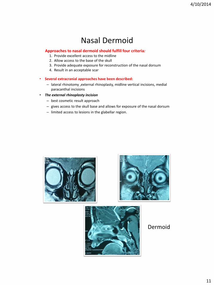
Nasal DermoidApproaches to nasal dermoid should fulfill four criteria:
1. Provide excellent access to the midline2. Allow access to the base of the skull3. Provide adequate exposure for reconstruction of the nasal dorsum4. Result in an acceptable scar
• Several extracranial approaches have been described:
– lateral rhinotomy ,external rhinoplasty, midline vertical incisions, medial paracanthal incisions
• The external rhinoplasty incision
– best cosmetic result approach
– gives access to the skull base and allows for exposure of the nasal dorsum
– limited access to lesions in the glabellar region.
Dermoid
4/10/2014
12
4/10/2014
13
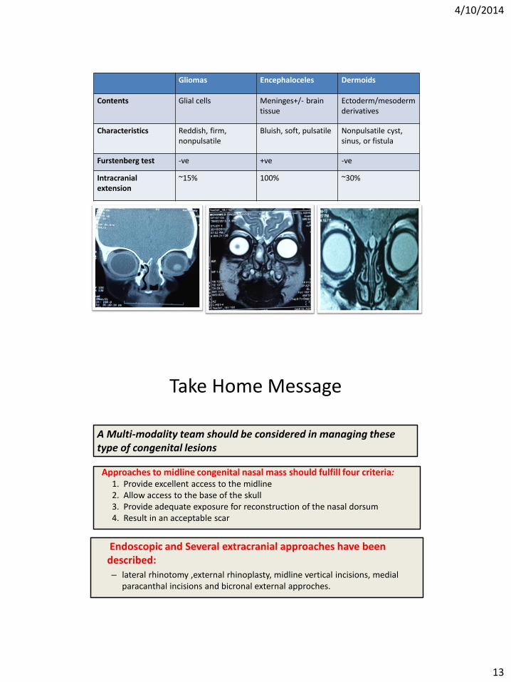
Gliomas Encephaloceles Dermoids
Contents Glial cells Meninges+/- brain tissue
Ectoderm/mesodermderivatives
Characteristics Reddish, firm, nonpulsatile
Bluish, soft, pulsatile Nonpulsatile cyst, sinus, or fistula
Furstenberg test -ve +ve -ve
Intracranial extension
~15% 100% ~30%
Take Home Message
Approaches to midline congenital nasal mass should fulfill four criteria:1. Provide excellent access to the midline2. Allow access to the base of the skull3. Provide adequate exposure for reconstruction of the nasal dorsum4. Result in an acceptable scar
Endoscopic and Several extracranial approaches have been described:– lateral rhinotomy ,external rhinoplasty, midline vertical incisions, medial
paracanthal incisions and bicronal external approches.
A Multi-modality team should be considered in managing these type of congenital lesions

4/10/2014
14
Thank You for your Attention