Embed Size (px)
Citation preview
BIODIVERSITAS ISSN: 1412-033X Volume 18, Number 2, April 2017 E-ISSN: 2085-4722 Pages: 733-740 DOI: 10.13057/biodiv/d180242
Endophytic bacteria producing antibacterial against methicillin-resistant Staphylococcus aureus (MRSA) in seagrass from Rote Ndao,
East Nusa Tenggara, Indonesia
DIAN SAGITA FITRI, ARTINI PANGASTUTI, ARI SUSILOWATI♥, SUTARNO Department of Bioscience, Graduate Program, Universitas Sebelas Maret. Jl. Ir. Sutami No. 36A Kentingan, Surakarta, 57126, Central Java, Indonesia.
Tel./Fax.: +62-271-632450, ♥email: [email protected], [email protected]
Manuscript received: 24 June 2016. Revision accepted: 13 April 2017.
Abstract. Fitri DS, Pangastuti A, Susilowati A, Sutarno. 2017. Endophytic bacteria producing antibacterial against methicillin-resistant Staphylococcus aureus (MRSA) in seagrass from Rote Ndao, East Nusa Tenggara, Indonesia. Biodiversitas 18: 733-740. Methicillin-resistant Staphylococcus aureus (MRSA) are bacteria that resistant to the various type of antibiotics and yet cannot be handled comprehensively. The discovery of new antibiotic from endophytic bacteria in seagrass of Rote Island is an option to overcome the resistance. The aims of this research were to screen endophytic bacteria inhibit MRSA from seagrass, to determine the species of the endophytic bacteria and the genetic relationship. Isolation of endophytic bacteria has carried out by inoculating surface sterilization seagrass leaves on Marine Agar (MA) medium. Selection of potential endophytic bacteria-producing anti-MRSA has done using overlay method against MRSA, gram-positive Bacillus subtilis, and gram-negative Escherichia coli. Identification of the endophytic bacteria based on the sequence of 16S rRNA encoding gene. The results showed that there were eight isolates of endophytic bacteria which have antibacterial activity against MRSA of seagrass Enhalus acoroides, Thalassia hemprichii, and Cymodocea rotundata in the Litianak and Oeseli Beaches, Rote Ndao. The diameter of inhibition zone was between 0.65-18.27 mm with narrow spectrum with broad spectrum antibacterial activity. The eight potential endophytic bacteria identity were Bacillaceae E2M1, Bacillaceae E2M3, Bacillus E2M4, Bacillus E2M7, Bacillaceae E2M8, Pseudomonadaceae C1M7, Shewanellaceae C2M3, and Rhodobacteraceae T1M3. Most of the isolates can be said to be a new species as the percent similarity of 16S rRNA gene sequence was less than 95% and promising to produce new antibacterial compounds. Phylogenetic relationship showed some isolates clustering in different groups that present the diverse groups of endophytic bacteria were found.
Keywords: Antibacteria, Bacillus subtilis, endophytic bacteria, Escherichia coli, MRSA, seagrass
INTRODUCTION
Since the imposition of antibiotics, bacteria have evolved mechanisms to resist the effects of antibiotics or often referred to as resistant bacteria. The evolution and the increasing spread of resistant bacteria species caused by several factors such as the widely used antibiotic and inaccurate application of antibiotics, as well as the use of antibiotics in animal feed extensively (Karchmer 2006). Staphylococcus aureus is one of the human pathogen most commonly found in various diseases ranging from mild skin infections, severe infections until disease that can threaten human safety such as endocarditis, pneumonia, and sepsis (Paterson et al. 2014). S. aureus resistance to the antibiotic penicillin was first reported in 1940. Then, methicillin-resistant strains of S. aureus (MRSA) was first reported in the early 1960s as the second leading cause of infection patients in hospitals and community; and become endemic worldwide including in Indonesia. Until now part of the problem of bacterial resistance that was never solved completely (Chamber 2001; Karchmer 2006).
Exploration of new sources becomes very important to find bioactive compounds that can be used as a new antibiotic (Clardy et al. 2006). Microorganisms have been the source of many useful compounds in medicine, the
pharmaceutical industry, and agriculture. However, most of these organisms derived from terrestrial habitats. After intensive research on terrestrial microorganisms, researchers are now focusing attention on other ecosystems, such as the sea. Microorganisms that associated by marine organisms has become a major target for the discovery of new bioactive compounds (Penesyan et al. 2010, 2011; Kang et al. 2015). One of them is the utilization of microbial endophyte. Endophyte is defined as microorganisms contained in asymptomatically in the plant tissue (Schulz et al. 2006). Some marine microorganisms survive in habitats with high-stress levels, undersea with low temperatures, limited light conditions with high pressure, grazing by herbivores on host plants, predation, and their competitors. These factors have led to the development of a unique metabolism, resulting in the production of new metabolites that differ from terrestrial microorganisms. Some endophytic microorganisms including bacteria can contribute to host defense by excretion of antibiotics and other bioactive substances. Thus, marine microorganisms offer an excellent resource for the discovery of new compounds with interesting biological activities, including antimicrobial, antifungal, antiprotozoal, antituberculosis and antiviral (Lu et al. 2010; Rahman et al. 2010).
B IODIVERSITAS 18 (2): 733-740, April 2017
734
Seagrass is an aquatic plant that can adapt to live and grow in the marine environment. Seagrass adapt to the unstable condition of the beach; seagrass vegetation provides many ecological functions for macroorganisms and microorganisms associated. One factor organisms to settle interest in the environment is classified as seagrass habitat productive, so as to provide food, a shelter for the survival of organisms associated (Gufran 2011).
In Rote Ndao, seagrass is distributed almost in all coastal areas such as Litianak and Oeseli beaches. Ecologically both beaches have differences, Litianak beach is a sandy beach and a bit muddy, while the Oeseli beach is rocky beaches mixed with sand. Although the presence of seagrass on both coasts knew their existence, both research and information about endophytic bacteria and potential in every beach have not been performed. Unique habitat of the beach is one of the important parameters to obtain new sources of endophytic bacteria that can produce secondary metabolites as antibiotics. The discovery of new antibiotic metabolites of endophyte is an important alternative to cope with increasing levels of drug resistance by human pathogens such as MRSA and the limited number of antibiotics that are effective against a variety of bacterial species. Therefore, it is necessary to do research on the potential of bioactive compounds owned by endophytic bacteria in seagrass in the Litianak and Oeseli beaches, Rote Ndao, East Nusa Tenggara, Indonesia.
MATERIALS AND METHODS
Media and culture condition Methicillin-resistant Staphylococcus aureus (MRSA),
Bacillus subtilis and Escherichia coli were obtained from the Laboratory of Clinical Microbiology, Faculty of
Medicine, Universitas Indonesia, Depok, West Java, Indonesia. The bacteria were stored on nutrient agar slant (Himedia). Endophytic bacteria were stored on marine agar media (Himedia). Stock cultures of the bacteria stored at-4°C. The bacteria were refreshed and stored at room temperature for anti-bacterial testing.
Seagrass sampling Seagrass was collected at Litianak and Oeseli Beaches,
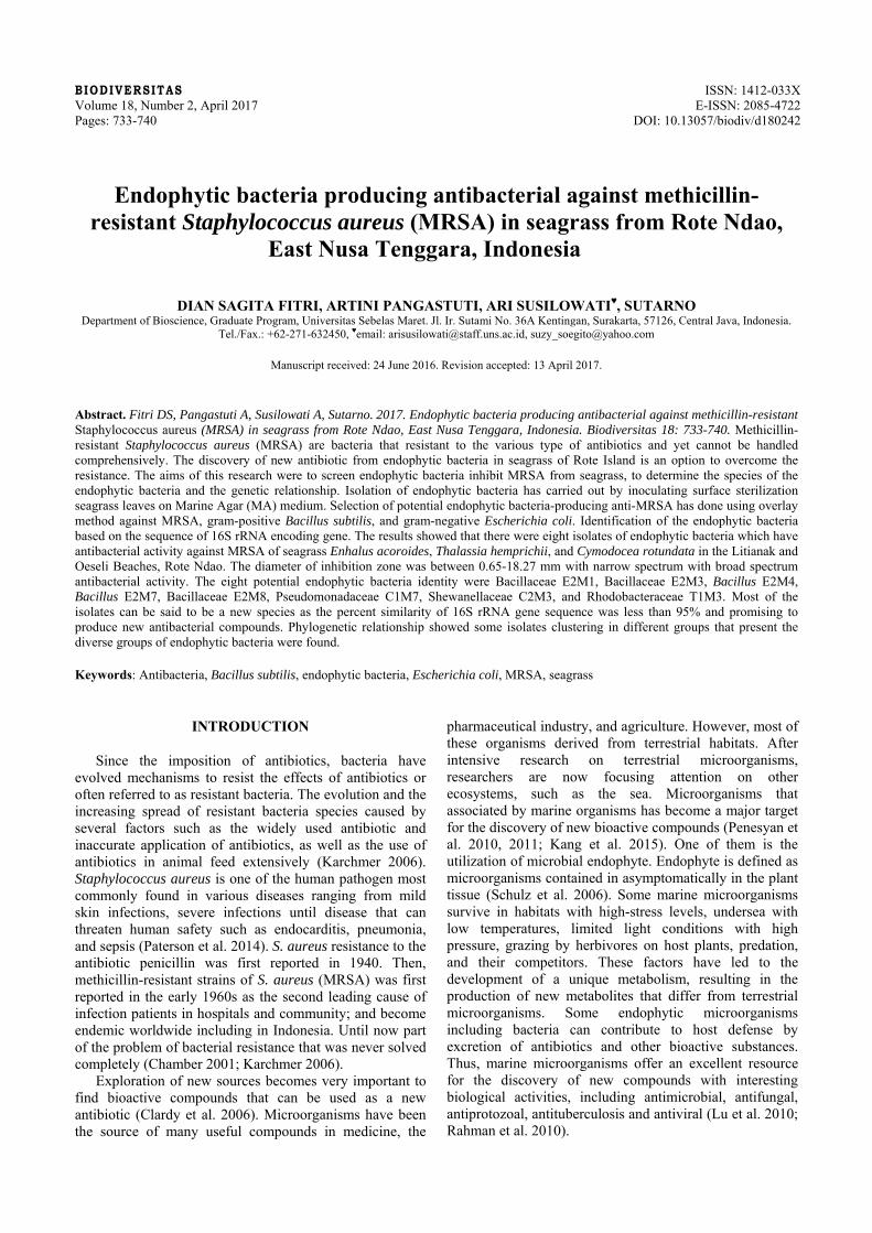
Rote Island of the Rote Ndao District, East Nusa Tenggara Province, Indonesia (Figure 1). Enhalus acoroides, Thalassia hemprichii, and Cymodocea rotundata were taken as a source of endophytic bacteria. Each seagrass was put in plastic bags and labeled contains information of species and the location, and then was placed in an ice box to be brought to the laboratory. The seagrass was identified by comparing the morphological character with a guide book of seagrass in tropical seagrasses in the Indo-West Pacific by Lanyon (1986) and Waycott et al. (2004).
Isolation of seagrass endophytic bacteria The seagrass leaves from each species were cleaned
with aquadest and then cut into 5 cm long. Surface sterilization was done by keeping a piece of the seagrass leaves into 70% ethanol for 1 minute, continued into 5.25% sodium hypochlorite solution for 1 minute, and washed with 70% ethanol for three times. The sterile leaves were sliced then planted in the marine agar media and incubated at room temperature (28-30oC) for 1-2 days. The plates were observed every day until some colony of the bacteria emerged (Ravikumar et al. 2010). Every single colony of the endophytic bacteria that showed different morphological colony was purified and stored as a pure culture.
Figure 1. Study site in Rote Island of the Rote Ndao District, East Nusa Tenggara Province, Indonesia. 1. Litianak Beach, 2. Oeseli Beach
1
2
FITRI et al. – Endophytic bacteria producing antibacterial against MRSA
735
Screening for endophytic bacteria producing bioactive anti-MRSA
Screening for seagrass endophytic bacteria which produced antibacterial conducted using overlay method (Sulistiyani et al. 2015). The pure culture of endophytic bacteria was inoculated into marine agar (MA) media and incubated at room temperature for six days or until the colony was formed with a diameter of ± 6 mm. Tested bacteria i.e., Methicillin-resistant S. aureus (MRSA), Gram-positive B. subtilis and Gram-negative E. coli were grown in nutrient broth medium and then incubated overnight in incubator shaker 100 rpm. A total of 0.1 mL nutrient broth culture with cell density ± 108 cells/mL put in 5 mL of Nutrient Agar media that have not been solidified (about 60°C), was homogenized and poured over MA media which has been overgrown with the endophytic bacteria and incubated for 24 hours at a temperature of 37ºC. If the endophytic bacteria produced anti-MRSA inhibition zone would be observed. The endophytic bacteria showed inhibition zone against the tested bacteria regarded as potential isolates.
Amplification of 16S rRNA encoding gene Genomic DNA of potential isolates of the endophytic
bacteria was extracted using Presto™ Mini gDNA kit (Geneaid). Concentration and purity of the extracted bacterial genomic DNA were analyzed using biophotometer. 16S rRNA gene amplification was conducted in a thermocycler machine (Veriti, Applied Biosystem) using a forward primer of 63f (5'-CAGGCCTAACACAT-GCAAGTC-3') and reverse primer of 1387r (5'-GGGCGGAWGTGTACAAGGC-3') (Marchesi et al. 1998). The PCR product was 1300 bp. PCR mixing reactions consisted of 12.5 µL of KAPA 2G Fast Ready Mix (buffer, dNTP mix, and DNA polymerase) (Kapa Biosystems), 1.25 µL of 63 forward primer (10 pmol), 1.25 µL of reverse primer 1387 (10 pmol), 2-5 µL of DNA template (genomic DNA of potential endophytic bacteria with concentrations <25 ng/µL); 1 mL of DNA template (for genomic DNA of potential endophytic bacteria with concentrations > 25 ng/µL); ddH2O up to 25 µL. The process of denaturation, annealing, and elongation consist of 30 cycles. Stages of the each cycle were comprised of pre-denaturation at a temperature of 94°C for 3 minutes, denaturation at 94°C for 15 seconds, annealing
at 55°C for 15 seconds, elongation of 72°C for 30 seconds, and finalizing at a temperature of 72°C for 2 minutes. The PCR products were determined by electrophoresis in 1% agarose on 85 volt power supply voltage, an amperage of 300 A for 60 minutes. Agarose gel was stained with ethidium bromide. DNA visualization was done using UV transilluminator and documented using gel documentation. The PCR product was then purified and sequenced at the 1stBase Laboratory, Singapore.
Data analysis The nucleotide sequences were aligned with the
GenBank data used the program of BLAST-N (Basic Local Alignment Search Tool Nucleotides) at the site of NCBI (National Center for Biotechnology Information) to determine the similarity of each of potential endophytic bacteria with 16S rRNA gene database that were stored in GenBank. Phylogenetic analysis of potential endophytic bacteria with other references bacteria was done by phylogenetic tree construction using Molecular Evolutionary Genetics Analysis (MEGA) 7 software (Kumar et al. 2015).
RESULTS AND DISCUSSION
Endophytic bacteria in seagrass with antibacterial activities towards MRSA
The isolation of endophytic bacteria of each seagrass species taken from Litianak Beach and Oeseli Beach, Rote Ndao District, East Nusa Tenggara, Indonesia results in 32 isolated endophytic bacteria and 8 of them have antibacterial activity against MRSA, B. subtilis, and E. coli. The result shows isolate of endophytic in seagrass has a potential as the antibacterial source against both irresistant and resistant pathogenic bacteria. From eight potential endophytic bacteria, five of them are isolated from seagrass E. acoroides, two of them are isolated from C. rotundata and 1of them are isolated from T. hemprichii. The eight screened isolates of endophytic bacteria in seagrass species i.e., E. acoroides, T. hemprichii, and C. rotundata from Litianak Beach and Oeseli Beach, which has antibacterial activities to MRSA bacteria, B. subtilis, and E. coli were provided in Table 1.
Table 1. Seagrass endophytic bacteria which have antibacterial activity against MRSA, Bacillus subtilis, and Escherichia coli. Isolate name
Origin of isolate (Seagrass species) Sampling area Clear Zone Diameter (mm)
MRSA Bacillus subtilis Escherichia coli E2M1 Enhalus acoroides Oeseli 915 119 - E2M3 Enhalus acoroides Oeseli 347 278 - E2M4 Enhalus acoroides Oeseli 875 316 - E2M7 Enhalus acoroides Oeseli 521 092 - E2M8 Enhalus acoroides Oeseli 235 065 - C2M3 Cymodocea rotundata Oeseli 167 124 - T1M3 Thalassia hemprichii Litianak 563 - - C1M7 Cymodocea rotundata Litianak 1827 1653 1341
B IODIVERSITAS 18 (2): 733-740, April 2017
736
The difference amount of potential isolate of endophyte
among three species of host plants from those two beaches caused by the difference of host plants and plant area which support endophytic bacteria in producing the antibacterial compound. The host plants are habitat for endophytic bacteria due to symbiotic mutualism as endophytic bacteria get nutrition which is the vitamin, polysaccharide and fatty acid from its host plant. On the other hand, the bacteria produce several products such as amino acid, antibiotics, and the toxin which are beneficial for growth, metabolism, and increasing chemical resistant of the host plant (Jeganathan et al. 2013). The infection due to pathogenic bacteria making the chemical resistant which have a self-protect for host plant become necessary. Thus, the antibacterial bioactive compound which produces by endophytic bacteria will be affected by host plant’s condition.
Oeseli and Litianak Beaches have different characteristic. Oeseli Beach is a rocky and sandy beach; the seagrass spreads more evenly and densely if it is compared to Litianak Beach which is sandy and muddy. The seagrass density indicates the more bacteria associated with the plant, the more space available for the bacteria to associate as well as endophytic bacteria, therefore the competition for getting space and nutrition for bacteria’s growth is bigger. This condition becomes one of the elements that boost the bacteria to produce bioactive compound as antibacterial which uses against the competitor.
Clear zone diameter that seen as antagonistic activity by the potential isolates between 0.65-18.27 mm (Table 1). This value is considered quite promising. For comparison, the previous research conducted by Handayani et al. (2015) shows an antagonistic activity of six strain of Bacillus sp. and four strain of Corynebacterium sp. sponge bacterial symbionts, Haliclona fascigera against MRSA bacteria,
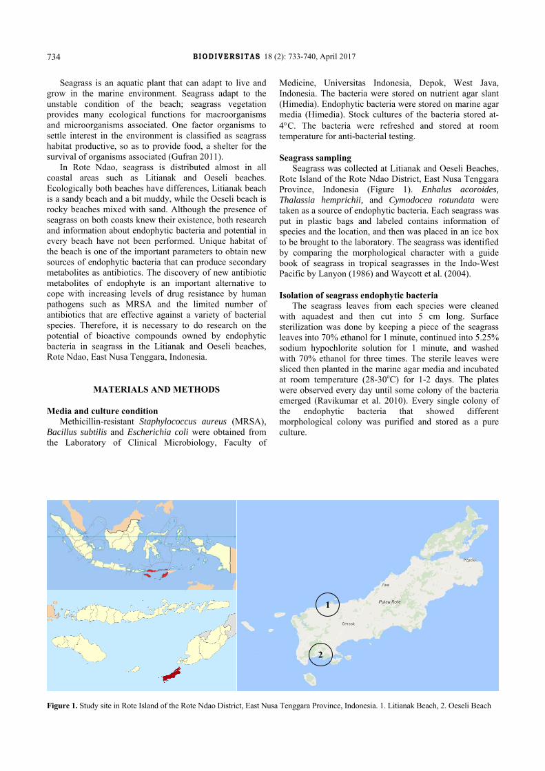
shows minimum clear zone is 6.67 mm, and the maximum is 15.17 mm. Besides, Susilowati et al. (2015) find that B. subtilis strain, bacterial symbionts of brown algae Sargassum sp., shows clear zone diameter 3.9 mm to MRSA. Virgibacillus salarius and Virgibacillus marismortui strain, bacterial symbionts of Sinularia sp. also have activity against MRSA, and the width of a clear zone are 1.9 mm and 2.8 mm. However, both of bacteria do not have antibacterial activity against E. coli (Sulistiyani et al. 2010). Based on that matter, it is known that potential endophytic bacteria which found in this research have a bigger and wider antibacterial ability not only to MRSA but also Gram-positive bacteria and Gram-negative bacteria if it is compared with the other research. The kind of antagonistic bacteria used against endophytic bacteria affects the forming of the clear zone. It is found that there are one isolated bacteria which forms clear area to MRSA only, two isolated bacteria to B. subtilis only, six isolated bacteria to both MRSA (Figure 1) and B. subtilis, and one isolated bacteria forms clear zone to MRSA, B. subtilis, and E. coli. Those results indicated that antibacterial produced by the bacteria shows narrow spectrum which is only active against Gram-positive bacteria and broad spectrum which is active against not only Gram-positive but also Gram-negative bacteria. Similar research undertaken by Ravikumar et al. (2010) shows five isolated endophytic bacteria in seagrass Syringodium isoetifolium and Cymodocea serrulata have antibacterial with broad spectrum against bacteria that are S. aureus, Klebsiella sp., Streptococcus pneumoniae, Streptococcus aeruginosa, and Pseudomonas aeruginosa. Sulistiyani et al. (2015) also finds 1 of 9 isolated symbionts bacteria in seagrass Enhalus sp. that belongs to Bacillus sp., has antibacterial activity against bacteria of Multi Drugs Resistant Tuberculosis (MDR-TB) came from their bioactive crude extracts.
Figure 1. Clear zone showing antibacterial activity formed by seagrass endophytic bacteria (isolate E2M1, left and E2M4, right) against MRSA bacteria in overlay method, incubation overnight at 37 C
FITRI et al. – Endophytic bacteria producing antibacterial against MRSA
737
Antibacterial produced by the isolate of endophytic
bacteria in seagrass in this research are suspected having different mechanism action regarding the activity against resistant bacteria such as MRSA, Gram-positive and Gram-negative bacteria. However, the mechanism involved in the activity against the bacteria as synergy from several bioactive compounds or one of the compounds is not determined yet because the bioactive compound extraction does not undertake yet to find out bioactive compound contained in antibacterial that produced by the bacteria. The previous research conducted by Darabpour et al. (2012) shows that bacteria Peseudoalteromonas piscicida strain PG-01 isolated from Persian Gulf waters had an antibacterial activity against MRSA. The antibacterial is bactericidal with a target to lyse cell wall of MRSA; it is known from TEM (Transmission Electron Micrographs) study on a dead MRSA bacteria. Moreover, Ling et al. (2015) have found new antibiotics named teixobactin from uncultured bacteria by using in situ method, and it can be used to overcome MRSA infection and Mycobacterium tuberculosis without creating resistance. Based on the 16S rRNA gene, the microorganism is Eleftheria terrae which belong to β-proteobacteria class. The mechanism of teixobactin regarding the activity against resistant bacteria is by obstructing cell wall synthetic by binding to lipid II (peptidoglycan precursor) and Lipid III (cell wall of the teichoic acid precursor) of the bacteria.
Identity of endophyte bacteria based on 16SrRNA encoding gene
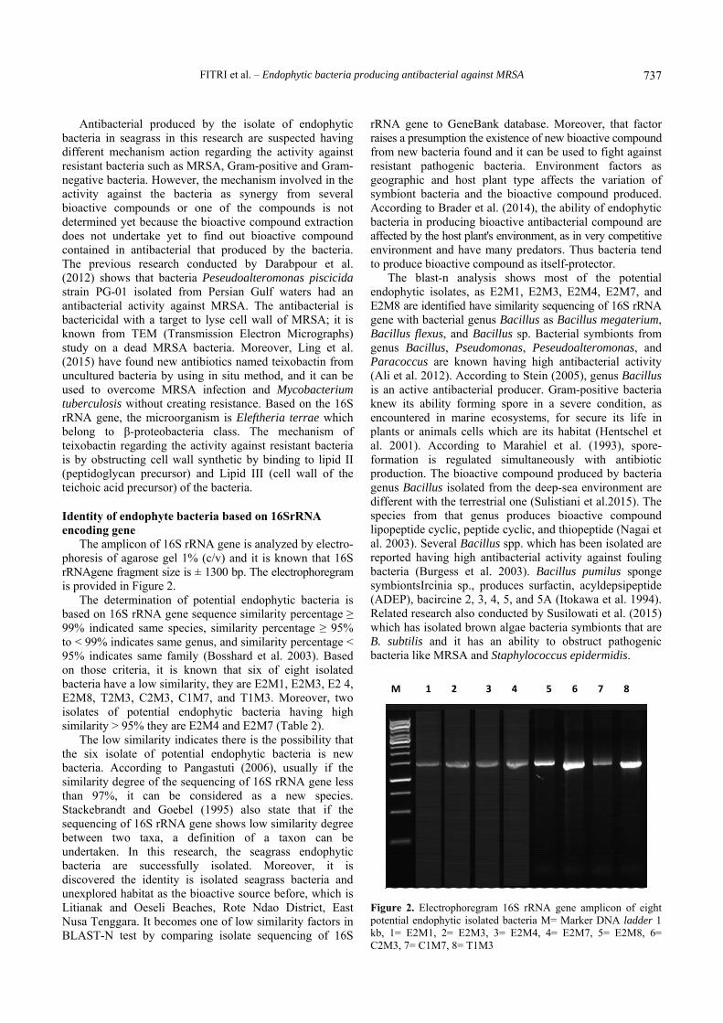
The amplicon of 16S rRNA gene is analyzed by electro-phoresis of agarose gel 1% (c/v) and it is known that 16S rRNAgene fragment size is ± 1300 bp. The electrophoregram is provided in Figure 2.
The determination of potential endophytic bacteria is based on 16S rRNA gene sequence similarity percentage ≥ 99% indicated same species, similarity percentage ≥ 95% to < 99% indicates same genus, and similarity percentage < 95% indicates same family (Bosshard et al. 2003). Based on those criteria, it is known that six of eight isolated bacteria have a low similarity, they are E2M1, E2M3, E2 4, E2M8, T2M3, C2M3, C1M7, and T1M3. Moreover, two isolates of potential endophytic bacteria having high similarity > 95% they are E2M4 and E2M7 (Table 2).
The low similarity indicates there is the possibility that the six isolate of potential endophytic bacteria is new bacteria. According to Pangastuti (2006), usually if the similarity degree of the sequencing of 16S rRNA gene less than 97%, it can be considered as a new species. Stackebrandt and Goebel (1995) also state that if the sequencing of 16S rRNA gene shows low similarity degree between two taxa, a definition of a taxon can be undertaken. In this research, the seagrass endophytic bacteria are successfully isolated. Moreover, it is discovered the identity is isolated seagrass bacteria and unexplored habitat as the bioactive source before, which is Litianak and Oeseli Beaches, Rote Ndao District, East Nusa Tenggara. It becomes one of low similarity factors in BLAST-N test by comparing isolate sequencing of 16S
rRNA gene to GeneBank database. Moreover, that factor raises a presumption the existence of new bioactive compound from new bacteria found and it can be used to fight against resistant pathogenic bacteria. Environment factors as geographic and host plant type affects the variation of symbiont bacteria and the bioactive compound produced. According to Brader et al. (2014), the ability of endophytic bacteria in producing bioactive antibacterial compound are affected by the host plant's environment, as in very competitive environment and have many predators. Thus bacteria tend to produce bioactive compound as itself-protector.
The blast-n analysis shows most of the potential endophytic isolates, as E2M1, E2M3, E2M4, E2M7, and E2M8 are identified have similarity sequencing of 16S rRNA gene with bacterial genus Bacillus as Bacillus megaterium, Bacillus flexus, and Bacillus sp. Bacterial symbionts from genus Bacillus, Pseudomonas, Peseudoalteromonas, and Paracoccus are known having high antibacterial activity (Ali et al. 2012). According to Stein (2005), genus Bacillus is an active antibacterial producer. Gram-positive bacteria knew its ability forming spore in a severe condition, as encountered in marine ecosystems, for secure its life in plants or animals cells which are its habitat (Hentschel et al. 2001). According to Marahiel et al. (1993), spore-formation is regulated simultaneously with antibiotic production. The bioactive compound produced by bacteria genus Bacillus isolated from the deep-sea environment are different with the terrestrial one (Sulistiani et al.2015). The species from that genus produces bioactive compound lipopeptide cyclic, peptide cyclic, and thiopeptide (Nagai et al. 2003). Several Bacillus spp. which has been isolated are reported having high antibacterial activity against fouling bacteria (Burgess et al. 2003). Bacillus pumilus sponge symbiontsIrcinia sp., produces surfactin, acyldepsipeptide (ADEP), bacircine 2, 3, 4, 5, and 5A (Itokawa et al. 1994). Related research also conducted by Susilowati et al. (2015) which has isolated brown algae bacteria symbionts that are B. subtilis and it has an ability to obstruct pathogenic bacteria like MRSA and Staphylococcus epidermidis.
Figure 2. Electrophoregram 16S rRNA gene amplicon of eight potential endophytic isolated bacteria M= Marker DNA ladder 1 kb, 1= E2M1, 2= E2M3, 3= E2M4, 4= E2M7, 5= E2M8, 6= C2M3, 7= C1M7, 8= T1M3
M 1 2 3 4 5 6 7 8
B IODIVERSITAS 18 (2): 733-740, April 2017
738
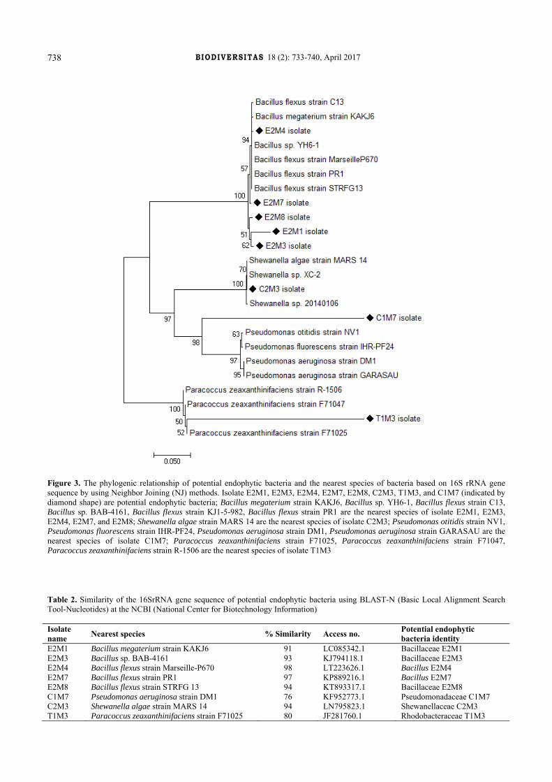
Figure 3. The phylogenic relationship of potential endophytic bacteria and the nearest species of bacteria based on 16S rRNA gene sequence by using Neighbor Joining (NJ) methods. Isolate E2M1, E2M3, E2M4, E2M7, E2M8, C2M3, T1M3, and C1M7 (indicated by diamond shape) are potential endophytic bacteria; Bacillus megaterium strain KAKJ6, Bacillus sp. YH6-1, Bacillus flexus strain C13, Bacillus sp. BAB-4161, Bacillus flexus strain KJ1-5-982, Bacillus flexus strain PR1 are the nearest species of isolate E2M1, E2M3, E2M4, E2M7, and E2M8; Shewanella algae strain MARS 14 are the nearest species of isolate C2M3; Pseudomonas otitidis strain NV1, Pseudomonas fluorescens strain IHR-PF24, Pseudomonas aeruginosa strain DM1, Pseudomonas aeruginosa strain GARASAU are the nearest species of isolate C1M7; Paracoccus zeaxanthinifaciens strain F71025, Paracoccus zeaxanthinifaciens strain F71047, Paracoccus zeaxanthinifaciens strain R-1506 are the nearest species of isolate T1M3 Table 2. Similarity of the 16SrRNA gene sequence of potential endophytic bacteria using BLAST-N (Basic Local Alignment Search Tool-Nucleotides) at the NCBI (National Center for Biotechnology Information) Isolate name Nearest species % Similarity Access no. Potential endophytic
bacteria identity E2M1 Bacillus megaterium strain KAKJ6 91 LC085342.1 Bacillaceae E2M1 E2M3 Bacillus sp. BAB-4161 93 KJ794118.1 Bacillaceae E2M3 E2M4 Bacillus flexus strain Marseille-P670 98 LT223626.1 Bacillus E2M4 E2M7 Bacillus flexus strain PR1 97 KP889216.1 Bacillus E2M7 E2M8 Bacillus flexus strain STRFG 13 94 KT893317.1 Bacillaceae E2M8 C1M7 Pseudomonas aeruginosa strain DM1 76 KF952773.1 Pseudomonadaceae C1M7 C2M3 Shewanella algae strain MARS 14 94 LN795823.1 Shewanellaceae C2M3 T1M3 Paracoccus zeaxanthinifaciens strain F71025 80 JF281760.1 Rhodobacteraceae T1M3
FITRI et al. – Endophytic bacteria producing antibacterial against MRSA
739
Moreover, C1M7 isolate has a similarity with bacteria family Pseudomonadaceae. Pseudomonas belongs to Gram-negative bacteria, non-fermentative, was found live in terrestrial and also on the deep-sea environment. Pseudomonas from deep-sea produces bioactive such as pyrroles, pseudopeptide, pyrrolidinedione, phthalate, andrimid, moiramides, zafrin, and bushrin, and also several bioactive components as antibacterial agents (Isnansetyo and Kamel 2009). Some species of that genus have been reported have antibacterial activity both against resistant bacteria and the irresistant one such as Pseudomonas bromoutilis (Burkholder et al. 1966), P. aeruginosa (Darabpour et al. 2010), and Pseudomonas fluorescens (Needham et al. 1994).
C2M3 has similarity with Shewanella algae. Nowadays, Shewanellaceae only has one genus that is Shewanella (MacDonell and Colwell 1985). S. algae is an anaerobe facultative bacteria which mostly has stem shape, Gram-negative, with oxidase-positive, and it is usually found in the deep-sea environment all over the world. This group of bacteria can be found in various sources such as active mud, sea invertebrate, red algae, seawater, sediment, and clinical sample (Lee et al. 2006). S. algae which are isolated from Callyspongia diffusa sponge has a high antibacterial activity to human and fish pathogenic bacteria as E. coli, E. cloacae, P. vulgaris, Salmonella typhi, and Vibrio sp. (Rachanamol et al. 2014).
However, T1M3 isolate has similarity with Paracoccus zeaxanthinifaciens. Paracoccus genus is Gram-negative bacteria short basil shaped, oxidase-positive and catalase-positive. Genus Paracoccus is from Rhodobacteraceae family and consists of 30 species. Members of genus Paracoccus can be isolated from soil, active mud, plant root, animal, and sea plants (Chen et al. 2011). The members of Paracoccus produce a beneficial metabolism for human as carotenoid zeaxanthin which is produced by P. zeaxanthinifaciens, and it has a significant role in preventing macular degeneration regarding aging which is the primary cause of blindness (Sajilata et al. 2008).
Phylogenetic relationship based on 16S rRNA gene sequence
The phylogenic relationship of potential endophytic bacteria of seagrass in this research and nearest species depicted in Figure 3. The construction of phylogenetic tree or known as dendrogram conducted using Molecular Evolutionary Genetics Analysis (MEGA) program version 7.0 (Kumar et al. 2015) by using Neighbor Joining (NJ) methods (Saitou et al. 1987).
According to a phylogenic tree, isolated bacterial E2M1, E2M3, E2M4, E2M7, and E2M8 have a very close phylogenetic relationship since clustering in one group. Those six of the endophytic bacteria were isolated together from the same seagrass E. acoroides which came from Oeseli Beach, and it belongs to genus Bacillus, the Bacillaceae family. Those six potential isolated endophytic bacteria have the furthest phylogenetic relationship with isolate T1M3 which is indicated as Rhodobacteraceae family. T1M3 is isolated from different species of seagrass and different growing area that is T. hemprichii from
Litianak Beach. Isolate C2M3 which belongs to Shewanellaceae family has near phylogenetic relationship and belongs to the same group with isolated C1M7 which belongs to Rhodobacteraceae even though each isolate are isolated from the different growing area and different species of seagrass. It is because the three group of the isolates belong to Gram-negative bacteria.
REFERENCES
Ali AIB, Bour ME, Ktari L, Bolhuis H, Ahmed M, Boudabbous A, Stal LJ. 2012. Jania Rubens associated bacteria: Molecular identification and antimicrobial activity. J Appl Phycol 24: 525-534.
Bosshard PP, Abels S, Zbinden R, Bottger ECB, Altwegg M. 2003. Ribosomal DNA Sequencing for Identification of Aerobic Gram-Positive Rods in the Clinical Laboratory (an 18 Month Evaluation). J Clin Microbiol 41: 4134-4140.
Brader G, Compant S, Mitter B, Trognitz F, Sessitsch A. 2014. Metabolic potential of endophytic bacteria. Curr Op Biotechnol 27: 30-37.
Burkholder PR, Pfister RM, Leitz FH. 1966. Production of a pyrrole antibiotic by a marine bacterium. Appl Microbiol 14: 649-653.
Chamber HF. 2001. The changing epidemiology of Staphylococcus aureus? Emerg Infect Dis 7: 178-182.
Chen MH, Sheu SY, Chen CA, Wang JT, Chen WM. 2011. Paracoccus isoporae sp. Nov., Isolated from the Reef Building Coral Isopora palifera. Intl J Syst Evol Microbiol 61: 1138-1143.
Clardy J, Fischbach MA, Walsh CT. 2006. New antibiotics from bacterial natural products. Nat Biotechnol 24: 1541-1550.
Darabpour E, Ardakani MR, Motamedi H, Ronagh MT. 2012. Isolation of a potent antibiotic producer bacterium, especially against MRSA, from Northern Region of the Persian Gulf. Bosnian J Basic Med Sci 12: 108-121.
Darabpour E, Motamedi H and Seyyed Nejad SM 2010. Antimicrobial properties of Teucrium polium against some clinical pathogens. Asian Pac J Tropic Med 3: 124-127.
Gufran HM, Kordi K. 2011. Seagrass Ecosystem. Jakarta, Rineka Cipta. [Indonesian]
Handayani D, Sandrawaty N, Murniati M, Regina R. 2015. Screening of Endophytic Bacteria Isolated from Marine Sponge Haliclona fascigera for Inhibition against Clinical Isolates of Methicillin-resistant Staphylococcus aureus (MRSA). J Appl Pharmaceut Sci 5: 139-142.
Hentschel U, Schmid M, Wagner M, Fieseler L, Gernert C, Hacker J. 2001. Isolation and phylogenetic analysis of bacteria with antimicrobial activities from the Mediterranean sponges Aplysina cavernicola and Aplysina aerophoba. FEMS Microbiol 35: 305-312.
Isnansetyo A, Kamei Y. 2009. Bioactive Substances Produced by Marine Isolates of Pseudomonas. J Ind Microbiol Biotechnol 36: 1239-1248.
Itokawa HT, Miyashita H, Morita K, Takeya T, Hirano M, Oka HK. 1994. Structural and conformational studies of [Ile] and [Leu] surfactins from Bacillus subtilis natto. Chem Pharm Bull 42: 604-607.
Jeganathan P, Rajasekaran KM, Devi ANK, Karuppusamy S. 2013. Antimicrobial activity and characterization of marine bacteria. Indian J Pharm Biol Res 1: 38-44.
Kang HK, Seo CH, Park Y. 2015. Marine peptides and their anti-infective activities. Mar Drugs 13: 618-654.
Karchmer AW. 2006. From theory to practice: resistance in Staphylococcus aureus and new treatments. Clin Microbiol Infect 12: 15-21.
Kumar S, Stecher G, Tamura K. 2015. MEGA7: Molecular Evolutionary Genetics Analysis Version 7.0 for Bigger Datasets. Mol Bio Evol DOI: 10.1093/molbev/msw054.
Lanyon J. 1986. Guide to the identification of seagrasses in the Great Barrier Reef region. Great Barrier Reef Marine Park Authority Special Publication Series (3). Nadicprint Services, Townsville, Australia.
Lee OO, Lau SCK, Tsoi MMY, Li X, Plakhotnikova I, Dobretsov S, Wu MCS, Wong PK, Weinbauer M, Qian PY. 2006. Shewanellairciniae sp. nov., a novel member of the family Shewanellaceae, isolated from the marine sponge Ircinia dendroides in the Bay of Villefranche, Mediterranean Sea. Intl J Syst Evol Microbiol 56: 2871-2877.
B IODIVERSITAS 18 (2): 733-740, April 2017
740
Ling LL, Schneider T, Peoples AJ, Spoering AL, Engels I, Onlon BP, Mueller A, Schäberle TF, Hughes DE, Epstein S, Jones M, Lazarides L, Steadman VA, Cohen DR, Felix CR, Etterman KA, Millett WP, Nitti AG, Zullo AM, Chen C, Lewis K. 2015. A new antibiotic kills pathogens without detectable resistance. Nature 517: 455-459.
Lu X, Gao X, Liu X, Jiao B. 2010. Marine microbes derived anti-bacterial agents-Mini review. Med Chem 10: 1077-1090.
MacDonell MT, Colwell RR. 1985. Phylogeny of the Vibrionaceae, and recommendation for two new genera, Listonella, and Shewanella. Syst Appl Microbiol 6: 171-182.
Marahiel M.A., Nakano M.M., Zuber P. 1993. Regulation of peptide antibiotic production in Bacillus. Mol Microbiol 7: 631-636.
Marchesi JR, Sato T, Weightman AJ, Martin TA, Fry JC, Hiom SJ, Wade WG. 1998. Design and evaluation of useful bacterium specific PCR primers that amplify genes coding for bacterial 16S rRNA. Appl Environ Microbiol 64: 795-799.
Nagai K, Kamigiri K, Arao N, Suzumura K, Kawano Y, Yamaoka M, Zhang H, Watanabe M, Suzuki K. 2003. YM-266183 and YM-266184, Novel thiopeptide antibiotics produced by Bacillus cereus isolated from marine sponge. J Antibiotics 56: 123-128.
Needham J, Kelly MT, Ishige M, Andersen RJ. 1994. Andrimid and moiramides A-C, metabolites produced in culture by a marine isolate of the bacterium Pseudomonas fluorescence structure elucidation and biosynthesis. J Org Chem 59: 2058-2063.
Pangastuti A. 2006. Species definition of prokaryotes based on 16S rRNA and protein-coding genes sequence. Biodiversitas 7: 292-296.
Paterson GK, Harrison EM, Holmes MA. 2014. The emergence of mecC methicillin-resistant Staphylococcus aureus. Trends Microbiol 22: 42-47.
Penesyan A, Kjelleberg S, Egan S. 2010. Development of novel drugs from marine surface associated microorganisms. Mar Drugs. 2010;8:438–459.
Penesyan A, Tebben J, Lee M, Thomas T, Kjelleberg S, Harder T, Egan S. 2011. Identification of the antibacterial compound produced by the marine epiphytic bacterium Pseudovibrio sp. D323 and related sponge-associated bacteria. Mar Drugs 9 (8): 1391-1402.
Rachanamol RS, Lipton AP, Thankamani V, Sarika AR, Selvin J. 2014. Molecular characterization and bioactivity profile of the tropical sponge-associated bacterium Shewanella algae VCDB. Helgol Mar Res 68: 263-269.
Rahman H, Austin B, Mitchell WJ, Morris PC, Jamieson AJ, Adams DR, Spragg AM, Schweizer M. 2010. Novel anti-infective compounds from marine bacteria. Mar Drugs 8: 498-518.
Ravikumar S, Thajuddin N, Suganthi P, Inbaneson SJ, Vinodkumar T. 2010. Bioactive potential of seagrass bacteria against human bacterial pathogens. J Environ Biol 31: 387-389.
Saitou N, Nei M. 1987. The Neighbor-joining method: A new method for reconstructing phylogenetic trees. Mol Biol Evol 4: 406-425.
Sajilata MG, Singhal RS, Kamat MY. 2008. The carotenoid pigment zeaxanthin-A review. Compr Rev Food Sci Food Saf 7: 28-49.
Schulz B, Boyle C. 2006. What are endophytes? Microbial Root Endophytes 9: 1-13.
Stackebrandt E, Goebel BM. 1995. A Place for DNA-DNA reassociation and 16S rRNA sequence analysis in the present species definition in bacteriology. Intl J Syst Bacteriol 44: 846-849.
Stein T. 2005. Bacillus subtilis antibiotics: Structures, syntheses, and specific functions. Mol Microbiol 56: 845-857.
Sulistiyani, Nugraheni AA, Khoeri MM, Sabdono A, Karna O, Radjasa. 2010.Antibacterial activity of bacterial symbionts of soft coral Sinularia sp. against pathogenic resistant bacteria. J Coastal 13: 113-118.
Sulistiyani, Wahjono H, Radjasa OK, Sabdono A, Khoeri MM, Karyana E. 2015. Antimycobacterial activities from seagrass Enhalus sp. associated baterai against Multi-Drug Resistance Tuberculosis Bacteria (MDR-TB). Procedia Environ Sci 23: 253-259.
Susilowati R, Sabdono A, Widowati I. 2015. Isolation and characterization of bacteria associated with brown algae Sargassum spp. from Panjang Island and their antibacterial activities. Procedia Environ Sci 23: 240-246.
Waycott M, McMahon K, Mellors J, Calladine A and Kleine D. 2004. A Guide to Tropical Seagrasses of the Indo-West Pacific. James Cook University, Townsville, Australia.


![An Endophytic Nodulisporium sp. Producing Volatile Organic ... · More recently, a novel endophytic fungus has been described and named Muscodor sp. [5]. To date, all members of this](https://img.dokumen.tips/doc/110x75/5e230c37437eab286d62d3af/an-endophytic-nodulisporium-sp-producing-volatile-organic-more-recently-a.jpg)


![Identification of Taxol-producing endophytic fungi ... · 8 BAPT-R 5-TCGCCATCTCTGCCATACTT-3 [25] Table 2 Fungal endophytes isolated from Salacia oblonga. Sample code Species Sample](https://img.dokumen.tips/doc/110x75/5f54415ded367c34ba072297/identification-of-taxol-producing-endophytic-fungi-8-bapt-r-5-tcgccatctctgccatactt-3.jpg)


![Genome sequencing and analysis of the paclitaxel-producing ......Taxus plant species [12-14]. Low productivity of paclitaxel in endophytic fungi prevents these organisms from being](https://img.dokumen.tips/doc/110x75/611d68fcf38d7737a3559f70/genome-sequencing-and-analysis-of-the-paclitaxel-producing-taxus-plant-species.jpg)