Embed Size (px)
Citation preview

ENDOCRINE PHYSIOLOGY
Aside: Overview of Endocrine Physiology Endocrine system:
- Defined as a collection of glands scattered throughout body that secrete hormones - These glands can include:
o (i) Endocrine organ comprised of specialised endocrine cells (Eg. thyroid gland) o (ii) Cluster of endocrine cells within an organ (Eg. Islets of Langerhans) o (iii) Individual endocrine cells scattered diffusely throughout an organ (Eg. RA –
ANP) - There are two types of gland systems:
o (i) Hypothalamic-pituitary-gland system (Eg. T3/T4, cortisol, androgens) o (ii) Free-standing endocrine gland system (Eg. insulin, RAAS, PTH)
Hormones:
- Chemical messenger that are synthesised and secreted by specific endocrine cell (≈ ductless glands) into the circulation in trace amounts
- Implicated in cell-to-cell communication → act on a specific target cell (generally distant from endocrine cell) via receptor binding → initiates specific IC pathways and signal amplification of IC processes → alters cellular activity
Types of hormones:
- (1) Peptides o Constitute MOST hormones (Eg. glucagon, insulin, PTH, ACTH, LH/FSH,
ADH, oxytocin, hypothalamic RFs) o Synthesised like any protein (transcription to mRNA in the nucleus, translated in
ER into polypeptide, and concentrated in the Golgi apparatus), and are stored in secretory granules (small peptides bind to specific binding proteins; large proteins are by themselves), where they are released via Ca2+-dependent exocytosis
o Medium-large size (3-200 a.a.) H2O-soluble hormones → act via cell-surface receptors → either: (a) GPRC:
(i) Gs → activate AC to ↑ IC [cAMP] → activates PKA → phosphorylates IC proteins to mediate cellular activity
(ii) Gi → opposite of Gs (inhibits AC) (iii) Gq → activate PLC to cleave PIP2 from cell membrane →
forms IP3 (cause Ca2+ release from ER/mitochondria) and DAG (activates DAG) → mediates cellular activity
(b) TK receptor – Paired glycoprotein receptor with kinase activity → with receptor binding, it ∆ conformation and phosphorylates itself at Tyr-molecules → then phosphorylates other proteins to exert cellular activity
- (2) Amines o Derived from either (i) Tyr (T3/T4, DA, NAd, Adr) or (ii) Trp (5-HT, melatonin)
→ synthesised within cytoplasm and stored in storage granules (attached to specific binding proteins (Eg. catecholamines) or as part of thyroglobulin within colloid follicle) → secreted via exocytosis
o Small H2O-soluble hormones → can act via:
Aside – Intercellular communication can occur:- (1) Directly → via gap junctions - (2) Indirectly → via intercellular chemical messengers → include:
o (a) Hormones (see above) o (b) Neurotransmitters (Eg. ACh) – Chemical messengers released from neurons →
act on adjacent target cells (neurons, muscle, glandular cells) o (c) Autocoids (Eg. PG) – Chemical messengers that act locally to site of release → (i)
Paracrine (acts on surrounding cells) or (ii) Autocrine (acts on own cell)
(i) Cell-surface receptors – Catecholamines via GPCR (α → Gq; β → Gs) (ii) IC receptors – T3/T4 bind nuclear receptors that ∆ gene transcription
- (3) Steroids o Synthesised from cholesterol and released immediately (Ie. not stored) o Small fat-soluble hormones (GC, MC, androgens, sex hormones, vitamin D) →
enter cells and bind cytoplasmic receptors → forms steroid-receptor complex which enters nucleus → stimulates gene transcription to alter protein synthesis
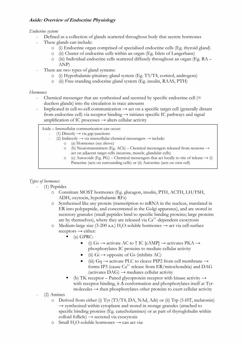
Regulation of hormones:
- Hormones are generally regulated via –ve feedback mechanism via → (i) Long loop feedback, (ii) Short loop feedback, or (iii) Ultra short loop feedback
- Regulation of hormone receptors: o “Down-regulation” – ↓ responsiveness of target tissue due to ↓ active receptor
numbers (caused by ↓ production or inactivation of part of receptor) → causes ↓ hormone effect
o “Upregulation” – ↑ responsiveness of target tissue due to ↑ active receptor numbers (caused by ↑ synthesis or activation of receptor molecules) → causes ↑ hormone effect
(a) To describe the actions of pancreatic hormones and the control of their secretion. (I) Pancreas: Islets of Langerhans
- “Islets of Langerhans” forms 1-2% of pancreatic mass (as the “endocrine” portion) → consists of scattered spherical clusters containing 4 types of APUD cells:
o (i) α cells (20% of islet cells) → secrete glucagon o (ii) β cells (70% of islet cells) → secrete insulin o (iii) δ cells (5% of islet cells) → secrete somatostatin o (iv) F cells (5% of islet cells) → secrete pancreatic polypeptide
- These hormones are secreted into → portal venous system (II) Insulin: Structure of insulin:
- 51 a.a. polypeptide hormone (MWT 5000 Da) → consists of 2x polypeptide chains (21 a.a. A-chain and 30 a.a. B-chain) linked by a disulphide bridge
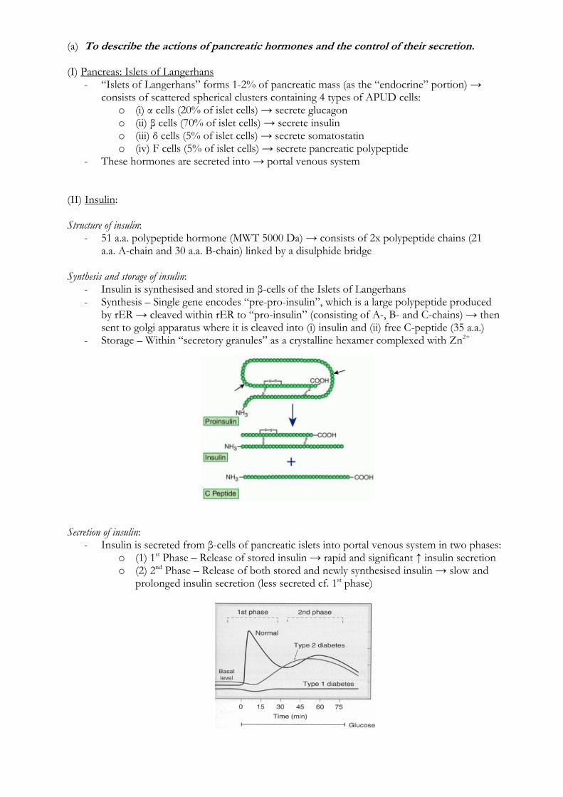
Synthesis and storage of insulin:
- Insulin is synthesised and stored in β-cells of the Islets of Langerhans - Synthesis – Single gene encodes “pre-pro-insulin”, which is a large polypeptide produced
by rER → cleaved within rER to “pro-insulin” (consisting of A-, B- and C-chains) → then sent to golgi apparatus where it is cleaved into (i) insulin and (ii) free C-peptide (35 a.a.)
- Storage – Within “secretory granules” as a crystalline hexamer complexed with Zn2+ Secretion of insulin:
- Insulin is secreted from β-cells of pancreatic islets into portal venous system in two phases: o (1) 1st Phase – Release of stored insulin → rapid and significant ↑ insulin secretion o (2) 2nd Phase – Release of both stored and newly synthesised insulin → slow and
prolonged insulin secretion (less secreted cf. 1st phase)
- It is secreted via Ca2+-induced exocytosis → extracellular Ca2+ enters β-cells → binds to calmodulin → causes exocytosis of insulin from secretory granules
- Control of secretion: o (1) Metabolic substrates:
(a) Glucose (most VITAL stimulus) → ↑ release when BGL > 5 mmol/L
(b) Amino acids → ↑ release with ↑ plasma [a.a.] (c) FAs and ketone bodies → ↑ release with ↑ plasma [FA] or [KB]
o (2) Hormonal factors: (a) Pancreatic hormones
(i) Glucagon → ↑ release (ii) Somatostatin → ↓ release
(b) GI hormones (GIP, gastrin, secretin, CCK) → ↑ release (c) Pituitary hormones (GH, ACTH, TSH) → ↑ release
o (3) Neural factors (via unmyelinated postganglionic SNS and PNS fibres) mAChR → ACh ↑ release (Nb. with food intake → ↑ vagus activity!) α2-receptor → NAd ↓ release
Metabolism of insulin:
- Insulin has a plasma t ½ 5 mins → 80% enzymatic degradation (into separate A and B chains) in liver and kidneys
Functions of insulin:
- Cellular mechanism of action: o Insulin receptor is a large transmembrane heterodimer glycoprotein complex →
consists of 2x α-subunits and 2x β-subunits joined by disulphide bridges: α-subunit → entirely extracellular → binds insulin β-subunit → within cell membrane → has tyrosine kinase activity
o Process of receptor activation: Insulin binds to α-subunit → activates tyrosine kinase in β-subunit β-subunit then phosphorylates (i) other β-subunit (autophosphorylation),
and (ii) other membrane-bound proteins → activates them Through an IC cascade, these activated proteins then phosphorylate and
activate several IC protein kinases and phosphatases → either (i) mediate IC enzyme activity (Ie. recruit GLUT4 to membrane), or (ii) regulate transcription of DNA → alter protein synthesis (Ie. ↑ glycogen synthase)
o Process of receptor inactivation: β-subunit phosphorylation causes ↓ receptor affinity for insulin, aggregation
of occupied receptors into clusters, and then internalisation into vesicles Occupied receptors are processed in lysosomes → free receptor recycled
back to cell membrane - Effects of insulin:
Important to note: Glucose as a trigger for insulin release - β-cells have ATP-sensitive K+ channels → maintain RMP of β-
cells by allowing continued K+ efflux - With ↑ BGL → glucose enters β-cells via GLUT-2 facilitated
transport (insulin-independent) → glucose is then metabolised to produce ATP
- ↑ IC ATP inhibits the ATP-sensitive K+ channels → causes β-cell depolarisation by inhibiting K+ efflux → opens VG-Ca2+ channels and permits influx of Ca2+ → causes insulin release via Ca2+-induced exocytosis
Affects insulin-sensitive tissues (1°ly liver, muscle and adipose) → exerts an anabolic effect (↑ uptake and utilisation of metabolic substrates, and ↓ breakdown of their tissue stores)
CHO metabolism Uptake of glucose intracellularly → cause ↓ BGL as follows: - (i) Liver – ↑ glucose uptake 2° to ∆ IC glucose metabolism (cf. ∆ activity
of membrane GLUT4) → keeps IC [glucose] low (via processes below) to maintain a [ ] gradient for glucose to be taken up by GLUT4
o (i) ↑ glucokinase activity → converts glucose to G6P o (ii) ↑ PFK activity → ↑ oxidation of glucose via glycolysis o (iii) ↑ glycogen synthase activity (and ↓ glycogen phosphorylase
activity) → causes ↑ glycogen synthesis o (iv) Inhibits PEPCK activity → ↓ gluconeogenesis o (v) ↓ G6P phosphatase activity → prevents glucose release
- (ii) Muscle, adipose cells and other insulin-dependent cells o Upregulates GLUT4 to cell surface → ↑ facilitated diffusion of
glucose intracellularly o In muscle → ↑ glycogen synthesis and glycolysis o In adipose → ↑ glycolysis and TAG synthesis (see below)
Protein metabolism ↑ tissue protein stores due to: - (i) ↑ a.a. uptake and protein synthesis (esp liver, muscle and adipose) - (ii) ↓ protein catabolism and oxidation of a.a.
Fat metabolism ↑ tissue fat stores due to: - (i) ↑ glucose uptake into liver and adipocytes (via ↑ GLUT4 activity) →
this ↑ TAG synthesis (esp in liver) as glucose is converted to it substrates → (a) α-glycerophosphate, and (b) FA (via acetyl-CoA)
- (ii) ↑ lipoprotein lipase and LDL receptor activity → ↑ breakdown of LDL peripherally → ↑ uptake of FA/glycerol of TAG storage
- (iii) ↓ hormone-sensitive lipase activity → ↓ lipolysis peripherally - (iv) ↓ ketogenesis in liver
Cellular growth Mitogenic effect → stimulates cell proliferation (via effect on DNA transcription/protein synthesis) → vital for foetal development
Electrolytes ↑ cellular uptake of K+, Ca2+ and PO43- (2° to membrane hyperpolarisation caused by ↓ membrane permeability to Na+)
(II) Glucagon: Synthesis and storage of glucagon:
- Glucagon is a 29 a.a. polypeptide hormone synthesised and stored by α-cells of Islets of Langerhans
- It is produced from “Pre-pro-glucagon”, a large single polypeptide chain → then cleaved to pro-glucagon → then finally to glucagon → stored in storage vesicles
Secretion of glucagon:
- Glucagon secretion from α-cells is controlled by the following factors: o (1) Metabolic substrates
(a) Glucose (most VITAL stimulus) → ↑ release with ↓ BGL (b) Amino acids → ↑ release with ↑ plasma [a.a.] (c) FAs and KB → ↑ release with ↓ plasma [FA] or [KB]
Note – Glucose uptake by non-insulin dependent cells (see below) are INDIRECTLY affected 2° to ∆ BGL afforded by insulin-dependent cells
Important to note – Insulin-independent sites: - (1) Brain (EXCEPT for hypothalamus – satiety centre) - (2) Intestinal mucosa - (3) Renal tubules - (4) RBC - (5) Placenta
o (2) Hormonal factors (a) GIT hormones (CCK, gastrin, secretin) → ↑ release (b) Glucocorticoids → ↑ release (c) β-adrenoceptor stimulation → ↑ release (d) Somatostatin → ↓ release (e) Insulin → ↓ release
o (3) Others Infection, exercise, stress → ↑ release
Metabolism of glucaon:
- Glucagon has a short plasma t ½ 5-10 mins → metabolised by the liver Functions of glucagon:
- Cellular mechanism of action → via GPCR (Gs) - Effects → exerts catabolic effect on insulin-sensitive tissues (liver, muscle, adipose)
CHO metabolism ↑ BGL via – (i) ↑ hepatic glycogenolysis (↑ glycogen phosphorylase activity), and (ii) ↑ hepatic gluconeogenesis (↑ glucogenic a.a. and glycerol uptake by hepatocytes, ↑ PEPCK and pyruvate carboxylase activity)
Protein metabolism ↑ protein catabolism of tissue protein stores via – (i) ↑ a.a. oxidation (Ie. a.a. for GCN) and (ii) ↑ protein breakdown
Fat metabolism ↑ plasma FA and KB levels via – (i) ↑ lipolysis in adipocytes (↑ hormone-sensitive lipase activity) → liberates FFA/glycerol, (ii) ↑ β-oxidation of FFAs, (iii) ↑ hepatic ketogenesis (in absence of insulin)
Endocrine ↑ release of insulin, GH and somatostatin (III) Somatostatin and Pancreatic Polypeptide: Somatostatin:
- 14 a.a polypeptide that is produced from δ-cells ofIslets of Langerhans (BUT also from (i) Hypothalamus and (ii) Gastric D-cells)
- Stimuli for release – (i) ↑ plasma levels of glucose, a.a. and FA, and (ii) Acidic gastric environment
- Effects: o (i) ↓ secretion of pancreatic hormones (insulin, glucagon, pancreatic polypeptide) o (ii) ↓ GH release from pituitary o (iii) ↓ gastric, duodenal and GB motility o (iv) ↓ secretion and absorption in GIT (Eg. gastric acid secretion)
- It has a plasma t ½ of 3 minutes Pancreatic polypeptide:
- Polypeptide secreted by F-cells of Islets of Langerhans → influence GIT function (Ie. ↑ enzyme secretion, ↓ intestinal motility, Etc.)
(b) To explain the control of blood glucose levels. Overview of blood glucose levels (BGL):
- Normal BGL ~ 4-7 mmol/L in adults (can ↓ to 3-3.5 mmol/L with fasting) - It is tightly regulated despite variations in glucose intake → homeostatic mechanisms
prevent (i) hyperglycaemia post-prandially and (ii) hypoglycaemia with fasting - Basis for tight regulation:
o Hypoglycaemia → depresses function of organs that use glucose obligately and whose means of glucose uptake is dependent on adequate BSL (Ie. insulin-independent tissues) → esp CNS/brain
o Hyperglycaemia → (i) osmotic effect (cellular dehydration, osmotic diuresis with loss of H2O/electrolytes from body), and (ii) glycosuria (lose major metabolic substrate as waste)
Glucose body storage and utilisation:
- Glucose body stores: o (1) Glycogen (main store) → total 500g (2000 kcal energy) → 12-24 hr supply
(i) Liver glycogen (100 g) can be used to maintain BGL → has ability to take up and release glucose from circulation via balance of glycogenolysis vs glycogen synthesis (Nb. it possesses glucose-6-phosphatase)
(ii) Muscle glycogen (400 g) cannot be used to maintain BGL → lacks glucose-6-phosphatase (cf. liver) and cannot release glucose from its stores
o (2) Circulation → 10 g in ECF (5 g in BV) (40 kcal energy) → small supply, but circulatory store vital for transporting glucose to tissue sites of need
- Glucose utilisation in body: o ALL cells can utilises glucose (via glycolysis), while MOST cells can use FFAs (and
KBs) for metabolism – EXCEPT (i) RBC and (ii) CNS/brain → these are obligate glucose users
Control of BGL:
- BGL is dependent on the balance of: o (1) Glucose input into blood:
(i) Small bowel – Glucose is absorbed from small bowel SB → BGL fluctuates with dietary intake (↑ post-prandially; ↓ with fasting)
(ii) Liver – Glucostat function buffers ∆ in BGL via hormonal control (see below)
o (2) Glucose output from blood: (i) Utilisation by body cells – (a) Insulin-dependent cells (esp
muscle/adipose) → take up and utilise glucose under hormonal influence (esp insulin), (b) Non-insulin dependent cells → take up glucose as per local metabolic need
(ii) Urine – ↑ BGL causes glycosuria → limits further ↑ in BGL - Thus, BGL control is determined by factors that regulate the balance of glucose
input/output from blood → these are: o (1) Hormonal factors
Insulin → main regulatory hormone for ∆ in BGL during normal state Released when BGL > 5 mmol/L → causes (i) ↑ glucose uptake
into insulin-sensitive cells, (ii) ↑ glucose utilisation (esp glycolysis and FFA synthesis), (iii) ↑ liver/muscle glycogen synthesis, (v) ↓ liver gluconeogenesis
When BSL < 3 mmol/L (during fasting state), the following hormones are secreted to ↑ BGL:
(i) Glucagon → ↑ hepatic glycogenolysis and gluconeogenesis
(ii) Glucocorticoids → ↑ hepatic gluconeogenesis and ↓ peripheral uptake and utilisation of glucose (due to ↑ peripheral FFA utilisation instead)
(iii) GH → Anti-insulin effect via ↓ peripheral glucose uptake/utilisation
(iv) Catecholamines → ↑ hepatic glycogenolysis and ↓ peripheral glucose uptake/utilisation
(v) T3/T4 → ↑ GIT absorption of glucose, ↑ hepatic glycogenolysis and ↑ hepatic gluconeogenesis
o (2) Glucostat function of liver ↓ BGL → ↑ glucagon (and counter-regulatory hormones) and ↓ insulin →
stimulates hepatic glycogenolysis and gluconeogenesis (inhibits glycogen synthesis and glycolysis) → causes glucose release from liver
↑ BGL → opposite effects produced → so glucose is taken up by liver Control of BGL post-prandially and with fasting:
- (1) Post-prandial o ↑ BGL due to ↑ SB absorption of glucose → causes β cells of islets to release
insulin (causes ↓ α cell secretion of glucagon, ↓ GC, GH, T3/T4, catecholamines) o Effect:
(i) Liver – ↑ glucose uptake (due to ∆ IC metabolic processes – see above) → ↑ glycogen synthesis, ↑ glycolysis (→ FA synthesis), ↓ gluconeogenesis
(ii) Muscle/adipose – ↑ glucose uptake (due to ↑ GLUT4 transporter) → ↑ glycolysis, ↑ glycogen synthesis (muscle) and ↑ glycerol synthesis (in adipose → TAG synthesis with FFA)
- (2) Fasting o ↓ BGL due to ↑ glucose uptake/utilisation by cells → causes α cell secretion of
glucagon, ↑ release of GC, GH, T3/T4, catecholamines (also ↓ insulin release by β cells)
o Effect: (i) Liver – ↑ glycogenolysis, ↑ gluconeogenesis, ↑ glucose-6-phosphatase
(and ↓ glycolysis) → causes ↑ glucose release from liver (ii) Muscle/adipose – ↓ glucose uptake → ↓ glycogen and TAG synthesis,
↑ lipolysis and protein breakdown → releases substrates (a.a,, FFA, glycerol) for hepatic gluconeogenesis
Note: Insulin and glucagon are important hormones in controlling BGL → as they have opposing effects, net effect on BGL is determined by their relative levels of hormones in blood
- Fed state → I/G ratio = 30 - Overnight fast → I/G ratio = 2 - Prolonged fast → I/G ratio = 0.5
(c) To describe the role of the hypothalamus in the integration of neuro-hormonal responses.
Hypothalamus is an important neuro-hormonal regulation centre → it controls (i) autonomic functions, and (ii) hormonal output from the pituitary gland Control of ANS functions:
CVS function - Stimulation of anterior hypothalamus → ↓ BP and HR - Stimulation of posterior hypothalamus → ↑ BP and HR
Thermoregulation - ↑ blood temperature → stimulates anterior hypothalamus → causes sweating and cutaneous vasodilation - ↓ blood temperature → stimulates posterior hypothalamus → causes shivering and cutaneous vasoconstriction
Feeding - Stimulation of the lateral hypothalamic nucleus (“hunger centre”) → hunger - ↑ BGL → stimulates ventromedial nucleus (“satiety centre”) → ↓ feeding by inhibiting the “hunger centre”
Water balance - Paraventricular nucleus (“thirst centre”) → ↑ [ ] of electrolytes at PVN cells → thirst response - Osmoreceptors at supraoptic nucleus and PVN → ↑ plasma osmolality → triggers release of ADH
Defence reactions Regulates SNS activity → affects catecholamines release by the adrenal medulla Sexual behaviour Sensitive to sex hormones (oestrogen and androgens)
Control of hormonal output from the pituitary gland:
- (1) Direct neural control of posterior pituitary gland: o “Magnocellular neurons” (located in SON/PVN) → extend their axons to the
posterior lobe of pituitary gland → secrete ADH and Oxytocin into circulation o Control – ↑ ADH/oxytocin secretion with ACh; ↓ with NAd
- (2) Indirect control of anterior pituitary gland by releasing/inhibiting factors released into the “hypothalamic-hypophysial portal system”:
o “Parvocellular neurons” (located in cell bodies near median eminence) → forms tuberoinfundibular tract that secretes “hypophysiotrophic hormones” (see below) into hypothalamic-hypophysial portal system → affects anterior pituitary gland
o Controlled by NAd, DA, 5-HT
Aside: “Hypothalamic-hypophysial portal system”- Median eminence of hypothalamus secretes hypophysiotrophic hormones into the “Primary
capillary plexus” (formed by branches of superior hypophysial artery) → this then forms the lesser portal veins → gives rise to the “Secondary capillary plexus” in anterior lobe of pituitary gland (Nb. this supplies 90% of anterior pituitary)
- Note → Posterior lobe of pituitary gland is supplied by capillary plexus from inferior hypophysial artery
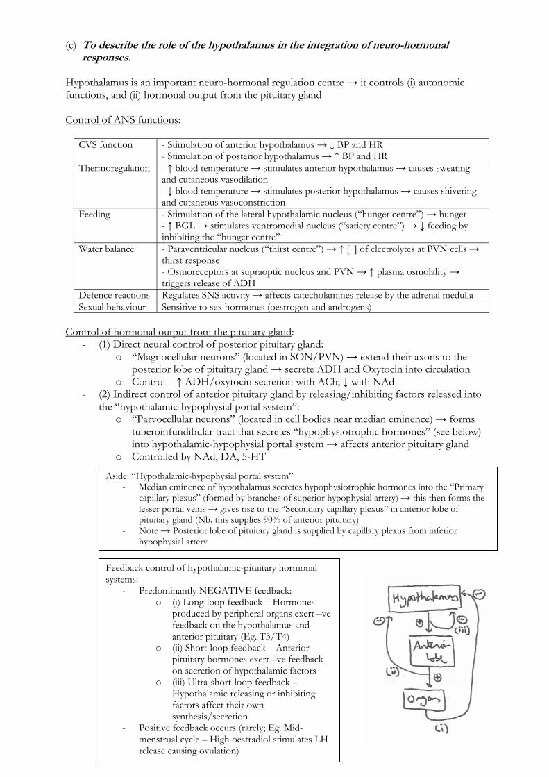
Feedback control of hypothalamic-pituitary hormonal systems:
- Predominantly NEGATIVE feedback: o (i) Long-loop feedback – Hormones
produced by peripheral organs exert –ve feedback on the hypothalamus and anterior pituitary (Eg. T3/T4)
o (ii) Short-loop feedback – Anterior pituitary hormones exert –ve feedback on secretion of hypothalamic factors
o (iii) Ultra-short-loop feedback – Hypothalamic releasing or inhibiting factors affect their own synthesis/secretion
- Positive feedback occurs (rarely; Eg. Mid-menstrual cycle – High oestradiol stimulates LH release causing ovulation)
(d) To describe the control of secretion and functions of pituitary hormones. (I) Anterior pituitary hormones: Overview of anterior pituitary gland:
- Anterior lobe of pituitary gland (aka. adenohyophysis) → forms 80% of pituitary gland → consists of 3 parts:
o (i) Pars tuberalis o (ii) Parts intermedius (intermediate lobe) o (iii) Pars distalis (anterior lobe) → produces most anterior pituitary hormones
- Derived from buccal ectoderm → it extends upwards from the epithelial lining of the primitive mouth cavity (as Rathke’s pouch), then fuses with the downward growing infundibulum of the hypothalamus (which forms the posterior pituitary)
- Consists of non-neural secretory epithelial cells connected INDIRECTLY to the hypothalamus via the “hypothalamic-hypophysial portal system”:
o (1) Non-neural secretory epithelium: (a) Granular secretory cells (chromophils)
(i) Acidophils (80%) → red-staining cells → synthesise and store large peptide hormones in secretory granules
o Lactotropes → PRL o Somatoropes → GH (somatotropins)
(ii) Basophils (20%) → blue-staining cells → synthesise and store glycoprotein hormones in secretory granules
o Gonadotropes → FSH and LH o Thyrotropes → TSH o Corticotropes → ACTH (and MSH)
(b) Agranular secretory cells (chromophobes) Weakly staining cells → inactive or degranulated secretory cells →
does not synthesise any hormones o (2) Hypothalamic-hypophysial portal system
Branches of superior hypophysial artery → forms “1° capillary plexus” near median eminence of hypothalamus → then forms lesser portal veins that gives rise to “2° capillary plexus” in anterior lobe of pituitary gland
Functions of portal venous system – (i) Blood supply of 90% of anterior pituitary (and infundibulum), (ii) Pathway for hypophysiotropic hormones (from median eminence of hypothalamus) to reach the anterior pituitary gland, and (iii) Allows hormones secreted by anterior pituitary gland to enter systemic circulation
- Blood supply → 90% supplied by hypothalamic-hypophysial portal system (from superior hypophysial artery)
Hypophysiotrophic hormones: Control of anterior pituitary gland
- Hypophysiotrophic hormones are small peptides (EXCEPT for DA) produced by cell bodies (containing “parvocellular neurons”) in the hypothalamus located near the median eminence → small amounts are secreted from the tuberoinfundibular tract into the hypothalamic-hypophysial portal system → reaches anterior pituitary where it exerts either an “excitatory” or “inhibitory” effect on non-neural secretory epithelial cells
- Nature of release from hypothalamus: o Pulsatile manner → can vary with circadian rhythm, but induced in the presence
of specific stimuli (Eg. cold, stress, Etc.) o Regulated by –ve feedback (esp from hormones secreted by peripheral organs)
EXCEPT PRL (see below)
Nb. Only trace levels of these hormones are found in systemic circulations b/c → (i) small amounts are secreted, and (ii) they are secreted into the hypothalamic-hypophysial portal system
- Types of hormones: o (1) Thyrotrophin-releasing hormone (TRH) → tripeptide (3 a.a.) → stimulates
TSH secretion from thyrotropes o (2) Corticotrophin-releasing hormone (CRH) → 41 a.a. peptide → stimulates
ACTH secretion from corticotropes o (3) Gonadotrophin-releasing hormone (GnRH) → 10 a.a. peptide → stimulates
LH and FSH secretion from gonadotropes o (4) Growth-hormone releasing hormone (GHRH) → 43 a.a. peptide →
stimulates GH secretion from somatoropes o (5) Somatostatin (GHIH) → 14 a.a. peptide → suppresses GH secretion from
somatotropes (also ACTH, TSH and PRL release; also inhibits insulin/glucagon) o (6) Prolactin releasing factor (PRF) → stimulates PRL secretion from lactotropes o (7) Dopamine (DA) → suppresses PRL secretion from lactotropes
Hormones of the anterior pituitary gland: (1) ACTH
- 39 a.a. peptide hormone synthesised and stored in secretory granules of corticotropes → control of secretion:
o ↑ secretion → (i) ↑ CRF (with stress, ↓ BGL, infection, injury, exercise, Etc.), (ii) catecholamines, and (iii) ADH
o ↓ secretion → -ve feedback with ↑ GC and androgen levels - Acts on adrenal cortex → via GPCR (Gs) on adrenocortical cells to cause:
o (i) ↑ GC synthesis and release (from all 3 layers of cortex → esp zona fasciculata and reticularis)
o (ii) ↑ androgen synthesis and release (from zona fasciculata and reticularis) o (iii) +ve trophic actions on adreno-cortical cells
(2) TSH
- Glycoprotein (α and β subunits) synthesised and stored in secretory granules of thyrotropes → control of secretion:
o ↑ secretion → (i) ↑ TRH levels (via Gq) 2° to cold exposure, (ii) oestrogen o ↓ secretion → (i) -ve feedback with ↑ free thyroid hormones levels (esp FT3), (ii)
↓ TRH levels 2° to trauma/stress, (iii) Glucocorticoids, (iv) Somatostatin, (v) Dopamine
- Acts on thyroid gland → via GPCR (Gs) on thyroid follicular cells to cause: o (i) ↑ synthesis and secretion of thyroid hormone (T4 [90%] and T3 [10%]) o (ii) ↑ size (hyperplasia and hypertrophy of follicular cells) and vascularity of
thyroid gland → facilitates further thyroid hormone synthesis (3) Gonadotrophins: LH and FSH
- LH and FSH are glycoproteins (α and β subunits) synthesised and stored in secretory granules of gonadotropes → ↑ secretion with GnRH
- Effects: o LH → Ovulation and luteinisation of ovarian follicles (females); testosterone
secretion (males) o FSH → Ovarian follicle development (females); spermatogenesis (males)
(4) Prolactin
- 198 a.a. peptide hormone synthesised and stored in secretory granules of lactotropes - It is under tonic inhibitory control by DA from hypothalamus → PRL secretion occurs
with (i) PRF release (due to suckling response), or (ii) TRH release - Effect
o (i) During pregnancy → promotes mammary gland and ductal development o (ii) Following delivery → (a) promotes lactation (↑ production of fat and lactose)
and (b) causes amenorrhoea (PRL suppresses LH secretion)
(5) GH
- 191 a.a. peptide hormone (with 2x disulphide bridges) synthesised and stored in somatotropes as a precursor molecule (pre-pro-GH)
- Released as a diurnal cycle (peaks at 1-2 hrs after sleep) → controlled as follows: o ↑ secretion → GHRH, nutritional factors (starvation, ↓ BGL, ↓ plasma FFA),
stress, exercise, trauma o ↓ secretion → GHIH, -ve feedback by somaomedins (IGF-1)
- Effects → maintains normal growth in conjunction with effects of thyroid, adrenocortical and sex hormones:
o (i) Releases somatomedins (IGF-1 from liver) → anabolic actions on skeletal/cartilage growth → causes proliferation of chondrocytes and osteoblasts and protein uptake by osteogenic cells → epiphyseal growth or periosteal growth (if epiphyses fused)
o (ii) Protein metabolism → ↑ a.a. uptake into cells and ↑ protein synthesis o (iii) Fat metabolism → ↑ lipolysis in adipose tissue, ↑ utilisation of FFA for energy
in peripheral tissues (via β-oxidation), ↑ ketogenesis o (iv) CHO metabolism → anti-insulin effect via ↑ glycogenesis and ↓ glucose
uptake by tissues (II) Posterior pituitary hormones: Overview of posterior pituitary gland:
- Posterior lobe of pituitary gland (aka. neurohypophysis) → forms 20% of pituitary gland - Derived from neuroendoderm (floor of diencephalon) → it extends as a downward
outgrowth from the hypothalamus as the “infundibulum”, and then fuses with the upward growing Rathke’s pouch (which forms the anterior pituitary)
- Neurosecretory cells (as unmyelinated “magnocellular neurons”) within the supraoptic nucleus (SON) and paraventricular nucleus (PVN) of the hypothalamus are DIRECTLY connected to the posterior pituitary gland via the infundibulum:
o SON and PVN cell bodies within hypothalamus → synthesise ADH and oxytocin o These hormones bind specific transport proteins (neurophysins) within the cell
bodies of SON/PVN → neurophysin-hormone complex then transported in vesicles along the nerve axon → then forms “storage granules” in nerve terminals within posterior pituitary gland → release contents via exocytosis
- Blood supply → By capillary plexus derived from inferior hypophysial artery Hormones of the posterior pituitary gland: (1) ADH:
- Nonapeptide (9 a.a.) synthesized 1°ly in cell body of SON (some also in PVN) of the hypothalamus → transported to posterior pituitary via infundibulum where it is stored
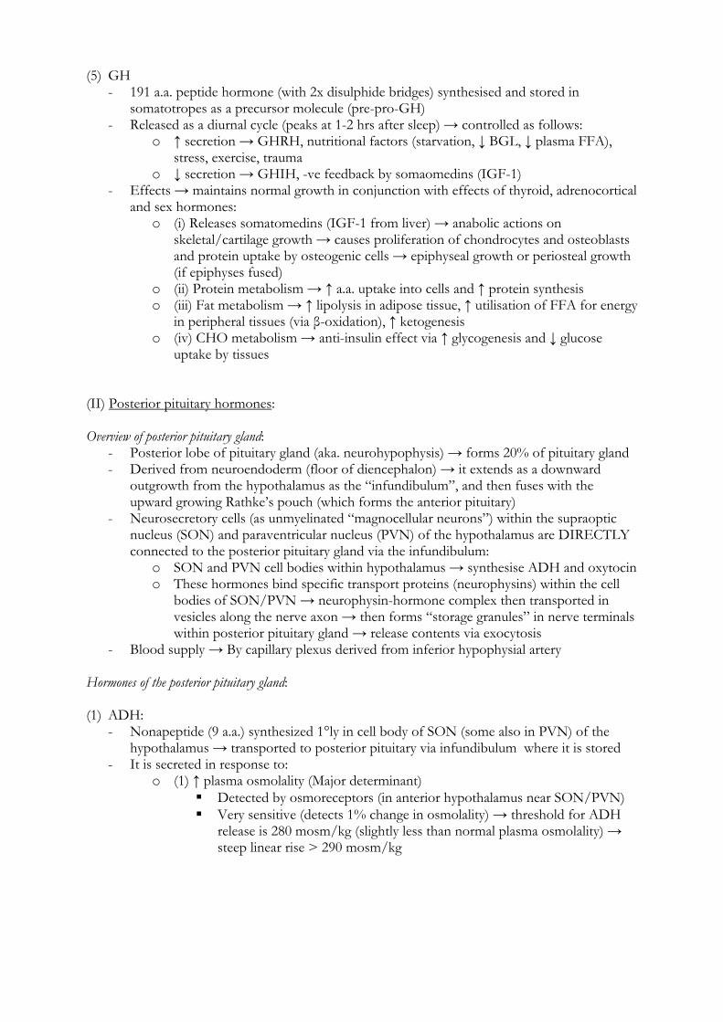
- It is secreted in response to: o (1) ↑ plasma osmolality (Major determinant)
Detected by osmoreceptors (in anterior hypothalamus near SON/PVN) Very sensitive (detects 1% change in osmolality) → threshold for ADH
release is 280 mosm/kg (slightly less than normal plasma osmolality) → steep linear rise > 290 mosm/kg
o (2) Non-osmotic stimuli: (i) Haemodynamic changes
↓ PV → ↓ MAP that is sensed by baroreceptors (mainly low-pressure BR in atrium) → cause ↑ ADH release
↓ sensitivity cf. osmotic stimuli (detects 5-10% change in PV) BUT very potent → overrides osmoreceptors (in terms of control
of ADH secretion) when there are LARGE changes in PV!!! (ii) ↑ AII (iii) Pain (iv) Nausea/vomiting (powerful stimuli) (v) Exercise (vi) Drugs (↑ release – morphine, nicotine, barbiturate; ↓ release – EtOH)
- Effects: o (1) V1 receptor (GPCR via Gq → activates PLC to ↑ IP3 → ↑ IC [Ca2+] →
smooth muscle contraction) → this causes: (a) Contraction of vascular SM cells (potent vasoconstrictor effect) → ↑
TPR and MAP (b) Renal afferent arteriolar constriction and contraction of renal
mesangial cells → ↓ GFR/RBF (c) Platelet aggregation and degranulation
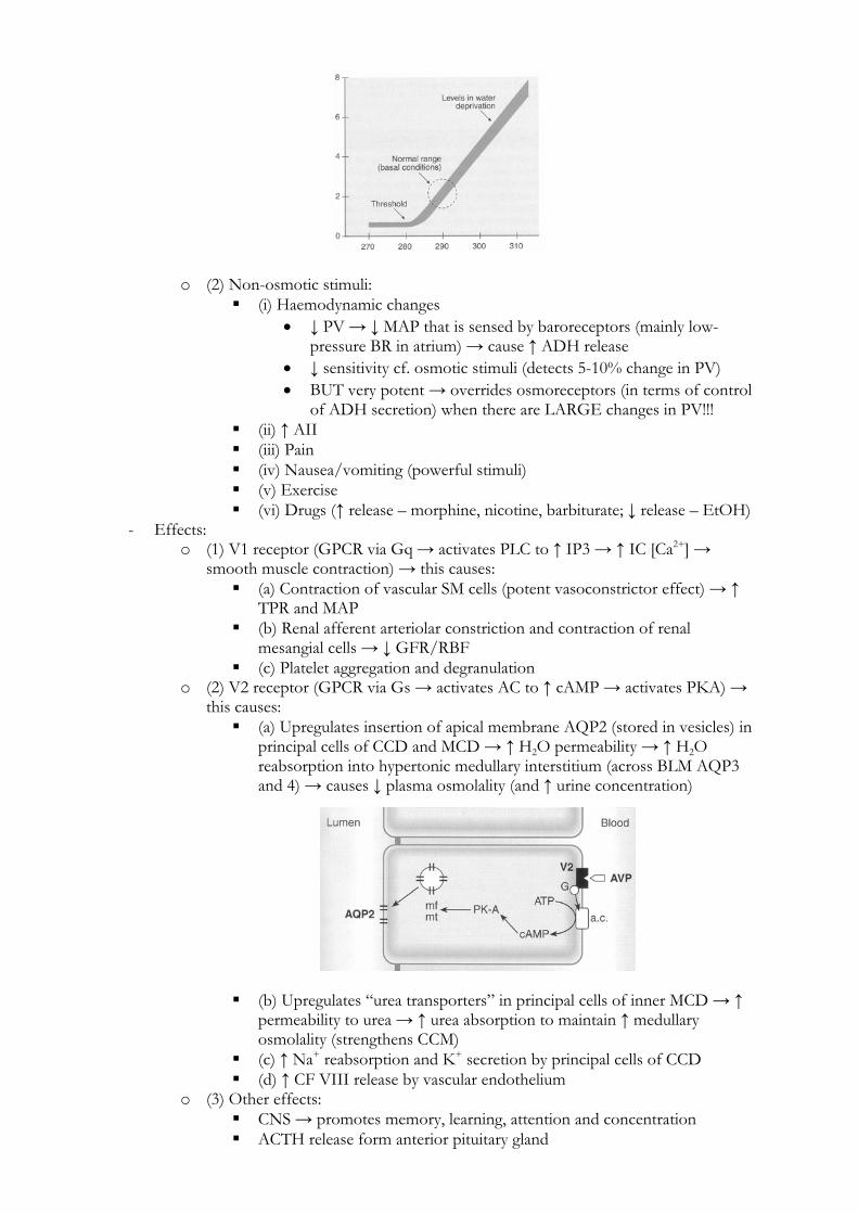
o (2) V2 receptor (GPCR via Gs → activates AC to ↑ cAMP → activates PKA) → this causes: (a) Upregulates insertion of apical membrane AQP2 (stored in vesicles) in
principal cells of CCD and MCD → ↑ H2O permeability → ↑ H2O reabsorption into hypertonic medullary interstitium (across BLM AQP3 and 4) → causes ↓ plasma osmolality (and ↑ urine concentration)
(b) Upregulates “urea transporters” in principal cells of inner MCD → ↑ permeability to urea → ↑ urea absorption to maintain ↑ medullary osmolality (strengthens CCM)
(c) ↑ Na+ reabsorption and K+ secretion by principal cells of CCD (d) ↑ CF VIII release by vascular endothelium
o (3) Other effects: CNS → promotes memory, learning, attention and concentration ACTH release form anterior pituitary gland
- ADH’s effect is very short-lived → short t ½ ~ 20 mins (rapidly inactivated by tissue peptidases → excreted by liver and kidney)
(2) Oxytocin:
- Nonapeptide (9 a.a.) synthesised 1°ly in cell body of PVN (some also in SON) of the hypothalamus → transported via infundibulum to be stored in posterior pituitary gland
- Control of secretion – (i) ↑ release in response to → cholinergic stimulation, (ii) ↓ release in response to → β-adrenergic activity, EtOH, enkephalins
- Effects: o (1) Ejection of milk – Somatic touch stimulation of nipple (Ie. suckling)
stimulates “let-down reflex” → cause oxytocin release to induce contraction of myoepithelium of lactating mammary glands → milk secretion
o (2) Myometrial contraction of pregnant uterus → during late pregnancy, a neuroendocrine reflex loop causes ↑ both oxytocin secretion and oxytocin receptor population → role in inducing labour/delivery
o (3) Uterine secretion/contractions during coitus → facilitates propulsion of semen to fallopian tubes
o (4) Various behavioural effects
(e) To describe the synthesis and functions of thyroid hormones and how their secretion is regulated.
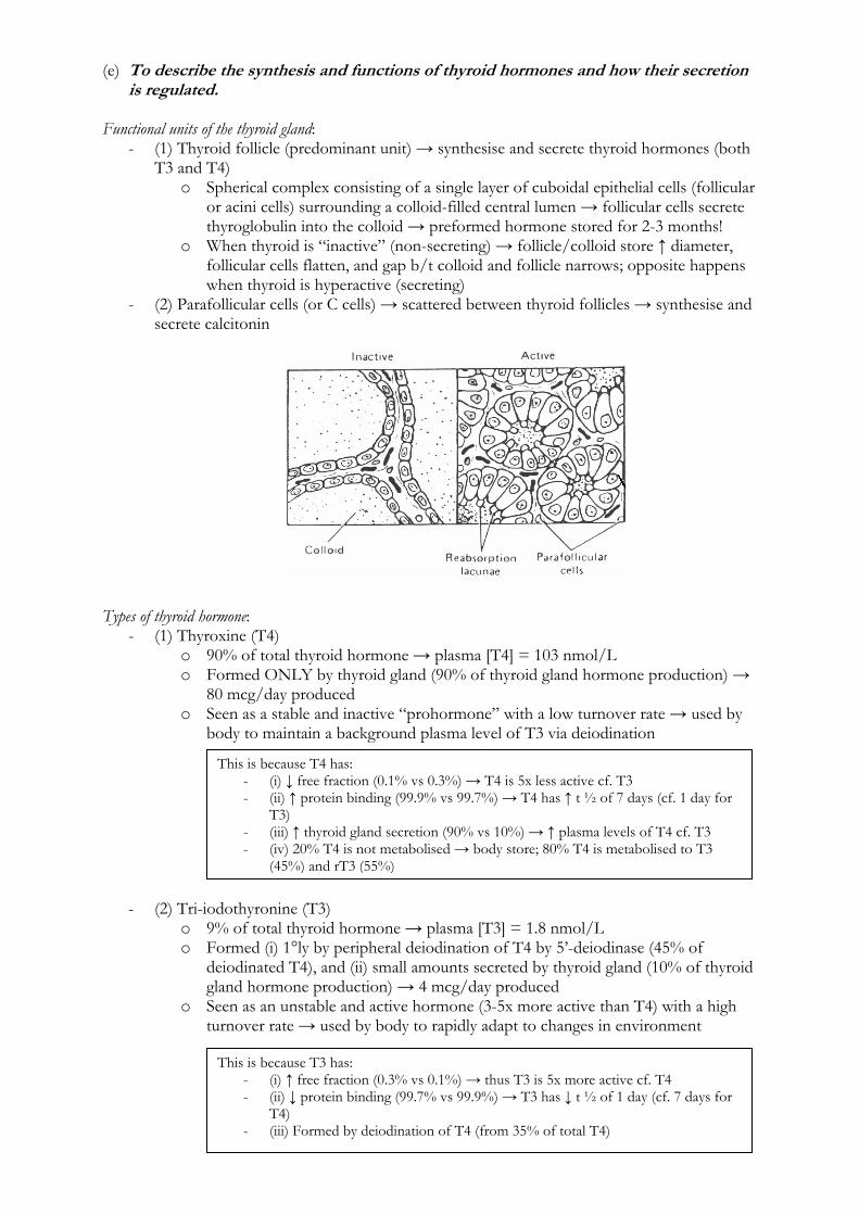
Functional units of the thyroid gland:
- (1) Thyroid follicle (predominant unit) → synthesise and secrete thyroid hormones (both T3 and T4)
o Spherical complex consisting of a single layer of cuboidal epithelial cells (follicular or acini cells) surrounding a colloid-filled central lumen → follicular cells secrete thyroglobulin into the colloid → preformed hormone stored for 2-3 months!
o When thyroid is “inactive” (non-secreting) → follicle/colloid store ↑ diameter, follicular cells flatten, and gap b/t colloid and follicle narrows; opposite happens when thyroid is hyperactive (secreting)
- (2) Parafollicular cells (or C cells) → scattered between thyroid follicles → synthesise and secrete calcitonin
Types of thyroid hormone:
- (1) Thyroxine (T4) o 90% of total thyroid hormone → plasma [T4] = 103 nmol/L o Formed ONLY by thyroid gland (90% of thyroid gland hormone production) →
80 mcg/day produced o Seen as a stable and inactive “prohormone” with a low turnover rate → used by
body to maintain a background plasma level of T3 via deiodination
- (2) Tri-iodothyronine (T3) o 9% of total thyroid hormone → plasma [T3] = 1.8 nmol/L o Formed (i) 1°ly by peripheral deiodination of T4 by 5’-deiodinase (45% of
deiodinated T4), and (ii) small amounts secreted by thyroid gland (10% of thyroid gland hormone production) → 4 mcg/day produced
o Seen as an unstable and active hormone (3-5x more active than T4) with a high turnover rate → used by body to rapidly adapt to changes in environment
This is because T4 has: - (i) ↓ free fraction (0.1% vs 0.3%) → T4 is 5x less active cf. T3 - (ii) ↑ protein binding (99.9% vs 99.7%) → T4 has ↑ t ½ of 7 days (cf. 1 day for
T3) - (iii) ↑ thyroid gland secretion (90% vs 10%) → ↑ plasma levels of T4 cf. T3 - (iv) 20% T4 is not metabolised → body store; 80% T4 is metabolised to T3
(45%) and rT3 (55%)
This is because T3 has: - (i) ↑ free fraction (0.3% vs 0.1%) → thus T3 is 5x more active cf. T4 - (ii) ↓ protein binding (99.7% vs 99.9%) → T3 has ↓ t ½ of 1 day (cf. 7 days for
T4) - (iii) Formed by deiodination of T4 (from 35% of total T4)
- (3) Reverse T3 (rT3) o 1% of total thyroid hormone o Formed by peripheral deiodination of T4 by 5’-deiodinase (55% of deiodinated
T4) → 2 mcg/day produced o Biologically inactive hormone → is deaminated and conjugated with glucuronic
acid for clearance via renal and hepatic routes Thyroid gland: Synthesis and secretion of thyroid hormone
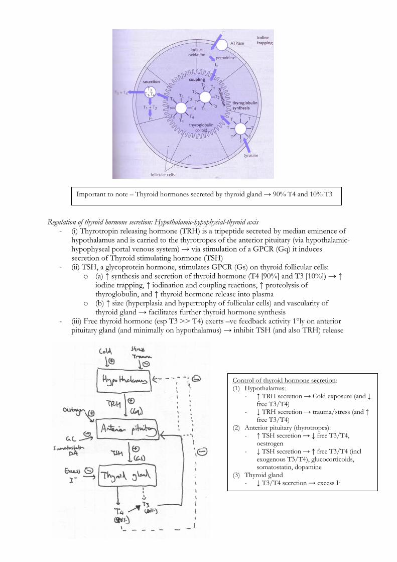
- Thyroid hormones are 1°ly synthesised in colloid (cf. follicular cell) as follows: o (1) Thyroglobulin synthesis
Tyrosine in follicular cell → converted to 100 Tyr-residue glycoprotein (thyroglobulin) → then exocytosed into colloid
o (2) Iodide trapping (*rate-limiting step*) Follicular cells actively take up plasma iodide (I-) across its basolateral
membrane via 2° active transport (Na+/I- symporter and Na+/K+ ATPase) → so IC [I-] is 30x ↑ cf. plasma [I-]
This process is – (i) Stimulated by TSH, and (ii) Inhibited by ↑ plasma [I-] (Wolff-Chaikoff effect), thiocyanate and perchlorate
o (3) Iodide oxidation Within follicular cell (near apical membrane), I- is oxidised to iodine (I2)
by thyroid peroxidise → ↑ its reactivity in binding thyroglobulin o (4) Iodination of thyroglobulin
Within colloid (near apical membrane of follicular cells), I2 rapidly binds to Tyr-molecules on thyroglobulin using iodinase to form mono-iodotyrosine (MIT) → then again to form di-iodotyrosine (DIT)
o (5) Coupling Within colloid, Tyr-molecules within thyroglobulin are coupled together
using a Peroxidase system (via oxidative condensation) – (i) 2x DIT → T4, and (ii) DIT + MIT → T3
T3 and T4 are then stored in colloid → bound to thyroglobulin moiety - TSH stimulation of the thyroid gland then causes secretion of T4 and T3 as follows:
o Iodinated thyroglobulin in colloid is taken into follicular cell by endocytosis at the apical membrane into lysosomes → proteinases degrade thyroglobulin to release free MIT, DIT, T3 and T4
o MIT and DIT are processed for I2 recycling, while T3 and T4 are released across the basolateral membrane into plasma
Aside: Iodine balance- Normal diet has 500 μg iodine per day (min intake of 20-150 μg/day) →
from salt, seafood, meat and some vegetables → absorbed in upper SB - Follicular cells are the only cell in body that can uptake/utilise iodine →
120 μg/day is taken up by thyroid gland (80 μg incorporated into stored T3/T4; 40 μg returned to plasma)
- Iodine is recycled in the body (via breakdown of T3/T4) and renally-excreted
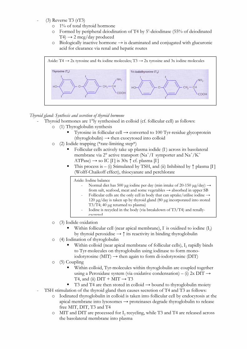
Aside: T4 → 2x tyrosine and 4x iodine molecules; T3 → 2x tyrosine and 3x iodine molecules
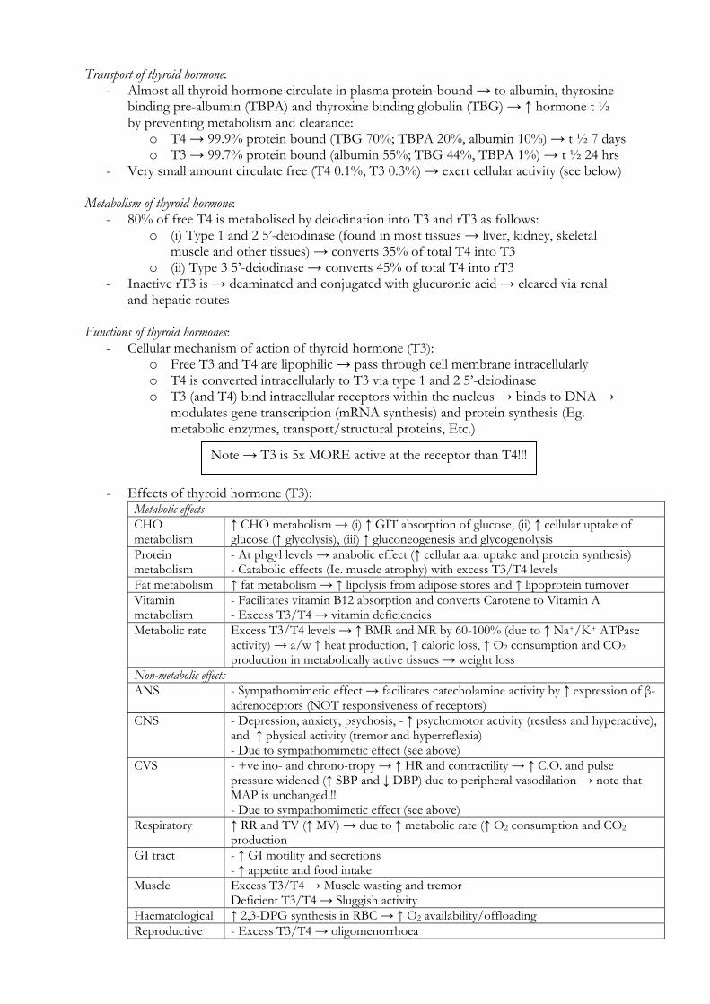
Regulation of thyroid hormone secretion: Hypothalamic-hypophysial-thyroid axis
- (i) Thyrotropin releasing hormone (TRH) is a tripeptide secreted by median eminence of hypothalamus and is carried to the thyrotropes of the anterior pituitary (via hypothalamic-hypophyseal portal venous system) → via stimulation of a GPCR (Gq) it induces secretion of Thyroid stimulating hormone (TSH)
- (ii) TSH, a glycoprotein hormone, stimulates GPCR (Gs) on thyroid follicular cells: o (a) ↑ synthesis and secretion of thyroid hormone (T4 [90%] and T3 [10%]) → ↑
iodine trapping, ↑ iodination and coupling reactions, ↑ proteolysis of thyroglobulin, and ↑ thyroid hormone release into plasma
o (b) ↑ size (hyperplasia and hypertrophy of follicular cells) and vascularity of thyroid gland → facilitates further thyroid hormone synthesis
- (iii) Free thyroid hormone (esp T3 >> T4) exerts –ve feedback activity 1°ly on anterior pituitary gland (and minimally on hypothalamus) → inhibit TSH (and also TRH) release
Important to note – Thyroid hormones secreted by thyroid gland → 90% T4 and 10% T3
Control of thyroid hormone secretion:(1) Hypothalamus:
- ↑ TRH secretion → Cold exposure (and ↓ free T3/T4)
- ↓ TRH secretion → trauma/stress (and ↑ free T3/T4)
(2) Anterior pituitary (thyrotropes): - ↑ TSH secretion → ↓ free T3/T4,
oestrogen - ↓ TSH secretion → ↑ free T3/T4 (incl
exogenous T3/T4), glucocorticoids, somatostatin, dopamine
(3) Thyroid gland - ↓ T3/T4 secretion → excess I-
Transport of thyroid hormone:
- Almost all thyroid hormone circulate in plasma protein-bound → to albumin, thyroxine binding pre-albumin (TBPA) and thyroxine binding globulin (TBG) → ↑ hormone t ½ by preventing metabolism and clearance:
o T4 → 99.9% protein bound (TBG 70%; TBPA 20%, albumin 10%) → t ½ 7 days o T3 → 99.7% protein bound (albumin 55%; TBG 44%, TBPA 1%) → t ½ 24 hrs
- Very small amount circulate free (T4 0.1%; T3 0.3%) → exert cellular activity (see below) Metabolism of thyroid hormone:
- 80% of free T4 is metabolised by deiodination into T3 and rT3 as follows: o (i) Type 1 and 2 5’-deiodinase (found in most tissues → liver, kidney, skeletal
muscle and other tissues) → converts 35% of total T4 into T3 o (ii) Type 3 5’-deiodinase → converts 45% of total T4 into rT3
- Inactive rT3 is → deaminated and conjugated with glucuronic acid → cleared via renal and hepatic routes
Functions of thyroid hormones:
- Cellular mechanism of action of thyroid hormone (T3): o Free T3 and T4 are lipophilic → pass through cell membrane intracellularly o T4 is converted intracellularly to T3 via type 1 and 2 5’-deiodinase o T3 (and T4) bind intracellular receptors within the nucleus → binds to DNA →
modulates gene transcription (mRNA synthesis) and protein synthesis (Eg. metabolic enzymes, transport/structural proteins, Etc.)
- Effects of thyroid hormone (T3): Metabolic effects CHO metabolism
↑ CHO metabolism → (i) ↑ GIT absorption of glucose, (ii) ↑ cellular uptake of glucose (↑ glycolysis), (iii) ↑ gluconeogenesis and glycogenolysis
Protein metabolism
- At phgyl levels → anabolic effect (↑ cellular a.a. uptake and protein synthesis) - Catabolic effects (Ie. muscle atrophy) with excess T3/T4 levels
Fat metabolism ↑ fat metabolism → ↑ lipolysis from adipose stores and ↑ lipoprotein turnover Vitamin metabolism
- Facilitates vitamin B12 absorption and converts Carotene to Vitamin A - Excess T3/T4 → vitamin deficiencies
Metabolic rate Excess T3/T4 levels → ↑ BMR and MR by 60-100% (due to ↑ Na+/K+ ATPase activity) → a/w ↑ heat production, ↑ caloric loss, ↑ O2 consumption and CO2 production in metabolically active tissues → weight loss
Non-metabolic effects ANS - Sympathomimetic effect → facilitates catecholamine activity by ↑ expression of β-
adrenoceptors (NOT responsiveness of receptors) CNS - Depression, anxiety, psychosis, - ↑ psychomotor activity (restless and hyperactive),
and ↑ physical activity (tremor and hyperreflexia) - Due to sympathomimetic effect (see above)
CVS - +ve ino- and chrono-tropy → ↑ HR and contractility → ↑ C.O. and pulse pressure widened (↑ SBP and ↓ DBP) due to peripheral vasodilation → note that MAP is unchanged!!! - Due to sympathomimetic effect (see above)
Respiratory ↑ RR and TV (↑ MV) → due to ↑ metabolic rate (↑ O2 consumption and CO2 production
GI tract - ↑ GI motility and secretions - ↑ appetite and food intake
Muscle Excess T3/T4 → Muscle wasting and tremor Deficient T3/T4 → Sluggish activity
Haematological ↑ 2,3-DPG synthesis in RBC → ↑ O2 availability/offloading Reproductive - Excess T3/T4 → oligomenorrhoea
Note → T3 is 5x MORE active at the receptor than T4!!!
- Deficient T3/T4 → menorrhagia/polymenorrhoea, ↓ libido - Due to direct gonadal effects and feedback mechanisms on anterior pituitary control of sexual functions
Endocrine ↑ metabolic turnover of other hormones (Ie. ↑ cortisol production) Growth and development
- T3/T4 is vital for cell division/maturation → normal CNS/skeletal development - Deficiency → Mental retardation, ↓ brain development and bone growth stunting
(f) To describe the control of secretion and functions of adreno-cortical hormones. (I) Overview of adreno-cortical hormones: Types of adreno-cortical hormones:
- There are 3 main classes → all are steroid-based hormones: o (1) Mineralocorticoid (1°ly aldosterone) o (2) Glucocorticoid (1°ly cortisol and corticosterone) o (3) Androgens (1°ly DHEA and androsteindione)
- Adrenal cortex (70% of adrenal gland; cf. adrenal medulla – 30% adrenal gland) is divided into 3 zones of different cell types → secrete different hormones:
Zone Part of adrenal cortex Hormones Zona glomerulosa Outer (15% volume) Aldosterone and corticosterone Zona fasciculata Middle (80% volume) Corticosterone, cortisol and androgens Zona reticularis Inner (5% volume) Corticosterone, cortisol and androgens
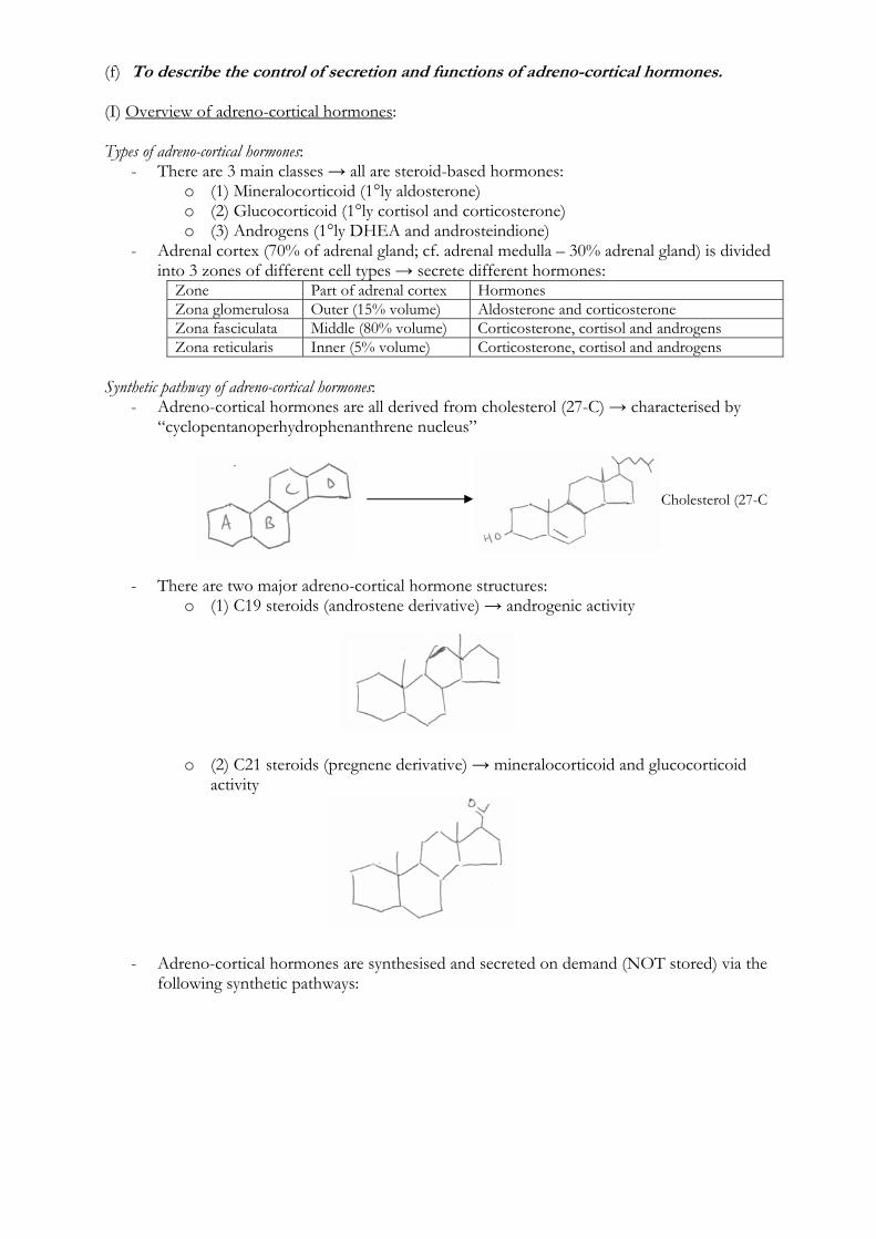
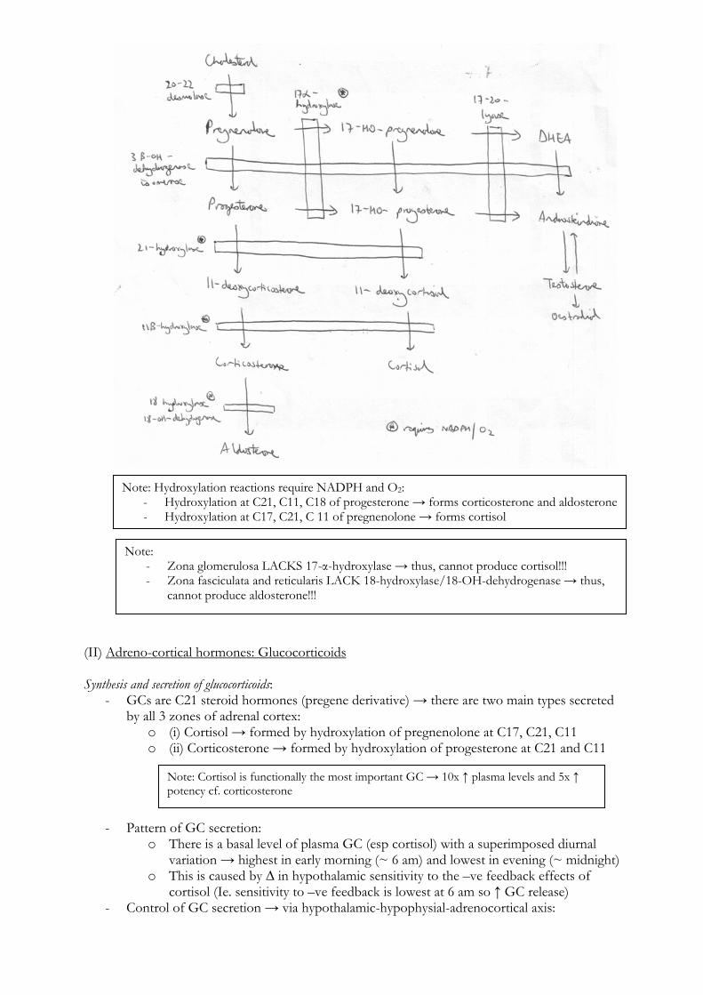
Synthetic pathway of adreno-cortical hormones:
- Adreno-cortical hormones are all derived from cholesterol (27-C) → characterised by “cyclopentanoperhydrophenanthrene nucleus”
- There are two major adreno-cortical hormone structures: o (1) C19 steroids (androstene derivative) → androgenic activity
o (2) C21 steroids (pregnene derivative) → mineralocorticoid and glucocorticoid activity
- Adreno-cortical hormones are synthesised and secreted on demand (NOT stored) via the following synthetic pathways:
Cholesterol (27-C
(II) Adreno-cortical hormones: Glucocorticoids Synthesis and secretion of glucocorticoids:
- GCs are C21 steroid hormones (pregene derivative) → there are two main types secreted by all 3 zones of adrenal cortex:
o (i) Cortisol → formed by hydroxylation of pregnenolone at C17, C21, C11 o (ii) Corticosterone → formed by hydroxylation of progesterone at C21 and C11
- Pattern of GC secretion: o There is a basal level of plasma GC (esp cortisol) with a superimposed diurnal
variation → highest in early morning (~ 6 am) and lowest in evening (~ midnight) o This is caused by ∆ in hypothalamic sensitivity to the –ve feedback effects of
cortisol (Ie. sensitivity to –ve feedback is lowest at 6 am so ↑ GC release) - Control of GC secretion → via hypothalamic-hypophysial-adrenocortical axis:
Note: Cortisol is functionally the most important GC → 10x ↑ plasma levels and 5x ↑ potency cf. corticosterone
Note: Hydroxylation reactions require NADPH and O2: - Hydroxylation at C21, C11, C18 of progesterone → forms corticosterone and aldosterone- Hydroxylation at C17, C21, C 11 of pregnenolone → forms cortisol
Note: - Zona glomerulosa LACKS 17-α-hydroxylase → thus, cannot produce cortisol!!! - Zona fasciculata and reticularis LACK 18-hydroxylase/18-OH-dehydrogenase → thus,
cannot produce aldosterone!!!
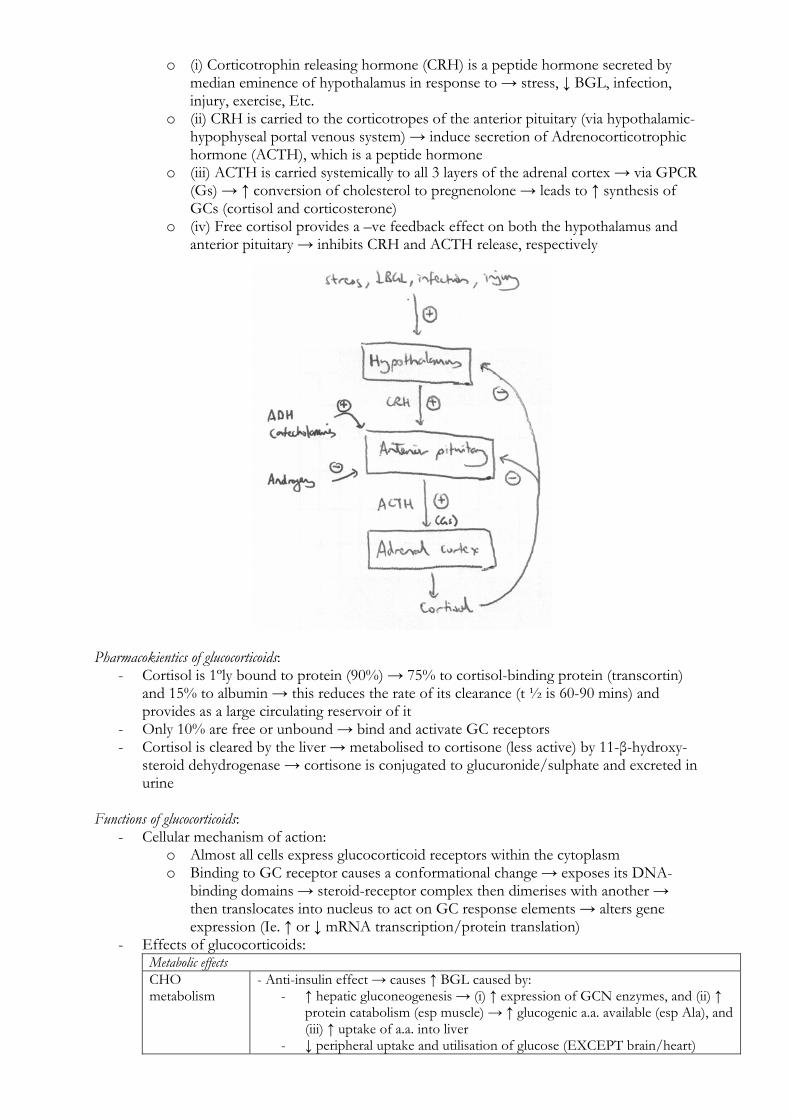
o (i) Corticotrophin releasing hormone (CRH) is a peptide hormone secreted by median eminence of hypothalamus in response to → stress, ↓ BGL, infection, injury, exercise, Etc.
o (ii) CRH is carried to the corticotropes of the anterior pituitary (via hypothalamic-hypophyseal portal venous system) → induce secretion of Adrenocorticotrophic hormone (ACTH), which is a peptide hormone
o (iii) ACTH is carried systemically to all 3 layers of the adrenal cortex → via GPCR (Gs) → ↑ conversion of cholesterol to pregnenolone → leads to ↑ synthesis of GCs (cortisol and corticosterone)
o (iv) Free cortisol provides a –ve feedback effect on both the hypothalamus and anterior pituitary → inhibits CRH and ACTH release, respectively
Pharmacokientics of glucocorticoids:
- Cortisol is 1ºly bound to protein (90%) → 75% to cortisol-binding protein (transcortin) and 15% to albumin → this reduces the rate of its clearance (t ½ is 60-90 mins) and provides as a large circulating reservoir of it
- Only 10% are free or unbound → bind and activate GC receptors - Cortisol is cleared by the liver → metabolised to cortisone (less active) by 11-β-hydroxy-
steroid dehydrogenase → cortisone is conjugated to glucuronide/sulphate and excreted in urine
Functions of glucocorticoids:
- Cellular mechanism of action: o Almost all cells express glucocorticoid receptors within the cytoplasm o Binding to GC receptor causes a conformational change → exposes its DNA-
binding domains → steroid-receptor complex then dimerises with another → then translocates into nucleus to act on GC response elements → alters gene expression (Ie. ↑ or ↓ mRNA transcription/protein translation)
- Effects of glucocorticoids: Metabolic effects CHO metabolism
- Anti-insulin effect → causes ↑ BGL caused by: - ↑ hepatic gluconeogenesis → (i) ↑ expression of GCN enzymes, and (ii) ↑
protein catabolism (esp muscle) → ↑ glucogenic a.a. available (esp Ala), and (iii) ↑ uptake of a.a. into liver
- ↓ peripheral uptake and utilisation of glucose (EXCEPT brain/heart)
Protein metabolism
- ↑ protein catabolism of protein stores (esp muscle and bone) → causes (i) ↑ plasma [ ] of a.a., (ii) net –ve N-balance, and (iii) ↓ tissue protein stores - Plasma a.a. are used for gluconeogensis (esp glucogenic a.a. → Ala)
Fat metabolism - Redistribution of fat from peripheral to central sites (Eg. central obesity, buffalo hump, Etc.) → due to ↑ peripheral lipolysis → (i) stimulates hormone-sensitive lipase, and (ii) potentiates lipolytic actions of other hormones (Eg. GH, glucagon, T3/T4, catecholamines) - ↑ cellular FA oxidation (and ketogenesis)
Non-metabolic effects Catecholamines and vasopressors
- Acts permissively with catecholamines and vasopressor agents (Eg. NAd, Adr, AII, Etc.) → ↑ CVS reactivity to them (Ie. ↑ +ve inotropy and MAP) - GCs ↑ synthesis of hormone-sensitive enzymes → catecholamines and vasopressors ↑ cAMP, which stimulate hormone-sensitive enzymes via ↑ cAMP-dependent kinase activity
GI tract ↑ gastric acid and pepsin secretion due to ↓ PG synthesis (risk of PUD) CNS - Influences cognitive and behavioural function (Ie. ↑ appetite, mania)
- Influences foetal/neonatal neuronal development Haematological - Mild ↑ in RBC, PMNL and platelets
- ↓ monocytes, lymphocytes, eosinophils, basophils Immunological - Immunosuppressive response due to ↓ lymphocytes, monocytes, basophils,
eosinophils, cytokine and complement levels - Caused by inhibition of PLA-2 expression (required for inflammatory eicosanoids) and cytokine/complement gene expression
Inflammation Anti-inflammatory response due to (i) stabilisation of lysosomal membranes (↓ proteolytic enzyme release), (ii) ↓ capillary permeability (↓ capillary leakage, histamine/bradykinin release), (iii) ↓ inflammatory mediators
Skeletal ↓ collagen synthesis by osteoblasts and ↑ collagen breakdown by collagenase (risk of osteoporosis)
Endocrine - Stress response → redistribution of metabolic fuel substrates for ↑ energy production and anabolism at damaged tissues - Suppresses anterior pituitary hormones release (ACTH, LH/FSH, TSH, GH)
(III) Adreno-cortical hormones: Aldosterone Synthesis and secretion of aldosterone:
- Aldosterone is a 21-C steroid hormone (pregnene derivative) → formed by hydroxylation of progesterone at C21, C11, C18 (see above)
- It is the main mineralocorticoid synthesised and secreted by zona glomerulosa → stimulus for synthesis/secretion include:
o (1) Angiotensin-II (most important) → via Gq effect → stimulates (i) synthesis (converts cholesterol to pregnenolone) and (ii) secretion
o (2) ↑ plasma [K+] → secreted with 1% ↑ [K+] → due to depolarisation of cell membrane
o (3) ACTH (mild effect) → via Gs effect → stimulates synthesis (converts cholesterol to pregnenolone)
o (4) ↓ plasma [Na+] → secreted with 10% ↓ [Na+] (Nb. this is overridden by ∆ ECFV → so it is not released with hypervolaemic hyponatraemia)
Pharmacokinetics of aldosterone:
- Aldosterone circulates bound to albumin (60%) and transcortin (or CBG; 10%) → 30% is free hormone (Ie. unbound) and readily degraded → thus, it has a short t ½ (20 mins)
- 90% eliminated via 1st pass through liver (by reduction of double-bond of A ring to form Tetrahydroaldosterone) → then conjugated with glucuronide and renally excreted
Functions of aldosterone:
- Cellular mechanism of action:
o Aldosterone binds to IC mineralocorticoid receptors (found ONLY in colon, kidney, bladder, sweat/salivary glands)
o Binding to MC receptor causes a conformational change → exposes its DNA-binding domains → steroid-receptor complex then dimerises with another → then translocates into nucleus to act on MC response elements → alters gene expression (Ie. mRNA transcription/protein translation)
- Effects of aldosterone: o (1) ↑ plasma Na+ (and ↑ ECVF)
(i) ↑ renal tubular Na+ reabsorption at Principal cells of DCT/CCD → upregulates basolateral Na+/K+ ATPase and luminal ENaC → MAIN determinant of Na+ tubular handling (reabsorbs 1-2% of filtered Na+)
(ii) ↑ Na+ reabsorption throughout the GI tract, sweat and salivary glands → upregulates Na+/K+ ATPase
(iii) ↑ renal tubular H2O reabsorption (a/w Na+ reabsorption) → ↑ ECFV 5-15% only → limited by “Escape phenomenon”, where initial ↑ ECFV is limited by natriuretic/diuretic effect of ANP secretion
o (2) ↓ plasma K+ (i) ↑ renal tubular K+ secretion at Principal cells of DCT/CCD →
upregulates basolateral Na+/K+ ATPase and luminal K+ channels → MAIN determinant of K+ tubular secretion
(ii) ↑ K+ secretion throughout the GI tract, sweat and salivary glands → upregulates Na+/K+ ATPase
(iii) ↑ IC uptake of K+ o (3) Alkalosis by renal H+ loss → ↑ H+ secretion (and HCO3
- reabsorption) by type A intercalated cells of CCD → upregulates H+ and H+/K+ ATPase
o (4) ↑ plasma Cl- → ↑ Cl- reabsorption in distal tubule (Nb. This can lead to hyperchloraemic acidosis with aldosterone excess)
(IV) Adreno-cortical hormones: Sex steroids
- Zona fasciculata and reticularis synthesise and secrete 3 androgenic hormones (male sex hormones) in response to ACTH:
o (1) Androsteinedione o (2) DHEA (and DHEA sulphate) o (3) Testosterone
- DHEA (and DHEA sulphate) and androsteinedione are weak androgens → converted peripherally to more active hormones:
o (i) DHEA (and DHEA sulphate) → converted to testosterone o (ii) DHEA (and DHEA sulphate) and androsteindione → converted to oestrogen
in fat, mammary glands and other tissues - Maturation of zona fasciculata/reticularis (and androgen secretion) begin a few years
before puberty (7-9 y.o.) during adrenarche → androgens are present in both genders: o In men → constitutes 5% of total adult androgen activity (minimal) o In women → constitutes 50% of total adult androgen activity (pubic and axillary
hair growth)
(g) To describe the control of secretion and functions of adrenal medullary hormones. Overview of adrenal medullary hormones:
- Chromaffin cells of adrenal medulla → synthesize/secrete following bioamine hormones: o (1) Dihydroxyphenolic amines
(a) Adrenaline (80%) → mainly produced in adrenal medulla (cf. brain) (b) Noradrenaline (20%) → less produced here (cf. NAd made in SNS
post-ganglionic peripheral nerve terminals and CNS nerve terminals) o (2) Metenkephalin
Synthesis, storage, and release of catecholamines:
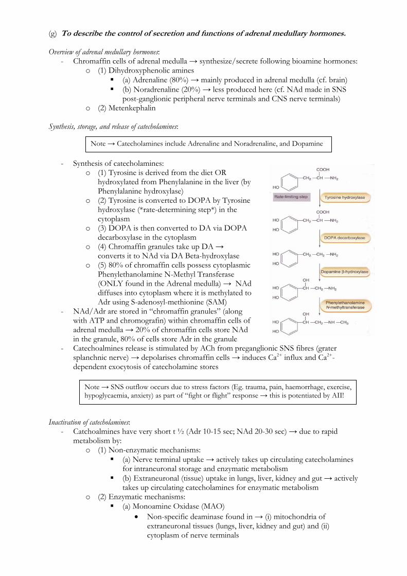
- Synthesis of catecholamines: o (1) Tyrosine is derived from the diet OR
hydroxylated from Phenylalanine in the liver (by Phenylalanine hydroxylase)
o (2) Tyrosine is converted to DOPA by Tyrosine hydroxylase (*rate-determining step*) in the cytoplasm
o (3) DOPA is then converted to DA via DOPA decarboxylase in the cytoplasm
o (4) Chromaffin granules take up DA → converts it to NAd via DA Beta-hydroxylase
o (5) 80% of chromaffin cells possess cytoplasmic Phenylethanolamine N-Methyl Transferase (ONLY found in the Adrenal medulla) → NAd diffuses into cytoplasm where it is methylated to Adr using S-adenosyl-methionine (SAM)
- NAd/Adr are stored in “chromaffin granules” (along with ATP and chromografin) within chromaffin cells of adrenal medulla → 20% of chromaffin cells store NAd in the granule, 80% of cells store Adr in the granule
- Catechoalmines release is stimulated by ACh from preganglionic SNS fibres (grater splanchnic nerve) → depolarises chromaffin cells → induces Ca2+ influx and Ca2+-dependent exocytosis of catecholamine stores
Inactivation of catecholamines:
- Catchoalmines have very short t ½ (Adr 10-15 sec; NAd 20-30 sec) → due to rapid metabolism by:
o (1) Non-enzymatic mechanisms: (a) Nerve terminal uptake → actively takes up circulating catecholamines
for intraneuronal storage and enzymatic metabolism (b) Extraneuronal (tissue) uptake in lungs, liver, kidney and gut → actively
takes up circulating catecholamines for enzymatic metabolism o (2) Enzymatic mechanisms:
(a) Monoamine Oxidase (MAO) Non-specific deaminase found in → (i) mitochondria of
extraneuronal tissues (lungs, liver, kidney and gut) and (ii) cytoplasm of nerve terminals
Note → Catecholamines include Adrenaline and Noradrenaline, and Dopamine
Note → SNS outflow occurs due to stress factors (Eg. trauma, pain, haemorrhage, exercise, hypoglycaemia, anxiety) as part of “fight or flight” response → this is potentiated by AII!
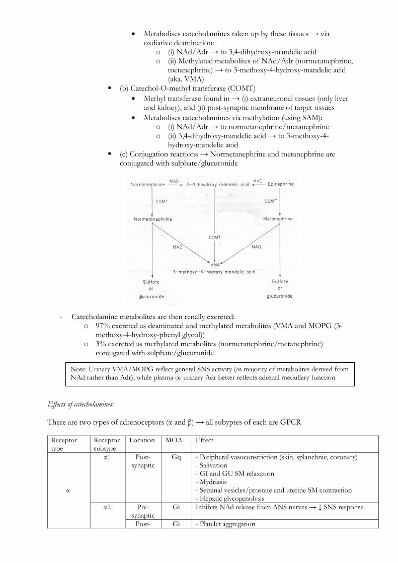
Metabolises catecholamines taken up by these tissues → via oxdiative deamination:
o (i) NAd/Adr → to 3,4-dihydroxy-mandelic acid o (ii) Methylated metabolites of NAd/Adr (normetanephrine,
metanephrine) → to 3-methoxy-4-hydroxy-mandelic acid (aka. VMA)
(b) Catechol-O-methyl transferase (COMT) Methyl transferase found in → (i) extraneuronal tissues (only liver
and kidney), and (ii) post-synaptic membrane of target tissues Metabolises catecholamines via methylation (using SAM):
o (i) NAd/Adr → to normetanephrine/metanephrine o (ii) 3,4-dihydroxy-mandelic acid → to 3-methoxy-4-
hydroxy-mandelic acid (c) Conjugation reactions → Normetanephrine and metanephrine are
conjugated with sulphate/glucuronide
- Catecholamine metabolites are then renally excreted: o 97% excreted as deaminated and methylated metabolites (VMA and MOPG (3-
methoxy-4-hydroxy-phenyl glycol)) o 3% excreted as methylated metabolites (normetanephrine/metanephrine)
conjugated with sulphate/glucuronide Effects of catecholamines: There are two types of adrenoceptors (α and β) → all subyptes of each are GPCR Receptor type
Receptor subtype
Location MOA Effect
α1 Post-synaptic
Gq - Peripheral vasoconstriction (skin, splanchnic, coronary) - Salivation - GI and GU SM relaxation - Mydriasis - Seminal vesicles/prostate and uterine SM contraction - Hepatic glycogenolysis
Pre-synaptic
Gi Inhibits NAd release from ANS nerves → ↓ SNS response
α
α2
Post- Gi - Platelet aggregation
Note: Urinary VMA/MOPG reflect general SNS activity (as majority of metabolites derived from NAd rather than Adr); while plasma or urinary Adr better reflects adrenal medullary function
synaptic - Hyperpolarisation of CNS neurons (sedation, analgesia) - ↓ insulin release from beta-pancreatic islet cells
β1 Post-synaptic
Gs - +ve inotropy and chronotropy (↑contractility/HR) - +ve dromotropy (↑ electrical conduction → arrhythmias) - GI SM relaxation - Renin secretion from JG cells in JGA (kidney)
β2 Post-synaptic
Gs - Bronchodilation and ↓ bronchial wall gland secretion - Vasodilation (esp skeletal muscle) - Uterine SM relaxation - GI SM relaxation - Muscle tremors - Hepatic glycogenolysis - Glucagon release from pancreas - Mast-cell inhibition of histamine release
β
β3 Post-synaptic
Gs Lipolysis and thermogenesis at adipose tissue (esp brown fat) and skeletal muscle
NAd and Adr have different affinities for each type of adrenoceptor: Target organ SNS effect CVS (i) +ve inotropic, chronotropic and dromotropic effects (β1)
(ii) Generally causes vasoconstriction (α1) in skin, splanchnic viscera, kidneys and mucosa, but can cause vasodilation (β2) in skeletal muscle (iii) AII release causing vasoconstriction (due to β1 effects on renin secretion at JG cells)
Lungs (i) Bronchial SM relaxation (β2) (ii) Pulmonary blood vessel vasoconstriction (iii) Reduced bronchial wall gland secretion (β2)
GI tract (i) Increased sphincteric tone (pylorus, ileocolic and rectal sphincters) (ii) Inhibition of peristalsis and reduced GI motility (iii) Reduced GI secretions (iv) Constrict splanchnic arterioles to redistribute BV * All above via α and β effect *
GU/reproductive tract (i) Relaxes bladder detrusor muscle (ii) SM contraction of seminal vesicles and prostate (ejaculation) (α1) (iii) Uterine contraction (α1) and relaxation (β2)
Eyes (i) Dilation of pupil (radial muscles) (α1) (ii) Retraction of eyelid
Cutaneous (i) Piloerection (M3) (ii) Vasconstriction (α1) (iii) Sweating (M3)
Musculoskeletal Arteriolar vasodilation and muscle tremor (β2) Metabolic (i) Hyperglycaemia – Hepatic glycogenolysis and glucagon release (β2),
Muscle glycogenolysis (β1), and reduced insulin release (α) (ii) Lipolysis (β1)
Haematological (i) Mast cell inhibition of histamine release (β2) (ii) Platelet-aggregation (α2)
α-receptors – NAd > Adr > Iso, and β-receptors – Iso > Adr > NAd
Note: - Adr → potent at both α and β receptors - NAd → more potent at α receptors and as effective as Adr on β-1 receptors (little effect
on β-2 receptors)
(h) To describe the control of secretion and the functions of renin and angiotensin. Renin:
- Glycoprotein hormone produced as pre-pro-hormone and stored in secretory granules within Juxtaglomerular cells in walls of afferent arterioles (part of JGA)
- It is cleaved into an active hormone and secreted in response to: o (1) Renal SNS NAd nerves and circulating catecholamines → via β1 effect o (2) ↓ renal arteriolar BP (as sensed by intrarenal baroreceptors) o (3) ↓ tubular [NaCl] in TAL/EDCT (as sensed by Macula densa as part of tubulo-
glomerular feedback) o (4) Prostacyclin o Nb. ADH and AII both INHIBIT renin secretion
- It has a t ½ ~ 40-120 mins Angiotensin I (AI):
- Renin cleaves Angiotensinogen (α2 globulin produced in liver) to an inactive 10 a.a. peptide AI
- This is the RATE LIMITING STEP of the RAAS pathway! Angiotensin II (AII):
- Angiotensin-Converting Enzyme (ACE) in the capillary endothelium (esp in lungs) converts AI to an active 8 a.a peptide AII
- AII acts via GPCR via Gq → activates PLC to ↑ IP3 → ↑ IC Ca2+ to cause: o (1) ↑renal tubular reabsorption of Na+ and Cl- (and ↑ ECVF) and ↑ tubular K+
secretion via → (i) Direct effects on the PCT, and (ii) Release of Aldosterone from the adrenal cortex
o (2) ↑ body H2O content via → (i) ↑ thirst/H2O intake (via hypothalamus), and (ii) ADH secretion (↑ H2O reabsorption in renal CD)
o (3) ↓ RBF and GFR (esp at ↑↑↑ levels of AII) via: (i) Renal afferent and efferent arteriolar vasoconstriction (↓ GFR and
RBF) (ii) Mesangial contraction (↓ GFR) Nb. at low levels of AII → it maintains GFR via renal afferent arteriolar
constriction alone! o (4) Net tubular fluid/electrolyte reabsorption → due to ↓ peritublar capillary
PHYDROSTATIC 2° to renal arteriolar vasoconstriction o (5) Potent vasoconstrictor → ↑ BP 2° to ↑ SVR o (6) ↑ SNS activity → via central and peripheral effects → ↑ HR, C.O., BP o (7) Inhibits renin release at JG cells (in JGA) → -ve feedback
- It has a t ½ ~ 1-3 mins Angiotensin III (AIII):
- Additional a.a. moieties are split from AII to produce AIII in some tissues → AIII has similar actions to AII, but with much less potency
(i) To describe the regulation of plasma calcium including the actions and control of vitamin D, parathyroid hormone, and calcitonin.
(I) Overview of Ca2+ distribution and balance: Distribution of Ca2+ in body:
- Total body Ca2+ → 25,000 mmol (400 mmol/kg): o (i) Readily exchangeable pool (1%) → ECF (esp plasma) → immediate reserve for
sudden ∆ plasma [Ca2+]: Total plasma [Ca2+] ~ 2.12-2.65 mmol/L → ionised [Ca2+] ~ 1.2 mmol/L There are two pools of Ca2+ in plasma:
(a) Diffusible Ca2+ (55%) → 45% free/ionised and 10% complexed (to citrate, carbonate, HPO4)
(b) Non-diffusible Ca2+ (45%) → protein bound (esp to albumin) → pH-dependent (↑ binding with ↑ pH)
o (ii) Poorly exchangeable pool (99%) → in bone/teeth (as hydroxyapatite, phosphates, carbonates) → not available for rapid mobilisation
Ca2+balance in body:
- Daily Ca2+ intake → 20 mmol/day (min 5 mmol/day) - Bone liberates/reabsorbs 500 mmol/day from the “readily exchangeable pool” - Daily Ca2+ loss via:
o (i) Kidneys (40%) → 2.5-7.5 mmol/day Glomerular filtration of 250 mmol/day → 95% reabsorbed by tubules
(PCT 65% with Na2+, TAL of LoH 20%, distal nephron 10%) → 5% excreted
Tubular reabsorption ↑ at LoH/distal nephron reabsorption due to (i) PTH and (ii) 1,25-dihydroxy-vitamin D
o (ii) GIT in faeces (60%) → 6-14 mmol/day (II) Regulation of plasma Ca2+: Three hormones are responsible for the homeostasis of plasma Ca2+ levels:
- (1) Parathyroid hormone (PTH) - (2) Vitamin D - (3) Calcitonin
These hormones act on:
- (1) Bone: o Osteoblasts – Form collage matrix with glycosaminoglycans mineralized by Ca2+
and PO43- that form hydroyapatite crystals
o Osteoclasts – Resorb bone releasing matrix, Ca2+ and PO43-
- (2) Kidney o 98% Ca2+ filtered at glomerulus is reabsorbed at (i) proximal tubule (60%), (ii)
loop and distal tubules (40%) - (3) GI tract
o Ca2+ is absorbed by both (a) passive (dependent on plasma [Ca2+]) and (b) active mechanisms (stimulated by 1,25-OH-Vitamin D)
Note: Corrected plasm Ca2+ → depends on plasma protein level (esp albumin):
- 0.02 mmol/L correction factor added to measured plasma Ca2+ per 1 g/L ↑ plasma albumin (up to 40 g/L)
- Eg. plasma Ca2+ of 1.83 and albumin 25 g/L → 1.83 + (40-25) x 0.02 = corrected Ca2+ of 2.13 mmol/L
(1) Parathyroid Hormone (PTH):
- PTH is a 84 a.a. peptide hormone (cleaved from 110 a.a. PreProPTH) secreted from Chief cells in 4x parathyroid gland (present at superior and inferior poles of both thyroid gland lobes) → in response to:
o (a) ↓ serum Ca2+ o (b) ↑ serum PO4
3- o (c) ↓ serum vitamin D levels
- Acts via GPCR (Gs → cAMP) → causes ↑ serum Ca2+ (and ↓ serum PO43-):
o (a) Bone → bone resorption releasing Ca2+ and PO43- in 2 phases – (i) Initial rapid
phase involving osteolysis, then (ii) Slow phase with osteoclastic activity o (b) Kidneys → ↓ proximal tubule PO4
3- reabsorption and ↑ distal tubule Ca2+ reabsorption
o (c) GI tract → ↑ proximal renal tubular activation of vitamin D (enhances 1α-hydroxylase activity) → indirectly ↑ Ca2+ absorption in intestines
(2) Calcitriol (Vitamin D):
- Calcitriol is a steroid hormone that is derived 1°ly in skin (via conversion of 7-dehydrocholesterol → cholecalciferol by UV light) → then hydroxylated in liver to form 25-OH-cholecalciferol → then hydroxylated in proximal nephrons of kidney (1α-hydroxylase) to form 1,25-OH-cholecalciferol (aka. calcitriol)
- Calcitriol is produced 2° to ↑ expression of renal α1-hydroxylase by:
o (a) ↓ serum Ca2+ o (b) ↓ serum PO4
3- o (c) Direct feedback of calcitriol (such as ↓ vitamin D levels) o (d) PTH
- Acts via IC receptor (alters mRNA transcription) → causes ↑ serum Ca2+ and PO43-:
o (a) Bone → Demineralization in the short term (to release Ca2+ and PO43-) → acts
synergistically with PTH to ↑ osteoclast activity; enhances mineralization and promotes bone formation chronically
o (b) Kidneys → ↑ renal reabsorption of Ca2+ in the distal renal tubules, and reabsorption of PO4
3- in the proximal renal tubules o (c) GI tract → ↑ ACTIVE intestinal absorption of Ca2+ & PO4
3- (by inducing Ca2+-binding protein in mucosa of small intestines)
(3) Calcitonin:
- Calcitonin is a 32 a.a. peptide hormone secreted from parafollicular cells (or C-cells) of the thyroid gland → Nb. it is NOT essential in humans!
- It is secreted in response to: o (a) ↑ serum Ca2+ o (b) Gastric hormones – Gastrin, CCK, Glucagon, Secretin
- Causes ↓ serum Ca2+ (and PO43-) levels by:
o (a) ↓ bone resorption (inhibits osteoclasts) o (b) ↑ renal Ca2+ and PO4
3- excretion o (c) Inhibiting jejunal absorption of Ca2+ and PO4
3-
Note – 25-OH-cholecalciferol is the DOMINANT form in circulation BUT is biologically inactive → 1,25-OH-cholecalciferol is biologically ACTIVE but present in small amounts
(j) To describe the role of prostaglandins and other autocoids. Overview of eicosanoids:
- Diverse group of molecules generated de novo from arachidonic acid → a 20-C polyunsaturated FA (with 4 double-bonds) found in cell membrane phospholipids
- Includes – (i) Prostaglandins (incl prostacyclin), (ii) Thromboxanes, (iii) Leukotrienes, (iv) Lipoxins, and (v) other related molecules (Eg. PAF, 5-HETE, 12-HETE, Etc.)
- Produced by all cells in body (esp kidney, heart, lungs, liver, pancreas, brain, reproductive organs) EXCEPT RBC → specific eicosanoids are made by various tissues (Ie. not all cells produce same type) → this is dictated by type of enzymes expressed by cells in tissue
- Synthesised in small quantities prior to release (NOT stored preformed in tissues) → exert 1°ly local paracrine effects → very potent activity → but rapidly inactivated (within seconds-minutes)
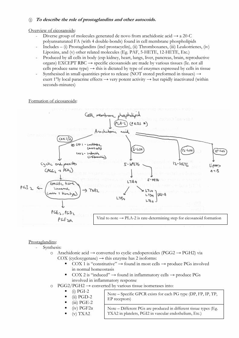
Formation of eicosanoids: Prostaglandins:
- Synthesis: o Arachidonic acid → converted to cyclic endoperoxides (PGG2 → PGH2) via
COX (cycloxygenase) → this enzyme has 2 isoforms: COX 1 is “constitutive” → found in most cells → produce PGs involved
in normal homeostasis COX 2 is “induced” → found in inflammatory cells → produce PGs
involved in inflammatory response o PGG2/PGH2 → converted by various tissue isomerases into:
(i) PGI-2 (ii) PGD-2 (iii) PGE-2 (iv) PGF2α (v) TXA2
Vital to note → PLA-2 is rate-determining step for eicosanoid formation
Note – Specific GPCR exists for each PG type (DP, FP, IP, TP, EP receptors)
Note – Different PGs are produced in different tissue types (Eg. TXA2 in platelets, PGI2 in vascular endothelium, Etc.)
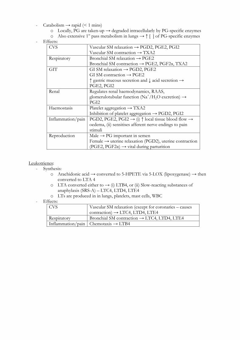
- Catabolism → rapid (< 1 mins)
o Locally, PG are taken-up → degraded intracellularly by PG-specific enzymes o Also extensive 1st pass metabolism in lungs → ↑ [ ] of PG-specific enzymes
- Effects: CVS Vascular SM relaxation → PGD2, PGE2, PGI2
Vascular SM contraction → TXA2 Respiratory Bronchial SM relaxation → PGE2
Bronchial SM contraction → PGE2, PGF2α, TXA2 GIT GI SM relaxation → PGD2, PGE2
GI SM contraction → PGE2 ↑ gastric mucous secretion and ↓ acid secretion → PGE2, PGI2
Renal Regulates renal haemodynamics, RAAS, glomerulotubular function (Na+/H2O excretion) → PGI2
Haemostasis Platelet aggregation → TXA2 Inhibition of platelet aggregation → PGD2, PGI2
Inflammation/pain PGD2, PGE2, PGI2 → (i) ↑ local tissue blood flow → oedema, (ii) sensitises afferent nerve endings to pain stimuli
Reproduction Male → PG important in semen Female → uterine relaxation (PGD2), uterine contraction (PGE2, PGF2α) → vital during parturition
Leukotrienes:
- Synthesis: o Arachidonic acid → converted to 5-HPETE via 5-LOX (lipoxygenase) → then
converted to LTA 4 o LTA converted either to → (i) LTB4, or (ii) Slow-reacting substances of
anaphylaxis (SRS-A) – LTC4, LTD4, LTE4 o LTs are produced in in lungs, platelets, mast cells, WBC
- Effects: CVS Vascular SM relaxation (except for coronaries – causes
contraction) → LTC4, LTD4, LTE4 Respiratory Bronchial SM contraction → LTC4, LTD4, LTE4 Inflammation/pain Chemotaxis → LTB4
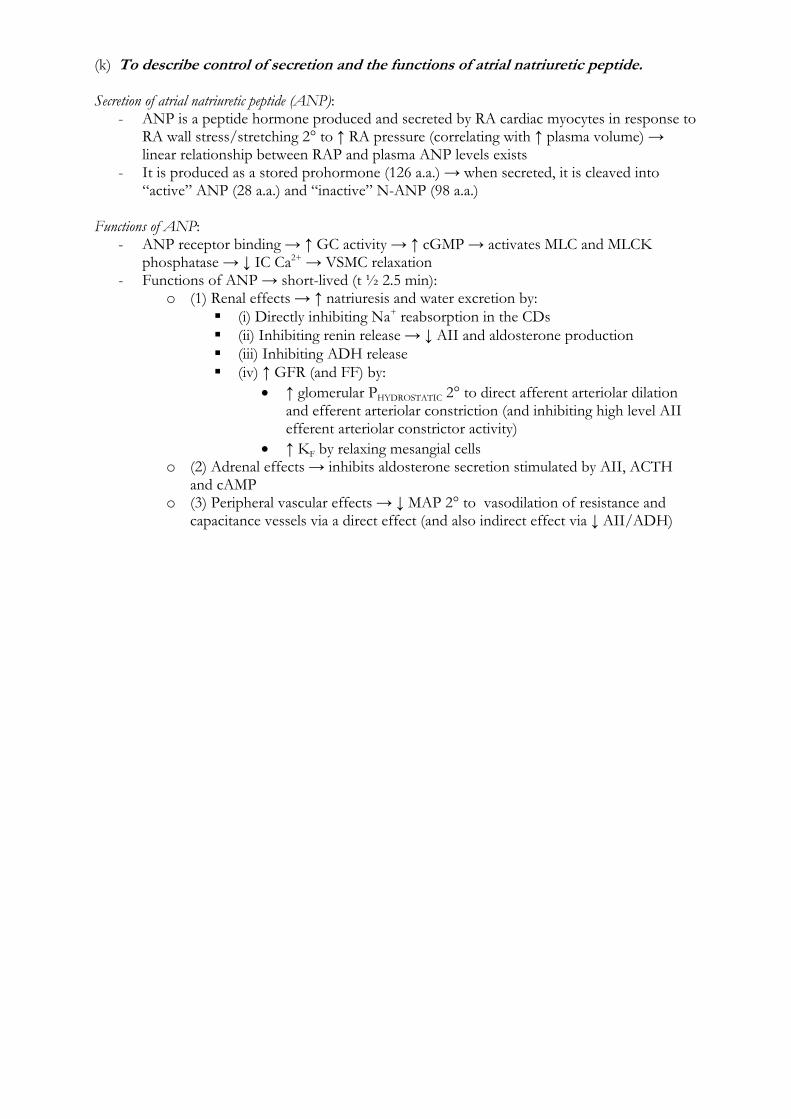
(k) To describe control of secretion and the functions of atrial natriuretic peptide. Secretion of atrial natriuretic peptide (ANP):
- ANP is a peptide hormone produced and secreted by RA cardiac myocytes in response to RA wall stress/stretching 2° to ↑ RA pressure (correlating with ↑ plasma volume) → linear relationship between RAP and plasma ANP levels exists
- It is produced as a stored prohormone (126 a.a.) → when secreted, it is cleaved into “active” ANP (28 a.a.) and “inactive” N-ANP (98 a.a.)
Functions of ANP:
- ANP receptor binding → ↑ GC activity → ↑ cGMP → activates MLC and MLCK phosphatase → ↓ IC Ca2+ → VSMC relaxation
- Functions of ANP → short-lived (t ½ 2.5 min): o (1) Renal effects → ↑ natriuresis and water excretion by:
(i) Directly inhibiting Na+ reabsorption in the CDs (ii) Inhibiting renin release → ↓ AII and aldosterone production (iii) Inhibiting ADH release (iv) ↑ GFR (and FF) by:
↑ glomerular PHYDROSTATIC 2° to direct afferent arteriolar dilation and efferent arteriolar constriction (and inhibiting high level AII efferent arteriolar constrictor activity)
↑ KF by relaxing mesangial cells o (2) Adrenal effects → inhibits aldosterone secretion stimulated by AII, ACTH
and cAMP o (3) Peripheral vascular effects → ↓ MAP 2° to vasodilation of resistance and
capacitance vessels via a direct effect (and also indirect effect via ↓ AII/ADH)