Embed Size (px)
Citation preview

www.newphytologist.org 247
Forum
Blackwell Publishing LtdOxford, UKNPHNew Phytologist0028-646X1469-8137© The Authors (2008). Journal compilation © New Phytologist (2008)May 200800??????LettersCommentary Commentary
Commentary
Regulation of cellulose synthesis – aNOther player in the game?
Cellulose is a central component in plant cell walls. In theprimary cell wall (deposited in cells that are still expanding),it is a vital component of the load-bearing network andbecause of its physical properties is important in determiningthe orientation of cell expansion. After a period of cellexpansion, some cells lay down a thick secondary cell wallinside the primary wall. The secondary cell wall provides plantswith the mechanical properties that allow them to standupright, and is a major component in properly functioningxylem vessels. Cellulose is one of the major components ofsecondary cell walls. The importance of cellulose in plantcell walls is reflected in it being the world’s most abundantbiopolymer, with an estimated 180 billion tonnes synthesizedannually (Englehardt, 1995). Despite this importance, ourunderstanding of how cellulose is synthesized, and how thissynthesis is regulated, is still incomplete. In this issue of NewPhytologist (pp. 386–396), Correa-Aragunde et al. describehow the signalling molecule nitric oxide (NO) modulatescellulose synthesis in tomato (Solanum lycopersicum) roots.
Pharmaceutical application of the NO donor sodiumnitroprusside (SNP) was used to investigate incorporation ofradiolabelled glucose into cellulose. Low (pmolar) concentrationsof NO increased incorporation of radiolabelled glucoseinto the cellulose fraction in roots, whereas higher (nmolar)concentrations reduced incorporation into cellulose. Theseeffects were transient and reversible, as determined by useof an NO scavenger. Microscopic analysis of root structuresuggested that these differences were caused by effects onprimary cell wall synthesis. Root length was reduced in plantstreated with higher concentrations of NO and was accompaniedby reduced cortical cell length and an apparent swelling of theroot, phenotypes that are frequently observed in Arabidopsismutants affected in primary cell wall cellulose synthesis.Three different cellulose synthase (CesA) catalytic subunits aregenerally considered to be required for cellulose synthesis,the three subunits being different in primary and secondarycell walls. Correa-Aragunde et al. identified three CesA tran-scripts from tomato cDNA libraries that are likely to beinvolved in primary cell wall cellulose synthesis, based on thesimilarity to three genes involved in this process in potato(Solanum tuberosum). The level of transcript of these threegenes was slightly reduced by treatment with high concentrations
of NO, suggesting that it may be at a transcriptional level thatNO affects cellulose synthesis. Nitric oxide signalling in plantsis an area in which there is still much to learn, and has beencovered in a recent review (Wilson et al., 2008). It is clear,however, that NO is important as a signalling molecule in anumber of processes in plants, and the description in this issueof its effect on modulating cellulose synthesis adds to agrowing list of pathways that are affected by NO.
‘It is the organization of multiple glucan chains into
microfibrils that is absolutely central to the physical
properties that they confer once deposited in the cell wall.’
Cellulose synthesis is central to plant development, but theway in which it is regulated is still unclear and is currently anarea of intense research. Cellulose is a simple polymer ofunbranched β-1,4-linked glucan chains, with successiveglucose residues inverted 180 degrees to form a flat ribbon inwhich the repeating unit is cellobiose. These parallel chainsare then able to form extensive hydrogen bonds betweenindividual glucan chains resulting in crystallization of multiplechains into cellulose microfibrils – insoluble, cable-likestructures. It is the organization of multiple glucan chains intomicrofibrils that is absolutely central to the physical propertiesthat they confer once deposited in the cell wall. This organizedstructure of the microfibril is a direct result of the organizationof the protein complex that synthesizes cellulose. The plasmamembrane-bound cellulose synthase complex (CSC) is a large(> 4 MDa) protein complex that includes multiple copies ofthree different CesA proteins. It is currently not known ifthere are any other protein components. The CSC can bevisualized at the plasma membrane in freeze-fracture electronmicroscopy as hexameric structures, which gives rise to thembeing known as rosettes. It is the organization of multiple copiesof the three CesA proteins into a defined arrangement thatallows the simultaneous synthesis of multiple β-1,4-linkedglucan chains in a conformation that allows them to hydrogenbond and crystallize into the functional unit of cellulose, themicrofibril (Fig. 1). The three CesAs required are different inprimary and secondary cell wall CSCs, and the presence andactivity of all three proteins is required for correct cellulose

Commentary
www.newphytologist.org © The Authors (2008). Journal compilation © New Phytologist (2008)
Forum248
synthesis (Gardiner et al., 2003; Taylor et al., 2003; Desprezet al., 2007; Persson et al., 2007).
Despite the importance of cellulose synthesis to plants,how it is regulated is not well understood. There are a numberof different levels at which regulation of cellulose synthesismay occur, and much of our understanding has come fromthe use of the model plant Arabidopsis. Our current under-standing of secondary cell wall cellulose synthesis is moreadvanced than that of primary cell wall synthesis, mainlybecause of the fact that plants lacking secondary cell wallcellulose are viable whereas plants lacking primary cell wallcellulose die at a very early stage of development. One con-sequence of a lack of secondary cell wall cellulose is a collapseof xylem vessels, a result of them being unable to withstandthe negative pressure generated by the transpiration stream(Fig. 2) (Turner & Somerville, 1997). In Arabidopsis, anetwork of transcription factors have been identified thatregulate secondary cell wall synthesis in different tissues (for arecent review of genes involved in regulating cellulose synthesissee Taylor, 2008 and references therein). These transcriptionfactors appear to co-ordinately regulate synthesis of all thecomponents of the secondary cell wall (cellulose, lignin andxylan). It is likely, however, that there is a cascade of transcriptionfactors regulating secondary cell wall synthesis, and that furtherdissection of these networks may identify transcription factorsinvolved in the activation of the individual pathways for thethree major secondary cell wall polymers, including cellulose.A second level of regulation is likely to exist in the assembly of
the CSC. The CSC is assembled in the endoplasmic reticulumand then transported intact to the plasma membrane. Theway in which the three catalytic subunits are assembled into therosette is currently unknown, but there is clearly a requirementfor defined and specific interactions between the subunits,and the organization of such a large protein complex islikely to require the assistance provided by as yet unidentifiedmolecular chaperones.
There is also a requirement for feedback regulation ofcellulose synthesis from the cell wall, where the cellulosemicrofibrils are deposited, and the cytosol, where the catalyticdomains of the proteins reside (Fig. 1a). One way in whichthis signalling across the plasma membrane could occur is byreceptor kinases. These proteins, of which there is a diversefamily of over 600 in plants, contain an extracellular ‘sensing’domain and a linked intracellular kinase domain that cantrigger a kinase cascade resulting in phosphorylation of targetproteins (for a recent review of cell wall signalling seeHumphrey et al., 2007). Phosphorylation sites on the cellulosesynthase catalytic subunits have been identified (Nuhse et al.,2004; Taylor, 2007) but the precise role of phosphorylation atthese sites has yet to be elucidated.
It is clear that there are multiple levels at which regulationof cellulose synthesis occurs. Despite its central role in plants,our knowledge of how cellulose synthesis is regulated is stillvery rudimentary and has to date mainly concentrated ondevelopmental regulation. The study by Correa-Aragundeet al. in this issue demonstrating that NO affects cellulosesynthesis in tomato roots contributes important knowledgeon environmental regulation. Resolving some of the unansweredquestions about cellulose synthesis is essential if we are tounderstand fundamental processes in plant development aswell as better utilize the vast quantities of sugars contained inplant cell walls as a source of bio-energy to combat global
Fig. 1 Model of the cellulose synthase rosette. (a) A ‘cross-section’ through a rosette is shown, each ‘lobe’ containing a number of catalytic subunits. The active sites of these subunits are cytosolic, and the extending cellulose microfibril crystallizes as it enters the cell wall. (b) Plan view. Each of the six ‘lobes’ is predicted to contain multiple copies of three different ‘types’ of cellulose synthase (CesA) proteins. The exact number of CesA proteins contained within a lobe, their stoichiometry and their specific interactions are currently unknown.
Fig. 2 Arabidopsis stem cross-sections of (a) wild-type and (b) a secondary cell wall cellulose-deficient mutant stained with toluidine blue. xe, xylem elements. Note the collapsed xylem elements in (b). Bars represent 0.05 mm.
New Phytologist (2008) 179: 247–249 www.newphytologist.org © The Author (2008). Journal compilation © New Phytologist (2008)

Letters
© The Authors (2008). Journal compilation © New Phytologist (2008) www.newphytologist.org
Forum 249
climate change. Recent developments have given us hopethat one day we may better understand the synthesis ofcellulose, and how it is regulated both developmentally andenvironmentally.
Acknowledgements
NGT is supported by a Royal Society University ResearchFellowship.
Neil G. Taylor
Centre for Novel Agricultural Products, Department ofBiology, University of York, York YO10 5DD,
UK (tel +44 1904 328756; fax +44 1904 328762;email [email protected])
References
Correa-Aragunde N, Lombardo C, Lamattina L. 2008. Nitric oxide: an active nitrogen molecule that modulates cellulose synthesis in tomato roots. New Phytologist 179: 386–396.
Desprez T, Juraniec M, Crowell EF, Jouy H, Pochylova Z, Parcy F, Hofte H, Gonneau M, Vernhettes S. 2007. Organization of cellulose synthase complexes involved in primary cell wall synthesis in Arabidopsis thaliana. Proceedings of the National Academy of Sciences, USA 104: 15572–15577.
Englehardt J. 1995. Sources, industrial derivatives and commercial applications of cellulose. Carbohydrates in Europe 12: 5–14.
Gardiner JC, Taylor NG, Turner SR. 2003. Control of cellulose synthase complex localization in developing xylem. The Plant Cell 15: 1740–1748.
Humphrey TV, Bonetta DT, Goring DR. 2007. Sentinels at the wall: cell wall receptors and sensors. New Phytologist 176: 7–21.
Nuhse TS, Stensballe A, Jensen ON, Peck SC. 2004. Phosphoproteomics of the Arabidopsis plasma membrane and a new phosphorylation site database. The Plant Cell 16: 2394–2405.
Persson S, Paredez A, Carroll A, Palsdottir H, Doblin M, Poindexter P, Khitrov N, Auer M, Somerville CR. 2007. Genetic evidence for three unique components in primary cell-wall cellulose synthase complexes in Arabidopsis. Proceedings of the National Academy of Sciences, USA 104: 15566–15571.
Taylor NG. 2007. Identification of cellulose synthase AtCesA7 (IRX3) in vivo phosphorylation sites – a potential role in regulating protein degradation. Plant Molecular Biology 64: 161–171.
Taylor NG. 2008. Cellulose biosynthesis and deposition in higher plants. New Phytologist 178: 239–252.
Taylor NG, Howells RM, Huttly AK, Vickers K, Turner SR. 2003. Interactions among three distinct CesA proteins essential for cellulose synthesis. Proceedings of the National Academy of Sciences, USA 100: 1450–1455.
Turner SR, Somerville CR. 1997. Collapsed xylem phenotype of Arabidopsis identifies mutants deficient in cellulose deposition in the secondary cell wall. The Plant Cell 9: 689–701.
Wilson ID, Neill SJ, Hancock JT. 2008. Nitric oxide synthesis and signalling in plants. Plant, Cell & Environment 31: 622–631.
Key words: cell wall, cellulose, cellulose synthesis, nitric oxide, plant development, regulation.252710.1111/j.1469-8137.2008.02527.xMay 200800249???250???LettersLetters
Letters
Letters
Enchilada redux: how complete is your genome sequence?
In 2004, Jorgensen made a pungent argument for sequencingentire plant genomes, entitled ‘Sequencing maize: just samplethe salsa or go for the whole enchilada?’ (Jorgensen, 2004).For some of us, the genes in the genome are the enchilada, andthe 80–90% plus of repeats in genomes, like those of maizeand barley, amount to eight to nine orders of refried beans.Although we like these refritos more than most, we think thatscientists would digest the data better, and produce lessgaseous reports, if they mostly concentrated on the genes(as most do). Now, as multiple plant genomes proceedtowards completed or draft full-genome status, it seems atimely moment to revisit this issue. Although the question ofcompleteness is largely moot with current technology (no
higher eukaryotic genome has been fully sequenced, althoughthe nematode Caenorhabditis elegans and rice (IRGSP, 2005)come close), the argument has now shifted to consider whatdegree of incompleteness is tolerable. We think that thisdiscussion, still somewhat a matter of personal philosophy,needs to continue. However, most importantly, we feel thatinvestigators need to know to what degree the sequencethat has been generated and assembled actually approachescompletion. We propose here a simple and low-cost methodto determine the level of genome sequence completeness,using the Arabidopsis thaliana genome as an example.
Back in ancient genome-sequencing days (i.e. 2000), whenArabidopsis provided the first comprehensive plant genomeanalysis, most sequencing involved clone-by-clone (e.g.BAC-by-BAC) sequencing and assembly across a minimumtiling path (MTP). Some genome projects, like the ongoingmaize genome sequence, continue to follow this approach.However, it is now accepted that a certain level of shotgungenome sequence from whole-genomic DNA is a vitaladjunct to clone-by-clone approaches, in order to account for
New Phytologist (2008) 179: 249–250

Letters
www.newphytologist.org © The Authors (2008). Journal compilation © New Phytologist (2008)
Forum250
sequences not fully represented in the MTP. This idea had notyet been fully conceived when the Arabidopsis genome waspublished, so there was no such accompanying data. Theparticipants in the Arabidopsis Genome Initiative (2000)knew that their c. 115 Mb assembly of largely contiguoussequence did not cover all of the genome, and they estimatedthat approx. 10 Mb of DNA had been missed (mostly inpericentromeric, centromeric and ribosomal DNA regions).This estimate appears to have under-represented the true case.By generating a detailed physical map of Arabidopsis centro-meres, Hosouchi et al. (2002) predicted that > 30 Mb of thegenome had not yet been sequenced. By using an unrelatedapproach (nuclear microfluorescence, with flanking genomesize standards (c. 100 Mb of C. elegans and c. 175 Mb ofDrosophila melanogaster)), Bennett et al. (2003) predictedthat the Arabidopsis genome in the Columbia ecotype wasc. 157 Mb, indicating an absence of > 40 Mb of data, or> 25% of the genome! These two techniques have their ownissues, however, and might be inaccurate in either direction.
Another approach to determine genome sequence assemblycoverage is to compare the results of a whole-genome shotgunsequence analysis with the assembled sequence. If the shotgunsequence data are truly random and present in sufficientquantity, then the percentages of DNA in each sequence typewill be exactly identical to their percentages in the full-genome assembly, if it is complete. For instance, if 25% of agenome comprises some specific repeat (as shown by theshotgun analysis), then this repeat should comprise 25% ofthe completed assembly. In order to test this idea, we sheared,cloned and generated 1583 high-quality random reads fromthe same source of the Columbia Arabidopsis ecotype thatwas used for the full-genome sequence, generating c. 1.36 Mbof data (Liu, 2005). The sequences of these clones indicatedthat c. 18.3% of the genome contained identified repeats,compared with 7.7% in the Arabidopsis genome sequenceassembly that was available in 2005. If one assumes that all ofthe missed DNA comprises known repeats, then the minimumArabidopsis genome size can be calculated to be slightly largerthan 134 Mb. Of course, some of the missed sequences are likelyto be unknown (e.g. low-copy-number and/or centromere-specific) repeats, or even genes. Hence, our prediction is a mini-mum, but there is no possibility that the genome could be smallerthan this size. We therefore predict that the original genome sizeestimate for Arabidopsis was not too inaccurate, with somewhatover 19 Mb of missed sequence.
As a side benefit, a small additional analysis of the shot-gun genome sequence data also allows a prediction to be madeof how many genes may have been missed by an assembly.Once again, using Arabidopsis as an example, we found nogene candidates in our data set that were not identified inthe Arabidopsis genome sequence. From our reconstructionexperiments with known genes, using data with the sizedistribution present in our 1583 reads, we would have alikelihood of c. 66% of identifying genes that are present on
these reads. Taking these factors into account, we predict, witha 95% confidence level, that < 250 genes were missed in theArabidopsis genome-sequencing project, and we have a certaintyof > 70% that 100 genes or fewer were missed (Liu, 2005).
Although we believe that our approach provides an ironcladminimum estimate of sequences and genes missing from theArabidopsis genome assembly, we are also aware that specificbiases against the successful cloning of some sequences couldskew our analysis. For this reason, sequencing approachesthat do not involve cloning (e.g. 454 or Solexa technologies)might provide the most appropriate route to pursue suchconfirmation.
The ease of generating such an analysis of genome sequenceand assembly completion, particularly when most currentsequencing projects already contain large dollops of randomshotgun sequence data, is difficult to overstate. We proposethat such an analysis should be an absolute publicationrequirement for all future genome-sequencing projects(including those in plants) that describe a ‘completed’ genomesequence. Although we can, and should, argue about whetherwe want our genome projects to concentrate on the salsa,enchilada or frijoles, we should all agree that it is necessary toknow how much of the feast that we have ordered, and paidfor, has been set on the table.
Renyi Liu1 and Jeffrey L. Bennetzen2*
1Department of Botany and Plant Sciences, University ofCalifornia, Riverside, CA 92521, USA; 2Department of
Genetics, University of Georgia, Athens, GA 30602,USA(*Author for correspondence:
tel +1 706 542 3698; fax +1 706 583 0972;email [email protected])
References
Bennett MD, Leitch IJ, Price HJ, Johnston JS. 2003. Comparisons with Caenorhabditis (∼100 Mb) and Drosophila (∼175 Mb) using flow cytometry show genome size in Arabidopsis to be ∼157 Mb and thus ∼25% larger than the Arabidopsis Genome Initiative estimate of ∼125 Mb. Annals of Botany 91: 547–557.
Hosouchi T, Kumekawa N, Tsuroka H, Kotani H. 2002. Physical map-based sizes of the centromeric regions of Arabidopsis thaliana chromosomes 1, 2, and 3. DNA Research 9: 117–121.
International Rice Genome Sequencing Project. 2005. The map-based sequence of the rice genome. Nature 436: 793–800.
Jorgensen R. 2004. Sequencing maize: just sample the salsa or go for the whole enchilada? Plant Cell 16: 787–788.
Liu R. 2005. The evolution of gene composition in angiosperms. PhD dissertation, University of Georgia, USA.
The Arabidopsis Genome Initiative. 2000. Analysis of the genome sequence of the flowering plant Arabidopsis thaliana. Nature 408: 796–815.
Key words: Arabidopsis thaliana, DNA sequencing, gene discovery, genome finishing, repetitive DNA content, whole-genome shotgun sequence analysis.253810.1111/j. 1469-8137.2007.02538.xMay 200800249???250???MeetingsMeetings
Meetings
New Phytologist (2008) 179: 249–250

Meetings
© The Authors (2008). Journal compilation © New Phytologist (2008) www.newphytologist.org
Forum 251
Meetings
Plant–microbe and plant–insect interactions meet common grounds
International Conference on Biotic Plant Interactions, Brisbane, Australia, March 2008
Plant–microbe and plant–insect interactions are of globalimportance for agriculture and of high interest to many plantscientists, microbiologists and entomologists. Traditionally,plant–microbe and plant–insect interactions have been lookedat as two separate issues, but in recent years it has becomeclear that the underlying physiological pathways in plantsoverlap substantially (Koornneef & Pieterse, 2008). TheInternational Conference on Biotic Plant Interactions (ICBPI;www.uq.edu.au/plants/icbpi/) brought together scientists andstudents who are interested in plant pathology and in thebeneficial interactions of plants with other organisms, includingviruses, bacteria, fungi, oomycetes, nematodes, insects andother herbivores. To highlight this, two topics from thisyear’s conference – harmful biotic plant interactions, and theinteractions of plants with beneficial microbial communities– are discussed in this article.
‘... identifying the overlapping defence mechanisms
against pathogen and herbivore attack will reveal
new insights into plant function and their responses to
environmental pressures ...’
Harmful biotic plant interactions
Plants are sessile organisms that are exposed to a constantbarrage of environmental stresses which impact on growth,development and reproduction. Important traits, such asyield and the resistance to biotic stress (e.g. pests andpathogens) and abiotic stress (e.g. ultraviolet light, drought,
salinity, high temperature and nutrient starvation) depend oninternal physiological programs and their regulation by signaltransduction pathways. Plants are the major source of foodand biomaterials worldwide but their production is severelycompromised by pathogens that cause disease and reduceyield and quality. The International Panel on Climate Change(IPCC) in their ‘AR4 Synthesis report’ recently predicted thatstresses from climatic extremes will increase and imposesignificant difficulties, including higher susceptibility to pestsand diseases and leading to estimated yield declines of up to50%. In addition, the impact of climate change on plantdefence chemistry, as discussed by Ros Gleadow (MonashUniversity, Victoria, Australia), could reduce the nutritionalvalue of crops, and the anthropogenic increase in CO2 alsocompromises plant defence against invasive insects (Zavalaet al., 2008). Understanding how plants defend themselvesagainst pathogens and herbivores, and how that may be mani-pulated, is therefore of critical importance for successful andsustainable agriculture. Boosting the plant’s defence system bynatural means also means less reliance on environmentallydamaging pesticides.
As highlighted by many speakers at the ICBPI, identifyingthe overlapping defence mechanisms against pathogen andherbivore attack will reveal new insights into plant function andtheir responses to environmental pressures. It could also poten-tially lead to the discovery of unifying principles of plant stresstolerance. Currently, there is a worldwide search for genes thatcan improve crop performance to abiotic and biotic stresses,while plant genetic engineers and breeders increasingly aimtowards producing more robust crop plants with reliableyields (rather than just high yields). Indeed, plant scientistshave the opportunity to make a real impact, for instance bystudying the underlying network of signalling pathwaysand molecules involved in stress responses and how theseregulate both beneficial and harmful biotic interactions. Theconsequent gain in knowledge is critical in the developmentof new biotechnological approaches to benefit sustainableagriculture.
Disease resistance in plants is conferred by recognition, signal transduction and defence activation
Pathogen infection and attack by herbivores result in a numberof molecular and physiological changes in plants (Fig. 1). Thehypersensitive response (HR) is a form of programmed cell-death that is activated by plants after sensing challenge by an
New Phytologist (2008) 179: 251–256

Meetings
www.newphytologist.org © The Authors (2008). Journal compilation © New Phytologist (2008)
Forum252
avirulent pathogen. At the ICBPI the early recognition andsignal transduction cascades, triggered by pathogen-associatedmolecular patterns and leading to resistance, were discussed atthe molecular level for several pathogens, including viruses(tobacco mosaic virus), bacteria (Pseudomonas syringae andXanthomonas oryzae), fungi (powdery mildews and fluxrust), oomycetes (Phytophthora infestans) and insects. ShaunaSomerville (Energy Bioscience Institute, Berkeley, USA)highlighted in her presentation that the plant cell-wallcomposition and alterations in this offer a first line ofdefence. However, in addition, plants also synthesize varioussignalling molecules, such as salicylic acid (SA), jasmonic acid(JA) and ethylene (ETH), all of which orchestrate a complexand interactive network of signalling pathways (Jones &Dangl, 2006; de Wit, 2007; Koornneef & Pieterse, 2008). Asa result, pathogenesis-related (PR) proteins with directdefensive roles often accumulate in both pest/pathogen-challenged and unchallenged (systemic) tissue of the sameplant.
Plant defence responses that are most effective against agroup of pathogens differ depending on the life style ofparticular pathogens, which typically either require living cells(biotroph) or dead tissues (necrotroph) for proliferation, butmany pathogens also alter their life style during diseaseprogression. In Arabidopsis, an intact SA signalling pathwayis believed to mediate the resistance to biotrophic pathogens,such as viruses, fungi (e.g. Erysiphe orontii), oomycetes (e.g.Hyaloperonospora parasitica) and bacteria (e.g. P. syringae),whereas the JA–ETH signalling pathway is thought to benecessary for resistance to necrotrophic pathogens, such as thefungus Botrytis cinerea and the bacterium Erwinia carotovora(Thomma et al., 1998; Rojo et al., 2003). Richard Oliver(Murdoch University, Perth, Australia) alerted researchers tothe fact that many crops lack natural resistance genes againstnecrotrophic pathogens and often produce several toxins thatinteract with different host-susceptibility gene products.Similarly, as discussed by Corné Pieterse (Utrecht University,the Netherlands) and Karam Singh (CSIRO Plant Industry,
Fig. 1 Representation of the complexity of the plant’s induced defence response to pathogens and insect herbivores. The interaction of Arabidopsis thaliana with pathogens and insects with different lifestyles or feeding modes results in the production of different defence signal signatures and blends of volatile organic compounds (VOC). Cross-talk between salicylic acid (SA)-, jasmonic acid (JA)- and ethylene (ETH)-dependent signalling pathways shapes the direct induced defence response, while the VOCs play a role in indirect defence.
New Phytologist (2008) 179: 251–256
Meetings
© The Authors (2008). Journal compilation © New Phytologist (2008) www.newphytologist.org
Forum 253
Floreat, Australia), different insects induce very different pathwaysaccording to their feeding mechanism and behaviour. Forexample, phloem-feeding insects, such as aphids and whiteflies, have been shown to activate the SA pathway, while tissue-chewing insects, such as leaf hoppers and caterpillars, generallyactivate the JA pathway. Furthermore, different plant volatileorganic compounds (VOCs) may attract or repel insects and/or their predators, leading to defence activation in neighbouringplants (Myron Zalucki (University of Queensland, Australia),Alexandre Il-Ichev (Tatura Centre, Victoria, Australia), Kaplanet al., 2008). Kevin Gould (University of Otago, Dunedin,New Zealand) also discussed how red anthocyanin colorationwas believed to play a role in the protection of leaves againstthe ravages of insect herbivores. A new approach for insectresistance, involving plant-mediated insect gene silencing, waspresented by Xiao-Ya Chen (Chinese Academy of Sciences,Shanghai, China). He showed how Arabidopsis plants thatwere engineered to produce double-stranded RNA whichinterferes with a cotton bollworm P450 gene led to stuntedlarval growth.
For effective defence, plants need to mount a targetedresponse to pathogen/herbivore invasion which activates onlythe genes and pathways that are required, whereas others needto be suppressed to conserve resources (Koornneef & Pieterse,2008). Brigitte Mauch-Mani (Université de Neuchâtel, Swit-zerland) demonstrated this, showing how priming is a physi-ological state that may offer these advantages. Priming enablesplants to mount different cellular defence responses morestrongly or more rapidly when attacked by pathogens orinsects or in response to abiotic stress. Priming can also beinduced by treatment with natural and synthetic compoundsand by beneficial microorganisms (Harman et al., 2004;Beckers & Conrath, 2007). Well known, also at a commercial
level, is the use of some rhizobacteria and antagonistic fungiof the genus Trichoderma, as presented by Matteo Lorito(University of Naples, Italy).
Interactions of plants with microbial communities
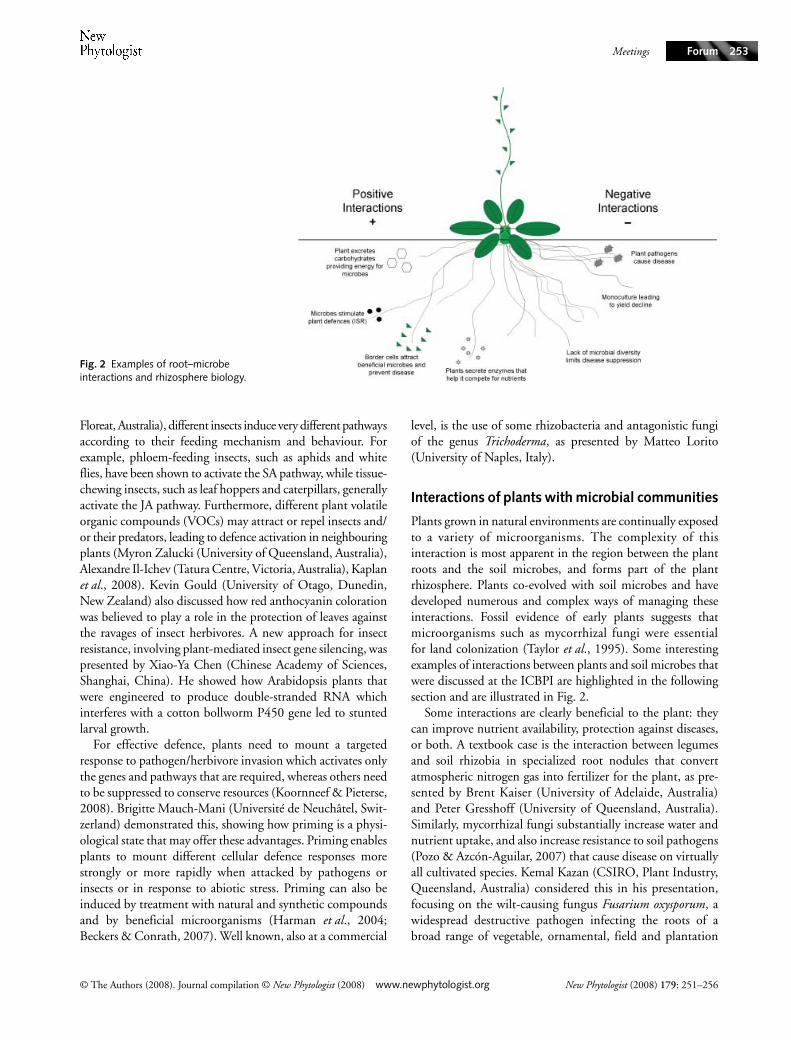
Plants grown in natural environments are continually exposedto a variety of microorganisms. The complexity of thisinteraction is most apparent in the region between the plantroots and the soil microbes, and forms part of the plantrhizosphere. Plants co-evolved with soil microbes and havedeveloped numerous and complex ways of managing theseinteractions. Fossil evidence of early plants suggests thatmicroorganisms such as mycorrhizal fungi were essentialfor land colonization (Taylor et al., 1995). Some interestingexamples of interactions between plants and soil microbes thatwere discussed at the ICBPI are highlighted in the followingsection and are illustrated in Fig. 2.
Some interactions are clearly beneficial to the plant: theycan improve nutrient availability, protection against diseases,or both. A textbook case is the interaction between legumesand soil rhizobia in specialized root nodules that convertatmospheric nitrogen gas into fertilizer for the plant, as pre-sented by Brent Kaiser (University of Adelaide, Australia)and Peter Gresshoff (University of Queensland, Australia).Similarly, mycorrhizal fungi substantially increase water andnutrient uptake, and also increase resistance to soil pathogens(Pozo & Azcón-Aguilar, 2007) that cause disease on virtuallyall cultivated species. Kemal Kazan (CSIRO, Plant Industry,Queensland, Australia) considered this in his presentation,focusing on the wilt-causing fungus Fusarium oxysporum, awidespread destructive pathogen infecting the roots of abroad range of vegetable, ornamental, field and plantation
Fig. 2 Examples of root–microbe interactions and rhizosphere biology.
New Phytologist (2008) 179: 251–256

Meetings
www.newphytologist.org © The Authors (2008). Journal compilation © New Phytologist (2008)
Forum254
crops (e.g. wheat, cotton, tomato and banana). However, themajority of plant–microbe interactions are far more subtleand often involve more than two partners. For example, yielddecline occurs when an agricultural crop has been grown asan exclusive monoculture on the same soil over a period ofyears. The effect manifests as a significant decrease in yieldswith each progressive season, with losses of more than 30% incrops, such as Australian sugarcane (Ken McGrath, Universityof Queensland, Australia). Yield decline has a major microbialcomponent, as fumigation of affected soils reverses the declinein biomass yield. The introduction of crop rotation, hencebreaking the monoculture lineage, can increase microbialdiversity and yield in subsequent seasons (Pankhurst et al.,2005).
Examples of beneficial rhizosphere interactions
Over the last few years, the field of rhizosphere biology hasrecognized the biological importance of root exudates inmediating interactions with other plants and microbes. Plantsconstantly secrete a diverse combination of antimicrobial rootexudates, which appears to limit the number of microbes thatcan form a compatible interaction with the plant, resulting indisease (Bais et al., 2005). Surprisingly, plant roots secreteup to 21% (and sometimes more) of all photosyntheticallyfixed carbon into the rhizosphere through root exudates(Marschner, 1995). Obviously, the plant is gaining a significantbenefit to warrant this large energy expense. For example,root-derived antimicrobial exudates from Arabidopsis conferredresistance to a wide range of bacterial pathogens, while apathogen that was resistant to these compounds blockedtheir synthesis and exudation, resulting in disease (Bais et al.,2005).
Plant roots actively compete for organic nitrogen sources,such as protein. While the plant invests time and energyattempting to manipulate the soil microbiome, the existingmicrobial population has a marked impact on the growth ofthe plant. Roots secrete significant amounts of proteases,which facilitate the uptake of organic sources of nitrogen,such as amino acids. Interestingly, plant roots are even ableto take up whole proteins, probably via endocytosis, thusactively competing with microbes for organic nitrogen sources(Paungfoo-Lonhienne et al., 2008).
Aside from direct exudation, the roots of some plants canrelease border cells from the root tips into the rhizosphereduring the normal growth process (Vicré et al., 2005). Thesecells remain alive, being separated from the main plant butacting as agents for the plant’s manipulation of the rhizo-sphere. For example, it has been shown that these cells canproduce compounds that can immobilize nematodes, as wellas alter the attachment of bacteria to the plant root (Vicréet al., 2005). While it is clear that plant roots and border cellsexude many compounds that affect certain microorganisms,very little is known about what effect this has on entire microbial
communities in the rhizosphere. Recently it has been shownthat root exudates can vividly change the composition of thesoil fungal community (Broeckling et al., 2008).
Some species of the microbial rhizosphere can interactwith the plant in a nonpathogenic manner to stimulate theproduction of plant defence responses. This effect, knownas induced systemic resistance (ISR), can provide dramaticincreases in resistance to a diverse range of plant pathogens,and has been shown to be effective under agricultural fieldconditions. Additionally, microbial populations from differ-ent soils can alter agronomic performance, resulting in changesin yields for several crops (Watt et al., 2006).
Several presentations given at the ICBPI, including thatby Chao-Ying Chen (National Taiwan University, Taiwan),focussed on the occurrence of soils that can actively preventdiseases from infecting pathogens. This rhizospheric effecthas recently been investigated more in detail and the so-called‘suppressive soils’ have been shown to prevent the infection ofmany soil-borne pathogens (Borneman & Becker, 2007). Ithas been demonstrated that a small amount of soil from asuppressive field can be used successfully to ‘inoculate’ othernonsuppressive fields, transferring this suppressive ability.Suppressive soils have been shown to be effective against manydiseases, including Fusarium wilt and nematode infestation.The molecular and microbiological basis of this phenomenonhas been partially clarified also with the aid of modernfunctional genomics techniques, including proteomics andmetabolomics (Marra et al., 2006). One of the most beneficialoutcomes is the selection of superior biocontrol strains, whichcan then be applied worldwide as bioagents. Highly effectiveisolates of Trichoderma, Pseudomonas, Agrobacterium, Bacillus,Streptomyces, Coniothyrium, Azospirillum and nonpathogenicFusarium species are the active ingredients in over 150 com-mercial formulations, acting in many cases as both biopesticidesand biofertilizers. The global use of these products is beginningto make a significant impact in agriculture by improvingyields while alleviating some of the negative effects such aspollution, loss of soil microflora and dependence on pesticides.Although the full potential of the natural germplasm frombeneficial rhizospheric agents is far from being fully understoodand exploited, it is certainly a tool that could be employed inthe future.
The ICBPI meeting in Brisbane clearly demonstrated thatplant defences against pathogens and insect herbivores areregulated by a network of interconnecting signalling pathways.The signalling networks that are activated by the plant inresponse to parasitic and beneficial organisms also overlap,which indicates that the regulation of the adaptive responseof the plant is finely balanced between protection againstaggressors and acquisition of benefits. Future research on howplants are able to cope with different harmful and beneficialbiotic interactions will certainly yield exciting new informa-tion that can be utilized for the development of novel crop-protection strategies. The next ICBPI meeting is planned for
New Phytologist (2008) 179: 251–256

Meetings
© The Authors (2008). Journal compilation © New Phytologist (2008) www.newphytologist.org
Forum 255
2010 in Shanghai and will no doubt provide an interestingupdate on the current progress being made.
Peer M. Schenk1*, Kenneth C. McGrath1, Matteo Lorito2
and Corné M. J. Pieterse3
1School of Integrative Biology, The University ofQueensland, St Lucia, Queensland 4105, Australia;
2University of Naples, Department of Arboriculture, Botanyand Plant Pathology, Via Università, 100, 80055 Portici
(NA), Italy; 3Utrecht University, Institute of EnvironmentalBiology, Plant-Microbe Interactions, Sorbonnelaan 16, 3584
CA Utrecht, the Netherlands
(*Author for correspondence:tel +61 7 33658817; fax +61 7 33651699; email
References
Bais HP, Prithiviraj B, Jha AK, Ausubel FM, Vivanco JM. 2005. Mediation of pathogen resistance by exudation of antimicrobials from roots. Nature 434: 217–221
Beckers GJ, Conrath U. 2007. Priming for stress resistance: from the lab to the field. Current Opinion in Plant Biology 10: 425–431.
Borneman J, Becker JO. 2007. Identifying microorganisms involved in specific pathogen suppression in soil. Annual Reviews in Phytopathology 45: 153–172.
Broeckling CD, Broz AK, Bergelson J, Manter DK, Vivanco JM. 2008. Root exudates regulate soil fungal community composition and diversity. Applied and Environmental Microbiology 74: 738–744.
Harman GE, Howell CR, Chet I, Viterbo A, Lorito M. 2004. Trichoderma species – opportunistic, avirulent plant symbionts. Nature Reviews Microbiology 2: 43–56.
Jones JDG, Dangl JL. 2006. The plant immune system. Nature 444: 323–329.
Kaplan I, Halitschke R, Kessler A, Sardanelli S, Denno RF. 2008. Constitutive and induced defenses to herbivory in above- and belowground plant tissues. Ecology 89: 392–406.
Koornneef A, Pieterse CMJ. 2008. Cross-talk in defense signaling. Plant Physiology 146: 839–844.
Marra R, Ambrosino P, Carbone V, Vinale F, Woo SL, Ruocco M, Ciliento R, Lanzuise S, Ferraioli S, Soriente I et al. 2006. Study of the three-way interaction between Trichoderma atroviride, plant and fungal pathogens by using a proteomic approach. Current Genetics 50: 307–321.
Marschner H. 1995. Mineral nutrition of higher plants, 2nd edn. London, UK: Academic Press.
Pankhurst CE, Stirling GR, Magarey RC, Blair BL, Holt JA, Bell MJ, Garside AL. 2005. Quantification of the effects of rotation breaks on soil biological properties and their impact on yield decline in sugarcane. Soil Biology and Biochemistry 37: 1121–1130.
Paungfoo-Lonhienne C, Lonhienne TGA, Rentsch D, Robinson N, Christie M, Webb RI, Gamage HK, Carroll BJ, Schenk PM, Schmidt S. 2008. Plants can use protein as a nitrogen source without assistance from other organisms. Proceedings of the National Academy of Science, USA 105: 4524–4529.
Pozo MJ, Azcón-Aguilar C. 2007. Unraveling mycorrhiza-induced resistance. Current Opinion in Plant Biology 10: 393–398.
Rojo E, Solano R, Sanchez-Serrano JJ. 2003. Interactions between signaling compounds involved in plant defense. Journal of Plant Growth Regulation 22: 82–98.
Taylor TN, Remy W, Hass H, Kerp H 1995. Fossil arbuscular mycorrhizae from the early Devonian. Mycologia 87: 560–573.
Thomma BPHJ, Eggermont K, Penninckx IAM, Mauch-Mani B, Vogelsang R, Cammue BPA, Broekaert WF. 1998. Separate jasmonate-dependent and salicylate-dependent defense-response pathways in Arabidopsis are essential for resistance to distinct microbial pathogens. Proceedings of the National Academy of Science, USA 95: 15107–15111.
Vicré M, Santaella C, Blanchet S, Gateau A, Driouich A. 2005. Root border-like cells of Arabidopsis. Microscopical characterization and role in the interaction with rhizobacteria. Plant Physiology 138: 998–1008.
Watt M, Kirkegaard JA, Passioura JB. 2006. Rhizosphere biology and crop productivity – a review. Australian Journal of Soil Research 44: 299–317.
de Wit PJGM. 2007. How plants recognize pathogens and defend themselves. Cellular and Molecular Life Sciences 64: 2726–2732.
Zavala JA, Casteel CL, Delucia EH, Berenbaum MR. 2008. Anthropogenic increase in carbon dioxide compromises plant defense against invasive insects. Proceedings of the National Academy of Science, USA 105: 5129–5133.
Key words: beneficial interactions, biopesticides, biotic plant interactions, ICBPI, microbial communities, plant defence, plant pathology, rhizosphere biology.
New Phytologist (2008) 179: 251–256

Meetings
www.newphytologist.org © The Authors (2008). Journal compilation © New Phytologist (2008)
Forum256
New Phytologist (2008) 179: 251–256
About New Phytologist
• New Phytologist is owned by a non-profit-making charitable trust dedicated to the promotion of plant science, facilitating projectsfrom symposia to open access for our Tansley reviews. Complete information is available at www.newphytologist.org.
• Regular papers, Letters, Research reviews, Rapid reports and both Modelling/Theory and Methods papers are encouraged.We are committed to rapid processing, from online submission through to publication ‘as-ready’ via OnlineEarly – our averagesubmission to decision time is just 29 days. Online-only colour is free, and essential print colour costs will be met if necessary. Wealso provide 25 offprints as well as a PDF for each article.
• For online summaries and ToC alerts, go to the website and click on ‘Journal online’. You can take out a personal subscription tothe journal for a fraction of the institutional price. Rates start at £135 in Europe/$251 in the USA & Canada for the online edition(click on ‘Subscribe’ at the website).
• If you have any questions, do get in touch with Central Office ([email protected]; tel +44 1524 594691) or, for a localcontact in North America, the US Office ([email protected]; tel +1 865 576 5261).