Embed Size (px)
Citation preview
Emerging Advancesin Rapid Diagnosticsof RespiratoryInfections
David R. Murdoch, MD, MSc, DTM&H, FRACP, FRCPAa,b,*,Lance C. Jennings, PhD, FRCPatha,b, Niranjan Bhat, MDc,Trevor P. Anderson, MScb
KEYWORDS
� Respiratory infection � Pneumonia � Polymerase chain reaction� Diagnostics � Antigen detection
Diagnostic laboratories play a central role in the recognition of new and emerginginfections. The identification of the severe acute respiratory syndrome (SARS) corona-virus in 2003 highlighted how modern diagnostic tools and collaboration between clini-cians, public health professionals, and laboratorians can lead to the rapidcharacterization of a new respiratory pathogen.1 Similarly, the development and rapiddissemination of a polymerase chain reaction (PCR) method to detect the novel H1N1influenza A strain by the US Centers for Disease Control and Prevention in 2009, reliedon the most recent diagnostic technology and played an important role in theresponse to the latest influenza pandemic.2 These events remind us of how muchrecent developments in diagnostics have improved our ability to identify respiratory
D.R.M. and N.B. performed part of this work under the Pneumonia Etiology Research for ChildHealth (PERCH) program at the Johns Hopkins Bloomberg School of Public Health funded bya grant from The Bill & Melinda Gates Foundation. N.B. is supported by grant1KL2RR025006-01 from the National Center for Research Resources (NCRR), a component ofthe National Institutes of Health (NIH), and NIH Roadmap for Medical Research. Its contentsare solely the responsibility of the authors and do not necessarily represent the official viewof NCRR or NIH. Information on NCRR is available at http://www.ncrr.nih.gov/. Informationon Reengineering the Clinical Research Enterprise can be obtained from http://nihroadmap.nih.gov/clinicalresearch/overview-translational.asp.a Department of Pathology, University of Otago Christchurch, PO Box 4345, Christchurch 8140,New Zealandb Microbiology Unit, Canterbury Health Laboratories, PO Box 151, Christchurch 8140, NewZealandc Division of Infectious Diseases, Department of Pediatrics, Johns Hopkins School of Medicine,600 North Wolfe Street, Baltimore, MD 21287, USA* Corresponding author. Department of Pathology, University of Otago Christchurch, PO Box4345, Christchurch 8140, New Zealand.E-mail address: [email protected]
Infect Dis Clin N Am 24 (2010) 791–807doi:10.1016/j.idc.2010.04.006 id.theclinics.com0891-5520/10/$ – see front matter ª 2010 Elsevier Inc. All rights reserved.
Murdoch et al792
viruses. For nonviral respiratory pathogens, developments in laboratory technologyhave been less profound in general, but have still led to modest improvements inthe diagnostic capability.
For the diagnostic microbiology laboratory, the routine evaluation of patients withsuspected respiratory infections continues to rely on methods that have been usedfor a long time: microscopy and culture of respiratory tract specimens, blood cultures,detection of antigens in urine and upper respiratory specimens, and serology. Recentadvances in pneumonia diagnostics have mostly occurred in the areas of antigen andnucleic acid detection. Despite these technological advances, there remain severalmajor challenges that hinder the search for the causes of respiratory infections, partic-ularly for pneumonia.3 These challenges include difficulty collecting lower respiratorytract specimens, problems distinguishing colonization from infection, poor clinical(diagnostic) sensitivity of assays, and often inadequate evaluation of new diagnostics.
This review focuses on recent advances in laboratory diagnostics that enable rapididentification of respiratory pathogens.
ANTIGEN DETECTION
Assays to detect microbial antigens in body fluids have been used for the diagnosis ofrespiratory infections for many years, using various formats such as immunofluores-cence, enzyme-linked immunosorbent assay (ELISA), latex agglutination, coagulation,and chromatographic immunoassay. These methods are the diagnostic tools mosteasily applied as near-patient tests, but development is reliant on the identificationof suitable antigens that are present in detectable quantities in clinical specimens.To date, commercial assays have been developed only for a limited range of patho-gens. The most widely available assays have focused on the detection of selectedbacterial pathogens in urine and the detection of viruses in respiratory specimens.
Among bacterial respiratory pathogens, assays for Streptococcus pneumoniae andLegionella pneumophila are the most developed. A newer generation immunochroma-tographic test that detects the C-polysaccharide cell wall antigen in urine (NOW) hasbeen an important advance in the diagnosis of pneumococcal disease.4 This test hasa sensitivity of 70% to 80% and a specificity of greater than 90% compared withconventional diagnostic methods for detection of pneumococcal pneumonia in adults.Unfortunately, the NOW test cannot be used reliably in children as it also detectspneumococcal carriage.5 Alternative pneumococcal antigens for diagnostic purposes,such as pneumolysin, have shown promising results, although none has been demon-strated to perform better than existing commercial C-polysaccharide antigenassays.6–8 The combination of a pneumolysin-specific antigen detection ELISAtogether with the NOW test may result in a better diagnostic yield because of thehigher specificity of the pneumolysin detection ELISA.7
Detection of soluble Legionella antigen in urine is an established and valuable toolfor the diagnosis of Legionnaires’ disease, although current commercial assays canonly reliably detect infection caused by Legionella pneumophila serogroup 1.9 Someassays have been intended to detect other legionellae,10 although the performanceis not as good as for L pneumophila serogroup 1.
Detection of respiratory viral antigens in respiratory secretions has become animportant diagnostic tool.11–13 Antigen detection using immunofluorescent techniqueswere pioneered in the 1970s, and commercial reagents are now widely used for thedetection of influenza viruses, respiratory syncytial virus (RSV), parainfluenza viruses,adenoviruses, and human metapneumovirus. These assays require technical exper-tise and have the advantage of allowing direct evaluation of specimen quality. More
Rapid Diagnostics 793
recently, commercial rapid diagnostic tests (RDTs) have become widely used for thedetection of influenza or RSV directly in respiratory specimens. These diagnostic testkits, produced as dipsticks, cassettes, or cards, contain internal controls, and a posi-tive result is signaled by a color change. Results are produced by these tests within5 to 40 minutes.
The sensitivity of rapid tests for the detection of seasonal influenza in clinical spec-imens ranges from 10% to 96%,14,15 and varies with virus type or subtype, timing ofspecimen collection, specimen type, patient age, and the test comparator.16,17 Withthe emergence of the pandemic influenza A (H1N1) 2009 virus, RDTs have been widelyused for patient triaging, although there are limited data available on their clinicalaccuracy.18,19 The sensitivity of these assays for detecting this new strain is 10% to69% compared with real-time PCR.20–22 Specificity of RDTs for seasonal influenzais 90% to 100% according to the available data for H1N1 2009.22 Commercial RSVRDTs have sensitivities of 71% to 95% and specificities of 80% to 100% comparedwith culture.23–25 Poorer performance has been observed in adults,26 which may berelated to the decreased viral titer in adults compared with children.
To correctly interpret results of RDTs, the prevalence of influenza or RSV disease ina community must be considered.15 During peak disease activity, positive predictivevalues are highest, but false-negative results more likely. The opposite is true duringtimes of low disease activity.17 When the disease prevalence is low or unknown,RDT results become difficult to interpret and of limited use.17
NUCLEIC ACID AMPLIFICATION TESTS
The use of nucleic acid amplification tests (NAATs) has transformed our understandingof respiratory infections, demonstrating the relevance of new agents such as humanmetapneumovirus, and providing new insights into previously recognized ones suchas rhinoviruses. The progressive commercialization and clinical application of thesemethods is placing them at the forefront of respiratory diagnostics.
NAATs possess several advantages over more traditional techniques for the detec-tion of respiratory pathogens.27 These tests have improved sensitivity for detectingorganisms that are fastidious, no longer viable, or present in small amounts. NAATscan provide rapid genetic information regarding sequence evolution, geographicvariation, or the presence of virulence factors or antibiotic resistance. Their rapid turn-around times allow them a more prominent role in patient management, and the abilityof NAATs to test for multiple pathogens simultaneously has aided in the diagnosis ofnonspecific respiratory syndromes, such as in outbreak settings. Within the labora-tory, NAATs offer enhanced opportunities for automation, and have a lower safetyrisk than culture for the detection of highly virulent pathogens.
Among the NAATs, the PCR is the most common and thoroughly evaluatedmethod.28,29 The PCR formats most relevant for respiratory diagnostics can be clas-sified into conventional, real-time, and multiplex platforms, with various amplicondetection methods such as gel analysis, ELISA, DNA hybridization, or the use of fluo-rescent dyes or chemical tags. Real-time PCR has several features that place it at anadvantage. First, the two steps of amplification and detection are combined in onereaction, increasing the speed and efficiency of testing and reducing the risks of oper-ator error and cross-contamination. Real-time PCR also allows for the possibility ofquantifying the amount of starting nucleic acid material.
Multiplex PCR systems, in which multiple PCR targets are sought after simulta-neously in one reaction, have gained wider acceptance, particularly among commer-cial assays. These systems have the advantage of increasing the number of
Murdoch et al794
pathogens tested for, without increasing the required amount of operator time orspecimen material. Multiplex assays have broadened the scope of respiratory surveil-lance studies, and have also led to the increasing recognition of dual or triple infectionsin the same individual.30 As noted in Table 1, these assays can be differentiated byeither their amplification or detection steps. In the amplification step, all multiplex plat-forms must balance the competing optimal PCR conditions for each individual target,and must overcome problems of competition and inhibition among the various primersand probes. Each platform uses a unique method to address these issues, such asnested primer combinations,30,31 complex primer structures,32,33 and nontraditionalnucleotides.34–36
These assays are even more varied in their detection stages, where the commontask is to differentially detect and report distinct populations of amplified targets.Several platforms involve solid-phase arrays, such as polystyrene microbead suspen-sions that use fluorescent dyes to differentiate targets,34,37,38 or the microchip formatsthat identify targets by binding to a specific physical location.39–41 The former hasbeen developed into platforms detecting 17 to 20 targets, whereas the latter can iden-tify between a few dozen to thousands of targets. The increased breadth of targetsafforded by microarrays, however, comes at the expense of decreased sensitivity.42
Multiplex PCR products can also be distinguished by their size, using resolutiontechniques such as agarose gel electrophoresis to differentiate by weight, andcapillary-based auto-sequencers that identify targets by length and sequence.32,33
Mass spectrometry can also be used for identification, either by the attachment ofhigh molecular weight tags to primers43–46 or by the analysis of specific nucleotidebase ratios that can be resolved by molecular weight.47
Regardless of the platform, all PCR assays require good primer design taking intoconsideration gene target, gene number, mobility of genes between species, stabilityof gene, and the presence of mutations. Bacteria have large genomes with manygenes for a fully functional organism, including their own genes for replication andenzyme product. Owing to the large genome size there are many targets availablefor specific detection of a bacterial species. Housekeeping genes, those genes thatare essential for the survival of the organism, are desirable gene targets becausethey have conserved regions and hypervariable regions (eg, the 16S rRNA gene).Genes found in multicopies will also increase the sensitivity of PCR assays. Thechoices of viral pathogen gene targets are limited because of the limited size of theviral genome. Genes that are highly conserved are desirable targets for PCR becausethey allow the detection of many strains. Other genes that process areas of nucleotidehypervariability caused by genetic mutation should be avoided because changes overprimer and probe sites can cause poor PCR efficiency and the potential for false-negative results. Table 2 lists some of the more common respiratory pathogen targetgenes.
The published literature on NAATs can be difficult to interpret, because studydesigns vary and rarely involve head-to-head comparisons among the differentassays. Calculation of clinical sensitivity and specificity is complicated becauseNAATs often are more sensitive than the reference culture-based standards. Compar-isons of study results are also problematic because of the use of different specimentypes that may have differential yields for pathogens.48 Finally, several NAAT platformsrequire investment in specialized equipment, the cost of which can only be recoveredthrough high-volume testing. Therefore, few NAAT assays for respiratory diagnosis arelicensed for clinical use, and their daily use in clinical practice remains uncommon. Topromote widespread adoption in the future, developers of NAAT diagnostics will needto standardize evaluation methods, particularly in comparison with reference
Rapid Diagnostics 795
techniques, reduce complexity and cost, and better demonstrate their utility in theclinical environment.
NAATs for Specific Respiratory Pathogens
Although NAATs have been developed for all important respiratory pathogens, theclinical application of these tests varies. Perhaps the area that NAATs have had thegreatest impact is for the diagnosis of infections caused by respiratory viruses.29,49,50
For most, if not all, respiratory viruses, detection of viral nucleic acid is the most sensi-tive diagnostic approach, and current ‘‘gold standards’’ (namely, culture and directimmunofluorescence) will be eventually replaced by NAATs.50 PCR has become thediagnostic test of choice for some respiratory viral infections (eg, for influenza duringthe current influenza H1N1 pandemic), and is a useful epidemiologic tool for charac-terizing the role of viruses in various disease states.51 NAATs can provide resultsrapidly and are able to detect many viral pathogens that are unable to be readilydetected by culture. Perhaps more so than for other respiratory pathogens, consider-able effort has been directed toward the development of multiplex assays to enablethe simultaneous detection of multiple viral pathogens. Given the increasingly largenumber of respiratory viruses, this can be a challenging task.
The need for improved diagnostic tools for pneumococcal disease has lead to theevaluation of several NAATs. For pneumonia, PCR has a sensitivity for detectingS pneumoniae in blood samples ranging from 29% to 100%,27 with a tendency forhigher sensitivity in children than adults. The finding of positive pneumococcal PCRresults from asymptomatic control subjects complicates interpretation.52–54 Whentesting sputum samples from adults with pneumonia, PCR positivity has rangedfrom 68% to 100%,27 although it is unclear how often this reflects upper respiratorytract colonization rather than infection.55 Further refinement of PCR assays, includingthe use of multiple targets, has increased the specificity,56 with lytA assays potentiallyoffering advantages over other assays.57,58 Quantification of S pneumoniae DNA loadmay provide additional diagnostic and prognostic information. Quantitative PCR mayhelp distinguish colonization from infection, with a higher bacterial burden in pneumo-coccal disease than in a carrier state.59 High pneumococcal DNA loads in blood havebeen recently shown to be associated with severe disease in various settings.60–62
NAATs have improved the ability of diagnostic laboratories to detect respiratorypathogens that are difficult to culture, such as Mycoplasma pneumoniae, Legionellaspecies, and Chlamydophila pneumoniae. An extensive evaluation of 13 antibodydetection assays using PCR as the comparator standard concluded that few commer-cial serologic assays for detection of M pneumoniae performed with sufficient sensi-tivity and specificity, and highlighted the increasing importance of NAATs.63 IndeedPCR is considered by many to be the method of choice for detection of M pneumoniaeinfection.64 Both upper and lower respiratory tract samples are suitable for testing forM pneumoniae by PCR, although throat swabs and nasopharyngeal samples may bepreferred because of high sensitivity, high specificity, and convenience. In practice,PCR has been successfully used to rapidly diagnose mycoplasma pneumonia duringoutbreaks, and was particularly useful in children, immunocompromised patients, andin early-stage disease.65,66
Legionnaires’ disease can be difficult to diagnose, and NAATs have proven a usefuladjunct to culture and antigen detection.9 PCR has repeatedly been shown to havesensitivity equal to or greater than culture when testing lower respiratory speci-mens.67–73 Legionella DNA can also be detected in nonrespiratory specimens, suchas urine, serum, and peripheral leukocytes,9 although testing these specimen typesis not well established.
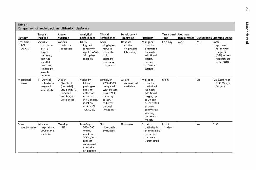
Table 1Comparison of nucleic acid amplification platforms
Platform
Targets
Included
Assays
Available
Analytical
Performance
Clinical
Performance
Development
Timeframe Flexibility
Turnaround
Time
Specimen
Requirements Quantitation Licensing Status
Real-time
PCR
(rtPCR)
Variable;
maximum
of 4–5
targets
per assay,
can run
parallel
reactions,
limited by
sample
volume
Various
in-house
protocols
Likely
highest
sensitivity,
eg, 1 pfu/mL,
10 copies/
reaction
Good;
singleplex
rtPCR is
often the
gold
standard
molecular
diagnostic
Depends
on the
originating
laboratory
Multiplex
must be
optimize
for each
addition
target,
limited
to 5 tota
targets
Half-day None Yes Some
approved
for in vitro
diagnosis
(IVD), others
research use
only (RUO)
Microbead
array
17–20 viral
or bacterial
targets in
each assay
QIagen
(Resplex I
[bacterial]
and II [viral]),
Luminex,
and Eragen
Biosciences
Varies by
kit and
pathogen;
limits of
detection
reported
at 60 copies/
reaction,
or 0.1–100
TCID50/mL
Sensitivity
72%–100%
compared
with culture
plus rtPCR;
varies by
target,
reduced
by dual
infections
All are
commercially
available
Multiplex
must be
optimize
for each
addition
target; u
to 30 can
be detec d
at once;
commerc l
kits may
be slow
modify
6–8 h No IVD (Luminex);
RUO (Qiagen,
Eragen)
Mass
spectrometry
All main
respiratory
viruses and
bacteria
MassTag,
IBIS
MassTag:
500–1000
copies/
reaction, 1
TCID50/mL;
IBIS: 50
copies/well
(basically
singleplex)
Not
rigorously
evaluated
Unknown Requires
optimiza on
of multip x;
detection
methods
unrestric d
Half to
1 day
No RUO
Mu
rdo
chet
al
796
d
al
l
d
al
p
te
ia
to
ti
le
te
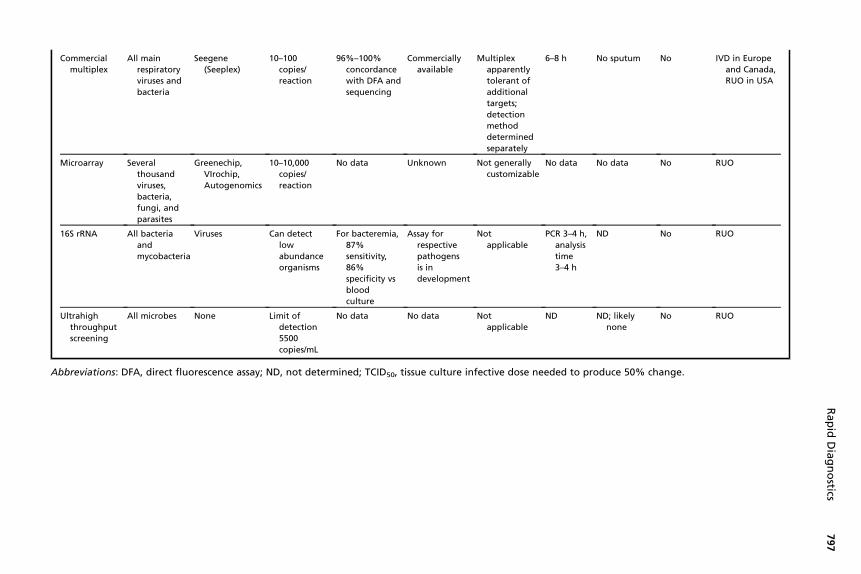
Commercial
multiplex
All main
respiratory
viruses and
bacteria
Seegene
(Seeplex)
10–100
copies/
reaction
96%–100%
concordance
with DFA and
sequencing
Commercially
available
Multiplex
apparently
tolerant of
additional
targets;
detection
method
determined
separately
6–8 h No sputum No IVD in Europe
and Canada,
RUO in USA
Microarray Several
thousand
viruses,
bacteria,
fungi, and
parasites
Greenechip,
VIrochip,
Autogenomics
10–10,000
copies/
reaction
No data Unknown Not generally
customizable
No data No data No RUO
16S rRNA All bacteria
and
mycobacteria
Viruses Can detect
low
abundance
organisms
For bacteremia,
87%
sensitivity,
86%
specificity vs
blood
culture
Assay for
respective
pathogens
is in
development
Not
applicable
PCR 3–4 h,
analysis
time
3–4 h
ND No RUO
Ultrahigh
throughput
screening
All microbes None Limit of
detection
5500
copies/mL
No data No data Not
applicable
ND ND; likely
none
No RUO
Abbreviations: DFA, direct fluorescence assay; ND, not determined; TCID50, tissue culture infective dose needed to produce 50% change.
Rap
idD
iag
no
stics797
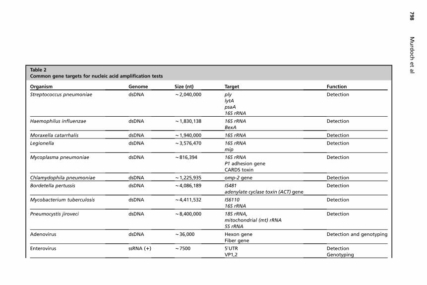
Table 2Common gene targets for nucleic acid amplification tests
Organism Genome Size (nt) Target Function
Streptococcus pneumoniae dsDNA w2,040,000 plylytApsaA16S rRNA
Detection
Haemophilus influenzae dsDNA w1,830,138 16S rRNABexA
Detection
Moraxella catarrhalis dsDNA w1,940,000 16S rRNA Detection
Legionella dsDNA w3,576,470 16S rRNAmip
Detection
Mycoplasma pneumoniae dsDNA w816,394 16S rRNAP1 adhesion geneCARDS toxin
Detection
Chlamydophila pneumoniae dsDNA w1,225,935 omp-2 gene Detection
Bordetella pertussis dsDNA w4,086,189 IS481adenylate cyclase toxin (ACT) gene
Detection
Mycobacterium tuberculosis dsDNA w4,411,532 IS611016S rRNA
Detection
Pneumocystis jiroveci dsDNA w8,400,000 18S rRNA,mitochondrial (mt) rRNA5S rRNA
Detection
Adenovirus dsDNA w36,000 Hexon geneFiber gene
Detection and genotyping
Enterovirus ssRNA (1) w7500 50UTRVP1,2
DetectionGenotyping
Mu
rdo
chet
al
798
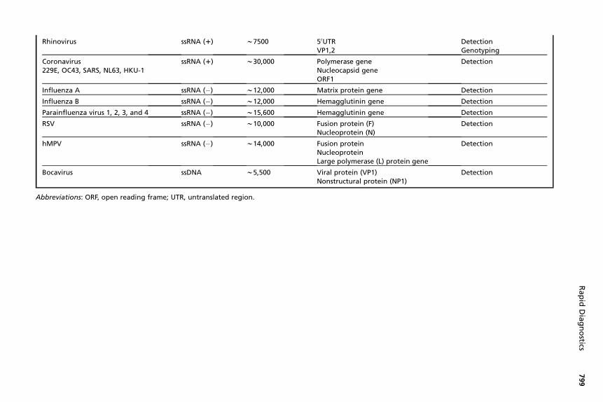
Rhinovirus ssRNA (1) w7500 50UTRVP1,2
DetectionGenotyping
Coronavirus229E, OC43, SARS, NL63, HKU-1
ssRNA (1) w30,000 Polymerase geneNucleocapsid geneORF1
Detection
Influenza A ssRNA (�) w12,000 Matrix protein gene Detection
Influenza B ssRNA (�) w12,000 Hemagglutinin gene Detection
Parainfluenza virus 1, 2, 3, and 4 ssRNA (�) w15,600 Hemagglutinin gene Detection
RSV ssRNA (�) w10,000 Fusion protein (F)Nucleoprotein (N)
Detection
hMPV ssRNA (�) w14,000 Fusion proteinNucleoproteinLarge polymerase (L) protein gene
Detection
Bocavirus ssDNA w5,500 Viral protein (VP1)Nonstructural protein (NP1)
Detection
Abbreviations: ORF, open reading frame; UTR, untranslated region.
Rap
idD
iag
no
stics799
Murdoch et al800
PCR has been extensively evaluated for the rapid diagnosis of C pneumoniae infectionusing various assays.74 A standardized approach to C pneumoniae diagnostic testingwas published in 2001 by the US Centers for Disease Control and Prevention and theCanadian Laboratory Center for Disease Control.75 However, there are still few eval-uations that have extensively used clinical samples, and the great variety in themethods used makes it difficult to make firm conclusions about performance. Tofurther complicate matters, significant interlaboratory discordance of detection rateshave been recorded for some assays.76,77
The diagnostic yield from PCR is consistently greater than for culture when testingnasopharyngeal samples for Bordetella pertussis.27 PCR remains positive for a longerperiod after the onset of symptoms and thus is useful for individuals who present latein their illness.78 In the investigation of a pertussis outbreak, the combination of PCRand culture for samples obtained 2 weeks or less after illness onset and PCR alone forsamples obtained more than 2 weeks after illness onset proved to be the most diag-nostically useful.79
PCR has greater sensitivity than cytologic methods for the detection of Pneumocys-tis jiroveci, although it has been difficult to interpret the common finding ofPCR-positive samples that are negative by standard methods.27 The latter may reflectP jiroveci colonization of uncertain clinical significance. The performance of PCR hasbeen shown to vary with different assays,80 although the results correlate well withclinical evidence of pneumocystis pneumonia.81
The need for improved diagnostic methods for tuberculosis has focused attentionon the potential role of NAATs. Advances in this area have been relatively slow, withNAATs for mycobacteria failing to provide greater sensitivity than culture-basedmethods. The relatively high false-negative rate with NAATs for Mycobacterium tuber-culosis probably reflects a combination of the paucibacillary nature of samples, pres-ence of inhibitors in samples, and suboptimal DNA extraction methods. The situationis changing, with new developments in rapid diagnosis and antibiotic susceptibilitytesting.82 For direct detection of M tuberculosis in respiratory samples, all commercialassays have high specificity (>98%), but variable sensitivities: 90% to 100% forsmear-positive samples and 33% to 100% for smear-negative samples.27 Conse-quently, it is recommended that use of these tests is restricted to only smear-positive samples. Evaluations of PCR for the diagnosis of tuberculosis inhigh-prevalence populations have been promising.83–86 Alternative strategies underdevelopment to diagnose tuberculosis by molecular tools include detection of myco-bacterial DNA in urine87,88 and direct detection in respiratory specimens bymicroarray.89
New Pathogen Discovery
The NAATs discussed herein target known pathogens. When NAATs fail to identify anagent, additional tools are needed to pursue an etiologic diagnosis as might beindicated in an outbreak setting. These additional methods include microarrays andhigh-throughput sequencing.42,90,91 Proteomics also has a potential for being devel-oped as a tool for pathogen discovery.92
BREATH ANALYSIS
Breath analysis is an exciting new area with enormous diagnostic potential.93–95 Alve-olar breath contains many biomarkers derived from the blood by passive diffusionacross the alveolar membrane,93 and also contains direct markers of lung injury.96–98
Breath testing is noninvasive, easily repeatable, and requires minimal specimen
Rapid Diagnostics 801
workup. Various testing methodologies and sample types have been used in breathresearch, usually involving the measurement of exhaled permanent gases, detectionof volatile organic compounds, or analysis of exhaled breath condensate.
The use of breath analysis for the investigation of respiratory infections has not yetbeen extensively evaluated. Electronic nose devices detect volatile molecules as theyinteract with chemical sensor assays.99–101 Based on the reactivity of multiple sensorsto the volatile molecules, an electronic signature is generated. Testing of exhaledbreath by a portable electronic nose has been used to diagnose pneumonia inmechanically ventilated patients.102–104 The clinical impact of this device needs furtherevaluation, but it could be used as a trigger for further diagnostic studies in pneumoniasuch as bronchoscopy.
Microorganisms produce volatile metabolites that may be used as biomarkers.105
Detection of these biomarkers in breath samples by gas chromatography/mass spec-troscopy or similar methods may provide an etiologic diagnosis of respiratory tractinfection. Ideally, specific biomarkers need to be identified, and it may be difficult todiscover unique markers for each pathogen produced in sufficient quantities to enabledetection. Potential biomarkers have been reported for some respiratory pathogens,such as Aspergillus fumigatus106,107 and M tuberculosis,108,109 but it is still uncertainwhether they will prove to be useful as clinical diagnostic tools.
FUTURE PROSPECTS
Diagnostic tests for respiratory infections will continue to evolve and become moreuser-friendly. Antigen-detection assays in immunochromatographic or similar formatsare rapid, simple to perform, and are most easily developed as near-patient tests.These methods are among the most attractive diagnostic tools, but further develop-ment is reliant on the discovery of suitable antigens that can be reliably detected inreadily obtained specimens. NAATs have now been developed to a stage where multi-plex assays that detect the common respiratory pathogens are commercially avail-able, although not all have been rigorously evaluated in clinical settings. Furtherimprovements in design and performance are expected, and an emphasis shouldbe placed on clarifying the clinical usefulness of NAATs, developing standardizedmethods, producing even more user-friendly platforms, and exploring the role ofquantitative assays. New approaches for respiratory pathogen detection are desper-ately needed. Breath analysis is an exciting new area with enormous potential, and itwill be interesting to follow progress in this area over the next few years.
REFERENCES
1. Peiris JSM, Yuen KY, Osterhaus A, et al. Current concepts: the severe acuterespiratory syndrome. N Engl J Med 2003;349:2431–41.
2. WHO Collaborating Center. CDC protocol of real-time RT-PCR for influenzaA (H1N1), 2009. Available at: http://www.who.int/csr/resources/publications/swineflu/CDCRealtimeRTPCR_SwineH1Assay-2009_20090430.pdf. AccessedMay 22, 2010.
3. Murdoch DR, O’Brien KL, Scott JAG, et al. Breathing new life into pneumoniadiagnostics. J Clin Microbiol 2009;47:3405–8.
4. Werno AM, Murdoch DR. Laboratory diagnosis of invasive pneumococcaldisease. Clin Infect Dis 2008;46:926–32.
5. Dowell SF, Garman RL, Liu G, et al. Evaluation of Binax NOW, an assay for thedetection of pneumococcal antigen in urine samples, performed among pedi-atric patients. Clin Infect Dis 2001;32:824–5.
Murdoch et al802
6. Cima-Cabal MD, M�endez FJ, V�azquez F, et al. Immunodetection of pneumolysinin human urine by ELISA. J Microbiol Methods 2003;54:47–55.
7. Garc�ıa-Su�arez MDM, Cima-Cabal MD, Villaverde R, et al. Performance ofa pneumolysin ELISA assay for the diagnosis of pneumococcal infections.J Clin Microbiol 2007;45:3549–54.
8. Rajalakshmi B, Kanungo R, Srinivasan S, et al. Pneumolysin in urine: a rapidantigen detection method to diagnose pneumococcal pneumonia in children.Ind J Med Microbiol 2002;20:183–6.
9. Murdoch DR. Diagnosis of Legionella infection. Clin Infect Dis 2003;36:64–9.10. Harrison T, Uldum S, Alexiou-Daniel S, et al. A multicenter evaluation of the
Biotest legionella urinary antigen EIA. Clin Microbiol Infect 1998;4:359–65.11. Abanses JC, Dowd MD, Simon SD, et al. Impact of rapid influenza testing at
triage on management of febrile infants and young children. Pediatr EmergCare 2006;22:145–9.
12. Jafri HS, Ramilo O, Makari D, et al. Diagnostic virology practices for respiratorysyncytial virus and influenza virus among children in the hospital setting:a national survey. Pediatr Infect Dis J 2007;26:956–8.
13. Jennings LC, Skopnik H, Burckhardt I, et al. Effect of rapid influenza testing onthe clinical management of paediatric influenza. Influenza Other Respi Viruses2009;3:91–8.
14. Hurt AC, Alexander R, Hibbert J, et al. Performance of six influenza rapid tests indetecting human influenza in clinical specimens. J Clin Virol 2007;39:132–5.
15. Uyeki TM. Influenza diagnosis and treatment in children: a review of studies onclinically useful tests and antiviral treatment for influenza. Pediatr Infect Dis J2003;22:164–77.
16. Smit M, Beynon KA, Murdoch DR, et al. Comparison of the NOW Influenza A &B, NOW Flu A, NOW Flu B, and Directigen Flu A1B assays, and immunofluores-cence with viral culture for the detection of influenza A and B viruses. DiagnMicrobiol Infect Dis 2007;57:67–70.
17. World Health Organization. WHO recommendations on the use of rapid testingfor influenza diagnosis. WHO, 2005; Available at: http://www.who.int/csr/disease/avian_influenza/guidelines/rapid_testing/en/index.html. Accessed May22, 2010.
18. Chan KH, Lai ST, Poon LL, et al. Analytical sensitivity of rapid influenza antigendetection tests for swine-origin influenza virus (H1N1). J Clin Virol 2009;45:205–7.
19. Hurt AC, Baas C, Deng YM, et al. Performance of influenza rapid point-of-caretests in the detection of swine lineage A(H1N1) influenza viruses. InfluenzaOther Respir Viruses 2009;3:171–6.
20. Centers for Disease Control and Prevention. Evaluation of rapid influenza diag-nostic tests for detection of novel influenza A (H1N1) virus—United States, 2009.MMWR Morb Mortal Wkly Rep 2009;58:826–9.
21. Faix DJ, Sherman SS, Waterman SH. Rapid-test sensitivity for novel swine-origininfluenza A (H1N1) virus in humans. N Engl J Med 2009;361:728–9.
22. Ginocchio CC, Zhang F, Manji R, et al. Evaluation of multiple test methods for thedetection of the novel 2009 influenza A (H1N1) during the New York Cityoutbreak. J Clin Virol 2009;45:191–5.
23. Borek AP, Clemens SH, Gaskins VK, et al. Respiratory syncytial virus detectionby Remel Xpect, Binax Now RSV, direct immunofluorescent staining, and tissueculture. J Clin Microbiol 2006;44:1105–7.
24. Selvarangan R, Abel D, Hamilton M. Comparison of BD Directigen EZ RSV andBinax NOW RSV tests for rapid detection of respiratory syncytial virus from
Rapid Diagnostics 803
nasopharyngeal aspirates in a pediatric population. Diagn Microbiol Infect Dis2008;62:157–61.
25. Zheng X, Quianzon S, Mu Y, et al. Comparison of two new rapid antigen detec-tion assays for respiratory syncytial virus with another assay and shell vialculture. J Clin Virol 2004;31:130–3.
26. Casiano-Col�on AE, Hulbert BB, Mayer TK, et al. Lack of sensitivity of rapidantigen tests for the diagnosis of respiratory syncytial virus infection in adults.J Clin Virol 2003;28:169–74.
27. Murdoch DR. Molecular genetic methods in the diagnosis of lower respiratorytract infections. APMIS 2004;112:713–27.
28. Barken KB, Haagensen JAJ, Tolker-Nielsen T. Advances in nucleic acid-baseddiagnostics of bacterial infections. Clin Chim Acta 2007;384:1–11.
29. Ieven M. Currently used nucleic acid amplification tests for the detection ofviruses and atypicals in acute respiratory infections. J Clin Virol 2007;40:259–76.
30. Brunstein JD, Cline CL, McKinney S, et al. Evidence from multiplex molecularassays for complex multipathogen interactions in acute respiratory infections.J Clin Microbiol 2008;46:97–102.
31. Li H, McCormac MA, Estes RW, et al. Simultaneous detection and high-throughput identification of a panel of RNA viruses causing respiratory tractinfections. J Clin Microbiol 2007;45:2105–9.
32. Kim SR, Ki CS, Lee NY. Rapid detection and identification of 12 respiratoryviruses using a dual priming oligonucleotide system-based multiplex PCRassay. J Virol Methods 2009;156:111–6.
33. Roh KH, Kim J, Nam MH, et al. Comparison of the Seeplex reverse transcriptionPCR assay with the R-mix viral culture and immunofluorescence techniques fordetection of eight respiratory viruses. Ann Clin Lab Sci 2008;38:41–6.
34. Lee WM, Grindle K, Pappas T, et al. High-throughput, sensitive, and accuratemultiplex PCR-microsphere flow cytometry system for large-scale comprehen-sive detection of respiratory viruses. J Clin Microbiol 2007;45:2626–34.
35. Marshall DJ, Reisdorf E, Harms G, et al. Evaluation of a multiplexed PCR assayfor detection of respiratory viral pathogens in a public health laboratory setting.J Clin Microbiol 2007;45:3875–82.
36. Nolte FS, Marshall DJ, Rasberry C, et al. MultiCode-PLx system for multiplexeddetection of seventeen respiratory viruses. J Clin Microbiol 2007;45:2779–86.
37. Mahony J, Chong S, Merante F, et al. Development of a respiratory virus paneltest for detection of twenty human respiratory viruses by use of multiplex PCRand a fluid microbead-based assay. J Clin Microbiol 2007;45:2965–70.
38. Pabbaraju K, Tokaryk KL, Wong S, et al. Comparison of the Luminex xTAG respi-ratory viral panel with in-house nucleic acid amplification tests for diagnosis ofrespiratory virus infections. J Clin Microbiol 2008;46:3056–62.
39. Chiu CY, Urisman A, Greenhow TL, et al. Utility of DNA microarrays for detectionof viruses in acute respiratory tract infections in children. J Pediatr 2008;153:76–83.
40. Quan P-L, Palacios G, Jabado OJ, et al. Detection of respiratory viruses andsubtype identification of influenza A viruses by GreeneChipResp oligonucleo-tide microarray. J Clin Microbiol 2007;45:2359–64.
41. Raymond F, Carbonneau J, Boucher N, et al. Comparison of automated micro-array detection with real-time PCR assays for detection of respiratory viruses inspecimens obtained from children. J Clin Microbiol 2009;47:743–50.
42. Quan P-L, Briese T, Palacios G, et al. Rapid sequence-based diagnosis of viralinfection. Antiviral Res 2008;79:1–5.

Murdoch et al804
43. Briese T, Palacios G, Kokoris M, et al. Diagnostic system for rapid and sensitivedifferential detection of pathogens. Emerg Infect Dis 2005;11:310–3.
44. Dominguez SR, Briese T, Palacios G, et al. Multiplex MassTag-PCR for respira-tory pathogens in pediatric nasopharyngeal washes negative by conventionaldiagnostic testing shows a high prevalence of viruses belonging to a newlyrecognized rhinovirus clade. J Clin Virol 2008;43:219–22.
45. Lamson D, Renwick N, Kapoor V, et al. MassTag polymerase-chain-reactiondetection of respiratory pathogens, including a new rhinovirus genotype, thatcaused influenza-like illness in New York State during 2004-2005. J Infect Dis2006;194:1398–402.
46. Renwick N, Schweiger B, Kapoor V, et al. A recently identified rhinovirus geno-type is associated with severe respiratory-tract infection in children in Germany.J Infect Dis 2007;196:1754–60.
47. Ecker DJ, Sampath R, Massire C, et al. Ibis T5000: a universal biosensorapproach for microbiology. Nature Rev Microbiol 2008;6:553–8.
48. Loens K, Van Heirstraeten L, Malhotra-Kumar S, et al. Optimal sampling sitesand methods for detection of pathogens possibly causing community-acquired lower respiratory tract infections. J Clin Microbiol 2009;47:21–31.
49. Fox JD. Nucleic acid amplification tests for detection or respiratory viruses.J Clin Virol 2007;40(Suppl 1):S15–23.
50. Mahony JB. Detection of respiratory viruses by molecular methods. Clin Micro-biol Rev 2008;21:716–47.
51. Jennings LC, Anderson TP, Beynon KA, et al. Incidence and characteristics ofviral community-acquired pneumonia in adults. Thorax 2008;63:42–8.
52. Dagan R, Shriker O, Hazan I, et al. Prospective study to determine clinical rele-vance of detection of pneumococcal DNA in sera of children by PCR. J ClinMicrobiol 1998;36:669–73.
53. Rudolph KM, Parkinson AJ, Black CM, et al. Evaluation of polymerase chainreaction for diagnosis of pneumococcal pneumonia. J Clin Microbiol 1993;31:2661–6.
54. Salo P, Ortqvist A, Leinonen M. Diagnosis of bacteremic pneumococcal pneu-monia by amplification of pneumolysin gene fragment in serum. J Infect Dis1995;171:479–82.
55. Murdoch DR, Anderson TP, Beynon KA, et al. Evaluation of a PCR assay fordetection of Streptococcus pneumoniae in respiratory and nonrespiratorysamples from adults with community-acquired pneumonia. J Clin Microbiol2003;41:63–6.
56. Sheppard CL, Harrison TG, Morris R, et al. Autolysin-targeting LightCyclerassay including internal process control for detection of Streptococcus pneumo-niae DNA in clinical samples. J Med Microbiol 2004;53:189–95.
57. Carvalho MGS, Tondella ML, McCaustland K, et al. Evaluation and improvementof real-time PCR assays targeting lytA, ply, and psaA genes for detection ofpneumococcal DNA. J Clin Microbiol 2007;45:2460–6.
58. Smith MD, Sheppard CL, Hogan A, et al. Diagnosis of Streptococcus pneumo-niae infections in adults with bacteremia and community-acquired pneumonia:clinical comparison of pneumococcal PCR and urinary antigen detection.J Clin Microbiol 2009;47:1046–9.
59. Kais M, Spindler C, Kalin M, et al. Quantitative detection of Streptococcuspneumoniae, Haemophilus influenzae, and Moraxella catarrhalis in lower respi-ratory tract samples by real-time PCR. Diagn Microbiol Infect Dis 2006;55:169–78.
Rapid Diagnostics 805
60. Carrol ED, Guiver M, Nkhoma S, et al. High pneumococcal DNA loads are asso-ciated with mortality in Malawian children with invasive pneumococcal disease.Pediatr Infect Dis J 2007;26:416–22.
61. Peters RPH, de Boer RF, Schuurman T, et al. Streptococcus pneumoniae DNAload in blood as marker of infection in patients with community-acquired pneu-monia. J Clin Microbiol 2009;47:3308–12.
62. Rello J, Lisboa T, Lujan M, et al. Severity of pneumococcal pneumonia associ-ated with genomic bacterial load. Chest 2009;136:832–40.
63. Beersma MFC, Dirven K, van Dam AP, et al. Evaluation of 12 commercial testsand the complement fixation test for Mycoplasma pneumoniae-specific immu-noglobulin G (IgG) and IgM antibodies, with PCR use as the ‘‘gold standard’’.J Clin Microbiol 2005;43:2277–85.
64. Daxboeck F, Krause R, Wenisch C. Laboratory diagnosis of Mycoplasma pneu-moniae infection. Clin Microbiol Infect 2003;9:263–73.
65. Kim NH, Lee JA, Eun BW, et al. Comparison of polymerase chain reaction andthe indirect particle agglutination antibody test for the diagnosis of Mycoplasmapneumoniae pneumonia in children during two outbreaks. Pediatr Infect Dis J2007;26:897–903.
66. Liu F-C, Chen P-Y, Huang F-L, et al. Rapid diagnosis of Mycoplasma pneumo-niae infection in children by polymerase chain reaction. J Microbiol ImmunolInfect 2007;40:507–12.
67. Cloud JL, Carroll KC, Pixton P, et al. Detection of Legionella species in respira-tory specimens using PCR with sequencing confirmation. J Clin Microbiol 2000;38:1709–12.
68. Jaulhac B, Nowicki M, Bornstein N, et al. Detection of Legionella spp. in bron-choalveolar lavage fluids by DNA amplification. J Clin Microbiol 1992;30:920–4.
69. Jonas D, Rosenbaum A, Weyrich S, et al. Enzyme-linked immunoassay fordetection of PCR-amplified DNA of legionellae in bronchoalveolar fluid. J ClinMicrobiol 1995;33:1247–52.
70. Kessler HH, Reinthaler FF, Pschaid A, et al. Rapid detection of Legionellaspecies in bronchoalveolar lavage fluids with the EnviroAmp Legionella PCRamplification and detection kit. J Clin Microbiol 1993;31:3325–8.
71. Lisby G, Dessau R. Construction of a DNA amplification assay for detection ofLegionella species in clinical samples. Eur J Clin Microbiol Infect Dis 1994;13:225–31.
72. Matsiota-Bernard P, Pitsouni E, Legakis N, et al. Evaluation of commercial ampli-fication kit for detection of Legionella pneumophila in clinical specimens. J ClinMicrobiol 1994;32:1503–5.
73. Weir SC, Fischer SH, Stock F, et al. Detection of Legionella by PCR in respiratoryspecimens using a commercially available kit. Am J Clin Pathol 1998;110:295–300.
74. Kumar S, Hammerschlag MR. Acute respiratory infection due to Chlamydiapneumoniae: current status of diagnostic methods. Clin Infect Dis 2007;44:568–76.
75. Dowell SF, Peeling RW, Boman J, et al. Standardizing Chlamydia pneumoniaeassays: recommendations from the Centers for Diseases Control and Prevention(USA) and the Laboratory Centre for Disease Control (Canada). Clin Infect Dis2001;33:492–503.
76. Apfalter P, Assadian O, Blasi F, et al. Reliability of nested PCR for detection ofChlamydia pneumoniae DNA in atheromas: results from a multicenter studyapplying standardized protocols. J Clin Microbiol 2002;40:4428–34.
Murdoch et al806
77. Apfalter P, Blasi F, Boman J, et al. Multicenter comparison trial of DNA extractionmethods and PCR assays for detection of Chlamydia pneumoniae in endarter-ectomy specimens. J Clin Microbiol 2001;39:519–24.
78. Muller FM, Hoppe JE, Wirsing von Kohig CH. Laboratory diagnosis of pertussis:state of the art in 1997. J Clin Microbiol 1997;35:2435–43.
79. Sotir MJ, Cappozzo DL, Warshauer DM, et al. Evaluation of polymerase chainreaction and culture for diagnosis of pertussis in the control of a county-wideoutbreak focused among adolescents and adults. Clin Infect Dis 2007;44:1216–9.
80. Robberts FJL, Liebowitz LD, Chalkley LJ. Polymerase chain reaction detectionof Pneumocystis jiroveci: evaluation of 9 assays. Diagn Microbiol Infect Dis2007;58:385–92.
81. Azoulay E, Bergeron A, Chevret S, et al. Polymerase chain reaction for diag-nosing pneumocystis pneumonia in non-HIV immunocompromised patientswith pulmonary infiltrates. Chest 2009;135:655–61.
82. Balasingham SV, Davidsen T, Szpinda I, et al. Molecular diagnostics intuberculosis: basis and implications for therapy. Mol Diagn Ther 2009;13:137–51.
83. Kibiki GS, Mulder B, van der Ven AJ, et al. Laboratory diagnosis of pulmonarytuberculosis in TB and HIV endemic settings and the contribution of real timePCR for M. tuberculosis in bronchoalveolar lavage fluid. Trop Med Int Health2007;12:1210–7.
84. Kivihya-Ndugga L, van Cleeff M, Juma E, et al. Comparison of PCR with theroutine procedure for diagnosis of tuberculosis in a population with high preva-lences of tuberculosis and human immunodeficiency virus. J Clin Microbiol2004;42:1012–5.
85. Ani A, Okpe S, Akambi M, et al. Comparison of a DNA based PCR method withconventional methods for the detection of M. tuberculosis in Jos, Nigeria.J Infect Dev Ctries 2009;3:470–5.
86. Ben Kahla I, Ben Selma W, Marzouk M, et al. Evaluation of a simplified IS6110PCR for the rapid diagnosis of Mycobacterium tuberculosis in an area with hightuberculosis incidence. Pathol Biol. DOI:10.1016/j.patbio.2009.04.001.
87. Green C, Huggett JF, Talbot E, et al. Rapid diagnosis of tuberculosis through thedetection of mycobacterial DNA in urine by nucleic acid amplification methods.Lancet Infect Dis 2009;9:505–11.
88. Gopinath K, Singh S. Urine as an adjunct specimen for the diagnosis of activepulmonary tuberculosis. Int J Infect Dis 2009;13:374–9.
89. Chang HJ, Huang MY, Yeh CS, et al. Rapid diagnosis of tuberculosis directlyfrom clinical specimens by gene chip. Clin Microbiol Infect. DOI:10.1111/j.1469-0691.2009.03045.x.
90. Lipkin WI, Gustavo P, Thomas B. Diagnostics and discovery in viral hemorrhagicfevers. Ann N Y Acad Sci 2009;1171(S1):E6–11.
91. Palacios G, Druce J, Du L, et al. A new arenavirus in a cluster of fatal transplant-associated diseases. N Engl J Med 2008;358:991–8.
92. Ye Y, Mar E-C, Tong S, et al. Application of proteomics methods for pathogendiscovery. J Virol Methods 2010;163:87–95.
93. Cao W, Duan Y. Breath analysis: potential for clinical diagnosis and exposureassessment. Clin Chem 2006;52:800–11.
94. Corradi M, Mutti A. Exhaled breath analysis: from occupational to respiratorymedicine. Acta Biomed 2005;76(Suppl 2):20–9.
Rapid Diagnostics 807
95. Risby TH, Solga SF. Current status of clinical breath analysis. Appl Phys B 2006;85:421–6.
96. Majewska E, Kasielski M, Luczynski R, et al. Elevated exhalation of hydrogenperoxide and thiobarbituric acid reactive substances in patients with communityacquired pneumonia. Respir Med 2004;98:669–76.
97. Romero PV, Rodr�ıguez B, Mart�ınez S, et al. Analysis of oxidative stress inexhaled breath condensate from patients with severe pulmonary infections.Arch Bronconeumol 2006;42:113–9.
98. Sack U, Scheibe R, Wotzel M, et al. Multiplex analysis of cytokines in exhaledbreath condensate. Cytometry A 2006;69:169–72.
99. Nagle HT, Schiffman SS, Gutierrez-Osuna R. The how and why of electronicnoses. IEEE Spectrum 1998;35:22–34.
100. Pearce TC. Computational parallels between the biological olfactory pathwayand its analogue ‘The Electronic Nose’: Part I. Biological olfaction. Biosystems1997;41:43–67.
101. Thaler ER, Hanson CW. Medical applications of electronic nose technology.Expert Rev Med Devices 2005;2:559–66.
102. Hanson CW, Thaler ER. Electronic nose prediction of a clinical pneumoniascore: biosensors and microbes. Anesthesiology 2005;102:63–8.
103. Hockstein NG, Thaler ER, Lin Y, et al. Correlation of pneumonia score with elec-tronic nose signature: a prospective study. Ann Otol Rhinol Laryngol 2005;114:504–8.
104. Hockstein NG, Thaler ER, Torigian D, et al. Diagnosis of pneumonia with an elec-tronic nose: correlation of vapor signature with chest computed tomographyscan findings. Laryngoscope 2004;114:1701–5.
105. Allardyce RA, Langford VS, Hill AL, et al. Detection of volatile metabolitesproduced by bacterial growth in blood culture media by selected ion flowtube mass spectrometry (SIFT-MS). J Microbiol Meth 2006;65:361–5.
106. Syhre M, Scotter JM, Chambers ST. Investigation into the production of 2-pen-tylfuran by Aspergillus fumigatus and other respiratory pathogens in vitro andhuman breath samples. Med Mycol 2008;46:209–15.
107. Chambers ST, Syhre M, Murdoch DR, et al. Detection of 2-pentylfuran in thebreath of patients with Aspergillus fumigatus. Med Mycol 2009;47:468–76.
108. Syhre M, Chambers ST. The scent of Mycobacterium tuberculosis. Tuberculosis2008;88:317–23.
109. Syhre M, Manning L, Phuanukoonnon S, et al. The scent of Mycobacteriumtuberculosis—Part II: breath. Tuberculosis 2009;89:263–6.