Embed Size (px)
Citation preview


Emergency in CVT Surgery:Massive hemoptysis
Punnarerk Thongcharoen, MD
Department of Surgery
Faculty of Medicine Siriraj Hospital
Mahidol University

Massive hemoptysis
• Definition: varies
– > 600 ml in 24 hours
– > 150 ml bolus

Etiology
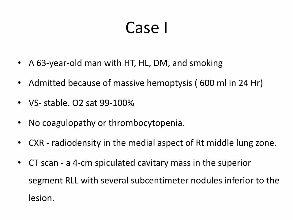
Case I
• A 63-year-old man with HT, HL, DM, and smoking
• Admitted because of massive hemoptysis ( 600 ml in 24 Hr)
• VS- stable. O2 sat 99-100%
• No coagulopathy or thrombocytopenia.
• CXR - radiodensity in the medial aspect of Rt middle lung zone.
• CT scan - a 4-cm spiculated cavitary mass in the superior
segment RLL with several subcentimeter nodules inferior to the
lesion.

• There is no consensus on the optimal diagnostic
approach to massive hemoptysis.
• CXR can identify the site of bleeding in 33–82%
• CT in 70–88.5%
– CT is much more efficient than bronchoscopy for
determining the cause of bleeding (60–77 vs. 2.5–8%)
– CT can replace bronchoscopy as a first-line investigational
approach because of its higher diagnostic yield


What would you do?

• Diagnostic workup for massive hemoptysis
should be undertaken as soon as airway
protection and hemodynamic status are
assessed and stabilized.

• FOB identifies the site of bleeding in 73–93%
– Significantly lower in cases of mild or moderate hemoptysis
– limitations of FOB in massive, life-threatening hemoptysis.
Interventional pulmonologists should not rely on FOB in
such situations
• Rigid bronchoscopy is more efficient at safeguarding airway
patency, preserving ventilation, and allowing better clearance of
the airways, therefore improving visualization
• FOB can be introduced through the rigid scope as it provides
easier access to the upper lobes and peripheral bronchi


LLL cavitary mass
• FOB was done. There was blood clot at the
orifice of RLL bronchus
• The FOB was advanced into the cavity. Forceps
biopsies led to bleeding from the orifice of the
superior segment.
• What is your initial action?

Aware of basic strategies to manage procedure-induced bleeding
• Biopsies in dependent areas of the lung
• Suctioning and frequent scope repositioning
should be kept to a minimum
• If massive bleeding is encountered, rigid
bronchoscopy

Rigid brochoscopy
• securing airway patency and safeguarding
ventilation, thereby preventing asphyxia.
• better suction of blood clots and secretions
through its large working channel, and
improved visualization of the airways
Bronchoscopic Treatment
• When expertise is available, bronchoscopic
treatment strategies ensure adequate control of
bleeding, therefore contributing to stabilization
of the patient’s hemodynamic status and
respiratory parameters.
• They are most often temporary measures for
early management

Bronchial blocking using devices and endobronchial treatment
• Cold-Saline Lavage
• Topical Vasoconstrictive Agents
– Topical epinephrine (1: 20,000)
– Topical antidiuretic hormone derivatives
• Tranexamic Acid (500–1,000 mg)
• Fibrinogen/Thrombin
• Balloon Tamponade
• Endobronchial Covered Stent Tamponade

Endobronchial Airway Blockade
• Silicone Spigot ( 6 mm)
• Bronchoscopy-Guided Topical Hemostatic Tamponade
• Endobronchial Sealing with Biocompatible Glue (n-
butyl cyanoacrylate)


• Laser Photocoagulation
• Argon Plasma Coagulation
• Electrocautery
• Other Treatments: Cryotherapy and
Brachytherapy

Laser

The initial action done

• Bleeding was initially controlled using a
combination of recombinant thrombin and
balloon bronchoplasty with a 4-Fr Fogarty
balloon.

• Two pieces of oxidized regenerated cellulose (Surgicell)
approximately 15x15 mm were folded and placed into the
jaws of a flexible biopsy forceps.
• The forceps were then withdrawn into the operating
channel of the bronchoscope, and the scope was reinserted
into the airways.
• The forceps were inserted into the cavity, and the ORC was
deployed.
• The bleeding stopped.

• The pathology on the forceps biopsy was
consistent with inflammatory changes

What to do next?

• He then underwent thoracotomy 1 week later
due to a continued concern for a malignancy.

Case II
• 21 years old woman
• Massive hemoptysis - 600 ml in 2 hr
• Hypovolemic shock
• What is your initial management?
• The initial approach for management of massive
hemoptysis involves protection of the airways
and volume resuscitation
• If the bleeding side is known, the patient should
be placed in a lateral decubitus position, with the
bleeding side down in order to prevent
aspiration into the unaffected lung.

• resuscitation + Bl transfusion
• How to deal with bleeding?

• Single –lumen intubation to secure airway and
remove clot bleeding decreased

• Intubated with a large-caliber ETT (No 8 or more),
and FOB should be immediately performed, to
suction blood clots and secretions.
• Alternatively, once the airways are cleared,
unilateral intubation can be performed to
– protect the non-bleeding lung from aspiration
– and to allow effective ventilation while awaiting
definitive treatment strategies.
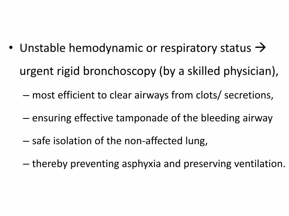
• Unstable hemodynamic or respiratory status
urgent rigid bronchoscopy (by a skilled physician),
– most efficient to clear airways from clots/ secretions,
– ensuring effective tamponade of the bleeding airway
– safe isolation of the non-affected lung,
– thereby preventing asphyxia and preserving ventilation.

• Lt lung bleeding Don’t selectively intubate the right main
bronchus
• as this procedure would occlude the right upper lobe
bronchus, further compromising gas exchange.
• Instead, tracheal intubation can be performed, followed by
insertion of a balloon catheter besides ETT through the
vocal cords, with subsequent introduction into the left main
bronchus under bronchoscopic visualization.


• Although double-lumen intubation also allows isolation
of the bleeding lung while preventing aspiration into
the unaffected lung,
• this procedure requires highly trained medical
personnel, and should only be performed after clearing
the airways.
• Once hemodynamic and respiratory conditions are
stabilized, urgent endovascular therapy should be
considered.

Back to case

• CXR and emergency CT show extensive
alveolar infiltrates both lung
• Emergency FOB was one

• Emergency FOB failed to localized bleeding
due to flooding of blood
• After resuscitation and stabilization,
hemodynamic OK, with O2 sat of 70%
• What to do next?

• Transferred to University Hospital
• Repeat FOB
• Airway could be cleared.
• A slightly elevated bluish structure in the Rt
main bronchus vascular malformation?
With continuous oozing

• Topical treatment with cold saline, epinephrine,
transamine
• Systemic treatment with desmopressin and transamine
• Bleeding was markedly reduced, however, not
completely stopped
• What to do next?

• If the underlying lesion is endoluminal, whether
in the central airways or in more peripheral
bronchi within the reach of the rigid scope, the
latter allows further management, including
– Local coagulation therapy (laser, electrocautery,
argon plasma coagulation, APC)

Bronchial Artery Embolization

The types of bronchial arterial supply
• Type I, two bronchial arteries on the left and one on the right as an intercostobronchial
trunk (ICBT) (40.6% );
• Type II, one on the left and one ICBT on the right (21.3%);
• Type III, two on the left and two on the right (one ICBT and one bronchial artery) (20.6%);
• Type IV, one on the left and two on the right (one ICBT and one bronchial artery) (9.7%).

• Massive hemoptysis usually originates from the high
pressure bronchial circulation (90%)
• Less frequently, the aorta (aortobronchial fistula,
ruptured aortic aneurysm) or nonbronchial systemic
circulation (ICS arteries, coronary arteries, thoracic
arteries originating from the axillary and subclavian
arteries and the upper and inferior phrenic arteries)

• Selective right internal mammary angiogram
shows an aberrant right bronchial artery

• Selective left bronchial angiogram demonstrates a large, hypervascular
lesion in the left upper lobe with an aneurysm (arrow). (b) On an
angiogram obtained after embolization with polyvinyl alcohol particles,
neither the hypervascular lesion nor the aneurysm is visualized.
A B

• a pathologic left bronchial artery (arrow) that
originates from the anterior wall of the
descending thoracic aorta

Embolic Materials
• Absorbable gelatin sponge
– widely used
– inexpensive, easy to handle, and has a controllable embolic size.
– disadvantages are its resolvability and lack of radiopacity.
• Polyvinyl alcohol particles
– nonabsorbable embolic materials
– particles 350–500 μm in diameter
– may prevent the early recurrence of hemoptysis due to recanalization
• It is essential to avoid the use of embolic materials that can pass through
the bronchopulmonary anastomosis (325 μm in the human lung)

• Liquid embolic agents (eg, isobutyl-2 cyanoacrylate,
absolute ethanol) are not currently used because of
the high risk of severe complications such as tissue
necrosis.
• Stainless steel platinum coils are generally not used for
BAE because they tend to occlude more proximal
vessels and may preclude repeat embolization if
hemoptysis recurs.

Result BAE
• The initial success rates - 73%–98%, (F/U 1 d to 1 mo)
• The long-term success rate - unfavorable.
• 10%–52%, with a mean F/U 1 to 46 mo
• Recurrent bleeding is more common in patients with
chronic tuberculosis, aspergilloma, or neoplasm.

• 58 patients (January 2000-February 2014)
• 40% had recurrent hemoptysis.
• A Kaplan-Meier analysis revealed an excellent
long-term survival that was 85% at 10 years


Back to the case

• Selective angiography of bronchial a., ICS a.,
IMA, aorta
– Two small aneyrysm and AVMs
• embolization completely stop bleeding
• On ETT. No bleeding. Hemodynamic – OK
• Severe hypoxia ECMO

• No further surgical intervention

• Emergency thoracotomy for massive hemoptysis is at high risk.
In case of bleeding from the arterial bronchial a., embolization
may enable to postpone surgery and operate secondarily.
• In case of bleeding from the pulmonary vessels (tumor
necrosis), surgical treatment must be immediate.

Surgery
• Emergency surgery has been gradually
abandoned because of the high morbidity and
mortality ranging between 20 and 30%
• In addition, surgical resection is not an option for
patients with poor functional status, moderate to
severe lung function impairment, bilateral
pulmonary disease or other comorbidities.


• Currently, surgery is mainly reserved for cases of
– technical failure of arteriography,
– early or repeated recurrences
– in extreme situations where the amount of bleeding
or the patient’s cardiopulmonary status are deemed
life-threatening and do not allow transfer to an
interventional radiology suite or any related delays in
management

• Surgery also remains the strategy of choice for
the management of massive hemoptysis caused
by diffuse and complex arteriovenous
malformations, iatrogenic PA rupture, chest
trauma, and mycetoma not responding to other
therapeutic strategies, or associated with
recurrent life-threatening hemoptysis

Mortality rate
• 71% in patients who lost ≽ 600 ml of blood in 4 h,
• 22% in patients with 6 600 ml within 4–16 h,
• 5% in those with 600 ml of hemoptysis within
16–48 h.

Surgical issues
• Preop cardiopulmonary assessment is difficult in
emergency situation. Use same principle as elective lung
resection
• Anatomical resection via conventional posterolateral
thoracotomy is recommended
• Positioning may be difficult because of “bleeding side up”
position
• Alternative approaches are available

Positioning
• If bleeding side up is not possible, consider
“prone position” thoracotomy

Non – anatomical resection
• The intrathoracic pathology may preclude
anatomical resection such as severe
intrapleural adhesion, destroyed lung with
calcified perihilar LN.
• Non-anatomical resection is acceptable.
• Non resection is also acceptable.


• A 57-year-old male with bronchiectasis with massive
hemoptysis.
• ETT was performed for airway protection.
• CXR showed increasing opacity in the left lower lobe with
bronchial dilatation
• Chest CT demonstrated bilateral bronchitis with cystic
dilatation in the LLL with numerous small vessels in the left
hilar region.

• Bronchoscopy revealed a large amount of
fresh blood over the lower trachea, and the
examination could not be completed.
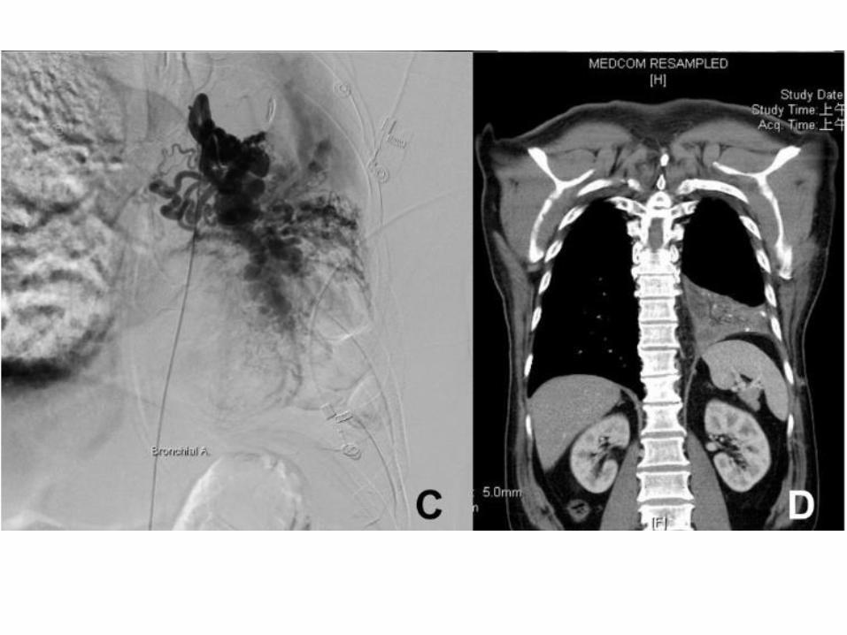
Pulmonary angiography demonstrated a
plexus of proliferating vessels around left
bronchus artery (Figure 1C).

• embolization of the left brachial artery was
performed. However, some proximal collateral
vessels which could not be embolized remained
• After the procedure, persistent hemoptysis was
noted, and therefore surgical intervention was
taken.
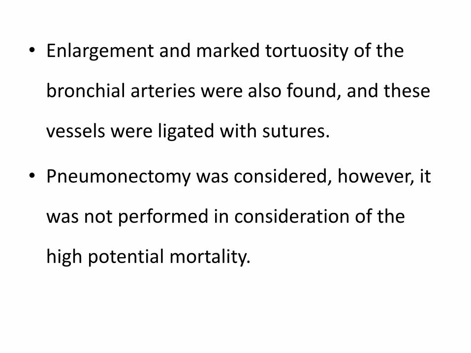
• Enlargement and marked tortuosity of the
bronchial arteries were also found, and these
vessels were ligated with sutures.
• Pneumonectomy was considered, however, it
was not performed in consideration of the
high potential mortality.

• The inferior pulmonary vein was transected to
allow identification of the left lower lobe
bronchus.
• The bronchus of the left lower lobe was then
transected to control the hemoptysis, and the
pulmonary artery was preserved.

Algorithm
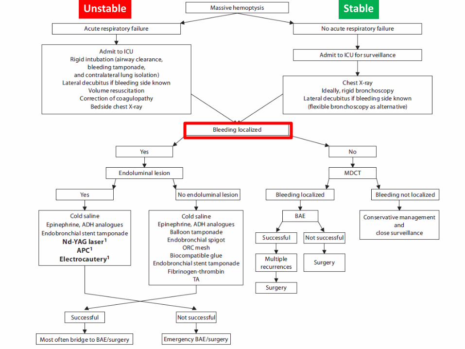
Unstable Stable


Take home messages
• Massive hemoptysis – stable/unstable
• Resuscitation and securing airway are the most
important
• Next question - Localized/ Diffuse
• Localization – FOB/ rigid bronchoscopy
• Diffuse - CT
• Investigation to identify the diagnosis and its definitive
treatment is crucial.

Treatment
• Endolumional lesion – endobronchial
treatmen: laser
• Localized + no endoluminal lesion –
endobronchial blokade/ hemostatic treatment

Endovascular treatment (Bronchial artery embolization)
• Failed endobronchial treatment for
endoluminal lesion
• Follow initial endobronchial treatment for
non-endoluminal lesion

Surgery
• High mortality. Aware of poor
cardiopulmonary reserve patient\
• Indications
– Failed BAE
– Recurrence
– Unstable, unable to do BAE…