Embed Size (px)
Citation preview
218 International Journal of Analytical and Bioanalytical Chemistry 2012; 2(4): 218-227
ISSN-2231-5012
Original Article
Electrochemistry of Hemoglobin I from Lucina pectinata immobilized on a
modified gold electrode with 3-mercaptopropionic acid
Mario Ortega-Núñez,† Rolando J. Tremont,
‡ Carmen A. Vega-Olivencia,
†* Juan López-Garriga,
†*
†Department of Chemistry, University of Puerto Rico at Mayagüez, PO BOX 9019,
Mayagüez, PR 00681-9019, United States ‡Department of Chemistry, University of Puerto Rico at Humacao,
Call Box 860, Humacao, PR 00792, United States
E-mail*: [email protected]; [email protected]
Received 26 August 2012; accepted 18 October 2012
Abstract
The extraordinary affinity of recombinant hemoglobin I from Lucina pectinata (rHbI) for hydrogen sulfide (H2S) allows us
to postulate it as substrate for the preparation of future hydrogen sulfide sensor. The modification of a gold surface with 3-
mercaptopropionic acid (3-MPA), nickel ion and recombinant hemoglobin I, and electrochemical kinetics parameters were
reported here. Cyclic Voltammetry and X-ray photoelectron spectroscopy were employed to characterize the electrode
modification process, while FTIR was used to verify the presence of hemoglobin on the surface. A pair of well-defined redox peaks for rHbIFe(III)–rHbIFe(II) was obtained. Electrochemical parameters of immobilized hemoglobin as formal
potential (Eo‟), charge transfer coefficient (α) and apparent heterogeneous electron transfer rate constant (ks) were estimated
by cyclic voltammetry data. Also, the electroactivity of this hemoglobin-electrode was exemplified at the prepared
electrode for association with hydrogen sulfide.
© 2012 Universal Research Publications. All rights reserved
Key words: Hemoglobin I, Lucina pectinata, Hydrogen sulfide, 3-mercaptopropionic acid, Gold electrode.
1. Introduction
Self-assembled monolayers are arrangements of organic
compounds on the surface of a solid. These monolayers are
formed by the interaction between terminal functional
groups and a surface, which can form the basis for
nanoelectronic applications and biosensors.
It has a particular interests for those molecules that show a
dual chemical functionality for the formation of these
monolayers. These molecules have a functional attaching group with high affinity for the substrate which forms a
covalent bond; a hydrocarbon chain that acts as a bridge
between the surface and the specie of interest, and a
terminal functional group that binds or interacts with the
chemical specie and determines the monolayer surface
properties.
Several studies have detailed behavior of monolayers based
on alkanethiols on gold surfaces formed by absorption from
the solution. In these works it is anticipated that short-chain
monolayers are less packed and less insulating than their
counterparts long-chain, which are highly ordered and
densely packed [1-5].
On the other hand, many investigations have been focused
on the immobilization of biomolecules using thiol
monolayers on gold in order to construct devices to
measure properties or chemical activity of biomolecules [6,
7]. In some cases, molecules such as enzymes or proteins
are immobilized because of Au-thiol SAMs complexing
properties for metal ions [8-10]. After forming complexes
with the monolayer, these metal ions have the ability to
coordinate with electron-rich groups that are found in proteins or enzymes [11]. In addition to the amino
terminus, some amino acids are especially suitable for
binding due to electron donor atoms in their side chains.
Many residues, such as Glu, Asp, Tyr, Cys, His, Arg, Lys
and Met, can participate in binding [12]. In previous works
the high affinity of metal complexes for amino groups from
lysine or histidine of the proteins or enzymes have been
demonstrated [13]. Hydrogen sulfide has been regarded as
a poisonous gas, with a wide spectrum of toxic effects. The
toxicity of H2S is comparable with that of hydrogen
cyanide; it forms a complex bond with iron in the
mitochondrial cytochrome c oxidase enzyme, thereby
Available online at http://www.urpjournals.com
International Journal of Analytical and Bioanalytical Chemistry
Universal Research Publications. All rights reserved
219 International Journal of Analytical and Bioanalytical Chemistry 2012; 2(4): 218-227
blocking oxygen from binding and stopping cellular
respiration. Under physiological conditions, 30% of H2S is
undissociated and 70% is dissociated to hydrosulfide ion,
solubilized in water and plasma, it can penetrate all types of
cells by simple diffusion [14].
H2S acts as a vasodilator and is also active in the brain where it increases the response of the N-methyl-D-aspartate
(NMDA) and facilitates long term potentiation, which is
involved in the formation of memory, acting as
neuromodulator and neuroprotector smooth muscle
relaxant. Eventually, the gas (H2S) is converted to sulfites
and further oxidized to thiosulfate and sulfate [15, 16].
In the environment, hydrogen sulfide has a very important
role in the geochemical cycle of sulfur, where a group of
bacteria produce H2S from sulfurs or sulfates, while others
use it as fuel [17]. It is usually found in volcanic gases,
natural gas and as a product of the biodegradation of
organic substances in soil and waters. Various methods and techniques (optical, electrochemical
and acoustic sensors) have been used for the
characterization of hydrogen sulfide [18-33]. Actually, the
most widely used sensor is based on polarographic
detection of HS- species that interacts with the couple redox
Fe3+/Fe2+ [34]; this HS- is obtained from the partial
dissociation of H2S dissolved in solution. One of the
disadvantages associated with this sensor is that it depends
on the selectivity provided by the membrane. Due to this
fact, it is impossible to be used directly in a wide range of
complex matrices without pre-treatment, where the H2S is mixed with other redox-active molecules that can pass
through the membrane and interact with ferric couple.
Most of the aforementioned methods and techniques
reported variable concentrations of H2S on the same
biological samples by the lack of standardization of
methods between laboratories [35]. Because of this, sensors
with high selectivity and specificity towards the analyte are
required. Biosensors are selective and specific devices
because the active principle is based on an enzyme or
protein that is specific to a substrate. This gives the
convenience of using them in complex systems because
they only respond to the interaction with a specific substrate. Because hemoglobin I from Lucina pectinata
presents this high affinity for hydrogen sulfide, was used as
the species that binds H2S in our electrode. The hemoglobin
I is a monomeric protein that has high affinity for hydrogen
sulfide. The association constant of ferric hemoglobin I for
H2S is extremely high (kon = 2.3 x 10⁵ M-1s-1) compared
with the dissociation (koff = 2.2 x 10⁻⁴ s-1). Several studies
show that this property is due to the unusual conformation
of amino acids in the heme pocket of hemoglobin I with
respect to other vertebrates‟ hemoglobins [14]. In this
work, a modified electrode based on 3-mercaptopropionic acid (3-MPA), Ni2+ ions and rHbI was developed. An
electrochemical examination on redox reaction of the
immobilized hemoglobin was done by measurements of
electrochemical kinetic parameters obtained from the
voltammetric data.
2. Materials and Methods
2.1. Materials and Instrumentation
Recombinant hemoglobin rHbI solutions were prepared by
dissolving it in a 0.1 M phosphate buffer (pH 6.70). 3-MPA
(Sigma, 99%), H2SO4 (99.999%), NiCl2 (Aldrich, 99.99%), sodium sulfide (Aldrich, 99.99%) and ethanol
(Sigma-Aldrich, 99.5%) were used as received. Water used in all experiments was previously distilled and
pumped through a deionization system to give an 18 M cm water. Electrochemical measurements were carried out
in a conventional three-electrode cell previously N2-purged
for 15 min. The nitrogen atmosphere was maintained
during the experiments. The measurements were performed
at room temperature on a BASi Epsilon for an Electrochemistry Potentiostat/Galvanostat system with EC
software, connected to a cell stand C-3 for voltammetry.
The cell is equipped with a platinum wire auxiliary
electrode (7.5 cm) with gold-plated connector (MW-1032),
and a 7.5 cm long RE-5B Ag/AgCl reference electrode with
a Vycor frit (the filling solution is aqueous 3M NaCl that
has been saturated with AgCl). The software EC epsilon
version 1.60.70 was used. Sample XPS analyses were
performed using the Al Ka (15.0 kV at 350 watts) source of
a PHI 5600 ci spectrometer. This instrument has a
hemispherical analyzer, a toroidal monochromator, and a multichannel detector. The base pressure in the chamber
during analysis was less than 1x10⁻⁹ Torr. The binding
energy values reported in this study were corrected using
the C 1s signal of the atmospheric contaminants (284.5
eV). Different experiment setups were used for the XPS
studies. Experimental conditions for each setup are
summarized in the results analysis section. FTIR
spectroscopy was used to corroborate the presence of the
protein in the final surface. Spectrum 100 FTIR
Spectrometer, from Perkin Elmer, equipped with a
Universal diamont ATR (uATR) top plate accessory one
refraction, was used. Software: Spectrum version V.6.0.2. 2.2. Electrodes and substrates
Polycrystalline gold electrodes from Bioanalytical Systems
(BASi) Inc., with a 1.6 mm diameter were employed as the
working electrodes for the electrochemical experiments,
whereas quartz crystal microbalance polycrystalline gold
crystal sensors from MAXTEK®, with a 2.54 cm diameter
were used for X-ray photoelectron spectroscopy (XPS) and
FTIR analysis. The cleaning procedure for the Au
electrodes consisted of polishing them on a velvet pad with
1, 0.3 and 0.05 m polishing alumina (BASi), and then
ultrasonically washing them for 5 min with deionized water. The electrochemical polishing pre-treatments of the
working electrodes are described elsewhere [36]. The
current-potential behavior characteristic of a clean gold
surface in a 0.1 M H2SO4 solution was used as a criterion of
surfaces cleanliness. After the pre-treatment procedures,
these electrodes were rinsed with deionized water and any
excess was removed with nitrogen. Before modifying a
gold electrode, cleanness of the electrode was verified by
cyclic voltammetry [36]. The reference and auxiliary
electrodes were Ag/AgCl 3 M NaCl and a platinum wire,
respectively.
2.3. Recombinant Hemoglobin I preparation Recombinant HbI (rHbI) was expressed in Escherichia coli
when the HbI cDNA was cloned into the pET28 (a+)
expression vector (transformed into BLi5 cells). The
220 International Journal of Analytical and Bioanalytical Chemistry 2012; 2(4): 218-227
induction was performed with 1 mM isopropyl β-D-1-
thiogalactopyranoside (IPTG) at 30 °C and nutrient
medium was supplemented with 30 g/mL hemin chloride
and 1% glucose. The expression yielded dark red cell
pellets that were lysed and centrifuged to separate the
soluble protein from the insoluble cell fractions as described previously [37]. The soluble protein fraction was
used for purification of the recombinant HbI in metal
affinity columns. Further purification of the sample was
achieved by fast performance liquid chromatography
(FPLC) in a Hi Load 26/60 Superdex 200 gel filtration
column.
2.4. Preparation of 3-MPA Self-assembled monolayer and
Ni2+ ions addition
Self-assembly of 3-MPA was done on gold surfaces using
MAXTEK® gold substrates and commercial Au disk
electrodes (BASi) of 1.6 mm of diameter. This monolayer
was prepared by immersing a clean gold electrode in a vial containing 0.5 mL of an ethanolic solution of 50 m of 3-
MPA for 24 h. Nitrogen was used for degassing the
solutions, and the vials were sealed and left unperturbed
during the modification period. Afterward, the substrate
obtained was washed with ethanol, dried under a nitrogen
flow and maintained in a desiccator for further
characterization. The surface is described as 3-MPA/Au for
the 3-MPA modifications on gold surfaces in the rest of the
manuscript, respectively.
2.5. Recombinant rHbI–modified electrode preparation
The surface 3-MPA/Au was immersed into 50 mM NiCl2 aqueous solution for 2 h. Then, the surface was dried under
nitrogen flow for further analysis. After, the modified
electrode (Ni2+/3-MPA/Au) was left into a 100 M rHbI
aqueous solution for 72 h to obtain the hemoglobin I
modified gold surface. The electrode was stored in
phosphate buffer pH 6.70 at 4C until electrochemical analysis, and the modified surfaces corresponding to
quartz-polycrystalline gold plates in a desiccator for surface
examination.
2.6. Electrochemical measurements
Cyclic voltammetry technique was used to identify changes
in current–potential shifts throughout the modification
process. Similarly, this technique was useful to evaluate the blocking effect with different surfaces obtained in the
modification process in a redox standard solution of
ferricyanide. Also current changes were evaluated in the
modified hemoglobin-electrode when aliquots of a Na2S
solution were added in order to provide H2S. In all cases a
0.1 M phosphate buffer solution pH 6.70 was used, and the
voltammograms were obtained at 100 mVs-1.
Amperometric analysis was carried out by applying
potential of 230 mV vs. Ag/AgCl on a stirred cell at 25°C.
The response was measured as the difference between total
and residual currents. The sodium sulfide solution was
prepared by adding the salt, previously purged with N2, to degassed and deoxygenated water to prevent oxygen
contamination.
3. Results and Discussion
3.1. XPS characterization
In order to characterize the binding mode 3-MPA
molecules on the Au surface, an XPS study of the layer
formed after 24 h of modification from the ethanolic
solutions of this substrate has performed. In addition the
formation of metal complexes between 3-MPA with Ni2+
ions when the modified surface (3-MPA/Au) was left in a
50 mM NiCl2 solution for 24 h, obtaining Ni2+/3-MPA/Au
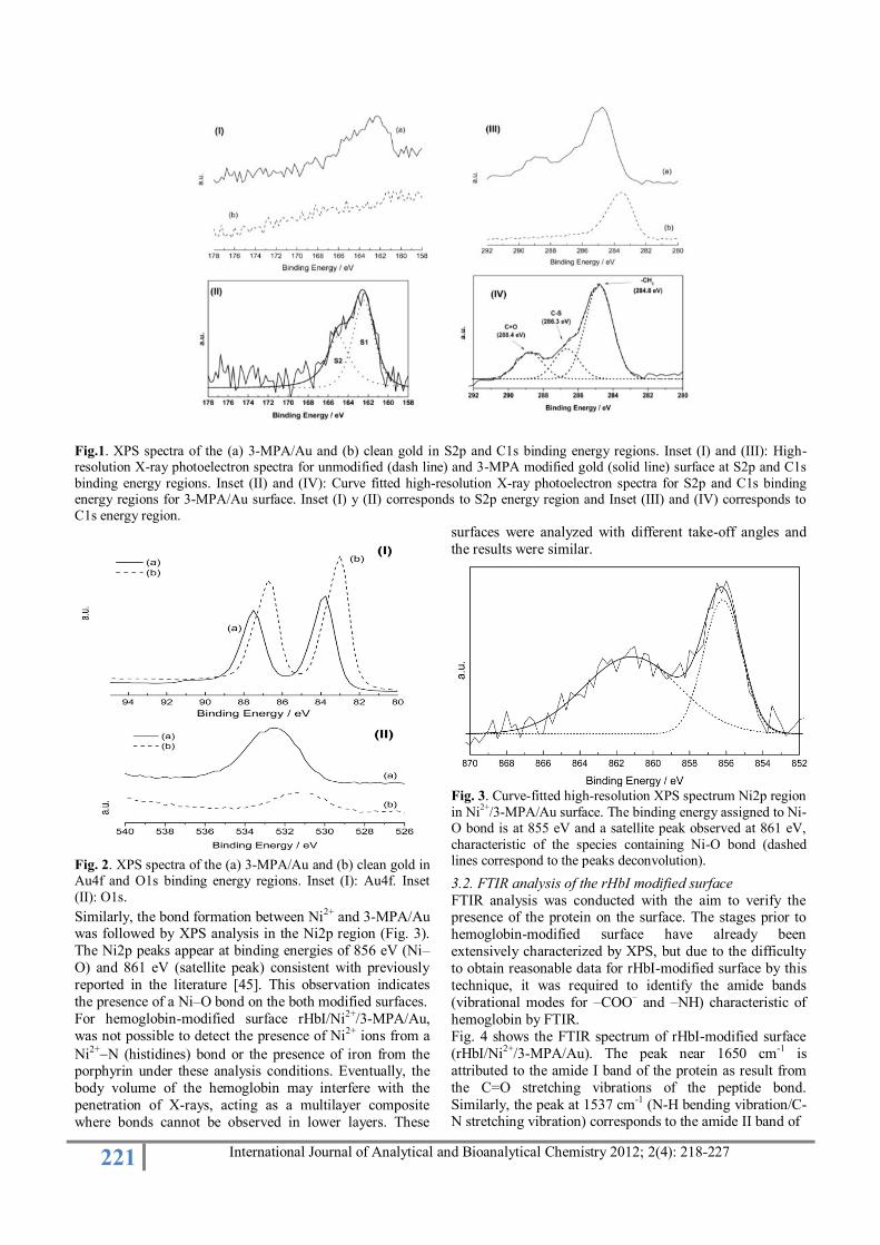
surface was studied. Figure 1 shows the comparison of the spectra between (a) 3-MPA modified surface and (b) clean
gold, in regions S2p and C1s.
In inset (I) in Figure 1 shows the difference in signals
between Au and 3-MPA/Au, due to appearance of a broad
peaks centered at approximately 163 eV in 3-MPA/Au
spectrum, corresponding to one doublet (S2p3/2 and S2p1/2)
with a peak separation of 1.2 eV and area ratio 2:1. These
peaks may deconvolution while maintaining the above
characteristics in terms of splitting and area ratio, using
Gaussian curves. This doublet consists of two components,
S1 and S2, where the main component S1 occurs at 162.3
eV while the minor one, S2, is found at 164.2 eV (Inset II, Fig. 1). The S1 component is attributed to 3-MPA
molecules chemisorbed on gold and indicates the formation
of a thiolate species. This component is characteristic of
organosulfur compounds on gold reported for cysteine [38-
40] and for thiols with unsubstituted alkyl chains and with
chains containing aromatic moieties [41, 42]. The S2
component, located at 164.0 eV could be assigned to
molecules not bound to gold, that is, physisorbed molecules
forming a partially occupied upper layer [42].
Inset III in Fig.1 shows the comparison of HRXPS spectra
of C1s region for the Au and 3-MPA/Au surfaces. In 3-MPA/Au spectrums, it is observed the formation of several
overlapping peaks that are not present in clean gold. Inset
IV in Fig. 1 shows the deconvolution to the peaks present
in the signal in the region C1s. It is evidence the presence
of characteristic peaks for the -CH2 groups (284.8 eV), C-S
(286.3 eV) and C=O (288.4 eV) [43, 44]. This confirms the
adsorption of the 3-MPA on the gold surface.
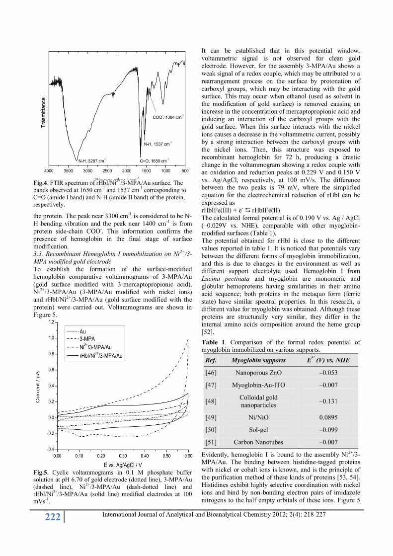
Furthermore, to confirm clearly the presence of 3-MPA on
the gold surface in 3-MPA/Au assembly, a high resolution
spectra in Au4f and O1s regions were performed. Inset I in
Fig. 2 shows the comparison between the spectra in the
region Au4f for (a) 3-MPA/Au surface and (b) clean gold. The spectrum corresponding to 3-MPA/Au surface (a)
shows a well-defined doublet with a peak separation of 3.6
eV and a half-width of 1.6 eV. The peak Au4f7/2 shows a
binding energy of 84.0 eV which is the typical value
expected when thiolate form self-assembled monolayers on
Au surfaces. Furthermore, decrease on the peak height and
area is an indication that these compounds are adsorbed on
the gold surface blocking the XPS detection of Au4f
photoelectrons. The high resolution XPS spectrum for O1s
region (Inset II in Fig. 2) shows a slightly asymmetric
broad peak centered about at 532 eV for 3-MPA/Au surface. This peak can be formed by the contribution of the
oxygen of the carboxyl and -OH groups, previously
reported in the literature [40, 43]. In addition, this slight
asymmetry may be is due to the presence of water
molecules coadsorbed by the surfaces. The oxygen (O1s)
and carbon (C1s) signals, which appear on the clean gold
surface, correspond to contaminants from the air, from the
cleaning process, or from handling of the substrate [38].
221 International Journal of Analytical and Bioanalytical Chemistry 2012; 2(4): 218-227
Fig.1. XPS spectra of the (a) 3-MPA/Au and (b) clean gold in S2p and C1s binding energy regions. Inset (I) and (III): High-resolution X-ray photoelectron spectra for unmodified (dash line) and 3-MPA modified gold (solid line) surface at S2p and C1s
binding energy regions. Inset (II) and (IV): Curve fitted high-resolution X-ray photoelectron spectra for S2p and C1s binding energy regions for 3-MPA/Au surface. Inset (I) y (II) corresponds to S2p energy region and Inset (III) and (IV) corresponds to
C1s energy region.
Fig. 2. XPS spectra of the (a) 3-MPA/Au and (b) clean gold in Au4f and O1s binding energy regions. Inset (I): Au4f. Inset
(II): O1s.
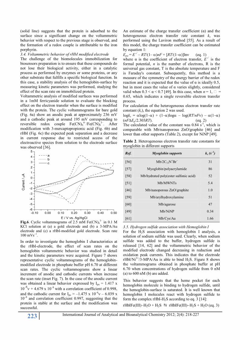
Similarly, the bond formation between Ni2+ and 3-MPA/Au was followed by XPS analysis in the Ni2p region (Fig. 3).
The Ni2p peaks appear at binding energies of 856 eV (Ni–
O) and 861 eV (satellite peak) consistent with previously
reported in the literature [45]. This observation indicates
the presence of a Ni–O bond on the both modified surfaces.
For hemoglobin-modified surface rHbI/Ni2+/3-MPA/Au,
was not possible to detect the presence of Ni2+ ions from a
Ni2+N (histidines) bond or the presence of iron from the porphyrin under these analysis conditions. Eventually, the
body volume of the hemoglobin may interfere with the
penetration of X-rays, acting as a multilayer composite
where bonds cannot be observed in lower layers. These
surfaces were analyzed with different take-off angles and
the results were similar.
Fig. 3. Curve-fitted high-resolution XPS spectrum Ni2p region
in Ni2+
/3-MPA/Au surface. The binding energy assigned to Ni-O bond is at 855 eV and a satellite peak observed at 861 eV,
characteristic of the species containing Ni-O bond (dashed lines correspond to the peaks deconvolution).
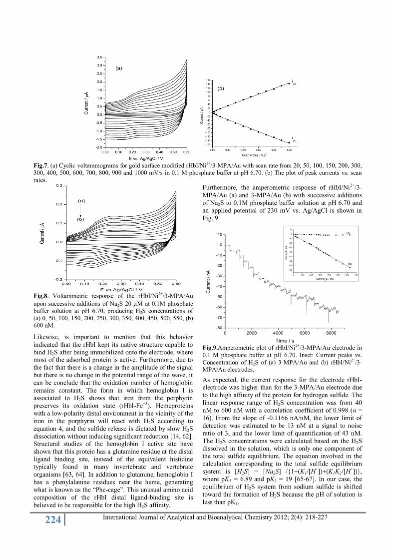
3.2. FTIR analysis of the rHbI modified surface
FTIR analysis was conducted with the aim to verify the presence of the protein on the surface. The stages prior to
hemoglobin-modified surface have already been
extensively characterized by XPS, but due to the difficulty
to obtain reasonable data for rHbI-modified surface by this
technique, it was required to identify the amide bands
(vibrational modes for –COO– and –NH) characteristic of
hemoglobin by FTIR.
Fig. 4 shows the FTIR spectrum of rHbI-modified surface
(rHbI/Ni2+/3-MPA/Au). The peak near 1650 cm-1 is
attributed to the amide I band of the protein as result from
the C=O stretching vibrations of the peptide bond.
Similarly, the peak at 1537 cm-1
(N-H bending vibration/C-N stretching vibration) corresponds to the amide II band of
222 International Journal of Analytical and Bioanalytical Chemistry 2012; 2(4): 218-227
Fig.4. FTIR spectrum of rHbI/Ni
2+/3-MPA/Au surface. The
bands observed at 1650 cm-1
and 1537 cm-1
corresponding to
C=O (amide I band) and N-H (amide II band) of the protein, respectively.
the protein. The peak near 3300 cm-1 is considered to be N-
H bending vibration and the peak near 1400 cm-1 is from
protein side-chain COO-. This information confirms the
presence of hemoglobin in the final stage of surface
modification. 3.3. Recombinant Hemoglobin I immobilization on Ni2+/3-
MPA modified gold electrode
To establish the formation of the surface-modified
hemoglobin comparative voltammograms of 3-MPA/Au
(gold surface modified with 3-mercaptopropionic acid),
Ni2+/3-MPA/Au (3-MPA/Au modified with nickel ions)
and rHbI/Ni2+/3-MPA/Au (gold surface modified with the
protein) were carried out. Voltammograms are shown in
Figure 5.
Fig.5. Cyclic voltammograms in 0.1 M phosphate buffer solution at pH 6.70 of gold electrode (dotted line), 3-MPA/Au
(dashed line), Ni2+
/3-MPA/Au (dash-dotted line) and rHbI/Ni
2+/3-MPA/Au (solid line) modified electrodes at 100
mVs-1.
It can be established that in this potential window,
voltammetric signal is not observed for clean gold
electrode. However, for the assembly 3-MPA/Au shows a
weak signal of a redox couple, which may be attributed to a
rearrangement process on the surface by protonation of
carboxyl groups, which may be interacting with the gold surface. This may occur when ethanol (used as solvent in
the modification of gold surface) is removed causing an
increase in the concentration of mercaptopropionic acid and
inducing an interaction of the carboxyl groups with the
gold surface. When this surface interacts with the nickel
ions causes a decrease in the voltammetric current, possibly
by a strong interaction between the carboxyl groups with
the nickel ions. Then, this structure was exposed to
recombinant hemoglobin for 72 h, producing a drastic
change in the voltammogram showing a redox couple with
an oxidation and reduction peaks at 0.229 V and 0.150 V
vs. Ag/AgCl, respectively, at 100 mV/s. The difference between the two peaks is 79 mV, where the simplified
equation for the electrochemical reduction of rHbI can be
expressed as
rHbIFe(III) + e- rHbIFe(II)
The calculated formal potential is of 0.190 V vs. Ag / AgCl
(–0.029V vs. NHE), comparable with other myoglobin-
modified surfaces (Table 1).
The potential obtained for rHbI is close to the different
values reported in table 1. It is noticed that potentials vary
between the different forms of myoglobin immobilization,
and this is due to changes in the environment as well as different support electrolyte used. Hemoglobin I from
Lucina pectinata and myoglobin are monomeric and
globular hemoproteins having similarities in their amino
acid sequence; both proteins in the metaquo form (ferric
state) have similar spectral properties. In this research, a
different value for myoglobin was obtained. Although these
proteins are structurally very similar, they differ in the
internal amino acids composition around the heme group
[52].
Table 1. Comparison of the formal redox potential of myoglobin immobilized on various supports.
Ref. Myoglobin supports E°’ (V) vs. NHE
[46] Nanoporous ZnO –0.053
[47] Myoglobin-Au-ITO –0.007
[48] Colloidal gold
nanoparticles –0.131
[49] Ni/NiO 0.0895
[50] Sol-gel –0.099
[51] Carbon Nanotubes –0.007
Evidently, hemoglobin I is bound to the assembly Ni2+/3-MPA/Au. The binding between histidine-tagged proteins
with nickel or cobalt ions is known, and is the principle of
the purification method of these kinds of proteins [53, 54].
Histidines exhibit highly selective coordination with nickel
ions and bind by non-bonding electron pairs of imidazole
nitrogens to the half empty orbitals of these ions. Figure 5
223 International Journal of Analytical and Bioanalytical Chemistry 2012; 2(4): 218-227
(solid line) suggests that the protein is adsorbed to the
surface since a significant change on the voltammetric
behavior with respect to the previous stage is observed, and
the formation of a redox couple is attributable to the iron
porphyrin.
3.4. Voltammetric behavior of rHbI modified electrode The challenge of the biomolecules immobilization for
biosensors preparation is to ensure that these compounds do
not lose their biological activity, either in a catalytic
process as performed by enzymes or some proteins, or any
other substrate that fulfills a specific biological function. In
this case, a stability analysis of the hemoglobin-surface by
measuring kinetic parameters was performed, studying the
effect of the scan rate on immobilized protein.
Voltammetric analysis of modified surfaces was performed
in a 1mM ferricyanide solution to evaluate the blocking
effect on the electron transfer when the surface is modified
with the protein. The cyclic voltammograms for bare gold (Fig. 6a) show an anodic peak at approximately 236 mV
and a cathodic peak at around 195 mV corresponding to
reversible redox couple Fe(CN)64–
/Fe(CN)63–
. After
modification with 3-mercaptopropionic acid (Fig. 6b) and
rHbI (Fig. 6c) the expected peak separation and a decrease
in current response due to restricted access of the
electroactive species from solution to the electrode surface
was observed [36].
Fig.6. Cyclic voltammograms of 2.5 mM Fe(CN)6
3– in 0.1 M
KCl solution at (a) a gold electrode and (b) a 3-MPA/Au
electrode and (c) a rHbI-modified gold electrode. Scan rate 100 mVs
–1.
In order to investigate the hemoglobin I characteristics at
the rHbI-electrode, the effect of scan rates on the
hemoglobin voltammetric behavior was studied in detail and the kinetic parameters were acquired. Figure 7 shows
representative cyclic voltammograms of the hemoglobin-
modified electrode in phosphate buffer pH 6.70 at different
scan rates. The cyclic voltammograms show a linear
increment of anodic and cathodic currents when increase
the scan rate (inset Fig. 7). In the case of the anodic current
was obtained a linear behavior expressed by Ipa = 1.417 x
10–6 + 4.679 x 10⁻⁸ with a correlation coefficient of 0.998,
and the cathodic current for Ipc = –1.475 x 10⁻⁶ – 6.039 x 10⁻⁸ and correlation coefficient 0.997, suggesting that the
protein is stable at the surface and the modification was
successful.
An estimate of the charge transfer coefficient (α) and the
heterogeneous electron transfer rate constant ks was
performed using the Laviron method [55]. As a result of
this model, the charge transfer coefficient can be estimated
by equation 1:
Epa = E˚’ – RT/(1–)nF + [RT/(1–)]ln (eq. 1) where α is the coefficient of electron transfer, E˚’ is the
formal potential, n is the number of electrons, R is the universal gas constant, T is the absolute temperature and F
is Faraday's constant. Subsequently, this method is a
measure of the symmetry of the energy barrier of the redox
reaction and it is expected that the value of α is ideally 0.5,
but in most cases the value of α varies slightly, considered
valid when 0.3 < α < 0.7 [49]. In this case, when n = 1, =
0.65, which indicates a single reversible electron transfer
process.
For calculation of the heterogeneous electron transfer rate
constant (ks), the equation 2 was used.
logks = log(1–) + (1–)log – log(RT/nF) – (1–)
(nFEp/2.303RT). (eq. 2) The calculated value of the constant was 0.84 s-1, which is
comparable with Mb/nanoporous ZnO/graphite [46] and lower than other supports (Table 2), except for NiNP [49].
Table 2. Heterogeneous electron transfer rate constants for
myoglobin in different supports
Ref. Myoglobin supports ks (s–1
)
[56] Mb/2C12N+Br− 31
[57] Myoglobin/polyacrylamide 86
[58] Mb/hydrated poly(ester sulfonic acid) 52
[51] Mb/MWNTs 5.4
[46] Mb/nanoporous ZnO/graphite 1.0
[59] Mb/arylhydroxylamine 51
[60] Mb/agarose 47
[49] Mb/NiNP 0.34
[61] Mb/Cys/Au 1.66
3.5. Hydrogen sulfide association with Hemoglobin I
For the H2S association with hemoglobin I analysis, a
solution of sodium sulfide was used. Clearly, when sodium sulfide was added to the buffer, hydrogen sulfide is
released [14, 62] and the voltammetric behavior of the
modified electrode changed decreasing in reduction and
oxidation peak currents. This indicates that the electrode
rHbI/Ni2+/3-MPA/Au is able to bind H2S. Figure 8 shows
the voltammograms obtained in phosphate buffer at pH
6.70 when concentrations of hydrogen sulfide from 0 nM
(a) to 600 nM (b) are added.
This behavior suggests that the heme pocket for each
hemoglobin molecule is binding to hydrogen sulfide, until
the hemoglobin-surface is saturated. It is well known that hemoglobin I molecules react with hydrogen sulfide to
form the complex rHbI-H2S according to eq. 3 [14]:
rHbIFe(III)–H2O + H2S rHbIFe(III)H2S + H2O (eq. 3)
224 International Journal of Analytical and Bioanalytical Chemistry 2012; 2(4): 218-227
Fig.7. (a) Cyclic voltammograms for gold surface modified rHbI/Ni
2+/3-MPA/Au with scan rate from 20, 50, 100, 150, 200, 300,
300, 400, 500, 600, 700, 800, 900 and 1000 mV/s in 0.1 M phosphate buffer at pH 6.70. (b) The plot of peak currents vs. scan
rates.
Fig.8. Voltammetric response of the rHbI/Ni
2+/3-MPA/Au
upon successive additions of Na2S 20 M at 0.1M phosphate buffer solution at pH 6.70, producing H2S concentrations of
(a) 0, 50, 100, 150, 200, 250, 300, 350, 400, 450, 500, 550, (b) 600 nM.
Likewise, is important to mention that this behavior
indicated that the rHbI kept its native structure capable to
bind H2S after being immobilized onto the electrode, where most of the adsorbed protein is active. Furthermore, due to
the fact that there is a change in the amplitude of the signal
but there is no change in the potential range of the wave, it
can be conclude that the oxidation number of hemoglobin
remains constant. The form in which hemoglobin I is
associated to H2S shows that iron from the porphyrin
preserves its oxidation state (rHbI-Fe+3). Hemeproteins
with a low-polarity distal environment in the vicinity of the
iron in the porphyrin will react with H2S according to
equation 4, and the sulfide release is dictated by slow H2S
dissociation without inducing significant reduction [14, 62]. Structural studies of the hemoglobin I active site have
shown that this protein has a glutamine residue at the distal
ligand binding site, instead of the equivalent histidine
typically found in many invertebrate and vertebrate
organisms [63, 64]. In addition to glutamine, hemoglobin I
has a phenylalanine residues near the heme, generating
what is known as the “Phe-cage”. This unusual amino acid
composition of the rHbI distal ligand-binding site is
believed to be responsible for the high H2S affinity.
Furthermore, the amperometric response of rHbI/Ni2+/3-
MPA/Au (a) and 3-MPA/Au (b) with successive additions
of Na2S to 0.1M phosphate buffer solution at pH 6.70 and
an applied potential of 230 mV vs. Ag/AgCl is shown in
Fig. 9.
Fig.9.Amperometric plot of rHbI/Ni
2+/3-MPA/Au electrode in
0.1 M phosphate buffer at pH 6.70. Inset: Current peaks vs.
Concentration of H2S of (a) 3-MPA/Au and (b) rHbI/Ni2+
/3-MPA/Au electrodes.
As expected, the current response for the electrode rHbI-electrode was higher than for the 3-MPA/Au electrode due
to the high affinity of the protein for hydrogen sulfide. The
linear response range of H2S concentration was from 40
nM to 600 nM with a correlation coefficient of 0.998 (n =
16). From the slope of -0.1166 nA/nM, the lower limit of
detection was estimated to be 13 nM at a signal to noise
ratio of 3, and the lower limit of quantification of 43 nM.
The H2S concentrations were calculated based on the H2S dissolved in the solution, which is only one component of
the total sulfide equilibrium. The equation involved in the
calculation corresponding to the total sulfide equilibrium
system is [H2S] = [Na2S] /{1+(K1/[H+])+(K1K2/[H
+])},
where pK1 = 6.89 and pK2 = 19 [65-67]. In our case, the
equilibrium of H2S system from sodium sulfide is shifted
toward the formation of H2S because the pH of solution is
less than pK1.
225 International Journal of Analytical and Bioanalytical Chemistry 2012; 2(4): 218-227
The restoration of the electrode for reuse was performed
leaving it at a constant potential of 0.0 V vs. Ag/AgCl for 1
h or until to observe that the current remains constant,
favoring an appropriate reducing environment and induce
the hemoglobin to release the H2S bonded Previous work
reported that when the concentration of H2S increases considerably (more than 3-10 times the concentration of
rHbI) generates a reducing environment induced by H2S
itself, favors the reduction of rHbI(III) to rHbIFe(II) and
H2S is released [62]. Therefore, a reducing environment is
generated at the electrode leaving a potential of 0 V and
promoting the release of gas. This electrode lost only 7% of
its initial activity after more than four successive
measurements and stored in 0.1M phosphate buffer solution
at pH 6.70 and 4 °C, when not in use. Moreover, the rHbI
electrode was tested in a buffer solution containing various
common ions to evaluate their response. The electrode
holds good response to a mixture of ions; 1 mM Ca2+, Na+, K+, NH4
+, NO3-, SO4
2-, Cl-, Br-, I- and 100 mM PO4-3 (from
buffer). It should be emphasized that higher ions
concentration leads to a loss of response, possibly due to
hemoglobin denaturation by salinity effect. In addition, an
erratic response is observed when a small aliquot of a
solution 1 mM CN- was added to the buffer solution.
Cyanide is a ligand that binds strongly to ferric ion forming
a very stable complex with hemoglobin and cannot be
displaced by ligands such as O2, NO or H2S. Some
publications in hemeproteins functionality and dynamic
confirm this fact [63, 64]. On the other hand, when a small aliquot of 1 mM H2O2 is added to a phosphate buffer
solution, a similar behavior is observed. Several recent
studies demonstrate that monomeric hemeproteins similar
to rHbI, may be associated with ferric ion of the porphyrin
[68], but as the peroxide concentration increases, it can
interact with any part of the assembly structure observing
an erratic response of the electrode. In conclusion, for H2S
analysis is recommended to remove hydrogen peroxide and
cyanide, both of which can greatly interfere with the
analysis. The problem is that iron from the porphyrin in
these hemeproteins binds to these ligands in the ferric state,
and the oxidation state does not change. The effective nuclear charge of ferric ion may change slightly when it
forms bonds with such substrates, but potential changes are
not so extreme as to be detected with the conditions
employed in this study. Finally, The relative response
current of rHbI-electrode was examined by checking
periodically the current generated at 230 mV when it was
exposed to a standard solution of Na2S in 0.1 M phosphate
buffer solution at pH 6.70. The modified electrode retained
95% of activity within a storage period of 30 days under
these conditions, but after a storage period of 2 months, the
electrode showed total loss of activity for H2S.
4. Conclusions
Recombinant Hemoglobin I from Lucina pectinata was
successfully immobilized on the surface of gold-
mercaptopropionic-nickel ion self- assembled monolayer,
according to the electrochemical results obtained by cyclic
voltammetry, amperometry, and kinetic parameters
calculated. In addition, the rHbI-modified gold electrode
showed good electrochemical activity to H2S without the
aid of any mediator. The resulting system rHbI/3-
MPA/Ni2+/Au has a promising potential for the
construction of a third-generation of electrochemical
biosensor based on the direct electrochemistry of other
histidine-tagged proteins or enzymes.
Acknowledgements The authors whish to acknowledge the use of the Materials
Characterization Center of the University of Puerto Rico,
Doctoral Student Ramonita Díaz-Ayala, Science on Wheels
Program at University of Puerto Rico at Mayagüez and
Department of Chemistry, University of Puerto Rico at
Humacao. This work was supported in part by funds from
the National Science Foundation, (MCB-0843608).
References [1] R. G. Nuzzo, B. R. Zegarski, L. H. Dubois, Fundamental
studies of the chemisorption of organosulfur compounds
on gold (111). Implications for molecular self-assembly on gold surfaces, J. Am. Chem. Soc. 109 (1987) 733–740.
[2] G. E. Poirier, E. D. Pylant, The Self-Assembly Mechanism of Alkanethiols on Au(111), Science 272
(1996) 1145-1148. [3] C. D. Bain, E. B. Troughton, Y. T. Tao, J. Evall, G. M.
Whitesides, R. G. Nuzzo, Formation of monolayer films by the spontaneous assembly of organic thiols from
solution onto gold, J. Am. Chem. Soc. 111 (1989) 321–335.
[4] M. D. Porter, T. B. Bright, D. L. Allara, C.E.D. Chidsey, Spontaneously Organized Molecular Assemblies. 4.
Structural Characterization of n-Alkyl Thiol Monolayers
on Gold by Optical Ellipsometry, Infrared Spectroscopy, and Electrochemistry, J. Am. Chem. Soc. 109 (1987) 109
3559-3568. [5] C.D. Bain, G.M. Whitesides, Correlations between
wettability and structure in monolayers of alkanethiols adsorbed on gold, J. Am. Chem. Soc. 110 (1988) 3665-
3666. [6] J. Zhang, H.E.M. Christensen, B.L. Ooi, J. Ulstrup, In situ
STM Imaging and Direct Electrochemistry of Pyrococcus furiosus Ferredoxin Assembled on Thiolate-Modified
Au(111) Surfaces, Langmuir 20 (2004) 10200-10207. [7] M. Matsunaga, T. Nakanishi, T. Asahi and T. Osaka,
Effect of surface coverage of gold (111) electrode with cysteine on the chiral discrimination of DOPA, Chirality
19 (2007) 295-299. [8] J.M. Zen, A.S. Kumar, J.C. Chen, Electrocatalytic
Oxidation and Sensitive Detection of Cysteine on a Lead Ruthenate Pyrochlore Modified Electrode, Anal.
Chem. 73 (2001) 1169-1175. [9] D. Arrigan and L. Bihan, A study of L-cysteine
adsorption on gold via electrochemical desorption and copper(II) ion complexation, Analyst 124 (1999) 1645-
1649. [10] W. Qian, J.H. Zhuang, Y.H. Wang, Z.X. Huang, The
effect of magnesium ion on the electrochemistry of cytochrome c and cytochrome b5 at a gold electrode
modified with cysteine, J. Electroanal. Chem. 447 (1998) 187-199.
[11] E. Baldrich, O. Laczka, F.J. Del Campo and F.J. Munoz,
Self-assembled monolayers as a base for immuno-functionalisation: unequal performance for protein and
bacteria detection, Anal. Bioanal. Chem. 390 (2008) 1557-1562.
226 International Journal of Analytical and Bioanalytical Chemistry 2012; 2(4): 218-227
[12] V. Gaberc-Porekar, V. Menart, Perspectives of
immobilized-metal affinity chromatography, J. Biochem. Bioph. Methods 49 (2001) 335-360.
[13] D.L. Johnson, L.L. Martin, Controlling protein orientation at interfaces using histidine tags: an alternative to
Ni/NTA, J. Am. Chem. Soc. 127 (2005) 2018-2019. [14] R. Pietri, E. Román-Morales and J. López-Garriga,
Hydrogen sulfide and hemeproteins: knowledge and mysteries, Antioxid. Redox Signaling 15 (2011) 393-404.
[15] M. Whiteman, J.S. Armstrong, S.H. Chu, S. Jia-Ling, B.S. Wong, N.S. Cheung, B. Halliwell and P.K. Moore,
The novel neuromodulator hydrogen sulfide: an endogenous peroxynitrite „scavenger‟?, J. Neurochem. 90
(2004) 765-768. [16] H. Kimura, Y. Nagai, K. Umemura, Y. Kimura,
Physiological roles of hydrogen sulfide: synaptic modulation, neuroprotection, and smooth muscle
relaxation, Antioxid. Redox Signaling 7 (2005) 795-803. [17] D.M. Whelpdale, G.R. Williams, Linking air issues with
dose-response and biogeochemical-cycle models, Environ. Monit. Assess. 46 (1997) 59-71.
[18] N.S. Lawrence, R.P. Deo, J. Wang, Electrochemical determination of hydrogen sulfide at carbon nanotube
modified electrodes, Anal. Chim. Acta 517 (2004) 131-137.
[19] S.J. Hawkins, N.M. Ratcliffe, A. Sagastizabal, The use of thin silver films for the detection of low concentrations of
hydrogen sulphide, Anal. Chim. Acta 359 (1998) 125-132.
[20] J. Rodríguez-Fernández, R. Pereiro, A. Sanz-Medel,
Optical fiber sensor for hydrogen sulphide monitoring in mouth air, Anal. Chim. Acta 471 (2002) 13-23.
[21] R.S. Niranjan, V.A. Chaudhary, I.S. Mulla, K. Vijayamohanan, A novel hydrogen sulfide room
temperature sensor based on copper nanocluster functionalized tin oxide thin films, Sens. Actuators, B:
Chemical 85 (2002) 26-32. [22] A. Bouzaza, A. Laplanche, S. Marsteau, Adsorption–
oxidation of hydrogen sulfide on activated carbon fibers: effect of the composition and the relative humidity of the
gas phase, Chemosphere 54 (2004) 481-488.
[23] K.J. Wallace, S.R. Cordero, C.P. Tan, V.M. Lynch, E.V. Anslyn, A colorimetric response to hydrogen sulfide,
Sens. Actuators, B: Chemical 120 (2007) 362-367. [24] O.S. Wolfbeis, W. Trettnak, Fluorescence quenching of
acridinium and 6-methoxyquinolinium ions by Pb2+
, Hg2+
, Cu
2+, Ag
+ and hydrogen sulphide, Spectrochim. Acta, Part
A 43 (1987) 405-408. [25] M.F. Choi and P. Hawkins, Development of sulphide-
selective optode membranes based on fluorescence quenching, Anal. Chim. Acta 344 (1997) 105-110.
[26] M.M.F. Choi and P. Hawkins, Development of an optical hydrogen sulphide sensor, Sens. Actuators, B: Chemical
90 (2003) 211-215. [27] E. Bramanti, L. D‟Ulivo, C. Lomonte, M. Onor, R.
Zamboni, G. Raspi, A. D‟Ulivo, Determination of hydrogen sulfide and volatile thiols in air samples by
mercury probe derivatization coupled with liquid
chromatography–atomic fluorescence spectrometry, Anal. Chim. Acta 579 (2006) 38-46.
[28] J. Blunden, V.P. Aneja, J.H. Overton, Modeling hydrogen sulfide emissions across the gas–liquid interface of an
anaerobic swine waste treatment storage system, Atmos.
Environ. 42 (2008) 5602-5611.
[29] H. Xu, J. Wu, C. Chen, L. Zhang and K. Ling, Detecting hydrogen sulfide by using transparent polymer with
embedded CdSe/CdS quantum dots, Sens. Actuators, B: Chemical, 143 (2010) 535-538.
[30] T. Ubuka, T. Abe, R. Kajikawa and K. Morino, Determination of hydrogen sulfide and acid-labile sulfur
in animal tissues by gas chromatography and ion chromatography, J. Chromatogr. B 757 (2001) 31-37.
[31] A. Tangerman, Measurement and biological significance of the volatile sulfur compounds hydrogen sulfide,
methanethiol and dimethyl sulfide in various biological matrices, J. Chromatogr. B 877 (2009) 3366-3377.
[32] M. Strianese, F. De Martino, C. Pellecchia, G. Ruggiero, S. D‟Auria, Myoglobin as a new fluorescence probe to
sense H2S, Protein Pept. Lett. 18 (2011) 282-286 [33] S. K. Pandey, K. Kim, K. Tang, A review of sensor-based
methods for monitoring hydrogen sulfide, Trends Anal. Chem. 32 (2012) 87-99.
[34] J.E. Doeller, T.S. Isbell, G. Benavides, J. Koenitzer, H. Patel, R.P. Patel, J.R. Lancaster Jr., V.M. Darley-Usmar
and D.W. Kraus, Polarographic measurement of hydrogen sulfide production and consumption by mammalian
tissues, Anal. Biochem. 341 (2005) 40-51. [35] M. Whiteman, S. Le Trionnaire, M. Chopra, B. Fox and J.
Whatmore, Emerging role of hydrogen sulfide in health and disease: critical appraisal of biomarkers and
pharmacological tools, Clin. Sci. 121 (2011) 459–488. [36] G. Sánchez-Pomales and C.R. Cabrera, Vertical
attachment of DNA–CNT hybrids on gold, J. Electroanal.
Chem. 606 (2007) 47–54. [37] R.G. León, H. Munier-Lehmann, O. Barzu, V. Baudin-
Creuza, R. Pietri, J. López-Garriga and C.L. Cadilla, High-level production of recombinant sulfide-reactive
hemoglobin I from Lucina pectinata in Escherichia coli. High yields of fully functional holoprotein synthesis in
the BLi5 E. coli strain, Protein Expression Purif. 38 (2004) 184-195.
[38] B.D. Ratner, D.G. Castner, in: Electron Spectroscopy for Chemical Analysis, ed. J.C. Vickerman, Surface Analysis
(Wiley, New York, 1997) Ch.3. [39] G. Dodero, L. De Michieli, O. Cavalleri, R. Rolandi, L.
Oliveri, A. Daccà, R. Parodi, L-Cysteine chemisorption on gold: an XPS and STM study, Colloids Surf. A 175
(2000) 121-128. [40] O. Cavalleri, L. Oliveri, A. Daccà, R. Parodi, R. Rolandi,
XPS measurements on L-cysteine and 1-octadecanethiol self-assembled films: a comparative study, App. Surf. Sci.
357 (2001) 175-176. [41] M. Zharnikov, S. Frey, K. Heister, M. Grunze,
Modification of Alkanethiolate Monolayers by Low Energy Electron Irradiation: Dependence on the Substrate
Material and on the Length and Isotopic Composition of the Alkyl Chains, Langmuir 16 (2000) 2697-2705.
[42] Y.W. Yang and L.J. Fan, High Resolution XPS study of decanethiol on Au (111): single sulfur-gold bonding
interaction, Langmuir 18 (2002) 1157-1164. [43] A. Abdureyim, S. Kera, H. Setoyama, K.K. Okudaira, R.
Suzuki, S. Masuda, N. Ueno, Y. Harada. Observation of
outermost surface layer of 4-mercaptohydrocynnamic acid self-assembled film on Au(111) by Penning
ionization electron spectroscopy, App. Surf. Sci. 144 (1999) 430-434.
[44] C.M. Whelan, M.R. Smyth and C.J. Barnes, HREELS,

227 International Journal of Analytical and Bioanalytical Chemistry 2012; 2(4): 218-227
XPS, and Electrochemical Study of Benzenethiol
Adsorption on Au(111), Langmuir 15 (1999) 116-126. [45] H.W. Nesbitt, D. Legrand and G.M. Bancroft,
Interpretation of Ni2p XPS spectra of Ni conductors and Ni insulators, Phys. Chem. Miner. 27 (2000) 357-366.
[46] G. Zhao, J. Xu, H. Chen, Interfacing myoglobin to graphite electrode with an electrodeposited nanoporous
ZnO film, Anal. Biochem. 350 (2006) 145-150. [47] J. Zhang, M. Oyama, Gold-nanoparticle-attached ITO as a
biocompatible matrix for myoglobin immobilization: direct electrochemistry and catalysis to hydrogen
peroxide, J. Electroanal. Chem. 577 (2005) 273-279. [48] W. Yang, Y. Li, Y. Bai and C. Sun, Hydrogen peroxide
biosensor based on myoglobin/colloidal gold nanoparticles immobilized on glassy carbon electrode by
a Nafion film, Sens. Actuators, B 115 (2006) 42-48. [49] M. R. Ganjali, A. B. Moghaddam, R. Dinarvand, S.
Ahadi, A. A. Saboury, Myoglobin immobilization on electrodeposited nanometer-scale nickel oxide particles
and direct voltammetry, Biophys. Chem. 134 (2008) 25-33.
[50] L. Gongxuan Lu, Q. Wang and B. Yang, Myoglobin/sol–gel film modified electrode: Direct electrochemistry and
electrochemical catalysis, Langmuir 20 (2004) 1342-1347.
[51] G.C. Zhao, L. Zhang, Q.W. Wei and Z.S. Yang, Myoglobin on multi-walled carbon nanotubes modified
electrode: direct electrochemistry and electro-catalysis, Electrochem. Commun. 5 (2003) 825–829.
[52] M. Rizzi, J. Wittenberg, A. Coda, P. Ascenzi, M.
Bolognesi, Structural bases for sulfide recognition in Lucina pectinata hemoglobin I, J. Mol. Biol. 258 (1996)
1-5. [53] E. Hemdan and J. Porath, Development of immobilized
metal affinity chromatography II. Interaction of amino acids with immobilized nickel iminodiacetate, J.
Chromatogr. A 323 (1985) 255-264. [54] G.L. Bush, A. Tassin, H. Friden and D. Meyer, Secretion
in yeast: purification and in vitro translocation of chemical amounts of prepro-alpha-factor, J. Biol. Chem.
266 (1991) 13811-13814. [55] E. Laviron, Adsorption, autoinhibition and autocatalysis
in polarography and in linear potential sweep voltammetry, J. Electroanal. Chem. 52 (1974) 355–393.
[56] A-E.F. Nassar, Z. Zhang, N. Hu, J.F. Rusling, T.F. Kumosinski, Proton- coupled electron transfer from
electrodes to myoglobin in ordered biomembrane-like
films, J. Phys. Chem. B 101 (1997) 2224–2231. [57] L. Shen, R. Huang, N. Hu, Myoglobin in polyacrylamide
hydrogel films: direct electrochemistry and electrochemical catalysis, Talanta 56 (2002) 1131–1139.
[58] N. Hu, J.F. Rusling, Electrochemistry and catalysis with myoglobin in hydrated poly(ester sulfonic acid) ionomer
films, Langmuir 13 (1997) 4119–4125. [59] S. Ashok Kumar, S.-M. Chen, Direct electrochemistry
and electrocatalysis of myoglobin on redox-active self-assembling monolayers derived from nitroaniline
modified electrode, Biosens. Bioelectron. 22 (2007) 3042–3050.
[60] H.H. Liu, Z.Q. Tian, Z.X. Lu, Z.L. Zhang, M. Zhang, D.W. Pang, Direct electrochemistry and electro-
-catalysis of heme-proteins entrapped in agarose hydrogel films, Biosensens. Bioelectron. 20 (2004) 294–304.
[61] T. F. Paulo, I.C. Diogenes, and H.D. Abruña, Direct electrochemistry and electrocatalysis of myoglobin
immobilized on L-cysteine self-assembled gold electrode, Langmuir 27 (2011) 2052-2057.
[62] R. Pietri, A. Lewis, R. León, G. Casabona, L. Kiger, S. Yeh, S. Fernandez-Alberti, M.C. Marden, C.L. Cadilla
and J. López-Garriga, Factors controlling the reactivity of hydrogen sulfide with hemeproteins, Biochemistry 48
(2009) 4881-4894. [63] D. Kraus and J. Wittenberg, Hemoglobins of the Lucina
pectinata/bacteria symbiosis I, J. Biol. Chem. 265 (1990) 16043-16053.
[64] D. Kraus and J. Wittenberg, J.F. Lu, J. Peisach,
Hemoglobins of the Lucina pectinata/bacteria symbiosis II, J. Biol. Chem. 265 (1990) 16054-16059.
[65] W. Giggenbach, Optical spectra of highly alkaline sulfide solutions and the second dissociation constant of
hydrogen sulfide, Inorg. Chem. 10 (1971) 1333-1338 [66] B. Meyer, K. Ward, K. Koshlap, L. Peter, Second
dissociation constant of hydrogen sulfide, Inorg. Chem. 22 (1983) 2345-2346.
[67] F.J. Millero, T. Plese, M. Fernandez, The dissociation of hydrogen sulfide in seawater, Limnol. Oceanogr. 33
(1988) 269-274. [68] L. Santiago-Rodriguez, J. Mendez, G.M. Flores-
Fernandez, M. Pagan, J.A. Rodriguez-Martinez, C.R. Cabrera and K. Griebenow, Enhanced stability of a
nanostructured cytochrome c biosensor by PEGylation, J. Electroanal. Chem. 663 (2011), 1-7.zzzzzzzzzzzzzzz
Source of support: Nil; Conflict of interest: None declared


![Manual Lucina[1]](https://img.dokumen.tips/doc/110x75/55cf9c2e550346d033a8ec0e/manual-lucina1.jpg)