Embed Size (px)
Citation preview
Efficacy, long-term toxicity, and mechanistic studies ofgold nanorods photothermal therapy of cancer inxenograft miceMoustafa R. K. Alia,1, Mohammad Aminur Rahmanb,1, Yue Wua, Tiegang Hanc, Xianghong Pengb, Megan A. Mackeya,Dongsheng Wangb, Hyung Ju Shind, Zhuo G. Chenb, Haopeng Xiaoa, Ronghu Wua, Yan Tangc, Dong M. Shinb,2,and Mostafa A. El-Sayeda,e,2
aSchool of Chemistry and Biochemistry, Georgia Institute of Technology, Atlanta, GA 30332; bDepartment of Hematology and Medical Oncology, WinshipCancer Institute, Emory University School of Medicine, Atlanta, GA 30322; cSchool of Biology, Georgia Institute of Technology, Atlanta, GA 30332; dQuestDiagnostics, Atlanta, GA 30308; and eSchool of Chemistry, King Abdul Aziz University, Jeddah 23218, Kingdom of Saudi Arabia
Contributed by Mostafa A. El-Sayed, February 25, 2017 (sent for review November 30, 2016; reviewed by Erin B. Dickerson and Chun Li)
Gold nanorods (AuNRs)-assisted plasmonic photothermal therapy(AuNRs-PPTT) is a promising strategy for combating cancer in whichAuNRs absorb near-infrared light and convert it into heat, causing celldeath mainly by apoptosis and/or necrosis. Developing a valid PPTTthat induces cancer cell apoptosis and avoids necrosis in vivo andexploring its molecular mechanism of action is of great importance.Furthermore, assessment of the long-term fate of the AuNRs aftertreatment is critical for clinical use. We first optimized the size, surfacemodification [rifampicin (RF) conjugation], and concentration (2.5 nM)of AuNRs and the PPTT laser power (2 W/cm2) to achieve maximalinduction of apoptosis. Second, we studied the potential mechanismof action of AuNRs-PPTT using quantitative proteomic analysis inmouse tumor tissues. Several death pathways were identified, mainlyinvolving apoptosis and cell death by releasing neutrophil extracellu-lar traps (NETs) (NETosis), which were more obvious upon PPTT usingRF-conjugated AuNRs (AuNRs@RF) than with polyethylene glycolthiol-conjugated AuNRs. Cytochrome c and p53-related apoptosismechanisms were identified as contributing to the enhanced effectof PPTT with AuNRs@RF. Furthermore, Pin1 and IL18-related signalingcontributed to the observed perturbation of the NETosis pathway byPPTT with AuNRs@RF. Third, we report a 15-month toxicity study thatshowed no long-term toxicity of AuNRs in vivo. Together, these datademonstrate that our AuNRs-PPTT platform is effective and safe forcancer therapy in mouse models. These findings provide a strongframework for the translation of PPTT to the clinic.
gold nanorods | plasmonic photothermal therapy | apoptosis |xenograft mice | long-term toxicity
Plasmonic photothermal therapy (PPTT) has recently drawnconsiderable attention for cancer treatment, because plasmonic
nanoparticles can effectively convert absorbed near-infrared (NIR)light into heat that ultimately kills cancer cells (1–4). Gold nanorods(AuNRs) show unique physicochemical properties compared withother gold nanoparticles (5, 6), in that they absorb and scatter NIRradiation (650−900 nm) strongly and have suitable aspect ratios(length divided by width). AuNRs have been found to be one of themost effective photothermal contrast agents (7, 8). PPTT can in-duce cell death mainly via two pathways: necrosis and apoptosis (9).During necrosis, the heat induced by PPTT (if above 50 °C) coulddisrupt the plasma membrane, causing the cellular components toleak out and cause inflammation, metastasis (10), and harm tosurrounding normal tissues. Apoptosis is a programmed cell deathpathway that triggers cancer cell death (11, 12), attenuates in-flammatory activities, and is regarded as a “cleaner” process of cellelimination. Skewing the response toward apoptotic cell deathrather than necrotic death will minimize inflammatory responsesthat may promote the damage or destruction of healthy tissues.Therefore, modulating PPTT to trigger apoptosis would be morefavorable in clinical settings (7, 13) It has been reported that dif-ferent intracellular locations or shapes of nanoparticles potentially
regulate the switch between necrosis and apoptosis (14). In thisstudy, we aimed to optimize the conditions of AuNRs-PPTT totrigger apoptosis rather than necrosis and examined the molecularimpact of AuNRs-PPTT in vivo using Western-blot analysis andMS-based proteomics.To optimize the efficacy of apoptosis induction, we considered
several aspects of the fabrication of AuNRs (i.e., the size, dose, andsurface modifications of AuNRs) to enhance the internalizationof AuNRs and the generation of heat. Effective internalizationof AuNRs in tumor tissue is a significant challenge (15–18). Toincrease the uptake of AuNRs, surface modification with ligandsthat assist endocytosis or block exocytosis has been used, aiming atgreater nanoparticle retention inside cancer cells (19). We havepreviously developed rifampicin (RF)-conjugated AuNRs for thepurpose of enhancing the internalization of AuNRs in tumor cells.RF was shown to enhance the entry of AuNRs into cells and de-crease exocytosis from the cells (20). However, detailed mecha-nisms of the behavior of RF-coated AuNRs in vivo have yet to beexplored. Heat generation and dose of the AuNRs must also beconsidered (21).To better understand the cellular responses to PPTT, more
systematic studies in vivo are required. Most previous reportsconcerning the mechanism of PPTT were only conducted in vitro(14) and focus on a single protein or pathway. Several mechanisms
Significance
This is a systematic in vivo study of gold nanorods (AuNRs)-assisted plasmonic photothermal therapy (AuNRs-PPTT) for can-cer. We have optimized the properties of our AuNRs and theconditions of PPTT to achieve maximal induction of tumor apo-ptosis. To examine themolecular mechanisms of action of AuNRs-PPTT, we used quantitative proteomics to study protein expres-sion levels in mouse tumor tissues and found the apoptosispathway to be significantly perturbed. We report a long-termtoxicity study (up to 15 months in the mouse model) that showedno toxicity of the AuNRs. Together, these data suggest that ourAuNRs-PPTT has potential as an approach to cancer therapy.
Author contributions: M.R.K.A., M.A.R., Z.G.C., D.M.S., and M.A.E.-S. designed research;M.R.K.A., M.A.R., Y.W., X.P., and M.A.M. performed research; M.R.K.A., T.H., D.W., H.J.S.,H.X., R.W., and Y.T. contributed new reagents/analytic tools; M.R.K.A., M.A.R., Y.W., T.H.,Z.G.C., D.M.S., and M.A.E.-S. analyzed data; and M.R.K.A., M.A.R., Y.W., D.M.S., andM.A.E.-S. wrote the paper.
Reviewers: E.B.D., University of Minnesota; and C.L., The University of Texas MD AndersonCancer Center.
The authors declare no conflict of interest.1M.R.K.A. and M.A.R. contributed equally to this work.2To whom correspondence may be addressed. Email: [email protected] or [email protected].
This article contains supporting information online at www.pnas.org/lookup/suppl/doi:10.1073/pnas.1619302114/-/DCSupplemental.
E3110–E3118 | PNAS | Published online March 29, 2017 www.pnas.org/cgi/doi/10.1073/pnas.1619302114
Dow
nloa
ded
by g
uest
on
Mar
ch 2
6, 2
020
have been proposed in vitro. Ali et al. (13) proposed heat shockprotein 70 as a major player. Pérez-Hernández et al. (14) reportedthat apoptosis during PTT using gold nanoprisms is mediated bythe proteins Bak and Bax, through the activation of the proteinBid. To better understand the mechanisms involved, systematicanalysis such as MS-based proteomics is necessary.The safety profile of AuNRs remains largely undefined (22).
Gold is a chemically inert material. Therefore, it is generally con-sidered biocompatible and has been used in some routine clinicalpractices for many years (e.g., in treating rheumatoid arthritis).Several studies have reported no significant short-term toxicity ofAuNPs (1 day to 3 months) (23–25). You et al. (26) have reporteda lack of both acute and chronic toxicity over 3 months followingmultiple injections of PEGylated hollow gold nanospheres in mice.However, other studies have contradictorily reported that thepresence of nanoparticles causes cytotoxicity such as actin stress(27), induces apoptosis, or causes inflammation in mouse livers(28). Some studies have explored how gold nanoparticle exposure(aside from any heat-induced effects) affects cells at the molecularlevel, affects pathways such as mitochondrial oxidative phosphor-ylation, and causes oxidative stress (29). The dose-dependent ef-fects of gold nanoparticles on biological systems have been widelyrecognized (30). A high dose of NPs or the desorbed surfactantsfrom their surface could result in “toxic” behavior (31, 32). Ofconcern for AuNRs is the incomplete purification of surfactantcetyltrimethylammonium bromide (CTAB), which may cause cy-totoxicity (33). Therefore, the optimal dose and purification ofAuNRs is of great importance in minimizing toxic effects. Thepreservation of spherical AuNPs without complete clearance over6 months has been reported (34). You et al. (26) also reported slowclearance of gold nanoparticles from the body during the 3-monthperiod. Such body deposition of metallic NPs over a long timeperiod raises significant concerns regarding their long-term safety.Therefore, a comprehensive investigation of the organ uptake,biodistribution, longer-term fate, and toxicity of AuNRs is essentialto fundamentally understand their in vivo biological interactionsand to use this knowledge to minimize their toxicity (24).Here, we report the long-term in vivo fate of AuNRs 15 months
after the initial administration, their biodistribution and associatedtoxicity in a BALB/c mouse model, and their efficacy in a headand neck squamous cell carcinoma (HNSCC) xenograft model.Our findings add to the current knowledge of AuNRs-PPTT invivo in regulating apoptosis signaling and in providing an effectiveand safe potential new treatment. This study provides a strongframework for the translation of this approach to the clinic.
ResultsEfficacy of AuNR@RF/PPTT in Cellular Growth Inhibition in Vitro(HNSCC Cells) and in Tumor Growth Reduction in an MDA686TUXenograft Model. AuNRs were synthesized using a seedless growthtechnique (35). AuNRs were conjugated with BSA as a protein carrierand linker to RF, as shown in schematic Fig. 1A. Detailed descrip-tions of AuNRs synthesis, conjugation, cellular uptake, and cytotox-icity measurements are in Supporting Information. Briefly, transmissionelectron microscope (TEM) images (Fig. S1A) show the averagesize of AuNRs [for small AuNRs: 25 (± 3) nm × 5.5 (± 0.8) nm(length × width), and for big AuNRs: 72 (± 7) nm × 16 (± 4) nm]and the UV-visible (UV-Vis) spectrum shows an absorbance centerednear 800 nm, corresponding to the longitudinal surface plasmonresonance band of AuNRs (Fig. S1B). Successful conjugation ofBSA/RF molecules to the surface of AuNRs (AuNRs@BSA@RF)was proven by red shift of the plasmon peak of AuNRs in the UV-Vis spectrum (Fig. S1B), fluorimetry (Fig. S1C), and zeta potential(Fig. S1D). Successful uptake of AuNRs@RF was observed by 3Dscanning differential interference contrast (DIC) microscopy (Fig.1B and Fig. S2), dark-field images (Fig. S1E), and UV-Vis ab-sorbance (Fig. S1F). After applying PPTT to AuNRs@RF, therelease of RF molecules from the surface of AuNRs, as shown in
schematic Fig. 1A, was demonstrated by UV-Vis spectra (Fig.S1B). After PPTT, the RF peaks of the particles (330 nm and470 nm) disappeared, accompanied by increased concentration ofRF in the supernatant, indicating the release of surface RF. Thenumber of RF molecules per AuNRs was calculated accordingto a previous study (36). From the calculation, we found 657RF molecules present on each AuNRs@BSA, most of whichwas released after PPTT as shown in Fig. S1B by the loss of RFpeaks on AuNRs. Furthermore, DIC images (Fig. 1B) show in-creases in the sizes of nanoparticle aggregates after PPTT, fur-ther supporting the release of surface ligands after PPTT (shownin Fig. 1A) which causes the particles to more easily aggregate.Similarly, AuNRs@PEG were also prepared, and their detailedcharacterization is described in Supporting Information (Fig. S3).The effectiveness of PPTT in the regulation of cell viability was
examined in five HNSCC cell lines, MDA686TU, Fadu, UD-SCC2, UM-SCC-47, and SqCC/Y1. We applied different concen-trations of AuNRs@PEG with 2 W/cm2 laser to optimize theAuNRs concentration. We found inhibition of cell viability (∼30–50%) following treatment with at least 2.5 nM AuNRs@PEGcompared with nontreated cell lines after 72 h (Fig. S4A). Fur-thermore, we compared the effectiveness of AuNRs@RF andAuNRs@PEG in MDA686TU cells. Interestingly, we observedthat AuNRs@RF/PPTT reduced cell viability more efficiently thanAuNRs@PEG/PPTT (Fig. S4B). We also observed that treatmentwith both AuNRs@RF and AuNRs@PEG mainly induced apo-ptosis without obvious necrotic death (Fig. S4C). These observa-tions prompted us to further investigate the mechanistic detailsof AuNRs@RF action. In MDA686TU cells treated withAuNRs@PEG or AuNRs@RF followed by laser treatment, RF-conjugated AuNRs induced approximately twice as much apoptosiscompared with AuNRs@PEG/PPTT alone (Fig. 1C and Fig. S4B).Western-blot analysis revealed that AuNRs-PPTT induced apoptoticsignaling molecules such as cleaved caspase 3 (effector caspase),followed by cleavage of substrate poly ADP ribose polymerase (PARP)(Fig. 1D). In addition, we observed that AuNRs@RF/PPTT re-duced cell viability and proliferation, evidenced by reduction ofAkt and Erk activation and up-regulation of the cell cycle inhibitorprotein p21 (Fig. 1D).To optimize AuNRs concentration and laser power and as-
sess anticancer therapeutic potential in vivo, we established anMDA686TU xenograft model. We applied three concentrations ofAuNRs (conjugated with PEG or RF), 2.5, 5, and 10 nM, alongwith three different powers of laser, 0.5, 1, and 2 W/cm2. In addi-tion, we compared two different sizes of AuNRs@PEG (thecharacterization of big AuNRs is shown in Fig. S5) due to theirdifferent heat conversion capacities. Nanoparticles (100 μL) wereinjected intratumorally and laser treatment was performed once onday 1. We included three control groups: PBS, AuNRs (withoutlaser), and laser alone. Tumor progression data over 25 days inmice from each treatment group are presented in Fig. 1E and Figs.S6 and S7, with the corresponding mice shown in Fig. 1F. Weobserved promising tumor growth inhibition with smaller-sizedAuNRs@PEG. Over 25 days, tumor growth was reduced signifi-cantly in mice treated with AuNRs@PEG at all three concentra-tions only when the laser power was 2 W/cm2 (Fig. 1E). Weobserved that treatment with 10 nM and 5 nM AuNRs@PEG with2 W/cm2 laser reduced tumor growth by manyfold compared withthe control groups (PBS, laser only, and AuNRs only) (Fig. 1E)[control groups (PBS, laser, or AuNRs) vs. treated groups (2.5, 5, or10 nMAuNRs@PEG-PPTT), P < 0.01]. However, we observed skinwounding effects in mice at these concentrations (Fig. 1F). Treat-ment with 2.5 nM AuNRs@PEG-PPTT had a moderate woundingeffect; however, tumor growth increased after 20 days (Fig. 1E). Incomparison with 2.5 nM small AuNRs@PEG (25-nm length),treatment with 2.5 nM large (72-nm length) AuNRs@PEG-PPTThad no obvious effect on tumor inhibition (Fig. 1E). BecauseAuNRs@RF-PPTT showed the most efficient apoptosis in vitro
Ali et al. PNAS | Published online March 29, 2017 | E3111
MED
ICALSC
IENCE
SCH
EMISTR
YPN
ASPL
US
Dow
nloa
ded
by g
uest
on
Mar
ch 2
6, 2
020
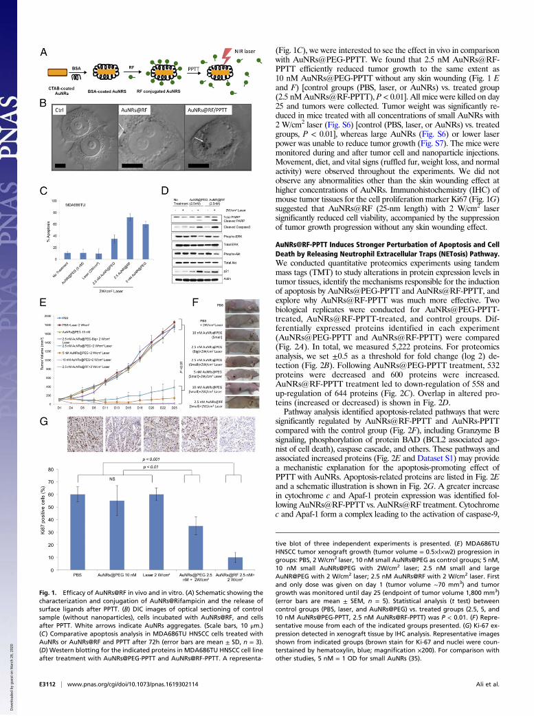
(Fig. 1C), we were interested to see the effect in vivo in comparisonwith AuNRs@PEG-PPTT. We found that 2.5 nM AuNRs@RF-PPTT efficiently reduced tumor growth to the same extent as10 nM AuNRs@PEG-PPTT without any skin wounding (Fig. 1 Eand F) [control groups (PBS, laser, or AuNRs) vs. treated group(2.5 nMAuNRs@RF-PPTT), P < 0.01]. All mice were killed on day25 and tumors were collected. Tumor weight was significantly re-duced in mice treated with all concentrations of small AuNRs with2 W/cm2 laser (Fig. S6) [control (PBS, laser, or AuNRs) vs. treatedgroups, P < 0.01], whereas large AuNRs (Fig. S6) or lower laserpower was unable to reduce tumor growth (Fig. S7). The mice weremonitored during and after tumor cell and nanoparticle injections.Movement, diet, and vital signs (ruffled fur, weight loss, and normalactivity) were observed throughout the experiments. We did notobserve any abnormalities other than the skin wounding effect athigher concentrations of AuNRs. Immunohistochemistry (IHC) ofmouse tumor tissues for the cell proliferation marker Ki67 (Fig. 1G)suggested that AuNRs@RF (25-nm length) with 2 W/cm2 lasersignificantly reduced cell viability, accompanied by the suppressionof tumor growth progression without any skin wounding effect.
AuNRs@RF-PPTT Induces Stronger Perturbation of Apoptosis and CellDeath by Releasing Neutrophil Extracellular Traps (NETosis) Pathway.We conducted quantitative proteomics experiments using tandemmass tags (TMT) to study alterations in protein expression levels intumor tissues, identify the mechanisms responsible for the inductionof apoptosis by AuNRs@PEG-PPTT and AuNRs@RF-PPTT, andexplore why AuNRs@RF-PPTT was much more effective. Twobiological replicates were conducted for AuNRs@PEG-PPTT-treated, AuNRs@RF-PPTT-treated, and control groups. Dif-ferentially expressed proteins identified in each experiment(AuNRs@PEG-PPTT and AuNRs@RF-PPTT) were compared(Fig. 2A). In total, we measured 5,222 proteins. For proteomicsanalysis, we set ±0.5 as a threshold for fold change (log 2) de-tection (Fig. 2B). Following AuNRs@PEG-PPTT treatment, 532proteins were decreased and 600 proteins were increased.AuNRs@RF-PPTT treatment led to down-regulation of 558 andup-regulation of 644 proteins (Fig. 2C). Overlap in altered pro-teins (increased or decreased) is shown in Fig. 2D.Pathway analysis identified apoptosis-related pathways that were
significantly regulated by AuNRs@RF-PPTT and AuNRs-PPTTcompared with the control group (Fig. 2F), including Granzyme Bsignaling, phosphorylation of protein BAD (BCL2 associated ago-nist of cell death), caspase cascade, and others. These pathways andassociated increased proteins (Fig. 2E and Dataset S1) may providea mechanistic explanation for the apoptosis-promoting effect ofPPTT with AuNRs. Apoptosis-related proteins are listed in Fig. 2Eand a schematic illustration is shown in Fig. 2G. A greater increasein cytochrome c and Apaf-1 protein expression was identified fol-lowing AuNRs@RF-PPTT vs. AuNRs@RF treatment. Cytochromec and Apaf-1 form a complex leading to the activation of caspase-9,
Fig. 1. Efficacy of AuNRs@RF in vivo and in vitro. (A) Schematic showing thecharacterization and conjugation of AuNRs@Rifampicin and the release ofsurface ligands after PPTT. (B) DIC images of optical sectioning of controlsample (without nanoparticles), cells incubated with AuNRs@RF, and cellsafter PPTT. White arrows indicate AuNRs aggregates. (Scale bars, 10 μm.)(C) Comparative apoptosis analysis in MDA686TU HNSCC cells treated withAuNRs or AuNRs@RF and PPTT after 72h (error bars are mean ± SD, n = 3).(D) Western blotting for the indicated proteins in MDA686TU HNSCC cell lineafter treatment with AuNRs@PEG-PPTT and AuNRs@RF-PPTT. A representa-
tive blot of three independent experiments is presented. (E) MDA686TUHNSCC tumor xenograft growth (tumor volume = 0.5×l×w2) progression ingroups: PBS, 2 W/cm2 laser, 10 nM small AuNRs@PEG as control groups; 5 nM,10 nM small AuNRs@PEG with 2W/cm2 laser; 2.5 nM small and largeAuNR@PEG with 2 W/cm2 laser; 2.5 nM AuNRs@RF with 2 W/cm2 laser. Firstand only dose was given on day 1 (tumor volume ∼70 mm3) and tumorgrowth was monitored until day 25 (endpoint of tumor volume 1,800 mm3)(error bars are mean ± SEM, n = 5). Statistical analysis (t test) betweencontrol groups (PBS, laser, and AuNRs@PEG) vs. treated groups (2.5, 5, and10 nM AuNRs@PEG-PPTT, 2.5 nM AuNRs@RF-PPTT) was P < 0.01. (F) Repre-sentative mouse from each of the indicated groups presented. (G) Ki-67 ex-pression detected in xenograft tissue by IHC analysis. Representative imagesshown from indicated groups (brown stain for Ki-67 and nuclei were coun-terstained by hematoxylin, blue; magnification ×200). For comparison withother studies, 5 nM = 1 OD for small AuNRs (35).
E3112 | www.pnas.org/cgi/doi/10.1073/pnas.1619302114 Ali et al.
Dow
nloa
ded
by g
uest
on
Mar
ch 2
6, 2
020
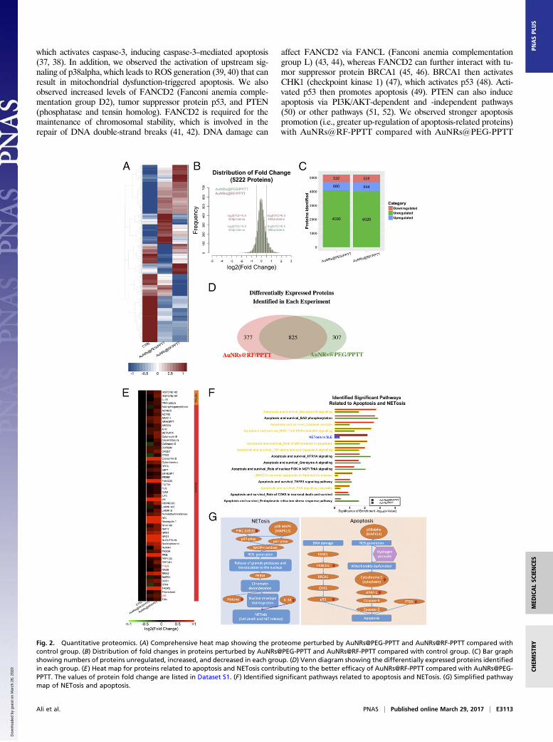
which activates caspase-3, inducing caspase-3–mediated apoptosis(37, 38). In addition, we observed the activation of upstream sig-naling of p38alpha, which leads to ROS generation (39, 40) that canresult in mitochondrial dysfunction-triggered apoptosis. We alsoobserved increased levels of FANCD2 (Fanconi anemia comple-mentation group D2), tumor suppressor protein p53, and PTEN(phosphatase and tensin homolog). FANCD2 is required for themaintenance of chromosomal stability, which is involved in therepair of DNA double-strand breaks (41, 42). DNA damage can
affect FANCD2 via FANCL (Fanconi anemia complementationgroup L) (43, 44), whereas FANCD2 can further interact with tu-mor suppressor protein BRCA1 (45, 46). BRCA1 then activatesCHK1 (checkpoint kinase 1) (47), which activates p53 (48). Acti-vated p53 then promotes apoptosis (49). PTEN can also induceapoptosis via PI3K/AKT-dependent and -independent pathways(50) or other pathways (51, 52). We observed stronger apoptosispromotion (i.e., greater up-regulation of apoptosis-related proteins)with AuNRs@RF-PPTT compared with AuNRs@PEG-PPTT
Fig. 2. Quantitative proteomics. (A) Comprehensive heat map showing the proteome perturbed by AuNRs@PEG-PPTT and AuNRs@RF-PPTT compared withcontrol group. (B) Distribution of fold changes in proteins perturbed by AuNRs@PEG-PPTT and AuNRs@RF-PPTT compared with control group. (C) Bar graphshowing numbers of proteins unregulated, increased, and decreased in each group. (D) Venn diagram showing the differentially expressed proteins identifiedin each group. (E) Heat map for proteins related to apoptosis and NETosis contributing to the better efficacy of AuNRs@RF-PPTT compared with AuNRs@PEG-PPTT. The values of protein fold change are listed in Dataset S1. (F) Identified significant pathways related to apoptosis and NETosis. (G) Simplified pathwaymap of NETosis and apoptosis.
Ali et al. PNAS | Published online March 29, 2017 | E3113
MED
ICALSC
IENCE
SCH
EMISTR
YPN
ASPL
US
Dow
nloa
ded
by g
uest
on
Mar
ch 2
6, 2
020
treatment (Fig. 2E). These observations at the molecular levelare in agreement with the phenotypic study showing thatAuNRs@RF-PPTT is more effective than AuNRs@PEG-PPTT in vivo and in vitro.Interestingly, our proteomics analysis also revealed that treat-
ment with AuNRs@RF-PPTT induced changes in the levels ofseveral proteins involved in the NETosis pathway more stronglythan AuNRs@PEG-PPTT (Fig. 2F). NETosis refers to a specificform of neutrophil cell death caused by pathogen infection,which releases NETs (neutrophil extracellular traps). It hasbeen recently reported that small NPs activate the NETosispathway (53). NETosis can be triggered by two pathways: di-rectly by IL-18 (54–56) or by chromatin decondensation (57–60).
In this study, proteomics analysis identified nine proteins in theNETosis pathway. Specifically, a clear elevation of histone H2,histone H4, IL-18, and Pin1 (peptidylprolyl cis/trans isomerase,NIMA-interacting 1) were observed following AuNRs@RF-PPTTtreatment; therefore, both pathways leading to NETosis werealtered (Fig. 2G). Because NETosis is related to endocytosis, wespeculate that AuNRs@RF may be better taken up by cells,resulting in enhanced NETosis. In summary, from proteomicsanalysis we determined that AuNRs@RF-PPTT can induce ap-optosis and NETosis more efficiently than AuNRs@PEG-PPTT.
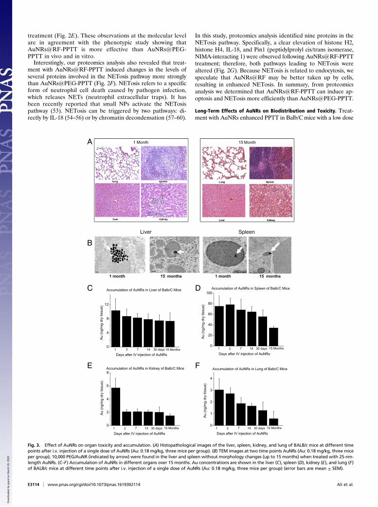
Long-Term Effects of AuNRs on Biodistribution and Toxicity. Treat-ment with AuNRs enhanced PPTT in Balb/C mice with a low dose
A
B
C D
E F
Fig. 3. Effect of AuNRs on organ toxicity and accumulation. (A) Histopathological images of the liver, spleen, kidney, and lung of BALB/c mice at different timepoints after i.v. injection of a single dose of AuNRs (Au: 0.18 mg/kg, three mice per group). (B) TEM images at two time points AuNRs (Au: 0.18 mg/kg, three miceper group); 10,000 PEG/AuNR (indicated by arrow) were found in the liver and spleen without morphology changes (up to 15 months) when treated with 25-nm-length AuNRs. (C–F) Accumulation of AuNRs in different organs over 15 months. Au concentrations are shown in the liver (C), spleen (D), kidney (E), and lung (F)of BALB/c mice at different time points after i.v. injection of a single dose of AuNRs (Au: 0.18 mg/kg, three mice per group) (error bars are mean ± SEM).
E3114 | www.pnas.org/cgi/doi/10.1073/pnas.1619302114 Ali et al.
Dow
nloa
ded
by g
uest
on
Mar
ch 2
6, 2
020
of NIR light (Fig. 1E), indicating their strong clinical potential.However, there is limited knowledge regarding several features ofthis new generation of AuNRs, including their biodistribution,long-term fate, and toxicity. To assess toxicity, the histopathologyof tissues from the liver, spleen, lung, and kidney of mice wasevaluated by a pathologist 1 month and 15 months after single i.v.injection of AuNRs@PEG. We did not observe any histopatho-logical abnormalities in any of the mouse organs (Fig. 3A). Fur-thermore, we monitored mice every week for 15 months followingAuNRs@PEG injection and did not observe any clinical signs oftoxicity, including ruffled fur, impeded movement, signs of abnormalconstitution, aberrant behavior, loss of weight, ocular or nasal dis-charge, respiratory distress, inability to walk, or diarrhea. TEM wasused to visually observe AuNRs@PEG particle uptake and organtissue microstructure. As seen in Fig. 3B, AuNRs@PEG remainedinside the cells without any structural changes. Measurement of Aulevels in the tissues demonstrated that AuNRs@PEG were presentmostly in the spleen and liver of mice (Fig. 3 C and D), and to alesser extent in the kidney and lung (Fig. 3 E and F). To measureany gold excretion, we tested the amount of Au in the feces of miceat three different time points (1, 14, and 30 days) (Fig. S8). Wefound that a very small portion of injected Au was excreted, whereasthe major portion of Au accumulated mainly in the spleen and liverand was sustained without any structural modification over a longperiod (observed up to 15 months). Au accumulated in mouseorgans from the first day of treatment and remained in these tissueseven 15 months later, without any evidence of toxicities.
DiscussionAuNRs-PPTT is widely recognized as a promising strategy forcombating cancer. Developing a valid PPTT in vivo that triggerscancer cell apoptosis (avoiding necrosis) and exploring its molecularmechanism of action is of great importance. In addition, exploringthe long-term fate of the AuNRs after treatment is critical for clinicaluse. In this systematic in vivo study, we (i) optimized the conditionsof AuNRs-PPTT to induce apoptosis, (ii) explored the molecularmechanisms of action of AuNRs-PPTT, and (iii) revealed the long-term (15 months) fate of AuNRs, which indicated their lack of toxicityin mouse models. To optimize PPTT conditions to maximize tumorapoptosis, we evaluated the size, surface modification, and concen-tration of AuNRs and the PPTT laser power both in vitro (fiveHNSCC cell lines) and in vivo (MDA686TU xenograft mice model).Cell and tumor growth inhibition and apoptosis were clearly observedin vitro and in vivo. It is worth noting that among the five HNSCC celllines we have used UM-SCC-47, which is a human papillomavirus-positive cell line. Our findings suggest that AuNRs-PPTT is equallyeffective against viral- and nonviral-derived tumors. Histopathologicalanalysis of mice tissue revealed that a lower concentration (2.5 nM) ofAuNRs@RF-PPTT significantly reduced cancer cell viability asshown by decreased Ki-67. Past attempts to induce apoptosis in vitrohave applied a moderate hyperthermia (61) or targeted differentcellular locations (7, 62, 63). Herein, by adjusting the surface modi-fication and heat generation, we were able to generate efficient tumorapoptosis in vivo without any skin wounding.In our study we have optimized our treatment dose based on the
tumor volume using very low doses (in nanomolar with 100 μLAuNPsinjection volume) and intratumoral injection, which is more realisticfor the clinical application of PPTT. In contrast, most earlier studiesinjected the nanoparticles intravenously, at doses based on animalbody weight (nanoparticle amount in milligrams per kilogram bodyweight) (8, 64, 65) Compared with other studies, we used a moderatelaser intensity and low exposure time (0.5–2 W/cm2 for 2 min) (66).The effect of heat shock (hyperthermia) on the induction of cell
apoptosis has been known for centuries. The mechanism ofhyperthermia-induced apoptosis has been largely unclear until re-cently (67). PPTT is not simply heat shock; instead, it could beregarded as a synergistic effect between nanoparticles and hyper-thermia. We observed that both apoptosis and NETosis pathways
were significantly affected after treatment with PPTT, especiallyAuNRs@RF-PPTT, which demonstrated a much stronger molec-ular impact on these pathways. Cytochrome c and p53-relatedapoptosis mechanisms were identified as contributing to the en-hanced effect of PPTT with RF-conjugated AuNRs. Furthermore,Pin1 and IL18-related signaling contribute to the observed per-turbation of the NETosis pathway by PPTT with RF-conjugatedAuNRs. Up-regulation of Pin1 has been shown to induce ROSproduction through phosphorylation of NADPH oxidase regula-tory subunits p47-phox and p67-phox (57–59). ROS productionfurther leads to the release of PERM.Histone H4 and histone H2 aresubsequently degraded, leading to chromatin decondensation, whichis further enhanced by PERM. Eventually the integrity of the nuclearenvelope is disrupted, resulting in cell rupturing (60). The greatereffect of AuNRs@RF-PPTT on NETosis may result from betteruptake of AuNRs into the cells when conjugated with RF. It has beenreported recently that cells entrap nanoparticles via formation ofNETs, which are formed immediately following rapid damage toplasma membranes and instability of the lysosomal compartmentinduced by nanoparticle stimulation (53). The significant effects ob-served in our study of AuNRs@RF-PPTT on the NETosis pathwaymay account for the greater efficacy of the RF conjugate.After PPTT, we observed the aggregation of AuNRs@RF
around cell nuclear membranes as shown by DIC microscopy (Fig.1B). Our proteomics study identified greater up-regulation of nuclearlamin proteins (Fig. 2E), which are responsible for nuclear shape andstructure. Therefore, AuNRs@RF may harm nuclear membraneintegrity through intrinsic cell defense mechanisms. However, laminsare known to impede cancer cell migration and invasion (68, 69)Lamin promotes cell-matrix adhesion and plays an important role inapoptosis by loosening epithelial cell contact with the extracellularmatrix (70). Lamins are targets for degradation in the apoptoticprocess and accordingly are often used as markers for apoptosis (71).There is to date limited knowledge regarding the biodistribution,
long-term fate, and toxicity of AuNRs. Thus, our studies of thetoxicity of AuNRs are important to develop safer treatments. Thisis a long-term toxicity study of AuNRs in mice lasting for 15 months.Based on our findings, we can conclude that these AuNRs have greatpotential to be used in PPTT for the local treatment of cancers,supporting the efficient translation of AuNRs into clinical settings.In summary, we have optimized the efficacy and studied the
molecular mechanisms of AuNRs-assisted PPTT and examined the15-month toxicity and fate of AuNRs in a mouse model. Together,these data demonstrate that our AuNRs-PPTT is highly effectiveand safe for local therapy for cancers. These findings provide astrong framework for translation of this approach to the clinic.
MethodsAuNRs Synthesis, Conjugation, and Characterization. A seedless growthmethodwas used for the synthesis of AuNRs with an average size of 25 ± 3 nm × 5.5 ±0.8 nm (length × width) (35). Briefly, HAuCl4 (5.0 mL, 1.0 mM) was added to5.0 mL of 0.2 M CTAB at room temperature. Then, 250 μL of 4.0 mM AgNO3,8 μL of 12 M HCl, and 70 μL of ascorbic acid (78.8 mM) was successively addedand the solution was gently mixed. Immediately afterward, 15 μL of 0.01 M ice-cold NaBH4 was injected into the unstirred growth solution and allowed toreact for 12 h. The synthesized AuNRs were then centrifuged at 21,000 × g for50 min and redispersed in deionized (DI) water, followed by a second centri-fugation at 19,000 × g for 40 min to remove the extra CTAB. A seed-mediatedgrowth method was used for the synthesis of AuNRs with an average size of72 ± 7 nm × 16 ± 4 nm, where 4.0 mM of silver nitrate and 78.8 mM of ascorbicacid were added to a growth solution consisting of 0.2 M CTAB, 1.0 mM ofHAuCl4, and 0.01 M of NaBH4. Then, CTAB stabilized AuNRs were purified bycentrifugation (10,000 × g for 50 min) and redispersed in DI H2O, followed by asecond centrifugation at 7,000 × g for 30 min. For AuNRs conjugated with PEG,we added mPEG-SH (1 mM) to the nanoparticles overnight to achieve about20,000 ligands on each particle. For AuNRs conjugated with RF, we first added50 mL of 0.5 mM BSA (Sigma-Aldrich) to 1 nM AuNRs solution, which was thenincubated for 3 h at room temperature. Then, 300 μL of 5 mM RF was added tothe AuNRs solution and incubated for 3 h. After conjugation, the particles werecentrifuged to remove the extra ligands. TEM was used to examine particle size
Ali et al. PNAS | Published online March 29, 2017 | E3115
MED
ICALSC
IENCE
SCH
EMISTR
YPN
ASPL
US
Dow
nloa
ded
by g
uest
on
Mar
ch 2
6, 2
020
and homogeneity. UV-Vis spectrometer and ZetaSizer 3000 (Malvern Instru-ments) were used to examine whether the conjugation was successful. For BSAfluorescent quenching experiments, excitation and emission measurementswere carried out on a Quanta Master 300 phosphorescence/fluorescence spec-trofluorometer. BSA was used at a concentration of 10−4 M. The number of RFmolecules per AuNRs was calculated according to Ali et al. (36), based on theUV-Vis spectra. The dose of AuNRs was presented as molar concentration. Forcomparison with other studies, 5 nM = 1 OD (35).
Dark-Field Images.MDA686TU cells were seeded on glass coverslips in completegrowth medium for 24 h to achieve 40% final confluence. Cells were incubatedwith 2.5 nM AuNRs with RF or PEG conjugation in supplemented DMEM cellculture medium for 24 h. Dark-field images were taken using an inverted mi-croscope equipped with a dark-field condenser and Lumenera Infinity2 CCDcamera; a 20× objective lens was used to collect scattered light from the samplesto produce dark-field images.
DIC Microscopy. MDA686TU cells were seeded on glass coverslips in completegrowth medium for 24 h then incubated with 2.5 nM AuNRs with RF or PEGconjugation in supplemented DMEM cell culture medium for 24 h. For DICimaging, an inverted Nikon Eclipse Ti-E microscope equipped with Perfect FocusSystem (PFS, 25-nm z-axial resolution) was used for imaging and z-stacks ac-quisitions under DICmicroscopy. TheDICmode used aDIC polarizer and analyzerpair, a high-resolution 100× I-R DIC slider, a high numerical aperture (N.A. 1.40)oil-immersion condenser lens, a Nikon CFI Apo TIRF 100× (N.A. 1.49) oil-immersion objective, and a 12 V/100 W halogen lamp as light source. Appro-priate bandpass filters were placed in the light path. Z-stack movies were takenby a Hamamatsu ORCA-Flash 4.0 V2 CMOS camera (C11440-22CU, pixel size6.5 μm × 6.5 μm) with Camera Link interface usingMicro-Manager and analyzedusing NIH ImageJ and reconstructed in Amira. Fixed Hey cells on 22- × 22-mmglass coverslips were rinsed with Dulbecco’s PBS at pH 7.4 and fabricated into asandwiched chamber with two pieces of double-sided tape and a cleaned glassslide. PBS solution was added into the chamber to fill the space and thechamber was sealed with clear nail polish. The sample slide was then placedunder the microscope for observation. Z-stacks were acquired using the multi-dimensional acquisition function in Micro-Manager. DIC optical sectioning(Movies S1 and S2) through the whole cell thickness was achieved by movingthe objective on the motorized nosepiece using PFS at 65 nm per step at 33-ms(30 frames per s) exposure time.
Cell Lines. Information and primary sources of HNSCC cell lines MDA686TU,Fadu, and SqCC/Y1were described elsewhere (72). UD-SCC2 andUM-SCC-47 celllines were obtained from R. L. Ferris, University of Pittsburgh Cancer Center,Pittsburgh. Cells were maintained in DMEM/F12 (1:1) medium supplementedwith 10% heat-inactivated FBS in a 37 °C, 5% CO2 humidified incubator.
Cell Viability Assay. Sulforhodamine B (SRB) assay was used for cell viabilitydetermination (73). Cells were seeded (5 × 10³ cells per well) in a 96-well plate.Twenty-four hours later cells were treated with AuNRs for 24 h and exposed tolaser for 2 min. Cells were fixed after another 24 h. Plates were stained withSRB and bound SRB was dissolved to assess OD at 492 nm using a microplatereader. The percentage of surviving cells was calculated based on the absor-bance values relative to the nontreated samples.
Apoptosis Assay. Apoptotic cells were identified and measured as describedelsewhere (74). Briefly, cells were collected and stained with AnnexinV-phycoerythrene and 7-AAD (BD Pharmingen) for 15 min at room tempera-ture. Samples were measured using a FACS caliber bench-top flow cytometer(Becton Dickinson). FlowJo software (Tree Star) was used for apoptosis analysis.
Western-Blot Analysis. Western blot was incubated with primary followed bysecondary antibodies and detected using enhanced chemiluminescencesystem as described (74). Primary antibodies were anti–phospho-Akt, anti-total Akt, anti–phospho-Erk, anti-total Erk, anti-caspase3, anti-caspase3, andanti-PARP from Cell Signaling; anti-p21 from Santa Cruz Biotechnology; andanti–β-actin from Sigma-Aldrich. Secondary antibodies were from Santa CruzBiotechnology. Western band quantification was performed using Image-Quant TL software (GE/Amersham Biosciences).
Nude Mice Bearing Human HNSCC Xenograft Tumor Model. Based on protocolsapproved by the Institutional Animal Care and Use Committee of EmoryUniversity, female nude mice (athymic nu/nu) aged 4–6 wk were purchasedfrom Taconic. Each mouse was injected with 5 × 10⁶ MDA686TU cells s.c. in theright flank, and tumor volume was monitored (volume = 0.5 × length ×width2).
Mice were randomized into groups once the tumors reached 70 mm3. Micewere injected intratumorally with PBS or different concentrations of AuNRs andexposed to different powers of NIR laser for 2 min (five mice per group). Tu-mors were monitored every other day and mice were killed on the 25th dayafter cancer cell transplantation. Tumors and organs were collected, measured,and processed for paraffin embedding.
IHC. Upon deparaffinization and rehydration, tissue sections were per-meabilizedwith 0.25%Triton-X-100/PBS for 5min. Tissue sectionswere blockedwith 2.5% horse serum for 30 min. To detect intracellular localization andexpression levels of Ki-67 proteins, we used mouse anti-human Ki-67 antibody(prediluted; Invitrogen) then counterstained cell nuclei using DAPI (Invitrogen).Mouse and rabbit IgG were used as negative controls.
Sample Preparation for Proteomics. MDA686TU cells (5 × 10⁶) were injectedinto the right flank of nude mice. We injected a single dose of AuNRs@PEG(2.5 nM) or AuNRs@RF (2.5 nM) intratumorally followed by 2 min of 2 W/cm2
NIR laser exposure when the tumors had reached 150 mm3. Twenty-four hourslater we collected tumors for proteomics analysis. Each experiment was re-peated twice. Mouse tumor tissues were homogenized, followed by ultrasonictissue ablation with ice-cold lysis buffer (50 mM Hepes, pH 7.8, 150 mM NaCl0.1% SDS, 0.5% sodium deoxycholate 1% Triton X-100 or Nonidet P-40, andphosphatase inhibitors). The cells were then scraped down and the obtainedmixtures homogenized with sonication and vortexing. Cell debris was re-moved by centrifugation at 18,000 × g for 20 min at 4 °C. Four volumes of ice-cold acetone/ethanol/acetic acid (vol/vol/vol = 50/50/0.1) was added to thesupernatant to precipitate the proteins at −20 °C overnight. After centrifu-gation, the protein pellet was redissolved in denaturing buffer (pH 8.0) con-taining 8 M urea and 50 mM Hepes, and the protein concentration was testedusing a Bradford assay. The disulfide bonds in the protein solution were re-duced by 2 mM DTT at 37 °C for 2 h and subsequently alkylated by adding6 mM iodoacetamide and incubation in darkness at room temperature for40 min (75). Purified peptides were labeled with 6-plex TMT reagents (Thermo)following the manufacturer’s protocol. Briefly, lyophilized peptides were dis-solved in 100 μL of 100 mM triethylammonium bicarbonate buffer, pH 8.5.Each channel of the TMT reagents was dissolved in 41 μL of anhydrous ACNand transferred into the peptide tube. The reaction was performed at roomtemperature for 1 h then was quenched by adding 8 μL of 5% hydroxylamine.Peptides from all six tubes were then mixed, desalted again using a tC18 Sep-Pak cartridge, and lyophilized overnight. For protein analysis, the peptidemixture was separated by high pH reversed-phase HPLC into 20 fractionswith a 40-min gradient of 5–55% acetonitrile (ACN) in 10 mM ammoniumacetate (pH 10).
Liquid Chromatography–Tandem MS Analysis and Database Search. After TMTlabeling and purification, lyophilized peptide samples were dissolved in 10 μLsolvent of 5% (vol/vol) ACN and 4% (vol/vol) formic acid (FA), and 4 μL of thedissolved sample were loaded onto a microcapillary column packed withC18 beads (Magic C18AQ, 3 μm 200 Å, 100 μL × 16 cm; Michrom Bioresources)by a Dionex WPS-3000TPLRS autosampler (UltiMate 3000 thermostated RapidSeparation Pulled Loop Wellplate Sampler). Peptides were separated byreversed-phase chromatography using an UltiMate 3000 binary pump with a90-min gradient of 4–30% (vol/vol) ACN (in 0.125% FA). Peptides were de-tected with a data-dependent Top15 method (76) in a hybrid dual-cell quad-rupole linear ion trap–Orbitrap mass spectrometer (LTQ Orbitrap Elite;ThermoFisher, with Xcalibur 3.0.63 software). For each cycle, one full MSscan (resolution 60,000) in the Orbitrap at 106 AGC target was followed by upto 15 MS/MS for the most intense ions. The selected ions were excluded fromfurther analysis for 90 s. Ions with single or unassigned charge were discarded.MS2 scans were performed in the orbitrap cell by activating with high energycollision dissociation at 40% normalized collision energy with 1.2 m/z isolationwidth. All MS2 spectra were converted into mzXML format and then searchedusing the SEQUEST algorithm (version 28) (77). Spectra were matched against adatabase containing sequences of all proteins in the UniProt Human (Homosapiens) database. The following parameters were used during the search:20 ppmmass tolerance; fully digested with trypsin; up to three missed cleavages;fixed modifications: carbamidomethylation of cysteine (+57.0214), TMT modifi-cation of lysine (+229.1629) and N terminus (+229.1629); variable modifications:oxidation of methionine (+15.9949). False discovery rates (FDRs) of peptide andprotein identifications were evaluated and controlled by the target-decoymethod (78). Each protein sequence was listed in both forward and reversedorders. Linear discriminant analysis, which is similar to other methods in theliterature (79), was used to control the quality of peptide (80). Peptides fewerthan seven amino acid residues in length were discarded. Furthermore, peptide
E3116 | www.pnas.org/cgi/doi/10.1073/pnas.1619302114 Ali et al.
Dow
nloa
ded
by g
uest
on
Mar
ch 2
6, 2
020
spectral matches were filtered to <1% FDR. An additional protein-level filter wasapplied in each dataset to reduce the protein-level FDRs (<1%).
Proteomics Data Analysis. For proteomics analysis each experiment was re-peated twice. The mean expression level of each protein was used fordownstream analysis. Raw data were normalized using supervised normali-zation of microarray (81). Variance due to biological replicates was adjustedby setting them as variables in the model. Variance explained by differentexperimental treatments (control, AuNRs@PEG-PPTT, and AuNRs@RF-PPTT)was fitted as a biological variable in the model. Hierarchical clustering wasperformed with statistical software R. Proteins identified as being affectedwere subjected to pathway analysis using the MetaCore from ThomsonReuters.
Short-Term and Long-Term Uptake Fate of PEGylated AuNRs. BALB/C mice(male), 6wk of age,were injected via the tail veinwith a 200-μL solution ofAuNRs(0.18 mg/kg). At specified time points of 1 day, 3, 7, 14, and 30 days and15 months mice were killed by pressurized CO2 asphyxiation, three mice in each
group. Liver, spleen, kidney, and lung were collected, rinsed with distilled water,and dried. The dried tissues were dissolved and assayed for Au using ICP-MS.
Histopathology Evaluation. Liver, spleen, kidney, and lung tissues were em-bedded in paraffin and cut at 5-μm thickness. The tissues were stained withhematoxylin and eosin (Sigma) to assess histological alterations via microscopy.
Statistical Analysis. All results represent the average of at least three separateexperiments and are expressed as mean ± SD or SE. Statistical analysis wasconducted using t test. P values less than 0.05 were considered statisticallysignificant.
ACKNOWLEDGMENTS. We thank Dr. Kuangcai Chen and Prof. Ning Fang(Georgia State University) for the DIC microscopic images and Dr. AntheaHammond for her assistance in critical reading and editing of the manuscript.This work was supported by National Cancer Institute Cancer NanotechnologyPlatform Partner Grant U01 CA151802 to Emory University and National ScienceFoundation Division of Chemistry Grant 1608801 to the Georgia Instituteof Technology.
1. Huang X, El-Sayed IH, Qian W, El-Sayed MA (2006) Cancer cell imaging and photo-thermal therapy in the near-infrared region by using gold nanorods. J Am Chem Soc128(6):2115–2120.
2. Dickerson EB, et al. (2008) Gold nanorod assisted near-infrared plasmonic photo-thermal therapy (PPTT) of squamous cell carcinoma in mice. Cancer Lett 269(1):57–66.
3. Hirsch LR, et al. (2003) Nanoshell-mediated near-infrared thermal therapy of tumorsunder magnetic resonance guidance. Proc Natl Acad Sci USA 100(23):13549–13554.
4. Abadeer NS, Murphy CJ (2016) Recent progress in cancer thermal therapy using goldnanoparticles. J Phys Chem C 120(9):4691–4716.
5. Steinmetz NF (2010) Review of “Cancer Nanotechnology: Methods and Protocols(Methods in Molecular Biology)” by Stephen R. Grobmyer (Editor), Brij M. Moudgil(Editor). BioMedical Engineering OnLine 9:55.
6. Alkilany AM, Thompson LB, Boulos SP, Sisco PN, Murphy CJ (2012) Gold nanorods:Their potential for photothermal therapeutics and drug delivery, tempered by thecomplexity of their biological interactions. Adv Drug Deliv Rev 64(2):190–199.
7. Ali MR, Ibrahim IM, Ali HR, Selim SA, El-SayedMA (2016) Treatment of natural mammarygland tumors in canines and felines using gold nanorods-assisted plasmonic photo-thermal therapy to induce tumor apoptosis. Int J Nanomedicine 11:4849–4863.
8. von Maltzahn G, et al. (2009) Computationally guided photothermal tumor therapyusing long-circulating gold nanorod antennas. Cancer Res 69(9):3892–3900.
9. Pattani VP, Shah J, Atalis A, Sharma A, Tunnell JW (2015) Role of apoptosis and ne-crosis in cell death induced by nanoparticle-mediated photothermal therapy.J Nanopart Res 17(1):1–11.
10. Bonfil RD, Bustuoabad OD, Ruggiero RA, Meiss RP, Pasqualini CD (1988) Tumor ne-crosis can facilitate the appearance of metastases. Clin Exp Metastasis 6(2):121–129.
11. Hanahan D, Weinberg RA (2011) Hallmarks of cancer: The next generation. Cell144(5):646–674.
12. Danial NN, Korsmeyer SJ (2004) Cell death: Critical control points. Cell 116(2):205–219.13. Ali MR, Ali HR, Rankin CR, El-Sayed MA (2016) Targeting heat shock protein 70 using
gold nanorods enhances cancer cell apoptosis in low dose plasmonic photothermaltherapy. Biomaterials 102:1–8.
14. Pérez-Hernández M, et al. (2015) Dissecting the molecular mechanism of apoptosisduring photothermal therapy using gold nanoprisms. ACS Nano 9(1):52–61.
15. Oh N, Park JH (2014) Endocytosis and exocytosis of nanoparticles in mammalian cells.Int J Nanomedicine 9(Suppl 1):51–63.
16. Iyer AK, Khaled G, Fang J, Maeda H (2006) Exploiting the enhanced permeability andretention effect for tumor targeting. Drug Discov Today 11(17-18):812–818.
17. Maeda H, Wu J, Sawa T, Matsumura Y, Hori K (2000) Tumor vascular permeability andthe EPR effect in macromolecular therapeutics: a review. J Controlled Release 65(1-2):271–284.
18. Liu X, et al. (2013) Enhanced retention and cellular uptake of nanoparticles in tumorsby controlling their aggregation behavior. ACS Nano 7(7):6244–6257.
19. Wilhelm S, et al. (2016) Analysis of nanoparticle delivery to tumours. Nature ReviewsMaterials 1:16014.
20. Ali MRK, Panikkanvalappil SR, El-Sayed MA (2014) Enhancing the efficiency of goldnanoparticles treatment of cancer by increasing their rate of endocytosis and cellaccumulation using rifampicin. J Am Chem Soc 136(12):4464–4467.
21. Mackey MA, Ali MRK, Austin LA, Near RD, El-Sayed MA (2014) The most effective goldnanorod size for plasmonic photothermal therapy: Theory and in vitro experiments.J Phys Chem B 118(5):1319–1326.
22. Alkilany AM, Murphy CJ (2010) Toxicity and cellular uptake of gold nanoparticles:What we have learned so far? J Nanopart Res 12(7):2313–2333.
23. Connor EE, Mwamuka J, Gole A, Murphy CJ, Wyatt MD (2005) Gold nanoparticles aretaken up by human cells but do not cause acute cytotoxicity. Small 1(3):325–327.
24. Axiak-Bechtel SM, et al. (2014) Gum arabic-coated radioactive gold nanoparticlescause no short-term local or systemic toxicity in the clinically relevant canine model ofprostate cancer. Int J Nanomedicine 9:5001–5011.
25. Chen H, et al. (2013) In vivo study of spherical gold nanoparticles: Inflammatory ef-fects and distribution in mice. PLoS One 8(2):e58208.
26. You J, et al. (2014) Pharmacokinetics, clearance, and biosafety of polyethylene glycol-coated hollow gold nanospheres. Part Fibre Toxicol 11:26.
27. Pernodet N, et al. (2006) Adverse effects of citrate/gold nanoparticles on humandermal fibroblasts. Small 2(6):766–773.
28. Cho YS, et al. (2009) Phosphorylation-driven assembly of the RIP1-RIP3 complex regu-lates programmed necrosis and virus-induced inflammation. Cell 137(6):1112–1123.
29. Qu Y, Huang Y, Lü X (2013) Proteomic analysis of molecular biocompatibility of goldnanoparticles to human dermal fibroblasts-fetal. J Biomed Nanotechnol 9(1):40–52.
30. Yildirimer L, Thanh NTK, Loizidou M, Seifalian AM (2011) Toxicology and clinicalpotential of nanoparticles. Nano Today 6(6):585–607.
31. Murphy CJ, et al. (2008) Gold nanoparticles in biology: Beyond toxicity to cellularimaging. Acc Chem Res 41(12):1721–1730.
32. Boisselier E, Astruc D (2009) Gold nanoparticles in nanomedicine: Preparations, im-aging, diagnostics, therapies and toxicity. Chem Soc Rev 38(6):1759–1782.
33. Niidome T, et al. (2006) PEG-modified gold nanorods with a stealth character for invivo applications. J Control Release 114(3):343–347.
34. Sadauskas E, et al. (2009) Protracted elimination of gold nanoparticles from mouseliver. Nanomedicine (Lond) 5(2):162–169.
35. Ali MRK, Snyder B, El-Sayed MA (2012) Synthesis and optical properties of small Aunanorods using a seedless growth technique. Langmuir 28(25):9807–9815.
36. Ali HR, et al. (2016) Gold nanorods as drug delivery vehicles for rifampicin greatlyimprove the efficacy of combating Mycobacterium tuberculosis with good bio-compatibility with the host cells. Bioconjug Chem 27(10):2486–2492.
37. Sakai T, et al. (2004) Nucling recruits Apaf-1/pro-caspase-9 complex for the inductionof stress-induced apoptosis. J Biol Chem 279(39):41131–41140.
38. Yin Q, et al. (2006) Caspase-9 holoenzyme is a specific and optimal procaspase-3processing machine. Mol Cell 22(2):259–268.
39. Geering B, Simon HU (2011) A novel signaling pathway in TNFα-induced neutrophilapoptosis. Cell Cycle 10(17):2821–2822.
40. Geering B, Gurzeler U, Federzoni E, Kaufmann T, Simon HU (2011) A novel TNFR1-triggered apoptosis pathway mediated by class IA PI3Ks in neutrophils. Blood 117(22):5953–5962.
41. Barroso E, et al. (2009) The Fanconi anemia family of genes and its correlation withbreast cancer susceptibility and breast cancer features. Breast Cancer Res Treat 118(3):655–660.
42. Montes de Oca R, et al. (2005) Regulated interaction of the Fanconi anemia protein,FANCD2, with chromatin. Blood 105(3):1003–1009.
43. Longerich S, San Filippo J, Liu D, Sung P (2009) FANCI binds branched DNA and ismonoubiquitinated by UBE2T-FANCL. J Biol Chem 284(35):23182–23186.
44. Yuan F, El Hokayem J, Zhou W, Zhang Y (2009) FANCI protein binds to DNA and in-teracts with FANCD2 to recognize branched structures. J Biol Chem 284(36):24443–24452.
45. Garcia-Higuera I, et al. (2001) Interaction of the Fanconi anemia proteins andBRCA1 in a common pathway. Mol Cell 7(2):249–262.
46. Taniguchi T, et al. (2002) S-phase-specific interaction of the Fanconi anemia protein,FANCD2, with BRCA1 and RAD51. Blood 100(7):2414–2420.
47. Yarden RI, Pardo-Reoyo S, Sgagias M, Cowan KH, Brody LC (2002) BRCA1 regulatesthe G2/M checkpoint by activating Chk1 kinase upon DNA damage. Nat Genet 30(3):285–289.
48. Goudelock DM, Jiang K, Pereira E, Russell B, Sanchez Y (2003) Regulatory interactionsbetween the checkpoint kinase Chk1 and the proteins of the DNA-dependent proteinkinase complex. J Biol Chem 278(32):29940–29947.
49. Norbury CJ, Zhivotovsky B (2004) DNA damage-induced apoptosis. Oncogene 23(16):2797–2808.
50. Weng L, Brown J, Eng C (2001) PTEN induces apoptosis and cell cycle arrest throughphosphoinositol-3-kinase/Akt-dependent and -independent pathways. Hum MolGenet 10(3):237–242.
51. Qi Y, et al. (2015) PTEN induces apoptosis and cavitation via HIF-2-dependentBnip3 upregulation during epithelial lumen formation. Cell Death Differ 22(5):875–884.
52. Zhao H, Dupont J, Yakar S, Karas M, LeRoith D (2004) PTEN inhibits cell proliferationand induces apoptosis by downregulating cell surface IGF-IR expression in prostatecancer cells. Oncogene 23(3):786–794.
Ali et al. PNAS | Published online March 29, 2017 | E3117
MED
ICALSC
IENCE
SCH
EMISTR
YPN
ASPL
US
Dow
nloa
ded
by g
uest
on
Mar
ch 2
6, 2
020
53. Muñoz LE, et al. (2016) Nanoparticles size-dependently initiate self-limiting NETosis-driven inflammation. Proc Natl Acad Sci USA 113(40):E5856–E5865.
54. Yu Y, Su K (2013) Neutrophil extracellular traps and systemic lupus erythematosus.J Clin Cell Immunol 4:4.
55. Kahlenberg JM, Carmona-Rivera C, Smith CK, Kaplan MJ (2013) Neutrophil extracel-lular trap-associated protein activation of the NLRP3 inflammasome is enhanced inlupus macrophages. J Immunol 190(3):1217–1226.
56. Mitroulis I, et al. (2011) Neutrophil extracellular trap formation is associated with IL-1β and autophagy-related signaling in gout. PLoS One 6(12):e29318.
57. Makni-Maalej K, et al. (2012) The TLR7/8 agonist CL097 primes N-formyl-methionyl-leucyl-phenylalanine-stimulated NADPH oxidase activation in human neutrophils:Critical role of p47phox phosphorylation and the proline isomerase Pin1. J Immunol189(9):4657–4665.
58. El Benna J, et al. (1996) Activation of p38 in stimulated human neutrophils: Phos-phorylation of the oxidase component p47phox by p38 and ERK but not by JNK. ArchBiochem Biophys 334(2):395–400.
59. Dang PM, Morel F, Gougerot-Pocidalo MA, El Benna J (2003) Phosphorylation of theNADPH oxidase component p67(PHOX) by ERK2 and P38MAPK: Selectivity of phos-phorylated sites and existence of an intramolecular regulatory domain in thetetratricopeptide-rich region. Biochemistry 42(15):4520–4526.
60. Papayannopoulos V, Metzler KD, Hakkim A, Zychlinsky A (2010) Neutrophil elastaseand myeloperoxidase regulate the formation of neutrophil extracellular traps. J CellBiol 191(3):677–691.
61. Li S, Chien S, Branemark PI (1999) Heat shock-induced necrosis and apoptosis in os-teoblasts. J Orthopaedic Res 17(6):891–899.
62. Wang L, et al. (2011) Selective targeting of gold nanorods at the mitochondria ofcancer cells: Implications for cancer therapy. Nano Lett 11(2):772–780.
63. Kang B, Mackey MA, El-Sayed MA (2010) Nuclear targeting of gold nanoparticles incancer cells induces DNA damage, causing cytokinesis arrest and apoptosis. J AmChem Soc 132(5):1517–1519.
64. Lal S, Clare SE, Halas NJ (2008) Nanoshell-enabled photothermal cancer therapy:Impending clinical impact. Acc Chem Res 41(12):1842–1851.
65. Melancon MP, et al. (2008) In vitro and in vivo targeting of hollow gold nanoshellsdirected at epidermal growth factor receptor for photothermal ablation therapy.MolCancer Ther 7(6):1730–1739.
66. Moon HK, Lee SH, Choi HC (2009) In vivo near-infrared mediated tumor destructionby photothermal effect of carbon nanotubes. ACS Nano 3(11):3707–3713.
67. Song AS, Najjar AM, Diller KR (2014) Thermally induced apoptosis, necrosis, and heat
shock protein expression in 3D culture. J Biomech Eng 136(7):071006-1–071006-10.68. Short B (2014) Lamin-A provides stiff resistance to cell migration. J Cell Biol 204(5):
626.69. van der Zee JA, et al. (2012) Tumour basement membrane laminin expression predicts
outcome following curative resection of pancreatic head cancer. Br J Cancer 107(7):
1153–1158.70. Esco MA, Wang Z, McDermott ML, Kurpakus-Wheater M (2001) Potential role for
laminin 5 in hypoxia-mediated apoptosis of human corneal epithelial cells. J Cell Sci
114(Pt 22):4033–4040.71. Broers JL, Ramaekers FC (2014) The role of the nuclear lamina in cancer and apoptosis.
Adv Exp Med Biol 773:27–48.72. Zhao M, et al. (2011) Assembly and initial characterization of a panel of 85 genomi-
cally validated cell lines from diverse head and neck tumor sites. Clin Cancer Res
17(23):7248–7264.73. Vichai V, Kirtikara K (2006) Sulforhodamine B colorimetric assay for cytotoxicity
screening. Nat Protoc 1(3):1112–1116.74. Rahman MA, et al. (2013) RRM2 regulates Bcl-2 in head and neck and lung cancers: A
potential target for cancer therapy. Clin Cancer Res 19(13):3416–3428.75. Wu Y, et al. (2014) Five-plex isotope dimethyl labeling for quantitative proteomics.
Chem Commun (Camb) 50(14):1708–1710.76. Xiao H, Wu R (2016) Quantitative investigation of human cell surface N-glycoprotein
dynamics. Chem Sci (Camb) 8:268–277.77. Eng JK, McCormack AL, Yates JR (1994) An approach to correlate tandem mass
spectral data of peptides with amino acid sequences in a protein database. J Am Soc
Mass Spectrom 5(11):976–989.78. Elias JE, Gygi SP (2007) Target-decoy search strategy for increased confidence in large-
scale protein identifications by mass spectrometry. Nat Methods 4(3):207–214.79. Käll L, Canterbury JD, Weston J, Noble WS, MacCoss MJ (2007) Semi-supervised learning
for peptide identification from shotgun proteomics datasets. Nat Methods 4(11):
923–925.80. Huttlin EL, et al. (2010) A tissue-specific atlas of mouse protein phosphorylation and
expression. Cell 143(7):1174–1189.81. Mecham BH, Nelson PS, Storey JD (2010) Supervised normalization of microarrays.
Bioinformatics 26(10):1308–1315.
E3118 | www.pnas.org/cgi/doi/10.1073/pnas.1619302114 Ali et al.
Dow
nloa
ded
by g
uest
on
Mar
ch 2
6, 2
020