Embed Size (px)
Citation preview
Effects of Previous Angina Pectoris in Patients With First Acute Myocardial Infarction
Not Receiving Thrombolytics Robert A. Kloner, MD, PhD, James Muller, MD, and Vicki Davis, DrPh,
for the MILE Study Group*
W e previously showed in the Thrombolysis in Myo- cardial Infarction-4 study that patients with a prc-
vious history of angina pcctoris who received throm- bolytics for acute myocardial infarction (AMI) had an acute in-hospital benefit, including better survival, less severe congestive heart failure or shock, and smaller infarct size assessed by crcatine kinase (CK) curves com- pared with patients without previous angina pectoris.’ This benefit was independent of visible epicardial coro- nary artcry collaterals assessed by angiography or the use of antianginal drugs. One potential explanation for the benelit is that previous angina caused ischemic pre- conditioning, the phenomenon whereby brief episodes of transient &hernia prior to a more prolonged duration of coronary occlusion markedly reduce myocardial infarct size.’ This phenomenon has been observed in all experimental animal models of repcrfused infarcts stud- ied: dogs, cats, rabbits, and pigs.” In animal models, however, the phenomenon is short-lived, although recent studies suggest that a second window of protection may
Fror- The Heart ‘ns’lt.jte of the Hospttal oi ihc Gocc Samo,i!an. Uni- vcrsity of Sowherr- Cali’ornio, Los A-geies, California, New tn_nlord Deaconess +spltat, Br’ghom uld Women’s Yospitol, and Harvard Medicnl Sch& Boston, Mcssochuxts, end Rescorcl- T.iongle Instl- tute. Rcseorch Tr’orgle Perk, North Ccro’lro. 3. I<loner’s currerv address is. heo? Institu;c Resew& Depxrtment Hospitcl of the God Sumorlton. 6 1 C: Sath Witmer Street, Los Angeles, Co ifornlo 903 17. Mowscr’pt :eceivco Augus; 29, i 994: &sed mo*luscript received November 14, 1994, and occeptcd November 15
l Appen&cs I:SI ng MIL:S lnvcsrlgotors cw be found In rcierewx 9 and :@.
occur 224 hours after the initial ischemic event4T5 and that preconditioning can be reinduced after its initial dis- sipation.6 If repcrfusion does not occur within 60 to 90 minutes of coronary occlusion in animals, the benefit of preconditioning is lost.“,7,8 To determine whether the pre- conditioning benefit in humans is also dependent on ear- ly reperfusion, we rctrospcctively analyzed data from the Multicenter Investigation of the Limitation of Infarct Size (MILIS) study. in which patients did not receive thrombolytic therapy. We sought to determine whether a history of previous angina lost its acute protective effect when thrombolytic therapy was not instituted. In a sub- group of patients with early CK-MB peaking of iI5 hours (in which presumed spontaneous thrombolysis occurred), we sought to determine whether a history of prior angina aff‘ected infarct size.
The MILE study was a nonthrombolytic multicen- ter study performed in the early 1980s to determine whether hyaluronidase or propranolol could limit myo- cardial infarct size.’ Although neither agent significant- ly afiected infarct size, a large database was created that has produced much valuable information. Previously, it was observed that 46% of infarcts were anterior, a third were inferior, and 12% were both. We reviewed data col- lectcd on the study forms regarding whether patients had a history of angina of more than or during the 3 weeks before a first qualifying AMI, and compared these find- ings with age at onset of AMI and several in-hospital outcomes: arrhythmias, congestive heart failure, infarct extension (assessed clinically and by CK curves), car- diogenic shock, ventricular scptal rupture, mitral regur-
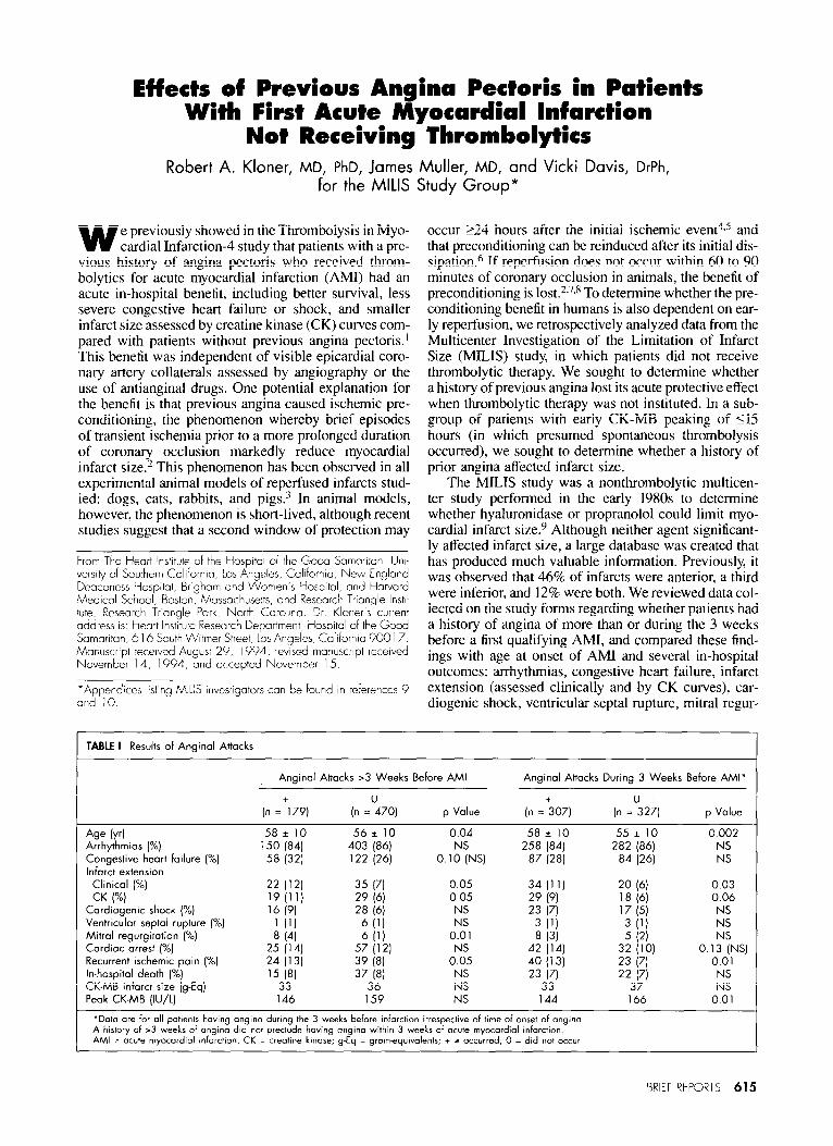
TABLE I Results of Angina1 Attacks
Angina1 Attacks >3 Weeks Before AMI Angina1 Attacks During 3 Weeks Before AMI’
(n =+179) (n =0470] p Value (n =+307] (n =0327) p Value
Age (~4 58* 10 56e 10 0.04 58i 10 55* 10 0.002 Arrhythmias (X) 150 (84) 403 (86) 258 (84) 282 (86) NS Congestive heart failure (%) 58 (32) 122 (26)
O.l!;NS] 87 (28) 84 (26) NS
Infarct extension Clinic01 (“A) 22 (12) 35 (71 0.05 34 (1 1) 20 (61 0.03 CK I”~) 19 (11) 29 (6) 0.05 29 (9) 18 (61 0.06
Cordiogenic shock (%) 16 (9) 28 (6) NS 23 (7) 17 (5) NS Ventricular septol (%] rupture 1 (11 6 ill NS 3 Ill 3 (‘1 NS
Mitral regurgitation (%) 8 (4) 6 ill 0.01 8 (3) 5 (21 Cardiac arrest (%) 25 (14) 57 (12) NS 42 (14) 32 (10) 0.1 ::NS) Recurrent ischemic pain (%) 24 (13) 39 (8) 0.05 40 (13) 23 (71 0.01 In-hospital death (%) ‘5 I81 37 I81 NS 23 (7) 22 171 NS CK-MB infarct size (g-Eq) 33 36 NS 33 37 NS Peak CK-MB (IU/L] 146 159 NS 144 166 0.01
‘Data ore for 011 patients having angina during the 3 weeks before infarction irrespective of time of onset of angina. A history of >3 weeks of angina did not preclude having angina within 3 weeks of acute myocordial infarction. AM = acute myocordiol Infarction. CK = creatine kinase; g-Eq = gramequivalents; + - occurred; 0 = did not occur
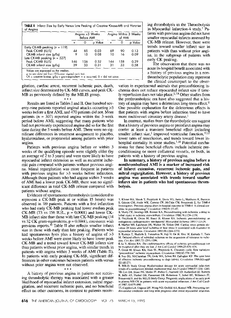
TABLE II Infarct Size by Early Versus late Peaking of Creatine Kinase-MB and Histories of Angina
Angino >3 Weeks Angina Within 3 Weeks Before AMI of AMI
+ 0 p Value + 0 p Value
Early CK-MB wakina in = 1191 Pebk CK-MB’(IU/L)- ’ ’ 44 85 0.03 69 90 0.13 CK-MB infarct size (g-Eq) 9 1.5 0.08 10 16 0.09
Late CK-MB peaking [n = 527) Peak CK-MB (IU/L) 146 156 0.32 144 158 0.29 CK-MB infarct size (g-Eq) 29 33 0.31 31 33 0.58
Values are expressed OS the median. p values obtained from Wilcoxon signed rank test. CK = crwtine kinare; g-Eq = gram-equivalent; + - occurred, 0 = did not occur
gitation, cardiac arrest, recurrent ischemic pain, death, infarct size determined by CK-MB curves, and peak CK- MB as previously defined by the MILIS group.
. . . Results are listed in Tables I and 11. One hundred sev-
enty-nine patients reported angina1 attacks occurring >3 weeks before a first AMI, and 470 patients did not. More patients (n = 307) reported angina within the 3-week period before AMI, suggesting that many patients who had not previously experienced angina did so for the first time during the 3 weeks before AMI. There were no sig- nificant differences in treatment assignment to placebo, hyaluronidase, or propranolol among patients with prior angina.
Patients with previous angina before or within 3 weeks of the qualifying episode were slightly older (by an average of 2 to 3 years) and were more likely to have myocardial infarct extension as well as recurrent ische- mic pain compared with patients without previous angi- na. Mitral regurgitation was more frequent in patients with previous angina for >3 weeks before infarction. Although those patients who had angina within 3 weeks of AM1 had a lower peak CK-MB, there was no signif- icant difference in total CK-MB release compared with patients without angina.
Evidence of spontaneous thrombolysis (considered to represent a CK-MB peak at or within 15 hours) was observed in 119 patients. Patients with a first infarction who had early CK-MB peaking had lower median peak CK-MB (73 vs 156 IU/L. p = O.ooOl) and lower CK- MB infarct size than those with late CK-MB peaking (14 vs 32 CK gram-e
8 uivalents, p = O.OWl), consistent with
previous reports.’ Table II also reflects smaller infarct size in those with early than late peaking. Patients who had spontaneous lysis plus a history of angina of >3 weeks before AM1 were more likely to have lower peak CK-MB and a trend toward lower CK-MB infarct size than patients without prior angina, with similar trends in patients with angina within 3 weeks of AM1 (Table II). In patients with early peaking CK-MB, significant dif- ferences in other outcomes between patients with versus without prior angina were not observed.
. . . A history of previous angina in patients not receiv-
ing thrombolytic therapy was associated with a greater likelihood of myocardial infarct extension, mitral regur- gitation, and recurrent ischemic pain, and no beneficial effect on other outcomes, in contrast to patients receiv-
616 THE AMERICAN JOURNAL OF CARDIOLOGY+’ V31 75
ing thrombolysis in the Thrombolysis in Myocardial Infarction-4 study.’ Pa- tients with previous angina did not have smaller myocardial infarcts assessed by CK-MB release. However, there were trends toward smaller infarct size in patients with than without prior angi- na, in the subgroup of patients with early CK peaking.
The observation that there was no acute in-hospital benefit associated with a history of previous angina in a non- thrombolytic population may represent the clinical counterpart to the obser-
vation in experimental animals that preconditioning is- chemia does not reduce myocardial infarct size if time- ly reperfusion does not take place.2,X8 Other studies from the prethrombolytic era have also suggested that a his- tory of angina may have a deleterious long-term etfect.” One possible explanation for the deleterious effects is that patients with angina before infarction tend to have more multivessel coronary artery disease.’
In contrast, studies from the thrombolytic era suggest that a history of previous angina or preinfarct angina may confer at least a transient beneficial effect including smaller infarct size,l improved ventricular function,‘2s’3 lower rates of reocclusion, and trends toward lower in- hospital mortality in some studies.‘T14 Potential mecha- nisms for these beneficial effects include ischemic pre- conditioning or more collateral vessels, or both, in patients with a history of previous angina.
In summary, a history of previous angina before a nonthrombolized AM1 is a marker of increased risk of infarct extension, recurrent ischemic pain, and mitral regurgitation. However, a history of previous angina was associated with trends toward smaller infarct size in patients who had spontaneous throm- bolysis.
1. Kloner KA. Shook l’, PrLyklcnk K, Daws VG. Junio I., Matthews K, Burstcin S, Gibson CM, Poole WK. Cannon CP, McCabe CH, Braunwald B. for TIMI- Investigaton. Previous angina alters in-hospital outcome in ‘IIMI-4. A climcal cor- relate to preconditioning? Circulafion lYY5:9 137-47. 2 Muny CE, Jennings KB, Rcimer KA. Preconditioning wth irhemia: a delay in lethal injury in irchemic mywardium. Ctrculafion lY86;74:1124-I 136. 3. Przyklenk K, Ovix M, Bauer B, Kloner RA. lwhemic preconditioning: an endogenous cardioprotcctive mechanism? ,Myxurdial lsrhemiu 1993:S: 13-24. 4. Ma&r MS, Latchman DS, Walker Xl, Yellon DM. Cardiac stre,s protein ele- \ation 24 hours after brief ischemia or heal stress is associated with resistance to myocardial infarction. Circulurion 19Y3;88:126+1272. 5. Kuzuya T. Hoshida S. Yamashiu N. Fuji H, Oe H, Hori M. Kamada T. Tda M. Delayed effects of sublethal &hernia on the acquisition of tolerance to Ische- mia. Circ Hrv 1993;72:1293-1299. 6. Li Y, Kloner RA. The cardioprowctwc effccls of ischemic precondmoninp can be recaptured after they are lost. I Am Golf Cnrdiof 1994;23:470-474 7. Ovize M. Kloner RA. II& SL, Przyklcnk K. Coronary cychc flow wariarionc “precondition” ischenw myccardium. Cwcularim 1992:85:77Y-789. 8. Nao BS. McClanahan TB, Ciroh MA. Schotr RJ. Gallagher KP. The hme lirmt of effective ischcmic preconditioning in dogs (ahstr). Circulorimr 19YQSZ(suppl 111):‘11-27’. 9. MILIS Study Group. Hyaluronidasc therapy for acute myxardial infarction: results of a randomired. blinded, multicenter trial. AmJ Cordiol 19X6:57: 12% 1243. 10. COY. DA, Stone PH. Muller JE. Parker C, Htiwell TD. Rutherford JD, Rohcns R. Jaffc AS, Hackel DB, Passamani ER. Robertson T. Sobcl BE, WilIerson JT, Braunwald E, and the MILIS Study Group. Prognostic Implications of an early peak in plasma MB CK in p&en’s with acute mywardial infarction. .I Am Co// Cm/i- d ‘987; lo:Y79-990. 11. CuppIes LA. Gagnon UP, Won8 ND, Ostfeld AA, Kannel WB. Preexisting car- diovascular conditions and Ion8-term prognosis after initial mywardinl infarction.
MARCH 15, 1995
‘lhe Framingham Study. Ant Hear/J 1993; 125:863 -872. of preinfarction angina for prexwation of left ventricular function in acute myu-
12. Matsuda Y. Oeawa II. hloritani K. Mawda .\I, Naito II, Mawwaki .M, Ikee cardial infarction. Am Ileor? J 1992; 124: I ‘&23. Y. Kuwkawa R. Effect\ of the prcsencc or absenrl: of precedme angina pcctoris 14. Muller DWM. Top01 W, Calif RM, Sigmon KK, Gormon I.. George 13s. ou left ventricular function afrer acute myocardial infarction. Am Ilrwrf J Kereiakez DJ, Lee KL. Elhs SG. and ‘rAMI Study Group. Relarionship hctw~~n 19X4;108:YS5 958. antecuient angina pwwris and shon term prognosis atier thrombolytic therapy for 13. Hirai ‘I‘. Fuiita ,M, Y.amanishl K. Ohno A, Miwa K, Sasayama S. Sqnifiwnce acue myocardial infarction. Am Ifmrf J 199tl1 19:22&231.
An Electrocardiographic Acuteness Score for Quantifying the Timing of a Myocardfal Infarction to
Guide Decisions Regarding Reperfusion Therapy Michelle L. Wilkins, BS, Aurora D. Pryor, BSE, Charles Maynard, PhD, Nancy B. Wagner, BA,
William J. Elias, MD, Paul E. Litwin, MS, Olle Pahlm, MD, Ronald H. Selvester, MD, W. Douglas Weaver, MD, Galen S. Wagner, MD, and Stanley T. Anderson, FRACP
ortality in acute myocardial infarction has been shown to be reduced by early administration of
thrombolytic therapy. I** When a patient presents with acute symptoms, the historical time of onset is the clin- ical criterion typically used to determine the duration of the pathophysiologic process. However, poor recall, atypical presentation, and intermittancy confound the accuracy of the patient history for determining the dura- tion of the thrombotic occlusion’ 3 (Raitt MH et al, per- sonal communication). The initial 1Zlead elcctrocardi- ogram (ECG) provides an alternate clinical method for timing the acuteness of a myocardial infarction. Exper- imental studies have dcscribcd the sequential changes in the T wave, ST segment, and QRS complex during the hyperacute, acute, subacute, and chronic phases of a myocardial infarction. 4A A clinical method has been developed in which each lead with ST-segment elcva- tion or abnormally tall T waves is assigned a score from 1 to 4 points, and these are added to produce a total score for the 12-lead ECG.’ Since this score considers the num- bcr of involved leads, it could be influenced by the extent, as well as the acuteness, of the infarction process. The present study introduces a modification of this method that controls for the number of involved Icads, and assesses the variability of acuteness scores between pre- hospital and hospital electrocardiographic recordings.
. . . Patients were selected liom those entered into the
Myocardial Infarction Triage and Intervention (MITI) project conducted in the greater Seattlc metropolitan area. Phase I of this study assessed the feasibility of prehos- pita1 initiation of thrombolytic therapy and phase 11 com- pared prehospital versus hospital initiation of throm- bolytic therapy.x*9 In these protocols, standard 12-lead ECGs were obtained from patients with symptoms sug- gestive of acute myocardial infarction on 2 occasions: at
tro- the Department of AAzdicine, Dv;tc Univcrslty Mcciical Center, DJrha~l. Acrt!. Carolira. and the Deprtmert of Med.cine, Uwer- si+y o! Washingto-, Scat.le, Wastlingtor; Tils s’udy was sqprted iI cart bv Grants RO’ HI3845404 and HS062C8C3 lrom *he Nationo’ heart, !ung, and Blood Institute, Bethesda, Mory!cnd, and by the D:.ke UI vewty Climcal Card’ovascular Study G,oup Dr Ga!en S Wag:.e,‘s cLrre?t address IL: Duke Uwerslty Med’ca’ Cer- ‘er, %30x 3636, I&am North Carolina 27710. MawscrIpt rcrelvcd August 10, 1994 rewed manuscript receiv& and accepf- ed Ncvember 28, I994
the time of prehospital evaluation by paramedics (ECG 1) and in the hospital emergency department (ECG 2). All patients from phase I of the MIT1 trial and those from phase II who were randomized to no prchospital throm- bolytic therapy were considered for the present study. The MITl protocol required ST-segment elevation on ECG 1 of 20.1 mV in 22 anatomically contiguous leads. Conti- guity in the frontal plant leads was considered accord- ing to the sequence: aVL, I, II, aVF, and III.
Eligibility for this study required: (1) confirmation of ST-segment elevation measured at the J point on ECG I; (2) no confounding factors including missing or mal- positioned leads, ventricular rhythm, 60 Hz artifact, unstable baseline, left or right bundle branch block, left or right ventricular hypertrophy, ventricular precxcita- tion, or heart rate > 110 beats/min; (3) no interval >2 hours between ECG 1 and 2; (4) no thrombolytic therapy between ECG 1 and 2; and (5) final diagnosis of AMI by the patient’s attending physician. The 154 patients who met all of these criteria constitute the study group.
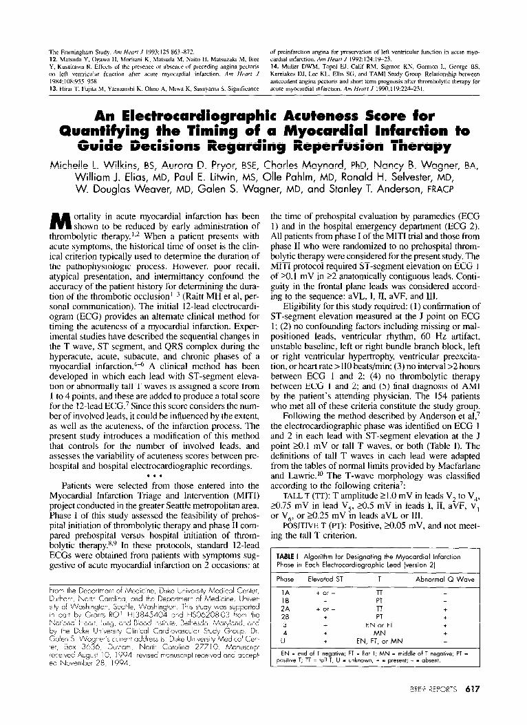
Following the method described by Anderson et al,7 the electrocardiographic phase was identified on ECG 1 and 2 in each lead with ST-segment elevation at the J point ZO.1 mV or tall T waves, or both (Table I). The definitions of tall T waves in each lead were adapted from the tables of normal limits provided by Macfarlane and Lawrie.‘” The T-wave morphology was classified according to the following criteria’:
TALL T (TT): T amplitude 2 1 .O mV in lcads V, to V,,, 20.75 mV in lead V,, 20.5 mV in leads 1, II, aVF, V, or V,, or a.25 mV m leads aVL or III.
POSITIVE T (fyT): Positive, a.05 mV, and not meet- ing the tall T criterion.
TABLE I Algorithm for Designating the Myocardial Infarction Phase in Each Electrocardiographic lead [version 2)
Phase Elevated ST T Abnormal Q Wave
1A + or - T-r 16 + PT 2A + or - l-r + 26 + PT + 3 + EN or FT +
u” + MN + + EN, FT, or MN -
EN = end of 1 negative; FT = flat T; MN = middle of T negative; PT = positive T; TT = toll 1; U - unknown; + = present; - - absent.
aRl:F REPCRTS 617


![Angina Pectoris Obstruction Decrease of blood irrigation Acute Myocardial Infarction (almost) Total occlusion Cellular necrosis [1] [1] Thygesen K, Alpert](https://img.dokumen.tips/doc/110x75/56649e7e5503460f94b8103d/angina-pectoris-obstruction-decrease-of-blood-irrigation-acute-myocardial-infarction.jpg)