Embed Size (px)
Citation preview

warwick.ac.uk/lib-publications
Original citation: Lemola, Sakari, Oser, Nadine, Urfer-Maurer, Natalie, Brand, Serge, Holsboer-Trachsler, Edith, Bechtel, Nina, Grob, Alexander, Weber, Peter and Datta, Alexandre N.. (2017) Effects of gestational age on brain volume and cognitive functions in generally healthy very preterm born children during school-age : a Voxel-based morphometry study. PLoS One, 12 (8). e0183519. Permanent WRAP URL: http://wrap.warwick.ac.uk/91497 Copyright and reuse: The Warwick Research Archive Portal (WRAP) makes this work of researchers of the University of Warwick available open access under the following conditions. This article is made available under the Creative Commons Attribution 4.0 International license (CC BY 4.0) and may be reused according to the conditions of the license. For more details see: http://creativecommons.org/licenses/by/4.0/ A note on versions: The version presented in WRAP is the published version, or, version of record, and may be cited as it appears here. For more information, please contact the WRAP Team at: [email protected]
RESEARCH ARTICLE
Effects of gestational age on brain volume and
cognitive functions in generally healthy very
preterm born children during school-age: A
voxel-based morphometry study
Sakari Lemola1☯*, Nadine Oser2☯, Natalie Urfer-Maurer3, Serge Brand4,5,6, Edith Holsboer-
Trachsler4, Nina Bechtel2, Alexander Grob3, Peter Weber2, Alexandre N. Datta2
1 Department of Psychology, University of Warwick, Coventry, United Kingdom, 2 Division of
Neuropediatrics and Developmental Medicine, University of Basel, Children’s Hospital Basel, Basel,
Switzerland, 3 Department of Psychology, University of Basel, Basel, Switzerland, 4 Center for Affective,
Stress and Sleep Disorders (ZASS), Psychiatric Clinics (UPK), University of Basel, Basel, Switzerland,
5 Department of Sport, Exercise and Health, Division of Sport and Psychosocial Health, Faculty of Medicine,
University of Basel, Basel, Switzerland, 6 Sleep Disorders Research Center, Kermanshah University of
Medical Sciences (KUMS), Kermanshah, Iran
☯ These authors contributed equally to this work.
Abstract
Objective
To determine whether the relationship of gestational age (GA) with brain volumes and cogni-
tive functions is linear or whether it follows a threshold model in preterm and term born chil-
dren during school-age.
Study design
We studied 106 children (M = 10 years 1 month, SD = 16 months; 40 females) enrolled in pri-
mary school: 57 were healthy very preterm children (10 children born 24–27 completed
weeks’ gestation (extremely preterm), 14 children born 28–29 completed weeks’ gestation,
19 children born 30–31 completed weeks’ gestation (very preterm), and 14 born 32 com-
pleted weeks’ gestation (moderately preterm)) all born appropriate for GA (AGA) and 49
term-born children. Neuroimaging involved voxel-based morphometry with the statistical
parametric mapping software. Cognitive functions were assessed with the WISC-IV. Gen-
eral Linear Models and multiple regressions were conducted controlling age, sex, and
maternal education.
Results
Compared to groups of children born 30 completed weeks’ gestation and later, children born
<28 completed weeks’ gestation had less gray matter volume (GMV) and white matter vol-
ume (WMV) and poorer cognitive functions including decreased full scale IQ, and process-
ing speed. Differences in GMV partially mediated the relationship between GA and full scale
IQ in preterm born children.
PLOS ONE | https://doi.org/10.1371/journal.pone.0183519 August 29, 2017 1 / 13
a1111111111
a1111111111
a1111111111
a1111111111
a1111111111
OPENACCESS
Citation: Lemola S, Oser N, Urfer-Maurer N, Brand
S, Holsboer-Trachsler E, Bechtel N, et al. (2017)
Effects of gestational age on brain volume and
cognitive functions in generally healthy very
preterm born children during school-age: A voxel-
based morphometry study. PLoS ONE 12(8):
e0183519. https://doi.org/10.1371/journal.
pone.0183519
Editor: Karen Lidzba, University Children’s Hospital
Tuebingen, GERMANY
Received: April 28, 2017
Accepted: August 4, 2017
Published: August 29, 2017
Copyright: © 2017 Lemola et al. This is an open
access article distributed under the terms of the
Creative Commons Attribution License, which
permits unrestricted use, distribution, and
reproduction in any medium, provided the original
author and source are credited.
Data Availability Statement: Participants were
assured that no individual data (including brain
volumes and cognitive functions) will be published.
Data will be provided in anonymised form for
researchers who meet the criteria for access to
confidential data from from the Clinical Trial Unit of
the University Hospital of Basel: https://dkf.unibas.
ch/de/departement/abteilungen/clinical-trial-unit;
https://dkf.unibas.ch/de/services/kontaktformular;
Tel. +41 61 328 6611.
Conclusions
In preterm children who are born AGA and without major complications GA is associated
with brain volume and cognitive functions. In particular, decreased brain volume becomes
evident in the extremely preterm group (born <28 completed weeks’ gestation). In preterm
children born 30 completed weeks’ gestation and later the relationship of GA with brain vol-
ume and cognitive functions may be less strong as previously thought.
Introduction
Very preterm birth is a risk for normal development of the brain [1,2] even in the absence of
major perinatal complications such as periventricular leukomalacia (PVL), intraventricular
haemorrhage (IVH), and periventricular haemorrhagic infarction (PHI) [3–6]. Compared to
term-born children, very preterm children had smaller brain volume including smaller gray
matter volume (GMV) and white matter volume (WMV) in childhood [6–8], adolescence
[9–12], and young adulthood [4,13]. Compared to their peers born at term, regional volume
differences in very preterm children, adolescents, and young adults were wide spread and
included decreases of GMV in all lobes (frontal [9,12,13,14], temporal [4,7,9,12,14], parietal
[7,9] and occipital lobes [12,13]). Further, decreased GMV was observed in the hippocampus
[9,11,15,16], thalamus [4,9,12,13,15], and cerebellum [12,17].
There is a large body of evidence showing that both gestational age [18] and brain volume
are related to cognitive function [9,12]. With regard to regional GMV one study showed that
in preterm children born between 30 and 34 weeks’ gestation and with low risk for neurologic
deficit or developmental difficulties, GMV in the temporal lobe was significantly reduced,
which in turn was related with decreased cognitive functions [19].
However, there is limited knowledge whether brain volume and cognitive function decrease
linearly with decreasing gestational age (GA) or whether there is a threshold of GA above
which the brain remains unaffected. Existing evidence on the association between gray matter
volume (GMV), white matter volume (WMV) and GA is inconsistent. Some studies found a
linear association of decreasing GMV and WMV with earlier GA [7,20], even in moderately
preterm children [21–23]. By contrast, one large study [9] showed no relationship between
birth weight and GMV and WMV above a birth weight of 1500g suggesting a threshold above
which maturity at birth (i.e., GA or birth weight) is no longer related to later brain size. Possi-
bly, studies indicating a linear relationship vs. studies indicating a threshold model of the rela-
tionship between GA and brain volume differed in sample composition. Such differences
between studies may involve the prevalence of other neonatal risk factors in their samples such
as the number of children with perinatal complications or children born small for gestational
age (SGA), which may play an important role for later brain development [24]. With regard to
the nature of the relationship between GA and cognitive function a recent study suggests the
existence of a nonlinear relationship. In children from the Bavarian Longitudinal Study the
association of GA with IQ and mathematic attainment became evident below a threshold of 34
weeks’ gestation while there was barely a relationship above this threshold [18].
The aim of the present study was to examine whether there is a linear decline in brain vol-
ume and cognitive functions with GA or whether the relationship is better described with a
threshold model involving a stronger relationship of GMV, WMV, and cognitive functions
with GA below a certain level of GA. Therefore, we compare five groups with different GA
with each other, children born in the 24–27 completed weeks’ gestation (extremely preterm
Cognitive and brain development after preterm birth
PLOS ONE | https://doi.org/10.1371/journal.pone.0183519 August 29, 2017 2 / 13
Funding: The present study was funded by the
Swiss National Science Foundation (projects:
Sleep, cognitive, and socio-emotional development
in preterm children during middle and late child-
hood, grant number: 143962; Socio-emotional
development and mental health of preterm
children: The role of HPA-axis function, sleep,
neuroplasticity, and physical exercise during the
transition to adolescence, grant number: 159362),
the Research Fund of the University of Basel
(project: Early origins of self-regulation and sleep,
grant number: DPE2083), and the Gottfried und
Julia Bangerter-Rhyner-Stiftung. The funders had
no role in study design, data collection and
analysis, decision to publish, or preparation of the
manuscript.
Competing interests: The authors have declared
that no competing interests exist.
children), 28–29 completed weeks’ gestation (very preterm children, earlier group), 30–31
completed weeks’ gestation (very preterm children, later group), 32 completed weeks’ gesta-
tion (moderately preterm children), and term born children. This approach allows to describe
brain and cognitive development in these subgroups and is therefore of interest for paediatri-
cians, educational services, and parents of preterm children. Although these groups represent
a considerable percentage of the newborn population in modern societies (in Germany [25]
and the USA [26] respectively, children born 24–27 completed weeks’ gestation account for
0.24% and 0.49% of the newborn population, children born 28–29 completed weeks’ gestation
account for 0.23% and 0.44%, children born 30–31 completed weeks’ gestation account for
0.36% and 0.76%, and children born 32 completed weeks’ gestation account for 0.30% and
0.59%), research comparing these subgroups of preterm children at school age regarding brain
development is missing. To exclude effects of other perinatal risk factors, only children with
low risk (i.e., without PVL, IVH, and PHI) and born appropriate for gestational age (AGA)
were studied. Moreover, we studied whether differences in GMV accounted for differences in
cognitive functions.
Materials and methods
Participants
Fifty-seven preterm children (24–32 completed weeks’ gestation; age: M = 10.0 years, SD = 1.3;
range: 7.8 to 12.3) and 49 term born children (�37 completed weeks’ gestation; age: M = 10.2
years, SD = 1.4; range: 7.9 to 12.8; t(104) = 0.87, P = 0.39) who successfully completed MRI
scans were included in the present study. Descriptive statistics are presented in Table 1. Pre-
term children were recruited from an initial cohort of 515 children born 24–32 completed
weeks’ gestation between January 1998 and December 2006 at the University Children’s Hos-
pital Basel (Switzerland). Inclusion criteria were enrollment in regular primary school in Swit-
zerland, no severe developmental delay, no evidence of major complications during the first
year of life (i.e. exclusion of children with PVL, IVH of grade 2 or higher, and PHI), being
born AGA (i.e., > 10th percentile of birth weight) [24], sufficient German language skills of
the parents to give informed consent, and residence in Switzerland and within 100 km from
the study center. Furthermore, because of MRI scanning, children with fixed dental braces
were excluded. Of 62 preterm children who originally had MRI scanning, 5 were excluded due
to movements during the MRI.
Compared to non-participants, the 57 preterm children with successful MRI scans had
modestly higher birth weight (1447g vs. 1286g, F(1, 512) = 6.07; P = 0.01) and higher GA (29.7
weeks vs. 29.1 completed weeks, F(1, 514) = 4.36; P = 0.04); gender did not differ (χ2(1) = 0.01;
P = 0.93). The participating preterm children included 10 children born 24–27 completed
weeks’ gestation, 14 children born 28–29 completed weeks’ gestation, 19 children born 30–31
completed weeks’ gestation, and 14 born 32 completed weeks’ gestation (see Table 1 for partic-
ipants’ characteristics). No child had PVL or PHI, while one child born 31 completed weeks’
gestation was diagnosed with mild IVH (grade 1).
Among the term born children three were excluded because of structural anomalies and
six due to movement during the MRI scanning. The term born control group finally con-
sisted of 49 children who were recruited from official birth notifications (n = 35; 71.4% of
the control group), children from hospital staff (n = 4; 8.2% of the control group), children
attending Children’s University (n = 2; 4.1% of the control group), one sibling of a preterm
child (n = 1; 2.0% of the control group), healthy siblings of participants from another study
(n = 3; 6.1% of the control group), and headache patients without structural abnormalities
(n = 4; 8.2% of the control group). Parents gave written informed consent for the children to
Cognitive and brain development after preterm birth
PLOS ONE | https://doi.org/10.1371/journal.pone.0183519 August 29, 2017 3 / 13
participate and assent was obtained from the child. The study was approved by the Ethics
Committee of Basel.
Procedure
Children visited the University Children’s Hospital Basel (Switzerland) for neuroimaging and
cognitive assessment was conducted by trained study personnel at the study center or at the
children’s homes. Mothers completed questionnaires to assess demographic data (no voca-
tional training, vocational training, university).
Measurement
Cognitive assessment. Cognitive functions were assessed using the German version of
the WISC-IV [27] which provides a full scale IQ representing a child’s global intellectual func-
tioning, as well as four index scores representing specific cognitive abilities: verbal comprehen-
sion, perceptual reasoning, working memory, and processing speed. The full scale IQ and the
index scores have a mean of 100 and a standard deviation of 15.
Neuroimaging procedure. Imaging of structural data was acquired using a 3-Tesla MRI
with a standard head coil (Magnetom VERIO, Siemens Healthcare, Erlangen, Germany).
Structural imaging was conducted with sagittal T1-weighted 3D high-resolution magnetization
prepared rapid gradient echo sequence (MPRAGE), with TR = 2000 ms, TE = 3,4 ms, TI =
1000 ms and an isotropic spatial resolution of 1x1x1 mm3.
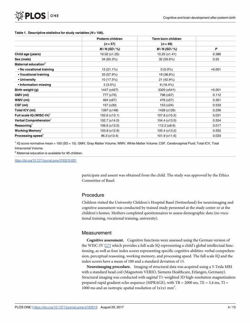
Table 1. Descriptive statistics for study variables (N = 106).
Preterm children Term born children
(n = 57) (n = 49)
M / N (SD / %) M / N (SD / %) P
Child age (years) 10.02 (±1.25) 10.25 (±1.41) 0.386
Sex (male) 34 (65.3%) 32 (59.6%) 0.55
Maternal education2
• No vocational training 12 (21.1%) 0 (0.0%) <0.001
• Vocational training 33 (57.9%) 19 (38.8%)
• University 10 (17.5%) 21 (42.9%)
• Information missing 2 (3.5%) 9 (18.4%)
Birth weight (g) 1447 (±427) 3320 (±541) <0.001
GMV (ml) 777 (±70) 798 (±67) 0.112
WMV (ml) 464 (±67) 476 (±57) 0.301
CSF (ml) 157 (±30) 153 (±24) 0.533
Total ICV (ml) 1397 (±148) 1428 (±126) 0.256
Full scale IQ (WISC-IV)1 102.6 (±13.1) 107.8 (±10.2) 0.031
Verbal Comprehension1 102.7 (±14.0) 104.4 (±13.5) 0.554
Reasoning1 106.6 (±13.0) 112.2 (±8.6) 0.017
Working Memory1 103.8 (±12.9) 105.4 (±13.2) 0.552
Processing speed1 96.3 (±13.4) 101.9 (±11.6) 0.033
1 IQ score normative mean = 100 (SD = 15). GMV, Gray Matter Volume; WMV, White Matter Volume; CSF, Cerebrospinal Fluid; Total ICV, Total
Intracranial Volume.2 Maternal education is available for 95 children.
https://doi.org/10.1371/journal.pone.0183519.t001
Cognitive and brain development after preterm birth
PLOS ONE | https://doi.org/10.1371/journal.pone.0183519 August 29, 2017 4 / 13
Image processing of voxel-based morphometric (VBM) analysis was conducted with the
statistical parametric mapping software (SPM8) (Wellcome Department of Imaging Neurosci-
ence, UCL Institute of Neurology, London, UK; http://www.fil.ion.ucl.ac.uk/spm/software/
spm8/) implemented in Matlab (Version 7.9.0, Mathworks Inc., USA). Structural images were
segmented into GMV and WMV, cerebrospinal fluid, bone, soft tissue and air/background
with the new-segment tool in SPM8. Default settings were used. A customized age- and sex-
matched Tissue Probability Map and a children’s T1-template using the average template
approach of the Template-O-matic toolbox [28] was calculated to create an average template
using the Diffeomorphic Anatomical Registration Through Exponentiated Lie Algebra
method (DARTEL). Subsequently GMV and WMV images were warped to this average tem-
plate and normalised into MNI space. Images were modulated non-linearly to correct for indi-
vidual brain size. GMV images were then smoothed with 8mm Gaussian kernel, WMV images
with 12mm. The quality check of the VBM8 toolbox (C. Gaser, University of Jena, Germany,
VBM8-Toolbox Manual, 2010; http://dbm.neuro.uni-jena.de/vbm8/VBM8-Manual.pdf) was
performed to check the accuracy of segmentation and normalization and to identify artefacts
and outliers. The homogeneity check indicated no outliners above or below 2 standard devia-
tions from the mean.
To examine regional GMV differences between groups, a full factorial design specification
was assessed in SPM8 with the five groups. An absolute threshold mask of 0.3 was set to gray
matter. All results were Family Wise Error (FWE) corrected at p = 0.05. Significant cluster of
GMV were further analysed using the VOI toolbox in SPM8 to calculate regional volumes per
child, again results were reported FWE corrected with a threshold of 0.05 in clusters > 30
voxel.
Statistical analyses
Mean differences of brain volumes and cognitive functions between groups with varying GA
were tested applying General Linear Models (GLM). First, multivariate GLM was conducted
with GMV and WMV as dependent variables, “gestational age-groups” as fixed factor, and
age, sex, and maternal education (dummy coded) as covariates (as potential further confound-
ers we also analyzed the association of paternal education, household income, and number of
children in the household with brain volume (GMV, WMV, CSF) and cognitive function
(IQ, verbal comprehension, perceptual reasoning, working memory, processing speed) con-
trolling age, sex, and maternal education; as none of these associations was significant (all p-
values>0.05) only age, sex, and maternal education were used as covariates). Then polynomial
tests and pairwise comparisons of the mean values of GMV and WMV between the five groups
24–27 completed weeks’ gestation, 28–29 completed weeks’ gestation, 30–31 completed weeks’
gestation, 32 completed weeks’ gestation, and term born children were conducted by boot-
strapping based on 1000 bootstrap samples. Second, multivariate GLM was conducted with
full scale IQ and the four index scores verbal comprehension, perceptual reasoning, working
memory, and processing speed as dependent variables, gestational age-groups as fixed factor,
and age, sex, and maternal education as covariates, which was again followed by polynomial
tests and pairwise comparisons between the five groups. Finally, mediation analysis to examine
whether GMV mediated the relationship between GA and cognitive functions was conducted
according to Baron and Kenny [29] controlling age, sex, and maternal education among the
preterm born children (24–32 completed weeks’ gestation). The nominal level of significance
was set at alpha < .05. Statistical analyses were performed with SPSS1 22.0 (IBM Corporation,
Armonk NY, USA) for Apple Mac1.
Cognitive and brain development after preterm birth
PLOS ONE | https://doi.org/10.1371/journal.pone.0183519 August 29, 2017 5 / 13
Results
GMV, WMV, and cognitive differences in children with different GA at
birth
The multivariate GLM with GMV and WMV as dependent variables showed significant
effects of the factors ‘groups of GA’ (F(4, 96) = 3.28, p = 0.015), age (F(2, 95) = 16.63,
p< 0.001), sex (F(2, 95) = 17.01, p< 0.001), but not maternal education (p-values for all
dummy variables > 0.05). Fig 1A and Table 2 present the brain volume differences in children
with different GA at birth. Polynomial contrasts revealed a significant linear trend for GMV
and WMV, as well as a significant quadratic trend for WMV (S1 Table, polynomial contrasts
and pairwise comparisons between five gestational age groups (P-values) based on 1000 boot-
strap samples and adjusted for age, sex, and maternal education). Pairwise comparisons
showed that children born 24–27 completed weeks’ gestation and 28–29 completed weeks’ ges-
tation had significantly smaller GMV compared to those born 30–31 completed weeks’ gesta-
tion and those born 32 completed weeks’ gestation. Children born 24–27 completed weeks’
gestation further differed significantly from term born children regarding GMV. Children
born 24–27 completed weeks’ gestation had also significantly smaller WMV compared to the
groups born 30–31 completed weeks’ gestation, 32 completed weeks’ gestation and term born
children. The group born 30–31 completed weeks’ gestation and the group born 32 completed
weeks’ gestation both showed non-significantly larger GMV and WMV than the term born
group (all P-values >.10; Fig 1A, Table 2, S1 Table). Moreover, no significant differences in
cerebrospinal fluid (CSF) volumes were found between the groups.
The multivariate GLM with full scale IQ, verbal comprehension, perceptual reasoning,
working memory, and processing speed as dependent variables showed significant effects of
the factors ‘groups of GA’ (F(5, 86) = 3.07, p = 0.013), age (F(5, 83) = 3.01, p = 0.015), sex (F(5,
83) = 3.27, p = 0.010), and maternal tertiary education (F(5, 83) = 2.39, p = 0.045). Fig 1B and
Table 2 present the differences regarding cognitive functions in children with different GA at
birth. Polynomial contrasts revealed a significant linear trend and quadratic trend for full scale
IQ and a significant linear trend for perceptual reasoning and processing speed (S1 Table).
Pairwise comparisons showed that children born 24–27 completed weeks’ gestation had signif-
icantly lower full-scale IQ than the groups born 30–31 completed weeks’ gestation, 32 com-
pleted weeks’ gestation, and term born children. Moreover, children born 28–29 completed
weeks’ gestation had significantly lower full-scale IQ than children born 32 completed weeks’
gestation. Regarding the four index scores representing specific cognitive abilities children
born 24–27 completed weeks’ gestation had significantly lower processing speed than the
groups born 30–31 completed weeks’ gestation, 32 completed weeks’ gestation, and term born
children as well as lower verbal comprehension compared to the group born 32 completed
weeks’ gestation. No significant differences were found between children born in the 30–31
completed weeks’ gestation and in the 32 completed weeks’ gestation compared to term born
children regarding cognitive functions (all P-values>.10; Fig 1B, Table 2, S1 Table).
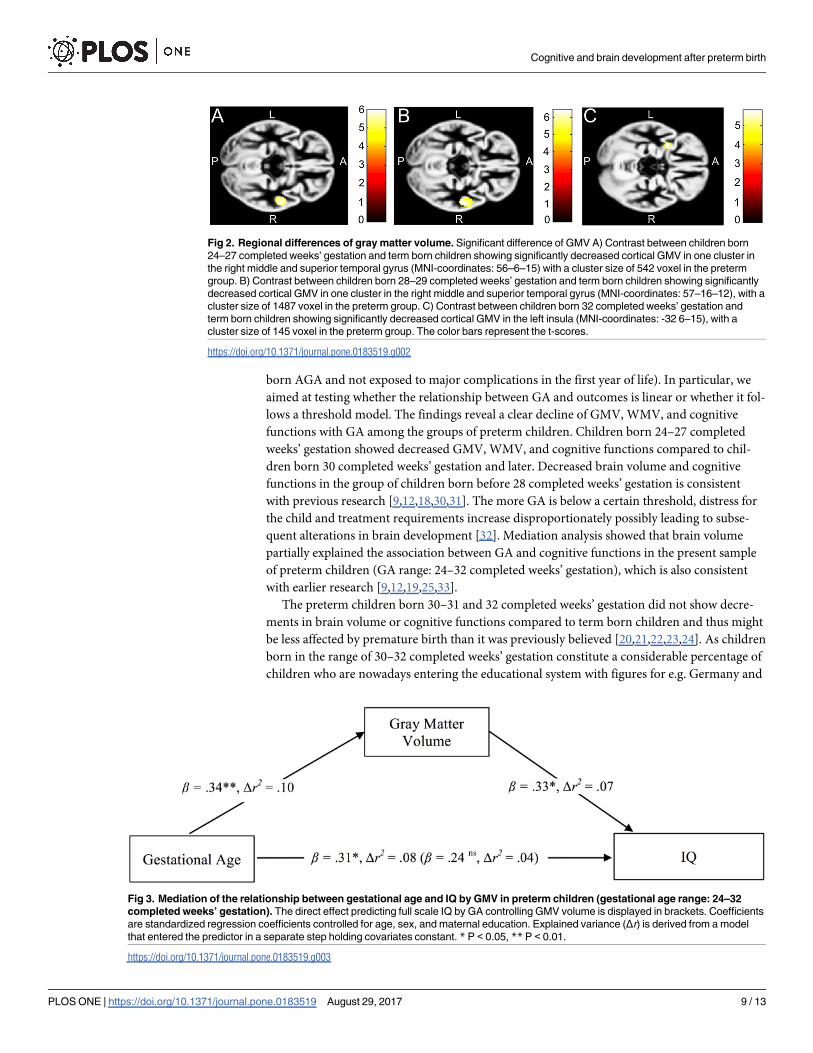
Regional GMV differences in children with different GA at birth
Fig 2 shows the analyses of regional cortical GMV differences. Compared to their term born
peers, children born 24–27 completed weeks’ gestation as well as children born 28–29 com-
pleted weeks’ gestation had both decreased GMV in one cluster in the right middle and supe-
rior temporal gyrus (MNI-coordinates: 56, -6, -15; cluster size: 542 voxel; MNI-coordinates:
57, -16, -12; cluster size: 1487 voxel, respectively). While the children born 30–31 completed
weeks’ gestation showed no significant differences from term born children, those born 32
Cognitive and brain development after preterm birth
PLOS ONE | https://doi.org/10.1371/journal.pone.0183519 August 29, 2017 6 / 13
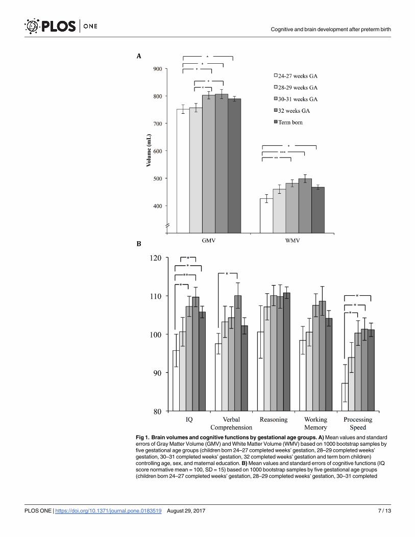
Fig 1. Brain volumes and cognitive functions by gestational age groups. A) Mean values and standard
errors of Gray Matter Volume (GMV) and White Matter Volume (WMV) based on 1000 bootstrap samples by
five gestational age groups (children born 24–27 completed weeks’ gestation, 28–29 completed weeks’
gestation, 30–31 completed weeks’ gestation, 32 completed weeks’ gestation and term born children)
controlling age, sex, and maternal education. B) Mean values and standard errors of cognitive functions (IQ
score normative mean = 100, SD = 15) based on 1000 bootstrap samples by five gestational age groups
(children born 24–27 completed weeks’ gestation, 28–29 completed weeks’ gestation, 30–31 completed
Cognitive and brain development after preterm birth
PLOS ONE | https://doi.org/10.1371/journal.pone.0183519 August 29, 2017 7 / 13
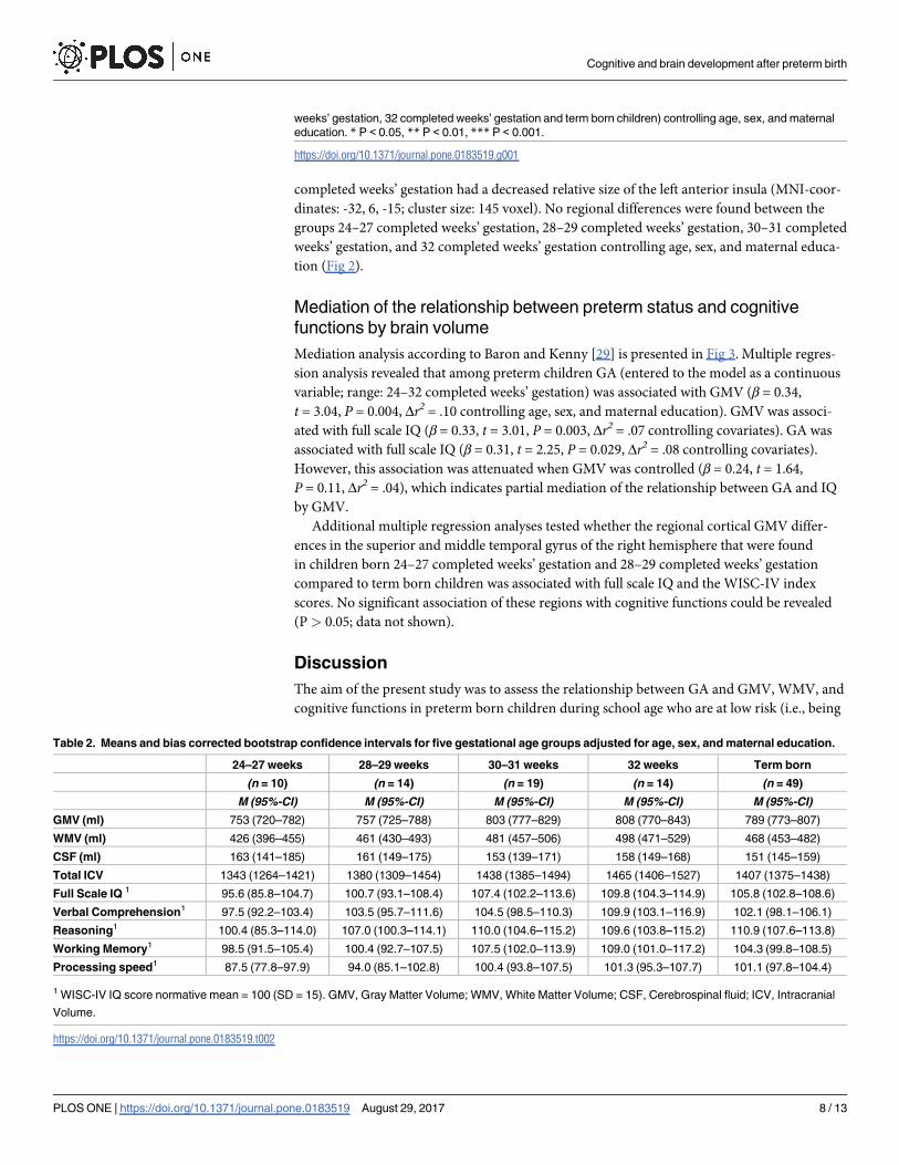
completed weeks’ gestation had a decreased relative size of the left anterior insula (MNI-coor-
dinates: -32, 6, -15; cluster size: 145 voxel). No regional differences were found between the
groups 24–27 completed weeks’ gestation, 28–29 completed weeks’ gestation, 30–31 completed
weeks’ gestation, and 32 completed weeks’ gestation controlling age, sex, and maternal educa-
tion (Fig 2).
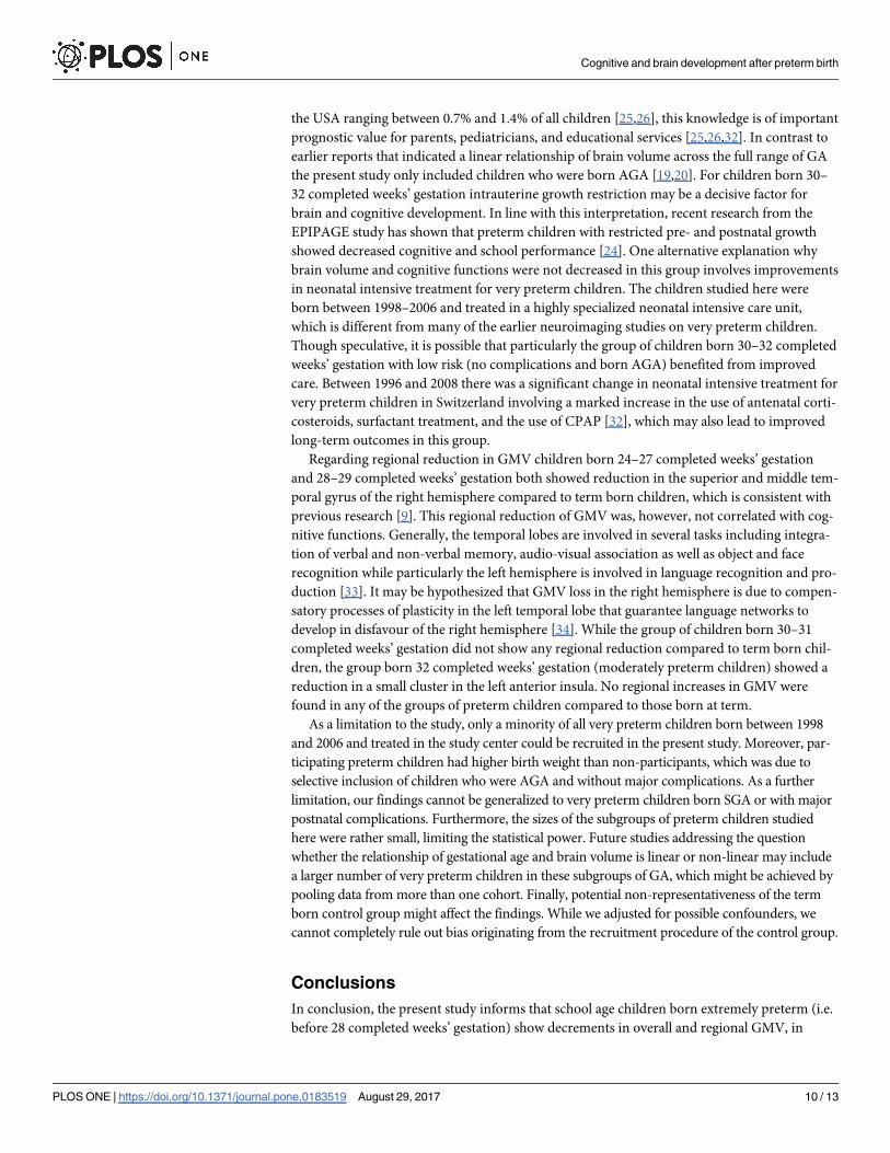
Mediation of the relationship between preterm status and cognitive
functions by brain volume
Mediation analysis according to Baron and Kenny [29] is presented in Fig 3. Multiple regres-
sion analysis revealed that among preterm children GA (entered to the model as a continuous
variable; range: 24–32 completed weeks’ gestation) was associated with GMV (β = 0.34,
t = 3.04, P = 0.004, Δr2 = .10 controlling age, sex, and maternal education). GMV was associ-
ated with full scale IQ (β = 0.33, t = 3.01, P = 0.003, Δr2 = .07 controlling covariates). GA was
associated with full scale IQ (β = 0.31, t = 2.25, P = 0.029, Δr2 = .08 controlling covariates).
However, this association was attenuated when GMV was controlled (β = 0.24, t = 1.64,
P = 0.11, Δr2 = .04), which indicates partial mediation of the relationship between GA and IQ
by GMV.
Additional multiple regression analyses tested whether the regional cortical GMV differ-
ences in the superior and middle temporal gyrus of the right hemisphere that were found
in children born 24–27 completed weeks’ gestation and 28–29 completed weeks’ gestation
compared to term born children was associated with full scale IQ and the WISC-IV index
scores. No significant association of these regions with cognitive functions could be revealed
(P> 0.05; data not shown).
Discussion
The aim of the present study was to assess the relationship between GA and GMV, WMV, and
cognitive functions in preterm born children during school age who are at low risk (i.e., being
weeks’ gestation, 32 completed weeks’ gestation and term born children) controlling age, sex, and maternal
education. * P < 0.05, ** P < 0.01, *** P < 0.001.
https://doi.org/10.1371/journal.pone.0183519.g001
Table 2. Means and bias corrected bootstrap confidence intervals for five gestational age groups adjusted for age, sex, and maternal education.
24–27 weeks 28–29 weeks 30–31 weeks 32 weeks Term born
(n = 10) (n = 14) (n = 19) (n = 14) (n = 49)
M (95%-CI) M (95%-CI) M (95%-CI) M (95%-CI) M (95%-CI)
GMV (ml) 753 (720–782) 757 (725–788) 803 (777–829) 808 (770–843) 789 (773–807)
WMV (ml) 426 (396–455) 461 (430–493) 481 (457–506) 498 (471–529) 468 (453–482)
CSF (ml) 163 (141–185) 161 (149–175) 153 (139–171) 158 (149–168) 151 (145–159)
Total ICV 1343 (1264–1421) 1380 (1309–1454) 1438 (1385–1494) 1465 (1406–1527) 1407 (1375–1438)
Full Scale IQ 1 95.6 (85.8–104.7) 100.7 (93.1–108.4) 107.4 (102.2–113.6) 109.8 (104.3–114.9) 105.8 (102.8–108.6)
Verbal Comprehension1 97.5 (92.2–103.4) 103.5 (95.7–111.6) 104.5 (98.5–110.3) 109.9 (103.1–116.9) 102.1 (98.1–106.1)
Reasoning1 100.4 (85.3–114.0) 107.0 (100.3–114.1) 110.0 (104.6–115.2) 109.6 (103.8–115.2) 110.9 (107.6–113.8)
Working Memory1 98.5 (91.5–105.4) 100.4 (92.7–107.5) 107.5 (102.0–113.9) 109.0 (101.0–117.2) 104.3 (99.8–108.5)
Processing speed1 87.5 (77.8–97.9) 94.0 (85.1–102.8) 100.4 (93.8–107.5) 101.3 (95.3–107.7) 101.1 (97.8–104.4)
1 WISC-IV IQ score normative mean = 100 (SD = 15). GMV, Gray Matter Volume; WMV, White Matter Volume; CSF, Cerebrospinal fluid; ICV, Intracranial
Volume.
https://doi.org/10.1371/journal.pone.0183519.t002
Cognitive and brain development after preterm birth
PLOS ONE | https://doi.org/10.1371/journal.pone.0183519 August 29, 2017 8 / 13
born AGA and not exposed to major complications in the first year of life). In particular, we
aimed at testing whether the relationship between GA and outcomes is linear or whether it fol-
lows a threshold model. The findings reveal a clear decline of GMV, WMV, and cognitive
functions with GA among the groups of preterm children. Children born 24–27 completed
weeks’ gestation showed decreased GMV, WMV, and cognitive functions compared to chil-
dren born 30 completed weeks’ gestation and later. Decreased brain volume and cognitive
functions in the group of children born before 28 completed weeks’ gestation is consistent
with previous research [9,12,18,30,31]. The more GA is below a certain threshold, distress for
the child and treatment requirements increase disproportionately possibly leading to subse-
quent alterations in brain development [32]. Mediation analysis showed that brain volume
partially explained the association between GA and cognitive functions in the present sample
of preterm children (GA range: 24–32 completed weeks’ gestation), which is also consistent
with earlier research [9,12,19,25,33].
The preterm children born 30–31 and 32 completed weeks’ gestation did not show decre-
ments in brain volume or cognitive functions compared to term born children and thus might
be less affected by premature birth than it was previously believed [20,21,22,23,24]. As children
born in the range of 30–32 completed weeks’ gestation constitute a considerable percentage of
children who are nowadays entering the educational system with figures for e.g. Germany and
Fig 2. Regional differences of gray matter volume. Significant difference of GMV A) Contrast between children born
24–27 completed weeks’ gestation and term born children showing significantly decreased cortical GMV in one cluster in
the right middle and superior temporal gyrus (MNI-coordinates: 56–6–15) with a cluster size of 542 voxel in the preterm
group. B) Contrast between children born 28–29 completed weeks’ gestation and term born children showing significantly
decreased cortical GMV in one cluster in the right middle and superior temporal gyrus (MNI-coordinates: 57–16–12), with a
cluster size of 1487 voxel in the preterm group. C) Contrast between children born 32 completed weeks’ gestation and
term born children showing significantly decreased cortical GMV in the left insula (MNI-coordinates: -32 6–15), with a
cluster size of 145 voxel in the preterm group. The color bars represent the t-scores.
https://doi.org/10.1371/journal.pone.0183519.g002
Fig 3. Mediation of the relationship between gestational age and IQ by GMV in preterm children (gestational age range: 24–32
completed weeks’ gestation). The direct effect predicting full scale IQ by GA controlling GMV volume is displayed in brackets. Coefficients
are standardized regression coefficients controlled for age, sex, and maternal education. Explained variance (Δr) is derived from a model
that entered the predictor in a separate step holding covariates constant. * P < 0.05, ** P < 0.01.
https://doi.org/10.1371/journal.pone.0183519.g003
Cognitive and brain development after preterm birth
PLOS ONE | https://doi.org/10.1371/journal.pone.0183519 August 29, 2017 9 / 13
the USA ranging between 0.7% and 1.4% of all children [25,26], this knowledge is of important
prognostic value for parents, pediatricians, and educational services [25,26,32]. In contrast to
earlier reports that indicated a linear relationship of brain volume across the full range of GA
the present study only included children who were born AGA [19,20]. For children born 30–
32 completed weeks’ gestation intrauterine growth restriction may be a decisive factor for
brain and cognitive development. In line with this interpretation, recent research from the
EPIPAGE study has shown that preterm children with restricted pre- and postnatal growth
showed decreased cognitive and school performance [24]. One alternative explanation why
brain volume and cognitive functions were not decreased in this group involves improvements
in neonatal intensive treatment for very preterm children. The children studied here were
born between 1998–2006 and treated in a highly specialized neonatal intensive care unit,
which is different from many of the earlier neuroimaging studies on very preterm children.
Though speculative, it is possible that particularly the group of children born 30–32 completed
weeks’ gestation with low risk (no complications and born AGA) benefited from improved
care. Between 1996 and 2008 there was a significant change in neonatal intensive treatment for
very preterm children in Switzerland involving a marked increase in the use of antenatal corti-
costeroids, surfactant treatment, and the use of CPAP [32], which may also lead to improved
long-term outcomes in this group.
Regarding regional reduction in GMV children born 24–27 completed weeks’ gestation
and 28–29 completed weeks’ gestation both showed reduction in the superior and middle tem-
poral gyrus of the right hemisphere compared to term born children, which is consistent with
previous research [9]. This regional reduction of GMV was, however, not correlated with cog-
nitive functions. Generally, the temporal lobes are involved in several tasks including integra-
tion of verbal and non-verbal memory, audio-visual association as well as object and face
recognition while particularly the left hemisphere is involved in language recognition and pro-
duction [33]. It may be hypothesized that GMV loss in the right hemisphere is due to compen-
satory processes of plasticity in the left temporal lobe that guarantee language networks to
develop in disfavour of the right hemisphere [34]. While the group of children born 30–31
completed weeks’ gestation did not show any regional reduction compared to term born chil-
dren, the group born 32 completed weeks’ gestation (moderately preterm children) showed a
reduction in a small cluster in the left anterior insula. No regional increases in GMV were
found in any of the groups of preterm children compared to those born at term.
As a limitation to the study, only a minority of all very preterm children born between 1998
and 2006 and treated in the study center could be recruited in the present study. Moreover, par-
ticipating preterm children had higher birth weight than non-participants, which was due to
selective inclusion of children who were AGA and without major complications. As a further
limitation, our findings cannot be generalized to very preterm children born SGA or with major
postnatal complications. Furthermore, the sizes of the subgroups of preterm children studied
here were rather small, limiting the statistical power. Future studies addressing the question
whether the relationship of gestational age and brain volume is linear or non-linear may include
a larger number of very preterm children in these subgroups of GA, which might be achieved by
pooling data from more than one cohort. Finally, potential non-representativeness of the term
born control group might affect the findings. While we adjusted for possible confounders, we
cannot completely rule out bias originating from the recruitment procedure of the control group.
Conclusions
In conclusion, the present study informs that school age children born extremely preterm (i.e.
before 28 completed weeks’ gestation) show decrements in overall and regional GMV, in
Cognitive and brain development after preterm birth
PLOS ONE | https://doi.org/10.1371/journal.pone.0183519 August 29, 2017 10 / 13
overall WMV, and in cognitive functions. By contrast, children born 30 completed weeks’ ges-
tation or later, who are at low risk (i.e. absence of major complications in the first year of life,
born AGA), may be less strongly affected by decreased GMV, WMV, and cognitive functions
than previously thought. This may indicate that the relationship of GA with brain volume and
cognitive functions follows a threshold model with more evident effects below 28 completed
weeks’ gestation.
Supporting information
S1 Table. Polynomial contrasts and pairwise comparisons between five gestational age
groups (P-values) based on 1000 bootstrap samples and adjusted for age, sex, and maternal
education. 1 Polynomial contrasts, 2 IQ score normative mean = 100 (SD = 15). GMV, Gray
Matter Volume; WMV, White Matter Volume; WISC-IV, Wechsler Intelligence Scale for Chil-
dren1–Fourth Edition. 24–27 completed weeks’ gestation: n = 10, 28–29 completed weeks’
gestation: n = 14, 30–31 completed weeks’ gestation: n = 19, 32 completed weeks’ gestation:
n = 14, Term born: n = 49.
(DOCX)
Acknowledgments
We thank all the families who took part in our study.
Author Contributions
Conceptualization: Sakari Lemola, Nadine Oser, Serge Brand, Alexander Grob, Peter Weber,
Alexandre N. Datta.
Data curation: Nadine Oser, Natalie Urfer-Maurer, Alexandre N. Datta.
Formal analysis: Sakari Lemola, Nadine Oser, Natalie Urfer-Maurer.
Funding acquisition: Sakari Lemola, Serge Brand, Edith Holsboer-Trachsler, Alexander Grob,
Peter Weber, Alexandre N. Datta.
Investigation: Nadine Oser, Natalie Urfer-Maurer, Nina Bechtel.
Methodology: Nadine Oser.
Project administration: Sakari Lemola, Alexandre N. Datta.
Resources: Sakari Lemola, Edith Holsboer-Trachsler, Alexander Grob, Peter Weber.
Software: Sakari Lemola.
Supervision: Sakari Lemola, Alexandre N. Datta.
Validation: Sakari Lemola, Nadine Oser, Alexandre N. Datta.
Visualization: Sakari Lemola, Nadine Oser, Natalie Urfer-Maurer.
Writing – original draft: Sakari Lemola, Nadine Oser.
Writing – review & editing: Sakari Lemola, Nadine Oser, Natalie Urfer-Maurer, Serge Brand,
Edith Holsboer-Trachsler, Nina Bechtel, Alexander Grob, Peter Weber, Alexandre N.
Datta.
Cognitive and brain development after preterm birth
PLOS ONE | https://doi.org/10.1371/journal.pone.0183519 August 29, 2017 11 / 13
References1. Volpe JJ. Brain injury in premature infants: a complex amalgam of destructive and developmental dis-
turbances. Lancet Neurol. 2009; 8(1):110–124. https://doi.org/10.1016/S1474-4422(08)70294-1 PMID:
19081519
2. Nosarti C, Froudist-Walsh S. Alterations in development of hippocampal and cortical memory mecha-
nisms following very preterm birth. Dev Med Child Neurol. 2016; 58(S4):35–45. https://doi.org/10.1111/
dmcn.13042 PMID: 27027606
3. Kapellou O, Counsell SJ, Kennea N, Dyet L, Saeed N, Stark J, et al. Abnormal cortical development
after premature birth shown by altered allometric scaling of brain growth. Plos Med. 2006; 3(8):e265.
https://doi.org/10.1371/journal.pmed.0030265 PMID: 16866579
4. Meng C, Bauml JG, Daamen M, Jaekel J, Neitzel J, Scheef L, et al. Extensive and interrelated subcorti-
cal white and gray matter alterations in preterm-born adults. Brain Struct Funct. 2015; 221(4):2109–
2121. https://doi.org/10.1007/s00429-015-1032-9 PMID: 25820473
5. Nosarti C. Structural and functional brain correlates of behavioral outcomes during adolescence. Early
Hum Dev. 2013; 89(4):221–227. https://doi.org/10.1016/j.earlhumdev.2013.02.002 PMID: 23477720
6. Monson BB, Anderson PJ, Matthews LG, Neil JJ, Kapur K, Cheong JLY, et al. Examination of the pat-
tern of growth of cerebral tissue volumes from hospital discharge to early childhood in very preterm
infants. JAMA pediatr. 2016; 170(8):772. https://doi.org/10.1001/jamapediatrics.2016.0781 PMID:
27368090
7. Soria-Pastor S, Gimenez M, Narberhaus A, Falcon C, Botet F, Bargallo N, et al. Patterns of cerebral
white matter damage and cognitive impairment in adolescents born very preterm. Int J Dev Neurosci.
2008; 26(7):647–654. https://doi.org/10.1016/j.ijdevneu.2008.08.001 PMID: 18765280
8. Zubiaurre-Elorza L, Soria-Pastor S, Junque C, Segarra D, Bargallo N, Mayolas N, et al. Gray matter vol-
ume decrements in preterm children with periventricular leukomalacia. Pediatr Res. 2011; 69(6):554–
560. https://doi.org/10.1203/PDR.0b013e3182182366 PMID: 21386751
9. Nagy Z, Ashburner J, Andersson J, Jbabdi S, Draganski B, Skare S, et al. Structural correlates of pre-
term birth in the adolescent brain. Pediatrics. 2009; 124(5):e964–e972. https://doi.org/10.1542/peds.
2008-3801 PMID: 19858152
10. Northam GB, Liegeois F, Chong WK, S. Wyatt J, Baldeweg T. Total brain white matter is a major deter-
minant of IQ in adolescents born preterm. Ann Neurol. 2011; 69(4):702–711. https://doi.org/10.1002/
ana.22263 PMID: 21391229
11. Nosarti C, Al-Asady MH, Frangou S, Stewart AL, Rifkin L, Murray RM. Adolescents who were born very
preterm have decreased brain volumes. Brain. 2002; 125(7):1616–1623. https://doi.org/10.1093/brain/
awf157
12. Nosarti C, Giouroukou E, Healy E, Rifkin L, Walshe M, Reichenberg A, et al. Grey and white matter dis-
tribution in very preterm adolescents mediates neurodevelopmental outcome. Brain. 2008; 131(1):205–
217. https://doi.org/10.1093/brain/awm282 PMID: 18056158
13. Nosarti C, Nam KW, Walshe M, Murray RM, Cuddy M, Rifkin N, et al. Preterm birth and structural brain
alterations in early adulthood. Neuroimage Clin. 2014; 6:180–191. https://doi.org/10.1016/j.nicl.2014.
08.005 PMID: 25379430
14. Zubiaurre-Elorza L, Soria-Pastor S, Junque C, Sala-Llonch R, Segarra D, Bargallo N, et al. Cortical
thickness and behavior abnormalities in children born preterm. PLoS One. 2012; 7(7):e42148. https://
doi.org/10.1371/journal.pone.0042148 PMID: 22860067
15. Gimenez M, Junque C, Narberhaus A, Caldu X, Salgado-Pineda P, Bargallo N, et al. Hippocampal gray
matter reduction associates with memory deficits in adolescents with history of prematurity. Neuro-
image. 2004; 23(3):869–877. https://doi.org/10.1016/j.neuroimage.2004.07.029 PMID: 15528087
16. Isaacs EB, Lucas A, Chong WK, Wood SJ, Johnson CL, Marshall C, et al. Hippocampal volume and
everyday memory in children of very low birth weight. Pediatr Res. 2000; 47(6):713–720. https://doi.org/
10.1203/00006450-200006000-00006 PMID: 10832727
17. Allin M, Matsumoto H, Santhouse AM, Nosarti C, Al-Asady MHS, Stewart AL, et al. Cognitive and motor
function and the size of the cerebellum in adolescents born very pre-term. Brain. 2001; 124(1):60–66.
https://doi.org/10.1093/brain/124.1.60
18. Wolke D, Strauss VYC, Johnson S, Gilmore C, Marlow N, Jaekel J. Universal gestational age effects on
cognitive and basic mathematic processing: 2 cohorts in 2 countries. J Pediatr. 2015; 66(6):1410–1416.
https://doi.org/10.1016/j.jpeds.2015.02.065 PMID: 25842966
19. Soria-Pastor S, Padilla N, Zubiaurre-Elorza L, Ibarretxe-Bilbao N., Botet F., Costas-Moragas C, et al.
Decreased regional brain volume and cognitive impairment in preterm children at low risk. Pediatrics.
2009; 124(6):e1161–e1170. https://doi.org/10.1542/peds.2009-0244 PMID: 19948618
Cognitive and brain development after preterm birth
PLOS ONE | https://doi.org/10.1371/journal.pone.0183519 August 29, 2017 12 / 13
20. Davis EP, Buss C, Muftuler LT, Head K, Hasso A, Wing DH, et al. Children’s brain development benefits
from longer gestation. Front Psychol. 2011; 2:1. https://doi.org/10.3389/fpsyg.2011.00001 PMID:
21713130
21. Munakata S, Okada T, Okahashi A, Yoshikawa K, Usukura Y, Makimoto M, et al. Gray matter volumet-
ric MRI differences late-preterm and term infants. Brain Dev. 2013; 35(1):10–16. https://doi.org/10.
1016/j.braindev.2011.12.011 PMID: 22285528
22. Brumbaugh JE, Conrad AL, Lee JK, DeVolder IJ, Zimmerman MB, Magnotta VA, et al. Altered brain
function, structure, and developmental trajectory in children born late preterm. Pediatr Res. 2016; 80
(2):197–203. https://doi.org/10.1038/pr.2016.82 PMID: 27064239
23. Rogers CE, Barch DM, Sylvester CM, Pagliaccio D, Harms MP, Botteron KN, et al. Altered gray matter
volume and school age anxiety in children born late preterm. J Pediatr. 2014; 165(5):928–935. https://
doi.org/10.1016/j.jpeds.2014.06.063 PMID: 25108541
24. Guellec I, Marret S, Baud O, Cambonie G, Lapillonne A, Roze JC, et al. Intrauterine growth restriction,
head size at birth, and outcome in very preterm infants. J Pediatr. 2015; 167(5):975–981. https://doi.
org/10.1016/j.jpeds.2015.08.025 PMID: 26384436
25. Voigt M, Jahrig K, Fusch C, Hartmann K, Rochow N, Renken C, et al. Analyse des Neugeborenenkol-
lektivs der Bundesrepublik Deutschland. Geburtshilfe und Frauenheilkunde. 2007; 67(03):256–260.
https://doi.org/10.1055/s-2006-924458
26. Martin JA, Osterman MJ, Kirmeyer SE, Gregory EC. Measuring Gestational Age in Vital Statistics Data:
Transitioning to the Obstetric Estimate. Natl Vital Stat Rep. 2015; 64(5):1–20. PMID: 26047089
27. Petermann F, Petermann U. Wechsler Intelligence Scale for Children®–Fourth Edition. 2011. http://
www.pearsonassessment.de/out/pictures/media/359501.pdf.
28. Wilke M, Holland SK, Altaye M, Gaser C. Template-O-Matic: A toolbox for creating customized pediatric
templates. NeuroImage. 2008; 41(3):903–913. https://doi.org/10.1016/j.neuroimage.2008.02.056
PMID: 18424084
29. Baron RM, Kenny DA. The moderator-mediator variable distinction in social psychological research:
Conceptual, strategic, and statistical consideration. J Pers Soc Psychol. 1986; 51(6):1173. http://dx.doi.
org/10.1037/0022-3514.51.6.1173 PMID: 3806354
30. Aarnoudse-Moens CSH, Weisglas-Kuperus N, van Goudoever JB, Oosterlaan J. Meta-analysis of neu-
robehavioral outcomes in very preterm and/or very low birth weight children. Pediatrics. 2009; 124
(2):717–728. https://doi.org/10.1542/peds.2008-2816 PMID: 19651588
31. Kerr-Wilson CO, Mackay DF, Smith GCS, Pell JP. Meta-analysis of the association between preterm
delivery and intelligence. J Public Health. 2012; 34(2):209–216.
32. Ruegger C, Hegglin M, Adams M, Bucher HU. Population based trends in mortality, morbidity and treat-
ment for very preterm- and very low birth weight infants over 12 years. BMC Pediatrics. 2012; 12(1):17.
https://doi.org/10.1186/1471-2431-12-17 PMID: 22356724
33. Gogtay N, Giedd JN, Lusk L, Hayashi KM, Greenstein D, Vaituzis AC, et al. Dynamic mapping of
human cortical development during childhood through early adulthood. Proc Natl Acad Sci USA. 2004;
101(21):8174–8179. https://doi.org/10.1073/pnas.0402680101 PMID: 15148381
34. Lidzba K, Staudt M, Wikle M, Kraegeloh-Mann I. Visuospatial deficits in patients with early left-hemi-
spheric lesions and functional reorganization of language: consequence of lesion or reorganization?
Neuropsychologia. 2006; 44:1088–94. https://doi.org/10.1016/j.neuropsychologia.2005.10.022 PMID:
16313931
Cognitive and brain development after preterm birth
PLOS ONE | https://doi.org/10.1371/journal.pone.0183519 August 29, 2017 13 / 13