Embed Size (px)
Citation preview
d e n t a l m a t e r i a l s 2 3 ( 2 0 0 7 ) 705–713
avai lab le at www.sc iencedi rec t .com
journa l homepage: www. int l .e lsev ierhea l th .com/ journa ls /dema
Effect of simulated pulpal pressure on dentin permeabilityand adhesion of self-etch adhesives
Salvatore Sauroa,f,∗, David H. Pashleyb, Marco Montanaria, Stefano Chersonia,Ricardo M. Carvalhoc, Manuel Toledanod, Raquel Osoriod, Franklin R. Tayb,e,Carlo Pratia
a Department of Oral Sciences, Alma Mater Studiorum University of Bologna, Bologna, Italyb Oral Biology and Maxillofacial Pathology, School of Dentistry, Medical College of Georgia, Augusta, GA 30912-1260, USAc Department of Operative Dentistry, Endodontics and Dental Materials, Bauru School of Dentistry, University of Sao Paulo,Bauru, Sao Paulo, Brazild Department of Dental Materials, University of Granada, Granada, Spaine Paediatric Dentistry and Orthodontics, Faculty of Dentistry, The University of Hong Kong, Prince Philip Dental Hospital,34 Hospital Road, Pokfulam, Hong Kong SAR, Chinaf Department of Biomaterials, Guy’s, King’s and St. Thomas’ Dental Institute, King’s College London, London, UK
a r t i c l e i n f o
Article history:
Received 26 February 2006
Accepted 19 June 2006
Keywords:
Permeability
Water uptake
Hydrophilic polymers
Hybrid layer
Dentin bonding systems
Microtensile bond strength
Water droplets
SEM
a b s t r a c t
Objectives. Dentin bonds made with one-bottle etch-and-rinse and self-etch adhesives are
affected by the formation of interfacial blisters, porosities and deterioration. The first objec-
tive of this study was to evaluate the fluid flow through resin–dentin interfaces created by
self-etching adhesives applied to deep dentin using a replica technique and by directly mea-
suring dentin permeability (P). The second objective was to examine the effect of intrapulpal
pressure on the microtensile bond strength of these adhesives.
Methods. A fluid-transport model was used to measure the fluid permeability (%P) through
different adhesives. Impressions of bonded dentin were taken with a polyvinylsiloxane
impression material to monitor fluid transudation from the surface of the adhesive. Pos-
itive replicas were fabricated for SEM examination. Two groups of resin-bonded specimens
(pulpal pressure versus no pulpal pressure) were created for microtensile bond strength
evaluation. Adhesive application was performed under 0 cm H2O. Pulpal pressure group
was submitted to 20 cm H2O of pulpal pressure during build-up procedures.
Results. Clearfil Protect Bond exhibited the lowest permeability and fewest numbers of fluid
droplets over the surface of the bonded dentin. G-Bond and Clearfil-S3 Bond were more
permeable than Clearfil Protect Bond. One Up Bond F was the most permeable adhesive.
A highly significant correlation was observed between the relative permeability of these
adhesives (%P) and the number of fluid droplets on the adhesive surfaces. The application
of pulpal pressure significantly reduced bond strength.
Significance. Resin–dentin bonds created by contemporary self-etch adhesives are susceptible
to fluid permeation induced by pulpal pressure. HEMA-based adhesives showed the largest
reductions in bond strengths after pulpal pressure application.
© 2006 Academy of Dental Materials. Published by Elsevier Ltd. All rights reserved.
∗ Corresponding author. Tel.: +39 05127024; fax: +39 051225208.E-mail address: [email protected] (S. Sauro).
0109-5641/$ – see front matter © 2006 Academy of Dental Materials. Published by Elsevier Ltd. All rights reserved.doi:10.1016/j.dental.2006.06.010
l s 2
706 d e n t a l m a t e r i a1. Introduction
Different classes of dentin–enamel bonding agents (DBAs) arenow available to clinicians. Although simplified DBAs reducethe number of clinical steps involved in bonding [1,2], manyof them are limited in their applications. In particular, most ofthese adhesives exhibited dramatic bond strength reductionsafter water storage [3,4].
Recent nanoleakage studies also demonstrated that simpli-fied DBAs exhibited fairly severe water sorption [5], as mani-fested by the extensive silver tracer deposits seen within thehybrid and adhesive layers [6]. Water sorption is enhanced bythe presence of hydrophilic and ionic resin or solvents. Waterplasticizes polymer chains and lowers the mechanical proper-ties of hydrophilic resins and promotes hydrolysis of resin andcollagen fibrillar components [5,7,8]. Fluid movement withinhybrid layers created by these DBAs has been demonstratedby the appearance of water droplets on the surface of cross-sections of polished resin–dentin interfaces [9]. Transudationof fluid across polymerized adhesives bonded to dentin hasalso been observed in vitro and in vivo when resin compos-ite build-ups were absent [10]. Water uptake and release werealso evident from restorative margins of cavities bonded withmany of these adhesives [11,12]. The outward movement ofdentinal fluid under a slight positive pulpal pressure can per-meate polymerized hydrophilic adhesives. This water mayinterfere with the subsequent coupling of resin composite tothese adhesives under a stimulated pulpal pressure.
The aim of this study was to evaluate the dentin permeabil-ity (P) and bond strength to deep dentin bonded with differentself-etching DBAs and subsequently submitted to simulatedpulpal pressure. The extent of fluid transudation across resin-bonded dentin was also examined with the use of an impres-sion material replica technique to identify the relationshipbetween adhesive permeability and its manifestation as fluiddroplets on the adhesive surfaces. Two null hypotheses weretested: the first was that there is no correlation between thepermeability of these DBAs and the quantity of fluid dropletsidentified on the surface of adhesives of bonded dentin. Thesecond was that simulated pulpal pressure has no effect onthe microtensile bond strength produced during the couplingof resin composite to self-etching adhesives.
2. Material and methods
2.1. Sample preparation
Extracted human molars (ages 20–40) were collected afterinformed consent had been obtained under a protocolapproved by the Institutional Review Board the Department ofDental Sciences of the University of Bologna, Italy. The teethwere stored in 4 ◦C water for no more than one month. Fortycrown segments, each with a minimal remaining dentin thick-ness of 0.7–0.8 mm, were obtained by first removing the roots
at 1 mm beneath the cementoenamel junction (CEJ) usinga slow-speed water-cooled diamond saw (Remet, Bologna,Italy). The occlusal enamel of each crown segment was sub-sequently removed with a parallel cut at 1.5 mm above the3 ( 2 0 0 7 ) 705–713
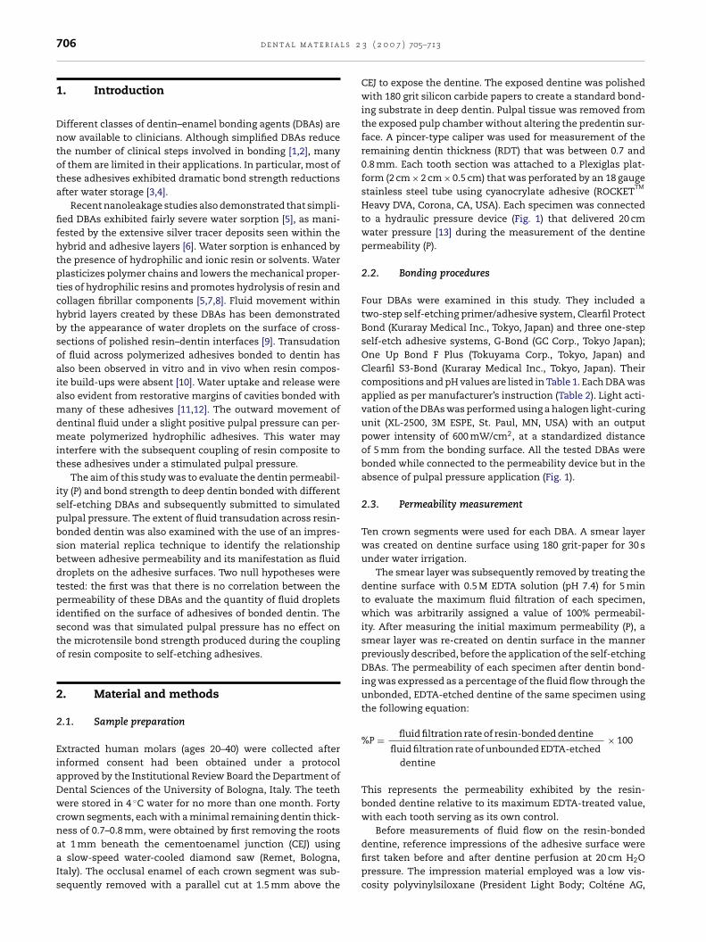
CEJ to expose the dentine. The exposed dentine was polishedwith 180 grit silicon carbide papers to create a standard bond-ing substrate in deep dentin. Pulpal tissue was removed fromthe exposed pulp chamber without altering the predentin sur-face. A pincer-type caliper was used for measurement of theremaining dentin thickness (RDT) that was between 0.7 and0.8 mm. Each tooth section was attached to a Plexiglas plat-form (2 cm × 2 cm × 0.5 cm) that was perforated by an 18 gaugestainless steel tube using cyanocrylate adhesive (ROCKET
TM
Heavy DVA, Corona, CA, USA). Each specimen was connectedto a hydraulic pressure device (Fig. 1) that delivered 20 cmwater pressure [13] during the measurement of the dentinepermeability (P).
2.2. Bonding procedures
Four DBAs were examined in this study. They included atwo-step self-etching primer/adhesive system, Clearfil ProtectBond (Kuraray Medical Inc., Tokyo, Japan) and three one-stepself-etch adhesive systems, G-Bond (GC Corp., Tokyo Japan);One Up Bond F Plus (Tokuyama Corp., Tokyo, Japan) andClearfil S3-Bond (Kuraray Medical Inc., Tokyo, Japan). Theircompositions and pH values are listed in Table 1. Each DBA wasapplied as per manufacturer’s instruction (Table 2). Light acti-vation of the DBAs was performed using a halogen light-curingunit (XL-2500, 3M ESPE, St. Paul, MN, USA) with an outputpower intensity of 600 mW/cm2, at a standardized distanceof 5 mm from the bonding surface. All the tested DBAs werebonded while connected to the permeability device but in theabsence of pulpal pressure application (Fig. 1).
2.3. Permeability measurement
Ten crown segments were used for each DBA. A smear layerwas created on dentine surface using 180 grit-paper for 30 sunder water irrigation.
The smear layer was subsequently removed by treating thedentine surface with 0.5 M EDTA solution (pH 7.4) for 5 minto evaluate the maximum fluid filtration of each specimen,which was arbitrarily assigned a value of 100% permeabil-ity. After measuring the initial maximum permeability (P), asmear layer was re-created on dentin surface in the mannerpreviously described, before the application of the self-etchingDBAs. The permeability of each specimen after dentin bond-ing was expressed as a percentage of the fluid flow through theunbonded, EDTA-etched dentine of the same specimen usingthe following equation:
%P = fluid filtration rate of resin-bonded dentine
fluid filtration rate of unbounded EDTA-etcheddentine
× 100
This represents the permeability exhibited by the resin-bonded dentine relative to its maximum EDTA-treated value,with each tooth serving as its own control.
Before measurements of fluid flow on the resin-bonded
dentine, reference impressions of the adhesive surface werefirst taken before and after dentine perfusion at 20 cm H2Opressure. The impression material employed was a low vis-cosity polyvinylsiloxane (President Light Body; Coltene AG,
d e n t a l m a t e r i a l s 2 3 ( 2 0 0 7 ) 705–713 707
Fig. 1 – Schematic showing how crown segments were created, attached to plexiglass, how fluid permeability wasmeasured under 20 cm H2O pressure, and how bonded composites were reduced to resin–dentin beams for storage and/orm
A3irtf(
prmwdws
aot(s
flbsdm
icrotensile bond tests.
ltstatten, Switzerland) with a relatively fast setting time ofmin. Just prior to taking impressions, the sticky, unpolymer-
zed oxygen-inhibition layer was removed from each bondedesin surface with a cotton pellet, to avoid interference withaking miniature impressions. These impressions were usedor fabrication of replicas for scanning electron microscopySEM).
Fluid transudation through the resin-bonded dentin waserformed according to the hydraulic conductance protocoleported by Pashley and Depew [14]. Briefly, fluid flow was
easured by following the movement of an air bubble trappedithin a 25 �L capacity glass capillary tube (0.7 mm insideiameter) (Microcaps, Fisher Scientific, Atlanta, GA, USA) thatas positioned between the pressure reservoir and the crown
egment (Fig. 1).The absolute fluid permeability (�L/min at 20 cm H2O)
nd the corresponding relative adhesive permeability (%P)btained for the four adhesives bonded to dentine were sta-istically compared using a one-way analysis of varianceANOVA) and Fisher’s PLSD test with statistical significanceet at ˛ = 0.05.
The experimental design involved measurement of fluidow across EDTA-treated dentin, smear layer-covered dentin
efore bonding and then after bonding, under 20 cm H2O pres-ure for a period of 10 min. Fluid flow measurement was con-ucted three times for each bonded specimen, from which theean fluid flow rate was determined.2.4. SEM examination
Positive replicas were fabricated from the polyvinylsiloxaneimpressions with a polyether impression material (PermadyneGarant, 3M/ESPE), according to the polyether replica tech-nique reported by Chersoni et al. [9]. As there is no chemicalreaction between polyether and polyvinylsiloxane, this replicatechnique has been shown to be an acceptable alternativeto the more time-consuming epoxy resin replica techniquefor replicating water transudation from dentin hybrid layers.The polyether positive replicas were subsequently coated withgold and examined using an SEM (Model 5400, JEOL, Tokyo,Japan) at 5–10 kV. The number of “spherical elevations” alongthe replica surface per 20 �m × 35 �m (i.e. 700 �m2 of sur-face are) at 3500× magnification, that corresponded to waterdroplets that accumulated on the surface of the resin-bondeddentin during the setting time (ca. 3 min) was recorded bytwo co-authors who did not participate in bonding and wereunaware of the group designations. Digital photographs weremade of the surface and a millimeter scale of each specimenafter EDTA-treatment, while measuring the maximum perme-ability. This permitted identification of the exact regions ofthe dentin surface that were most permeable [15]. After mak-
ing impressions of the entire flat coronal surface and castingreplicas, these same sites were located on the replicas fromthe digital photographs using digital micrometers. Four non-overlapping 20 �m × 35 �m micrographs were taken of each
708 d e n t a l m a t e r i a l s 2 3 ( 2 0 0 7 ) 705–713
Table 1 – Chemical composition, pH and source of tested adhesives
DBAs Chemical composition pH value
One Up Bond Plus (Tokuyama Corp., Tokyo, Japan) Methyl methacrylate ∼22-Hydroxyethyl methacrylateCoumarin dyeMethacryloyloxyalky acid phosphateMethacryloxyundacane 1,1 dicarboxylic acidMulti-functional methacrylic monomerFluoroaluminosilicate glass,Photoinitiator (aryl borate catalyst)Water
Clearfil S3-Bond (Kuraray Medical Inc., Tokyo, Japan) 2-Hydroxyethyl methacrylate 2.7Bis-phenol A diglycidylmethacrylate10-Methacryloyloxydecyl dihydrogen phosphateSilanated colloidal silicadl-CamphorquinoneEthyl alcoholWater
G-Bond (GC Corp., Tokyo, Japan) 4-Methacryloxyethyltrimellitate anhydride >2Triethylene glycol dimethacrylateUrethane dimethacrylateAcetone
Clearfil Protect Bond (Kuraray Medical Inc., Tokyo, Japan) 10-Methacryloyloxydecyl dihydrogen phosphate ∼2Bis-phenol A diglycidylmethacrylate2-Hydroxyethyl methacrylateHydrophobic dimethacrylatedl-CamphorquinoneN,N-Diethanol-p-toluidineSS
permeable region at 3500× magnification. Each dentin surfacehad either three or four such permeable regions depending onif it were a maxillary or mandibular third molar. Replicas madeon non-permeable sites such as the middle of the surfacedid not exhibit droplets even under positive pulpal pressure.Finally, the average and S.D. of water droplets was calculatedfor each specimen and for each adhesive group. The correla-tion between the mean relative adhesive permeability (%P) of
the four DBAs, and the mean number of fluid droplets thatappeared on the surfaces of the same resin-bonded dentinespecimens was investigated using linear regression analysiswith statistical significance set at ˛ = 0.05.Table 2 – Application procedures for the four DBAs investigated
DBAs Application mo
One Up Bond Plus Dispense equal amounts of Liquid A and Liquadhesive generously to thoroughly wet all cavfor at least 20 s. Spread the adhesive uniformloil-free air for 2 s. Light cure for 10 s.
Clearfil S3-Bond Apply the adhesive to dentin for 20 s. Air dry fLight cure for 20 s.
G-Bond Apply the adhesive using the micro-tip applic10 s. Dry thoroughly for 5 s with oil-free air unLight-cure for 10 s.
Clearfil Protect Bond Primer: apply two consecutive coats and leaveevaporate solvent for 10 s.Bonding: apply two consecutive coats, spreadstream of oil-free air for 2 s to spread the adhe
ilanated colloidal silicaurface treated sodium fluoride
2.5. Microtensile bond strength evaluation
Human extracted third molars were also used for measure-ment of microtensile bond strength. Two principal groups oftwenty specimens each were created (pulpal pressure versusno pulpal pressure) for each adhesive listed in Table 1. Since ithas been demonstrated the pulpal blood flow is significantlyreduced under local anaesthesia [16,17], the specimens in all
groups were bonded after connection to the pressure device(Fig. 1) but without any actual delivery of pulpal pressure. Com-posite build-ups were made using a light-cured flowable resincomposite (Gradia Direct LoFlo, GC Corp.) in five 1-mm incre-in the study
de Adhesive classification
id B and mix for 5 s. Apply theity surfaces. Leave undisturbedy using a gentle stream of
One-step self-etching system
or 5 s to evaporate the solvent. One-step self-etching system
ator and leave undisturbed forder maximum air pressure.
One-step self-etching system
undisturbed for at least 20 s, Two-step self-etching system
the adhesive using a gentlesive. Light cure for 20 s.
2 3 ( 2 0 0 7 ) 705–713 709
mmuwa
stfli
eseta(0eaiTdppw
3
3
Tdpo
l(df(
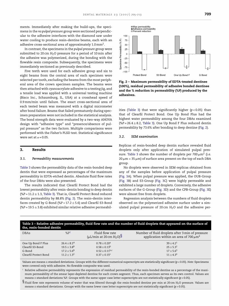
Fig. 2 – Maximum permeability of EDTA-treated dentinee(100%), residual permeability of adhesive bonded dentinee
d e n t a l m a t e r i a l s
ents. Immediately after making the build-ups, the speci-ens in the no pulpal pressure group were sectioned perpendic-
lar to the adhesive interfaces with the diamond saw underater cooling to produce resin–dentine beams, each with an
dhesive cross-sectional area of approximately 1.0 mm2.In contrast, the specimens in the pulpal pressure group were
ubmitted to 20 cm H2O pressure for a period of 10 min afterhe adhesive was polymerized, during the bonding with theowable resin composite. Subsequently, the specimens were
mmediately sectioned as previously described.Five teeth were used for each adhesive group and six to
ight beams from the central area of each specimen wereelected per tooth, excluding the beams from the most periph-ral area of the crown specimen samples. The beams werehen attached with cyanoacrylate adhesive to a testing jig, and
tensile load was applied with a universal testing machineBisco Inc., Schaumburg, IL, USA) at a crosshead speed of.9 mm/min until failure. The exact cross-sectional area ofach tested beam was measured with a digital micrometerfter bond failure. Beams that failed prematurely during spec-men preparation were not included in the statistical analysis.he bond strength data were evaluated by a two-way ANOVAesign with “adhesive type” and “presence/absence of pul-al pressure” as the two factors. Multiple comparisons wereerformed with the Fisher’s PLSD test. Statistical significanceere set at ˛ = 0.05.
. Results
.1. Permeability measurements
able 3 shows the permeability data of the resin-bonded deepentin that were expressed as percentages of the maximumermeability in EDTA-etched dentin. Absolute fluid flow ratesf the four DBAs were also presented.
The results indicated that Clearfil Protect Bond had theowest permeability after resin–dentin bonding to deep dentin
%P = 11.2 ± 1.3, Table 3). That is, Clearfil Protect Bond reducedentin permeability by 88.8% (Fig. 2). The resin–dentin inter-aces created by G-Bond (%P = 17.2 ± 5.6) and Clearfil S3-Bond%P = 19.5 ± 3.8) exhibited similar relative adhesive permeabil-Table 3 – Relative adhesive permeability, fluid flow rate and thethe, resin-bonded dentin
DBAs %P† Fluid flow rate(�L/min at 20 cm H2O
One Up Bond F Plus 26.4 ± 8.2A 0.78 ± 0.20a
Clearfil S3-Bond 19.5 ± 3.8B 0.58 ± 0.13b
G-Bond 17.2 ± 5.6B 0.52 ± 0.11b,c
Clearfil Protect Bond 11.2 ± 1.3C 0.37 ± 0.15c
Values are means ± standard deviations. Groups with the different numericwere covered only with adhesive. No flowable composite was used.† Relative adhesive permeability represents the expression of residual per
mum permeability of the smear layer-depleted dentine for each crownmeans ± standard deviations %P. Groups with the same upper case lette
§ Fluid flow rate represents volume of water that was filtered through thmeans ± standard deviations. Groups with the same lower case letter su
and the % reduction in permeability (%P) produced by theadhesives.
ities (Table 3) that were significantly higher (p < 0.05) thanthat of Clearfil Protect Bond. One Up Bond Plus had thehighest water permeability among the four DBAs examined(%P = 26.4 ± 8.2, Table 3). One Up Bond F Plus reduced dentinpermeability by 73.6% after bonding to deep dentine (Fig. 2).
3.2. SEM examination
Replicas of resin-bonded deep dentin surface revealed fluiddroplets only after application of simulated pulpal pres-sure. Table 3 shows the number of droplets per 700 �m2 (i.e.20 �m × 35 �m) of surface area present on the top of each DBAgroup.
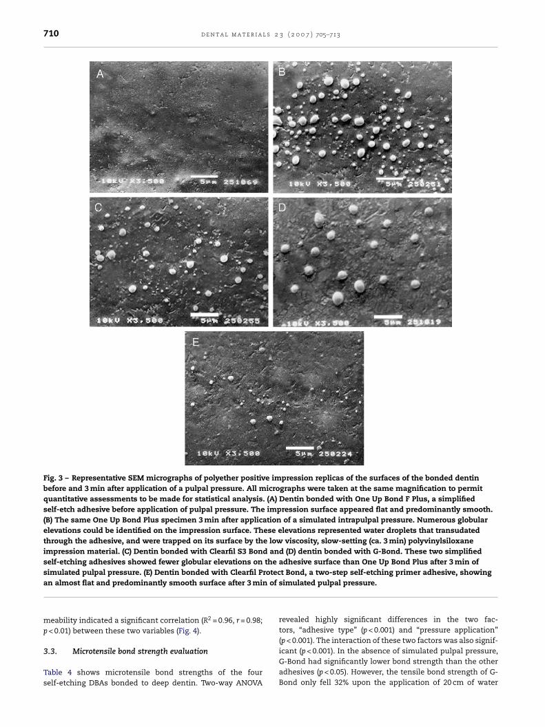
No droplets were observed in SEM-replicas obtained fromany of the samples before application of pulpal pressure(Fig. 3A). When pulpal pressure was applied, the OUB-Group(Fig. 3B) and S3-Group (Fig. 3C) were highly permeable andexhibited a large number of droplets. Conversely, the adhesivesurfaces of the G-Group (Fig. 3D) and the CPB-Group (Fig. 3E)
were almost free from droplets.Regression analysis between the numbers of fluid dropletsobserved on the polymerized adhesive surface under a sim-ulated pulpal pressure of 20 cm H2O and the adhesive per-
number of fluid droplets that appeared on the surface of
)§Number of fluid droplets after 3 min of pressure
application within an area of 700 �m2
39 ± 4.21
25 ± 5.32
17 ± 5.63
11 ± 4.34
al superscripts are statistically significant (p < 0.05). Note: Specimens
meability of the resin-bonded dentine as a percentage of the maxi-segment. Thus, each specimen serves as its own control. Values arer superscripts are not statistically significant (p > 0.05).
e resin-bonded dentine per min at 20 cm H2O pressure. Values areperscripts are not statistically significant (p > 0.05).
710 d e n t a l m a t e r i a l s 2 3 ( 2 0 0 7 ) 705–713
Fig. 3 – Representative SEM micrographs of polyether positive impression replicas of the surfaces of the bonded dentinbefore and 3 min after application of a pulpal pressure. All micrographs were taken at the same magnification to permitquantitative assessments to be made for statistical analysis. (A) Dentin bonded with One Up Bond F Plus, a simplifiedself-etch adhesive before application of pulpal pressure. The impression surface appeared flat and predominantly smooth.(B) The same One Up Bond Plus specimen 3 min after application of a simulated intrapulpal pressure. Numerous globularelevations could be identified on the impression surface. These elevations represented water droplets that transudatedthrough the adhesive, and were trapped on its surface by the low viscosity, slow-setting (ca. 3 min) polyvinylsiloxaneimpression material. (C) Dentin bonded with Clearfil S3 Bond and (D) dentin bonded with G-Bond. These two simplifiedself-etching adhesives showed fewer globular elevations on the adhesive surface than One Up Bond Plus after 3 min ofsimulated pulpal pressure. (E) Dentin bonded with Clearfil Protect Bond, a two-step self-etching primer adhesive, showing
n of
an almost flat and predominantly smooth surface after 3 mimeability indicated a significant correlation (R2 = 0.96, r = 0.98;p < 0.01) between these two variables (Fig. 4).
3.3. Microtensile bond strength evaluation
Table 4 shows microtensile bond strengths of the fourself-etching DBAs bonded to deep dentin. Two-way ANOVA
simulated pulpal pressure.
revealed highly significant differences in the two fac-tors, “adhesive type” (p < 0.001) and “pressure application”(p < 0.001). The interaction of these two factors was also signif-
icant (p < 0.001). In the absence of simulated pulpal pressure,G-Bond had significantly lower bond strength than the otheradhesives (p < 0.05). However, the tensile bond strength of G-Bond only fell 32% upon the application of 20 cm of water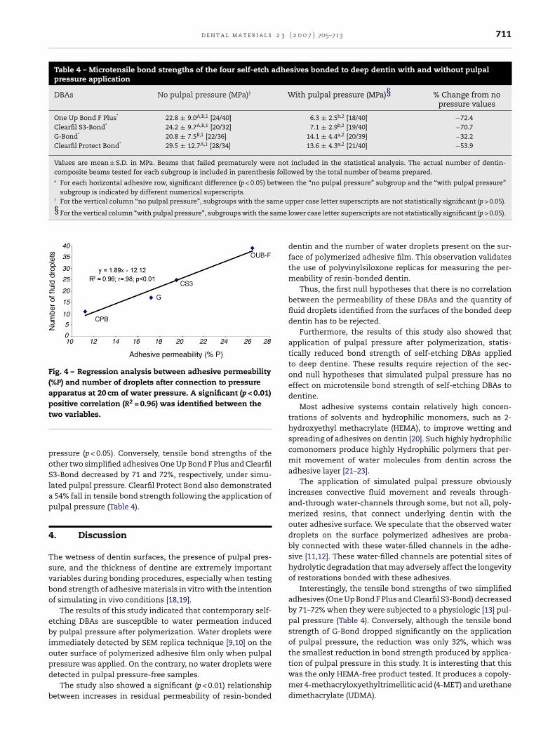
d e n t a l m a t e r i a l s 2 3 ( 2 0 0 7 ) 705–713 711
Table 4 – Microtensile bond strengths of the four self-etch adhesives bonded to deep dentin with and without pulpalpressure application
DBAs No pulpal pressure (MPa)† With pulpal pressure (MPa)§ % Change from nopressure values
One Up Bond F Plus* 22.8 ± 9.0A,B,1 [24/40] 6.3 ± 2.5b,2 [18/40] −72.4Clearfil S3-Bond* 24.2 ± 9.7A,B,1 [20/32] 7.1 ± 2.9b,2 [19/40] −70.7G-Bond* 20.8 ± 7.5B,1 [22/36] 14.1 ± 4.4a,2 [20/39] −32.2Clearfil Protect Bond* 29.5 ± 12.7A,1 [28/34] 13.6 ± 4.3a,2 [21/40] −53.9
Values are mean ± S.D. in MPa. Beams that failed prematurely were not included in the statistical analysis. The actual number of dentin-composite beams tested for each subgroup is included in parenthesis followed by the total number of beams prepared.∗ For each horizontal adhesive row, significant difference (p < 0.05) between the “no pulpal pressure” subgroup and the “with pulpal pressure”
subgroup is indicated by different numerical superscripts.† For the vertical column “no pulpal pressure”, subgroups with the same u§ For the vertical column “with pulpal pressure”, subgroups with the same l
Fig. 4 – Regression analysis between adhesive permeability(%P) and number of droplets after connection to pressureapparatus at 20 cm of water pressure. A significant (p < 0.01)positive correlation (R2 = 0.96) was identified between thet
poSlap
4
Tsvbo
ebiopd
b
wo variables.
ressure (p < 0.05). Conversely, tensile bond strengths of thether two simplified adhesives One Up Bond F Plus and Clearfil3-Bond decreased by 71 and 72%, respectively, under simu-
ated pulpal pressure. Clearfil Protect Bond also demonstrated54% fall in tensile bond strength following the application ofulpal pressure (Table 4).
. Discussion
he wetness of dentin surfaces, the presence of pulpal pres-ure, and the thickness of dentine are extremely importantariables during bonding procedures, especially when testingond strength of adhesive materials in vitro with the intentionf simulating in vivo conditions [18,19].
The results of this study indicated that contemporary self-tching DBAs are susceptible to water permeation inducedy pulpal pressure after polymerization. Water droplets weremmediately detected by SEM replica technique [9,10] on theuter surface of polymerized adhesive film only when pulpal
ressure was applied. On the contrary, no water droplets wereetected in pulpal pressure-free samples.The study also showed a significant (p < 0.01) relationshipetween increases in residual permeability of resin-bonded
pper case letter superscripts are not statistically significant (p > 0.05).
ower case letter superscripts are not statistically significant (p > 0.05).
dentin and the number of water droplets present on the sur-face of polymerized adhesive film. This observation validatesthe use of polyvinylsiloxone replicas for measuring the per-meability of resin-bonded dentin.
Thus, the first null hypotheses that there is no correlationbetween the permeability of these DBAs and the quantity offluid droplets identified from the surfaces of the bonded deepdentin has to be rejected.
Furthermore, the results of this study also showed thatapplication of pulpal pressure after polymerization, statis-tically reduced bond strength of self-etching DBAs appliedto deep dentine. These results require rejection of the sec-ond null hypotheses that simulated pulpal pressure has noeffect on microtensile bond strength of self-etching DBAs todentine.
Most adhesive systems contain relatively high concen-trations of solvents and hydrophilic monomers, such as 2-hydroxyethyl methacrylate (HEMA), to improve wetting andspreading of adhesives on dentin [20]. Such highly hydrophiliccomonomers produce highly Hydrophilic polymers that per-mit movement of water molecules from dentin across theadhesive layer [21–23].
The application of simulated pulpal pressure obviouslyincreases convective fluid movement and reveals through-and-through water-channels through some, but not all, poly-merized resins, that connect underlying dentin with theouter adhesive surface. We speculate that the observed waterdroplets on the surface polymerized adhesives are proba-bly connected with these water-filled channels in the adhe-sive [11,12]. These water-filled channels are potential sites ofhydrolytic degradation that may adversely affect the longevityof restorations bonded with these adhesives.
Interestingly, the tensile bond strengths of two simplifiedadhesives (One Up Bond F Plus and Clearfil S3-Bond) decreasedby 71–72% when they were subjected to a physiologic [13] pul-pal pressure (Table 4). Conversely, although the tensile bondstrength of G-Bond dropped significantly on the applicationof pulpal pressure, the reduction was only 32%, which wasthe smallest reduction in bond strength produced by applica-
tion of pulpal pressure in this study. It is interesting that thiswas the only HEMA-free product tested. It produces a copoly-mer 4-methacryloxyethyltrimellitic acid (4-MET) and urethanedimethacrylate (UDMA).
l s 2
r
712 d e n t a l m a t e r i a
Hydrophilic polymers such as 2-hydroxyethyl methacry-late (HEMA) are capable of imbibing large amount of waterwithin the adhesive and hybrid layers. Hence, water remainsentrapped at resin–dentine interface [6]. The presence of waterwithin adhesive film may degrade mechanical properties ofthe polymers, such as its ultimate tensile strength [8] and itsmodulus of elasticity [5] and may be responsible for reducedbond strength reported in this study. Conversely, a recentstudy has showed that experimental 4-MET-based adhesivesresult in less water uptake and phase separation when storedin water [24,25]. Apart from differences in chemical compo-sition and the presence of less hydrophilic co-monomers, thesuccess of G-Bond [26] may be due to its unique mode of appli-cation, as the manufacturer recommends a strong, continu-ous air-blast to remove the solvent prior to polymerization.Strong or longer air-blast apparently improves the quality ofthe adhesive film and reduces its permeability, thereby lower-ing the influence of water contamination [25,27].
Deep dentin is a highly permeable substrate [18,28] andmay supply excessive amounts of water to adhesives justprior to their polymerization. Water may rapidly dilute adhe-sive hydrophilic monomers and affect the resulting three-dimensional polymer structure. The ultimate tensile strength(UTS) of hydrophilic polymers was much lower than those ofhydrophobic polymers following one month of water storage[29]. In that same study, when the same polymers were storedin mineral oil instead of water, there was no change in theirUTS over time. A recent study by Ito et al. [5] demonstrated thatcommercial hydrophilic resins adsorbed between 5 and 12%water, which was associated with a drastic reduction in mod-ulus of elasticity caused by plasticization of the polymers bywater. In our study, all adhesives showed higher bond strengthwhen pulpal pressure was absent and no droplets formed onthe adhesive surface. In other words, it is evident that pul-pal pressure causes a drastic reduction in resin–dentine bondstrength.
The intrinsic wetness of dentin and the perfusion of fluidfrom the pulp chamber should be considered when bonding todeep dentin [28]. The density of water-filled dentin tubules andhence intrinsic water content of dentine increases with dentindepth [30] and these factors may reduce the bond strengthsand the longevity of the restoration [31–34]. Several investiga-tions have also demonstrated the sensitivity of various bond-ing systems to pulpal pressure [35] and dentin depth [36–38].
Recent studies [23,39] demonstrated that evaporative waterflux from dentin may induce water movement in adhesivefilms bonded to dentin. Water movement via crazing of thepolymerized adhesive matrix [8] may further generate addi-tional pathways for water penetration and result in increasedwater uptake. The inclusion of hydrophobic resin componentsin simplified self-etching DBAs may result in phase separationof the hydrophobic from the hydrophilic and ionic monomerson evaporation of their common solvent [25,40].
Using SEM and confocal microscopy, Sauro et al. [41]demonstrated that dentin adhesives are more susceptible towater uptake and lactic acid challenge than a two-steps self-
etching adhesive resulting in deterioration of the adhesives.Cadenaro et al. [42] demonstrated that insufficient polymer-ization of DBA may be one of the main causes of water dropletsformation and water uptake from dentin. Continuous water3 ( 2 0 0 7 ) 705–713
uptake from dentin tubules during the polymerization proce-dures may result in discontinuities in the structure of adhe-sive layer and the formation of an unstable, porous interfacewith water migrating from the dentine outward through theadhesive layer curing placement and polymerization of resincomposites.
Hence, over time, blisters and water channels of HEMA-based adhesives may increase their dimensions and creategreater stress at the interface between the adhesive film andthe overlying resin composite. Finally, the existence of micro-channels from dentin through the hybrid and adhesive layerscreate the conditions for a continuous water flux [12] anduncontrolled absorption and accumulation of proteins andother biomolecules from oral fluids that may further alter themechanical properties of adhesive film [43].
5. Conclusion
Within the limitations of this in vitro study, it was con-cluded that the hybrid layer and polymerized resin adhe-sive films of some self-etching all-in-one adhesives applied indeep dentin are extremely permeable after polymerizationwhen simulated pulpal pressure is applied. This results inreduced bond strength, especially when hydrophillic HEMA-based self-etching adhesives are used. Furthermore, it maypromote continuous bidirectional water movement acrossadhesive dentine interfaces over time that induces degrada-tion of resin–dentine bonds [44].
Thus, these materials and in particular all-in-one DBAs,must be avoided in deep dentin in favor of total-etch, three-step adhesives.
Acknowledgements
We would like to thank Dr. Paolo Ferrieri and Prof. RomanoMongiorgi for their support and assistance for the scanningelectron microscope and permeability apparatus. Materialswere generously supplied by Kuraray, Tokuyama and GC Corp.This study was supported by ‘Progetto Pluriennale’ UNIBOand RFO 2004 UNIBO from the University of Bologna, Italy,by CNPq# 474226/03-4, Brazil and by grant R01 DE14911 (PI D.Pashley) from the National Institute of Dental and CraniofacialResearch.
e f e r e n c e s
[1] Perdigao J. Dentin bonding as a function of dentin structure.Dent Clin N Am 2002;46:277–301.
[2] Van Meerbeek B, De Munck J, Yoshida Y, Inoue S, Vargas M,Vijay P, et al. Buonocore memorial lecture. Adhesion toenamel and dentine: current status and future challenges.Oper Dent 2003;28:215–35.
[3] Armstrong SR, Vargas MA, Fang Q, Laffoon JE. Microtensilebond strength of a total-etch 3-step, total-etch 2-step,
self-etch 2-step, and a self-etch 1-step dentine bondingsystem through 15-month water storage. J Adhes Dent2003;5:47–56.[4] De Munck J, Van Landuyt K, Peumans M, Poitevin A,Lambrechts P, Braem M, et al. A critical review of the
2 3
of charged groups on protein interactions with poly(HEMA)
d e n t a l m a t e r i a l s
durability of adhesion to tooth tissue: methods and results. JDent Res 2005;84:118–32.
[5] Ito S, Hashimoto M, Wadgaonkar B, Svizero N, Carvalho RM,Yiu C, et al. Effects of resin hydrophilicity on water sorptionand changes in modulus of elasticity. Biomaterials2005;26:6449–59.
[6] Tay FR, Pashley DH, Garcia-Godoy F, Yiu CK. Single-step,self-etch adhesives behave as permeable membranes afterpolymerization. Part II. Silver tracer penetration evidence.Am J Dent 2004;17:315–22.
[7] Hashimoto M, Tay FR, Ohno H, Sano H, Kaga M, Yiu CKY, etal. SEM and TEM analysis on water degradation of humandentine collagen. J Biomed Mater Res 2003;66:287–98.
[8] Yiu CKY, King NM, Carrilho MRO, Sauro S, Rueggeberg PratiC, Carvalho RM, et al. Effect of resin hydrophilicity andtemperature on water sorption of dental adhesive resins.Biomaterials 2005;26:6862–72.
[9] Chersoni S, Suppa P, Breschi L, Tay FR, Pashley DH, Prati C.Water movement in the hybrid layer after different dentinetreatments. Dent Mater 2004;20:796–803.
[10] Chersoni S, Suppa P, Grandini S, Goracci C, Monticelli F, YiuC, et al. In vivo and in vitro permeability of one-step selfetch adhesives. J Dent Res 2004;83:459–64.
[11] Frankenberger R, Tay FR. Self-etch vs etch-and-rinseadhesives: effect of thermo-mechanical fatigue loading onmarginal quality of bonded resin composite restorations.Dent Mater 2005;21:397–412.
[12] Prati C, Chersoni S, Acquaviva GL, Breschi L, Suppa P, Tay FR,et al. Permeability of marginal hybrid layers in compositerestorations. Clin Oral Investig 2005;9:1–7.
[13] Ciucchi B, Bouillaguet S, Holz J, Pashley DH. Dentine fluiddynamics in human teeth, in vivo. J Endodent 1995;21:191–4.
[14] Pashley DH, Depew DD. Effects of smear layer, copalite andoxalate on microleakage. Oper Dent 1986;11:95–102.
[15] Pashley DH, Adringa HJ, Derkson GD, Derkson ME, KalathorSR. Regional variability in the permeability of human dentin.Arch Oral Biol 1987;32:519–23.
[16] Chng HS, Pitt Ford TR, McDonald F. Effects of prilocaine localanaesthetic solutions on pulpal blood flow in maxillarycanines. Endod Dent Traumatol 1996;12:89–95.
[17] Premdas CE, Pitt Ford TR. Effect of palatal injections onpulpal blood flow in premolars. Endod Dent Traumatol1995;11:274–8.
[18] Prati C, Pashley DH. Dentine wetness, permeability andthickness and bond strength of adhesive systems. Am J Dent1992;5:33–8.
[19] Pioch T, Staehle HJ, Schneider H, Duschner H, Dorfer CE.Effect of intrapulpal pressure simulation in vitro on shearbond strengths and hybrid layer formation. Am J Dent2001;14:319–23.
[20] Tay FR, Pashley DH. Have dentine adhesives become toohydrophilic? J Can Dent Assoc 2003;69:724–31.
[21] Tay FR, Pashley DH, Suh BI, Carvalho RM, Itthagarun A.Single-step adhesives are permeable membranes. J Dent2002;30:371–82.
[22] Itthagarun A, Tay FR, Pashley DH, Wefel JS, Garcia-Godoy F,Wei SHY. Single-step, self-etch adhesives behave aspermeable membranes after polymerization. Part III.
Evidence from fluid conductance and artificial cariesinhibition. Am J Dent 2004;17:394–400.[23] Tay FR, Pashley DH, Suh BI, Hiraishi N, Yiu CKY. Buonocorememorial lecture. Water treeing in simplified dentineadhesives—Deja vu? Oper Dent 2005;30:561–79.
( 2 0 0 7 ) 705–713 713
[24] Unemori M, Matsuya Y, Matsuya S, Akashi A, Akamine A.Water absorption of poly(methyl methacrylate) containing4-methacryloxyethyl trimellitic anhydride. Biomaterials2003;24:1381–7.
[25] Van Landuyt KL, De Munck J, Snauwaert J, Coutinho E,Poitevin A, Yoshida Y, et al. Monomer-solvent phaseseparation in one-step self-etch adhesives. J Dent Res2005;84:183–8.
[26] Spreafico D, Semeraro S, Mezzanzanica D, Re D, Gagliani M,Tanaka T, et al. The effect of the air-blowing step on thetechnique sensitivity of four different adhesive systems. JDent 2006;34:237–44.
[27] Hashimoto M, Tay FR, Ito S, Sano H, Kaga M, Pashley DH.Permeability of adhesive resin films. J Biomed Mater Res2005;74:99–705.
[28] Itthagarun A, Tay FR. Self-contamination of deep dentin bydentinal fluid. Am J Dent 2000;13:195–200.
[29] Yiu CK, King NM, Pashley DH, Suh BI, Carvalho RM, CarrilhoMR, et al. Effect of resin hydrophilicity and water storage onresin strength. Biomaterials 2004;25:5789–96.
[30] Pashley DH. Clinical correlations of dentine structure andfunction. J Prosth Dent 1991;60:777–81.
[31] Prati C, Pashley DH, Montanari G. Hydrostatic intrapulpalpressure and bond strength of bonding systems. Dent Mater1991;7:57–8.
[32] Pereira PN, Okuda M, Sano H, Yoshikawa T, Burrow MF,Tagami J. Effect of intrinsic wetness and regional differenceon dentin bond strength. Dent Mater 1999;15:46–53.
[33] Perdigao J. Dentin bonding as a function of dentin structure.Dent Clin N Am 2002;6:277–301.
[34] Toledano M, Osorio R, Ceballos L, Fuentes MV, Fernandes CA,Tay FR, et al. Microtensile bond strength of several adhesivesystems to different dentin depths. Am J Dent 2003;16:292–8.
[35] Ozok AR, Wu M-K, De Gee AJ, Wesselink PR. Effect of dentinperfusion on the sealing ability and microtensile bondstrengths of a total-etch versus an all-in-one adhesive. DentMater 2004;20:479–86.
[36] Tagami J, Tao L, Pashley DH. Correlation among dentindepth, permeability and bond strength of adhesive resins.Dent Mater 1990;6:45–50.
[37] Tao L, Tagami J, Pashley DH. Pulpal pressure and bondstrengths of superbond and gluma. Am J Dent 1991;4:73–6.
[38] Ferrari M, Tay FR. Technique sensitivity in bonding to vitalacid-etched dentin. Oper Dent 2003;28:3–8.
[39] Hashimoto M, Ito S, Tay FR, Svizero NR, Sano H, Kaga M, etal. Fluid movement across the resin–dentin interface duringand after bonding. J Dent Res 2004;83:843–8.
[40] Spencer P, Wang Y. Adhesive phase separation at the dentininterface under wet bonding conditions. J Biomed Mater Res2002;62:447–56.
[41] Sauro S, Watson TF, Tay FR, Chersoni S, Breschi L, Bernardi F,et al. Water uptake of bonding systems applied on rootdentine surfaces: a SEM and confocal microscopic study.Dent Mater 2006;22:271–80.
[42] Cadenaro M, Antoniolli F, Sauro S, Tay FR, Di Lenarda R, PratiC, et al. Degree of conversion and permeability of dentaladhesives. Eur J Oral Sci 2005;113:525–30.
[43] Lord MS, Stenzel MH, Simmons A, Milthorpe BK. The effect
hydrogels. Biomaterials 2006;27:567–75.[44] Sano H, Yoshikawa T, Pereira PN, Kanemura N, Morigami M,
Tagami J, et al. Long-term durability of dentin bonds madewith a self-etching primer, in vivo. J Dent Res 1999;78:906–11.