Embed Size (px)
Citation preview
American Journal of Phytomedicine and Clinical Therapeutics www.ajpct.org
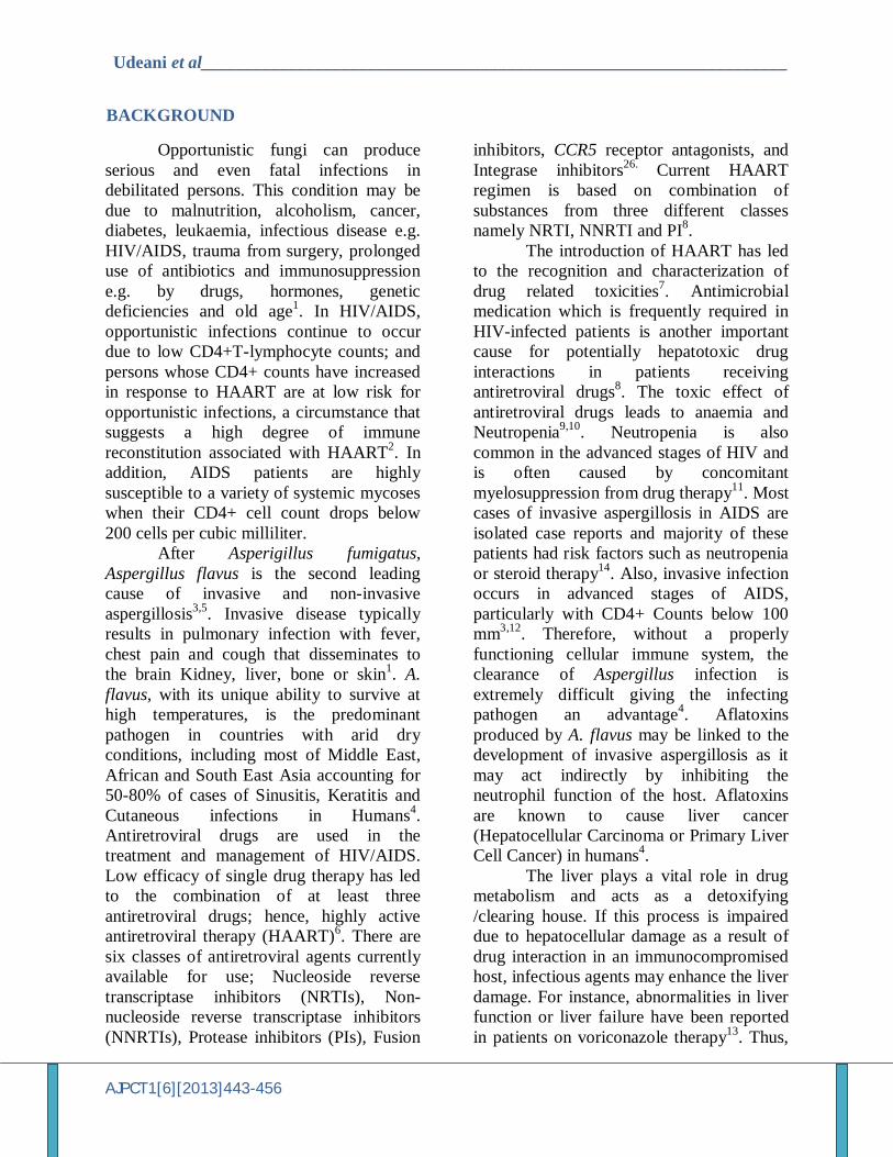
Original Article
Effect of Aspergillus flavus on the Liver of Experimental Rats Administered with Antiretroviral Drugs Theophilus K Udeani*, Chukwugozie N Okwuosa, Ezinne U Adiele
Department of Medical Laboratory Sciences, Faculty of Health Sciences, College of Medicine, University of Nigeria Enugu Campus, Enugu, Nigeria
ABSTRACT
Background: The aim of this study was to determine the association between antiretroviral (ARV) drugs and Aspergillus flavus in experimental rats. Methods: Inbred albino Wister rats, aged18-21weeks; weighing 200-250g were used. The experiment was carried out in two models, excluding antifungal drugs. In model 1; the rats were infected with Aspergillus flavus and treated with antiretroviral drugs; grouped into four of six rats each, based on their weights. Group A: Neat control while Groups B-D were administered with varying concentrations of Nevirapine, Zidovudine and Lamivudine, Zidovudine, Lamivudine and Nevirapine. In model 2, the animals were immunosuppressed with cyclophosphamide and then administered with ARV and A. flavus. In both models; pre and post inoculation blood samples were collected to assay for liver enzymes and the liver was harvested for histological assessment and recovery of A. flavus. Results: In model1, pre-inoculation liver enzyme values showed no statistical increase between control group and experimental groups (P> 0.05) while the post-inoculation liver enzyme values were statistically significant (P<0.05) between control and experimental group, with increase in values of ALT, AST and ALP. In model 2 ALT and ALP were increased in both pre-and post-inoculation of experimental group, when compared with control. Histological evidence of liver damage was observed in both models. Conclusions: The experimental administration of ARV drugs in rats may enhance the pathogenicity of opportunistic aspergillosis thereby impeding the metabolic functions of the liver.
Keywords: Antiretroviral drugs, Aspergillus flavus, liver, Liver enzymes, Neutropenia.
Address for Correspondence
Department of Medical Laboratory Sciences, Faculty of Health Sciences, College of Medicine, University of Nigeria Enugu Campus, Enugu, Nigeria E-mail: theoka2002@ yahoo.com
Udeani et al___________________________________________________________________
AJPCT1[6][2013]443-456
BACKGROUND
Opportunistic fungi can produce serious and even fatal infections in debilitated persons. This condition may be due to malnutrition, alcoholism, cancer, diabetes, leukaemia, infectious disease e.g. HIV/AIDS, trauma from surgery, prolonged use of antibiotics and immunosuppression e.g. by drugs, hormones, genetic deficiencies and old age1. In HIV/AIDS, opportunistic infections continue to occur due to low CD4+T-lymphocyte counts; and persons whose CD4+ counts have increased in response to HAART are at low risk for opportunistic infections, a circumstance that suggests a high degree of immune reconstitution associated with HAART2. In addition, AIDS patients are highly susceptible to a variety of systemic mycoses when their CD4+ cell count drops below 200 cells per cubic milliliter. After Asperigillus fumigatus, Aspergillus flavus is the second leading cause of invasive and non-invasive aspergillosis3,5. Invasive disease typically results in pulmonary infection with fever, chest pain and cough that disseminates to the brain Kidney, liver, bone or skin1. A. flavus, with its unique ability to survive at high temperatures, is the predominant pathogen in countries with arid dry conditions, including most of Middle East, African and South East Asia accounting for 50-80% of cases of Sinusitis, Keratitis and Cutaneous infections in Humans4. Antiretroviral drugs are used in the treatment and management of HIV/AIDS. Low efficacy of single drug therapy has led to the combination of at least three antiretroviral drugs; hence, highly active antiretroviral therapy (HAART)6. There are six classes of antiretroviral agents currently available for use; Nucleoside reverse transcriptase inhibitors (NRTIs), Non-nucleoside reverse transcriptase inhibitors (NNRTIs), Protease inhibitors (PIs), Fusion
inhibitors, CCR5 receptor antagonists, and Integrase inhibitors26. Current HAART regimen is based on combination of substances from three different classes namely NRTI, NNRTI and PI8.
The introduction of HAART has led to the recognition and characterization of drug related toxicities7. Antimicrobial medication which is frequently required in HIV-infected patients is another important cause for potentially hepatotoxic drug interactions in patients receiving antiretroviral drugs8. The toxic effect of antiretroviral drugs leads to anaemia and Neutropenia9,10. Neutropenia is also common in the advanced stages of HIV and is often caused by concomitant myelosuppression from drug therapy11. Most cases of invasive aspergillosis in AIDS are isolated case reports and majority of these patients had risk factors such as neutropenia or steroid therapy14. Also, invasive infection occurs in advanced stages of AIDS, particularly with CD4+ Counts below 100 mm3,12. Therefore, without a properly functioning cellular immune system, the clearance of Aspergillus infection is extremely difficult giving the infecting pathogen an advantage4. Aflatoxins produced by A. flavus may be linked to the development of invasive aspergillosis as it may act indirectly by inhibiting the neutrophil function of the host. Aflatoxins are known to cause liver cancer (Hepatocellular Carcinoma or Primary Liver Cell Cancer) in humans4.
The liver plays a vital role in drug metabolism and acts as a detoxifying /clearing house. If this process is impaired due to hepatocellular damage as a result of drug interaction in an immunocompromised host, infectious agents may enhance the liver damage. For instance, abnormalities in liver function or liver failure have been reported in patients on voriconazole therapy13. Thus,
Udeani et al___________________________________________________________________
AJPCT1[6][2013]443-456
the effect of HAART regimen and Aspergillus flavus infection on the liver of experimental rats was investigated, to understand their interaction in the absence of antifungal therapy. MATERIALS AND METHODS
Drugs The antiretroviral drugs; Nevirapine
(Heterodrugs LTD; Hyderabad, India), Zidovudine and Lamivudine (Biochem Pharmaceutical LTD, United Kingdom); were purchased from a standard pharmacy store in Enugu, Nigeria. Aspergillus flavus isolate
A. flavus isolate was obtained from Mycology unit, University of Nigeria Teaching Hospital (UNTH), Enugu. It was isolated from sputum of a HIV/AIDS patients suffering from pneumonia and with a CD4+ count of 210cells/ul. The isolate was further identified by sub-culture unto potato dextrose agar with Chloramphenicol and identified by needle mount using Lactophenol cotton blue15. The isolate was sub-cultured unto potato dextrose agar slant stored at +4oC until used. Experimental animals
Fourty-four (44) albino Wister rats were purchased from college of Medicine, Experimental Animal Unit; University of Nigeria Enugu campus. The experiment was conducted in the experimental animal unit. They were allowed to acclimatize for one week prior to the start of the experiment and fed ad libitum with standard feed and water. The investigation conforms to the Guide for the Care and Use of Laboratory Animals (National Institutes of Health Publication No. 85-23, revised 1996)
Experimental design and conduct Model 1
Twenty-four (24) albino Wister rats weighing 200-250g, were used. They were grouped into four (A-D) of six rats each based on their weights. Group A: Neat control, Group B were given 3.3mg/kg/bd.wt of Nevirapine, Group C were given 7.5mg/kg/bd.wt of Zidovudine and Lamivudine, group D were given 10.8mg/kg/bd.wt of Zidovudine, Lamivudine and Nevirapine; in accordance with human dosage for adults. These lasted for 21 days. On the 22nd day, 2mL of blood was collected from the retrorbital plexus of the median canthus of the eye using heparinised capillary tube into plain sterile tubes to obtain serum. One rat from each group was sacrificed and a portion of the liver harvested and fixed in 10% formal saline. One the 23rd day, 0.5mL containing 106 spores of A. flavus grown in Brain heart infusion broth was given to Groups B-D intra-peritoneal and the various drug dosages were continued. On the 39th day, 2mL of venous blood was collected into plain sterile tubes. The remaining animals were sacrificed and the liver harvested. A portion was collected into a plain sterile container while the remainder was fixed in 10% formol saline. Model 2
Twenty (20) rats weighing 200-250g, three months old were used. They were grouped into four cages (A-D) based on their weight; five rats in each group. Group A: neat control, Group B Zidovudine, Lamivudine and Nevirapine, Group C: Zidovudine, Lamivudine and Nevirapine plus Cyclophosphamide, Group D: Cyclophosphamide. Group B and C were given 10.8mg/kg/bd.wt of Zidovudine, Lamivudine and Nevirapine, Group C and D were given 2mg/kg of Cyclophosphamide. All drugs were given orally for three days.
Udeani et al___________________________________________________________________
AJPCT1[6][2013]443-456
One the 4th day, 2mL of venous blood was collected into plain sterile tubes to obtain serum. One the 5th days, 0.5mL containing 106 spores of A. flavus grown in BHIB were given to groups B-D intraperitoneally and the various drug dosages were continued. One the 30th day, 2mL of venous blood was collected into a plain sterile tube. The animals were sacrificed and the liver harvested. A portion was collected into a plain sterile container while the remainder was fixed in 10% formol saline. Re-isolation of A. flavus The liver tissues collected into plain sterile containers were cultured unto Saboraud’s dextrose agar and cornmeal agar; with Chloramphenicol in order to re-isolate the fungus. The fungus was identified based on its colonial appearance and morphological features by needle mount using Lactophenol cotton blue as described by Beneke and Rogers15. Serum analysis
The serum samples obtained were assayed for Alanine transaminase (ALT) and Aspartate transaminase (AST) using Reitman and Frankel method (16), Alkaline phosphatase using King and king method 17
and Total Bilirubin using Powell method18.
Histological analysis The liver tissues fixed in 10% formol-
saline were processed using standard histological techniques and stained by Haematoxylin and Eosin. Histopathological Study
After blood sampling, the rats were sacrificed and their livers were separated, sliced and washed in saline. The fragments from the liver tissues were preserved in 10% neutral formalin solution, embedded in paraffin wax, histological sections were prepared, stained with hematotoxylin (H) and
eosin (E), mounted and observed under light microscope. Statistical Analysis
Student t-test and one way analysis of variance (ANOVA) and the corresponding post hoc test were used for data analysis. All data were expressed as mean+ standard deviation and P < 0.05 was statistically significant. The SPSS (version 15.0) software was used for the analysis. RESULTS
Growth of Aspergilus flavus in the liver In the experimental animals, A. flavus
was re-isolated from groups B-D in both model1and 2. In control group A of both models, the fungus was not re-isolated. (Table1) Evaluation of biochemical parameters
In the experimental models, the following liver markers were assessed; alanine aminotransaminase (ALT),aspertate transaminase (AST), alkaline phosphatase (ALP) and Total bilirubin(TB). In model one, there was no appreciable increase (P> 0.05) in pre-inoculation, liver marker values of groups B, C and D when compared with group A (Table 2a). In post-inoculation analysis, there was a marked statistical increase (P< 0.05) in ALT, AST and ALP values of group B with 110.4 + 6.1U/L, 288.1 +11.3U/L, 377.3 + 1.2U/L when compared with control group A with 75.4 + 10.4U/L, 147.3 + 24.1U/L, 207.7 +24.5U/L respectively. Also, there was statistical increase (P< 0.05) in ALT values of group D with 106.2 + 3.4U/L when compared with 75.4 + 10.4U/L of control Group A (Table 2B). In model two, the pre-inoculation analysis showed a statistical increase in ALP values of group B,C and D with 504.5 + 80.8U/L, 692.0 + 43.0U/L, 418.4 + 32.4U/L when compared with control group A with 261.9 + 24.1U/L respectively. Also, there was
Udeani et al___________________________________________________________________
AJPCT1[6][2013]443-456
statistical increase (P< 0.05) in ALT value of group C with 97.5 + 2.6U/L when compared with control group A with 64.8 + 6.2U/L (Table 3A). From post-inoculation analysis results, there was statistical increase (P< 0.05) in ALT values of groups B,C and D with 154.7 + 1.7U/L, 166.6 + 9.0U/L, 158.7 + 22.6U/L when compared with control group A with 68.4 + 6.2U/L respectively. There was also statistical increase in ALP values of groups B,C and D with control group A with 552.0 + 18.4U/L, 776.8 + 25.4U/L, 584.1 + 59.8U/L when compared with control group A with 275.9 + 24.1U/L respectively (Table 3B). There was no significant increase in Total bilirubin of groups B, C and D when compared with control group A in both models. Histological assessment
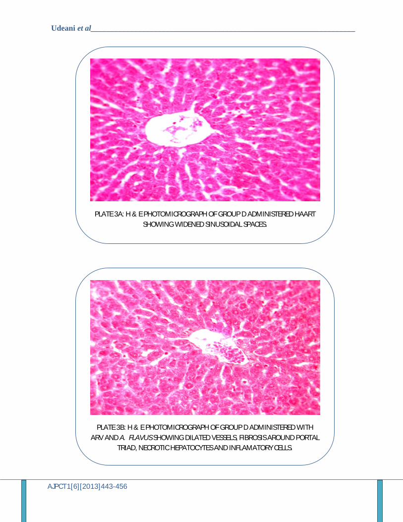
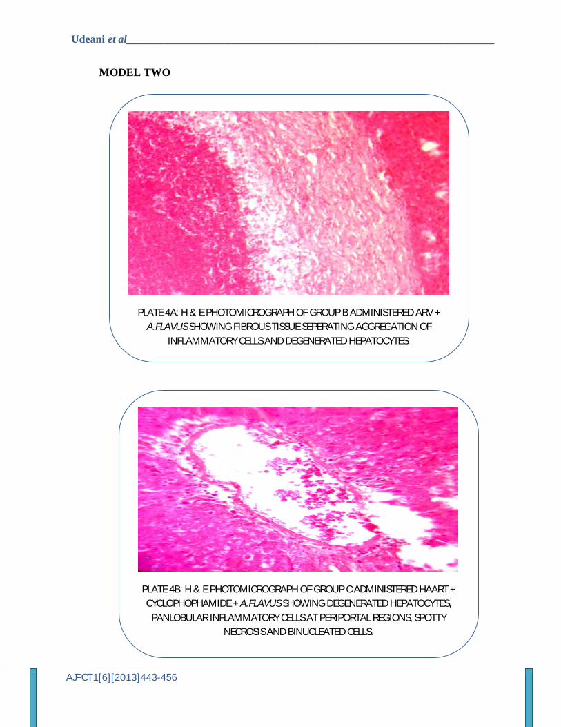
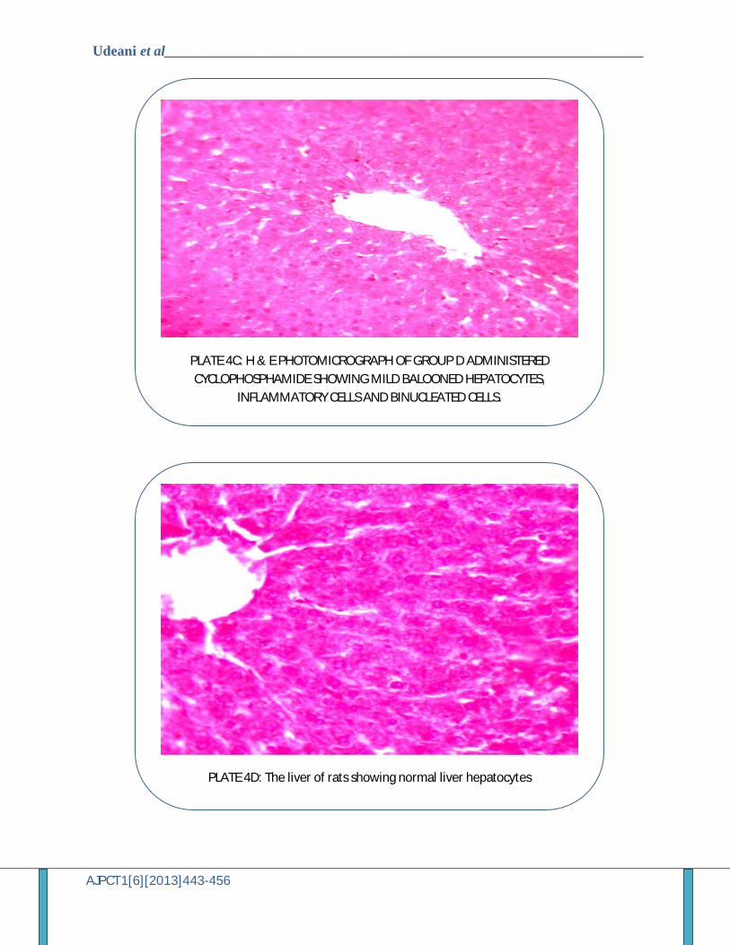
In model one pre-inoculation assessment, inflammatory cells with sinusoidal spaces and spotty necrosis was seen in group B, and mild formation of fibrous tissue around blood vessels in group C; while group D showed widened sinusoidal spaces (Plates 1a, 2a, 3a). Post-inoculation assessment showed fibrous tissues formation around vessels, necrotic hepatocytes, inflammatory cells within sinusoidal spaces and binucleated cells in groups B, C and D (Plates1b, 2b, 3b). Post-inoculation assessment in model two showed marked damage in group C, showing degenerated hepatocytes, panlobular inflammatory cells at periportal regions, binucleated cells when compared with group B showing fibrous tissue separating aggregates of inflammatory cells and degenerated hepatocytes and group C showing mild ballooned hepatocytes (Plates 4a, 4b, 4c). DISCUSSION
It has been ascertained that liver injury due to exposure to a drug or other non-infectious agents will lead to impaired liver
function that can result to clinical significant disease19, 27. This study was designed to evaluate the interaction between ARV drugs and A. flavus in experimental rats. Two experimental models were used to achieve this objective. The first model investigated the outcome of HAART regimen and A. flavus in the absence of immunosuppression; whereas the second model assessed the outcome of the HAART regimen in the experimental rats immunosuppressed with Cyclophosphamide. The functional mechanism of ARV drugs is based on the inhibition of certain stages of HIV viral replication either by competing with host cellular triphosphate substrates needed for pro-viral DNA synthesis, when incorporated, leads to viral DNA chain termination or by binding to the reverse transcriptase enzyme near its catalytic site and denaturing it 20.
From the results in the first model, it can be deduced that A. flavus disseminated to the liver in groups B, C and D respectively. The dissemination in these groups may have been due to neutropenia induced by ARV drugs given to the rats 21. Therefore it can be postulated that the damage to the liver of the rats due to ARV drugs allowed infiltration of A. flavus. Umar et al 21 reported that antiretroviral drugs, Lamivudine, Nevirapine and Stavudine administered independently to experimental rats caused a decrease in neutrophils and eosinophils. This effect may have helped in the establishment of infection in the liver. This was noticeable in the liver function markers as there was a significant increase in the ALT, AST and ALP as evidence of liver damage. Also, histological studies demonstrated liver damage in groups B, C and D as the liver showed marked fibrosis, necrotic hepatocytes, binucleated cells and inflammatory cells.
In the second model, there was translocation of A. flavus to the liver in groups B, C and D. This was evident in the re-isolation of A. flavus from the liver.
Udeani et al___________________________________________________________________
AJPCT1[6][2013]443-456
Immunosuppression with Cyclophosphamide induces generalized suppression of T-cell and B-cell function by destroying proliferating lymphoid cells 20. In these experimental rats, the destruction of lymphoid cells (T and B-cells), enhanced the destruction of the hepatocytes of the liver thereby permitting the infiltration of A. flavus. This was demonstrated in group C which showed that in the presence of immunosuppressive agent and ARV drugs, the liver damage was more as shown in the H and E staining with degenerated hepatocytes, necrosis, inflammatory and binucleated cells. The activities of serum ALT and ALP were increased in group C and this was significant when compared with group A. It therefore seems that serum enzyme alterations are an indication of liver damage. This agrees with Yin et al,22 postulated that in Carbon tetrachloride (CCL4) induced liver damage in experimental rats administered with aflatoxin B1, the serum enzyme alterations was an important factor in liver damage. This was reflected in the histology of group C where there was marked damage to the liver showing degenerated hepatocytes, spotty necrosis and panlobular inflammatory cells at periportal regions. This feature indicates non-regeneration/ non-proliferation of liver cells. This damage may be due to the interaction between ARV drugs used, in ARV drugs and A. flavus or its metabolic by-products in immunosuppresed rats. The changes observed in group B and D were mild when compared with group C. Of important note is the fibrosis in group B without immunosuppresive drug, a marked increase in ALT and ALP may be attributed to the effect of A. flavus and ARV drugs used in HAART. The deduction from this is that, in the presence of immunosuppresion, liver damage is enhanced 23, 24, 25.
In these experimental models, the administration of ARV drugs used in HAART may enhance the pathogenicity of
opportunistic aspergillosis thereby impeding the metabolic functions of the liver. In the report of Denning et al, 13, voriconazole have been reported to equally affect the liver in the presence of ARV drugs. In this experiment, antifungal drugs were not used but it is recommended that continuous monitoring of these drugs be advised.
REFERENCES 1. Willey, J. M., Sherwood, L. M. and
Woolverton, C. J. In: Prescott, Harley, and Klein’s Microbiology, Seventh Edition. McGraw-Hill companies, 2008; Pp 930-1017.
2. Kaplan, J. E., Hanson, D., Dworkin, M. S., Frederick, T., Bertolli, J., Loulindegren, M., Holmberg, S. and Jones, J. L. Epidemiology of Human Immunodeficiency Virus- Associated Opportunistic Infections in the United States in the Era of Highly Active Antiretroviral Therapy. Clin. Infect. Dis. 2000. 30 (1): S5-S14
3. Bica, I., McGovern, B., Dhar, R., Stone, D., McGowan, K., Scheib, R. and Snydman, D. R. Increasing mortality due to end-stage Liver disease in Patients with human Immunodeficiency virus infection. Clin. Infect. Dis.2001; 32:492-497.
4. Krishnan, S., Manavathu, E. K.and Chandrasekar P. H. Aspergillus flavus: an emerging non- Fumigatus aspergillus species of significance. Black well verlag GmbH, 2009; Pp 1-17.
5. Hedayati, M. T., Pasqualotto, A. C., Warn, P. A., Bowyer, P. and Denning, D. W. Aspergillus flavus: human pathogen; allergen and mycotoxin producer. Microbiology 2007; 153: 1677-1692.
6. Mocroft, A.,Youle, M., Moore, A., Sabin, C. A., Madge, S. and Lepri, A. C. Reasons for modification and discontinuation of antiretrovirals: results from a single treatment centre. AIDS; 2001; 15: 185-194.
7. Hammer, S. M., Kessler, H. A. and Saag, M. S. Issues in combination of antiretroviral therapy. Journal of Acquired Immune Deficrency Syndrome, 1994; 7: S24-S35.

Udeani et al___________________________________________________________________
AJPCT1[6][2013]443-456
8. Spengler, U., Lichterfeld, M. and Rockstroh, K. J. Anti-retroviral drug toxicity- a challenge for the hepatologist? J Hepatol.2002; 36: 283-294.
9. Janet, E., Benoit, S. C. and Jemsek, J. Treatment with Lamivudine, Zidovudine or both in HIV Positive patients. N Engl J Med, 1995; 323: 181-183.
10. Fische, M.C., Parker, C. B. and Pettineli C. A randomized controlled trial of reduced daily dose of zidovudine in Patients with AIDS. N Engl J Med.1990; 323: 200-202.
11. Van Leeuwen, R. Evaluation of safety and efficacy of Lamivudine in patients with a symptomatic or mildly symptomatic human immunodeficiency virus infection. J. Infect. Dis.1995; 26: 1166-1171.
12. Bodey, G. P. and Vartivarian, S. Aspergillosis. Eur. J Clinl Microbiol Infect Dis.1989; 8: 413-437.
13. Denning, D. W., Ribaud, P. and Milpied, M.. Efficacy and safety of voriconazole in the treatment of acute invasive aspergillosis. Clin Infect Dis.2002; 34: 563-571.
14. Meyohas, M.C., Roux, P., Poirot, J. L., Meynard J. L. and Frottier J. Aspergillosis in Acquired Immune Deficiency Syndrome. Pathol Biol (Paris) 1994; 42: 647-651.
15. Beneke, E. S. and Rogers, A. L. Medical Mycology and Human Mycoses. Star Publishing Company, California, 1996; Pp 32-33.
16. Reitman, S and Frankel, S. A colorimetric method for the determination of serum aspartate and alanine transaminases. American J Clin Pathol 1957; 28: 56-63.
17. King, P. R. N. and King, E. J. Estimation of plasma. Phosphatase by determination of hydrolysed phenol with aminoantipyrine. J. Clin. Pathol.1954; 7: 322- 326.
18. Powell, I. T. Estimation of Bilirubin in serum. Am. J Clin. Pathol.1944; 14: 55
19. Navarro, V. J. and Senior, J. R.) Drug-Related hepatotoxicity. N. Engl. J. Med.2006; 354: 731-739
20. Rang et al. Rang and Dale’s Pharmacology, Sixth Edition. Elsevier, 2007; Pp 679-690.
21. Umar, R. A., Ladan, M. J., Hassan, S. W., Sa’id, Y., Abbas, A. Y. and Oduolisaeme, I. B. Administration of antiretroviral drugs (Lamivudine, Nevirapine and Stavudine) has no untoward effect on haematological profile in Albino Wistar rats. Asian J. Biochem.2007; 2 : 147-151
22. Yin, S. J., Kao, M. C. and Lee, S. C Yin, S. J., Kao, M. C. and Lee, S. C. Sequential biochemical and histological changes in rats treated with aflatoxin B1. British J Cancer1980; 42: 319.
23. Mosquera, J., Warn, P. A. and Morrissey, J. Susceptibility testing of Aspergillus flavus: inoculum dependence with itraconazote and lack of correlation between susceptibility to amphotericin B in vitro and in vivo. Antimicrob. Agents Chemother.2001; 45: 1456-1462.
24. Denning, D. W., Venkateswarlu, K., Oakley, K. L., Anderson, M. J., Manning, N. J. Stevens, D. A., Warnock, D. W. and Kelly, S. I. Intraconazole resistance in Aspergillus fumigatus. Antimicrob Agents Chemother.1997; 41: 1364-1368.
25. Johnson, E. M., Oakley, K. L. and Radford, S. A. Lack of correlation of invitro amphotericin B susceptibility testing with outcome in a murine model of Aspergillus infection. J. Antimicrob. Chemother.2000; 45: 85-93.
26. Katzung, B. G., Masters, S. B. and Trevor, A. J. Basic and Clinical Pharmacology, 11th Edition. McGraw Hill Companies, 2009; Pp 630- 974.
27. Ferrer, E., Consiglio, E. Prodzamezer, D. Graki, I. Ramon J. M., Perez, J. L. and Gudiol, F. Analysis of the discontinuation of protease inhibitor therapy in routine clinical practice. Scand.J. Infect. Dis.1999; 31:4495-4499.
Udeani et al___________________________________________________________________
AJPCT1[6][2013]443-456
Table 1. Re-isolation of A. flavus from the liver of the experimental rats
- = NO GROWTH
+ = GROWTH
Table 2a. LIVER FUNCTION TEST VALUES AFTER 21 DAYS OF ARV DRUG
ADMINISTRATION AND A.FLAVUS PRE-INOCULATION
GROUP MODEL 1 MODEL 2
A - -
B + +
C + +
D + +
TEST A B C D
ALT(U/L) 75.0+ 13.9 74.8+ 9.1 91.9+ 36.8 80.4+31.5
AST(U/L) 136.2+13.7 146.5+13.8 144.3+24.6 148.1+33.8
ALP(U/L) 198.4+27.0 204.5+98.8 204.5+98.8 292.3+217.0
TB (umol/L) 0.4+ 0.2 0.8+ 0.4 0.5+ 0.2 0.7+ 0.2
Udeani et al___________________________________________________________________
AJPCT1[6][2013]443-456
Table 2b. LIVER FUNCTION TEST VALUES AFTER 38 DAYS OF ARV DRUG ADMINISTRATION AND A.FLAVUS POST-INOCULATION
*= P< 0.05
MODEL TWO
Table 3a. LIVER FUNCTION TEST VALUES AFTER 38 DAYS OF ARV DRUG ADMINISTRATION AND A.FLAVUS POST-INOCULATION
*= P< 0.05
Table 3b. LIVER FUNCTION TEST VALUES AFTER 14 DAYS OF ARV DRUG ADMINISTRATION AND A.FLAVUS POST-INOCULATION
* = P< 0.05
TEST A B C D
ALT (U/L) 75.4+10.4 110.4+6.1* 87.9+31.8 106.2+3.4*
AST (U/L) 147.3+24.1 288.1+11.3* 182.4+36.8 466.7+338.2
ALP (U/L) 207.7+24.5 377.3+1.2* 178.0+101.3 983.5+714.0
TB (umol/L) 0.5+0.2 0.5+0.2 0.5+0.4 0.4+0.2
TEST A B C D
ALT (U/L) 64.8+6.2 74.9+7.1 97.5+2.6* 68.6+10.0
ALP (U/L) 261.9+24.1 504.5+80.8* 692.0+43.0* 418.4+32.4*
TB (umol/L) 13.6+1.1 21.6+1.1 21.0+1.0 16.0+2.8
TEST A B C D
ALT (U/L) 68.4+6.2 154.7+1.7* 166.6+9.0* 158.7+22.6*
ALP (U/L) 275.9+24.1 552.0+18.4* 776.8+25.4* 584.1+59.8*
TB (umol/L) 14.2+1.1 20.4+1.9 21.0+1.1 16.7+1.9
Udeani et al___________________________________________________________________
AJPCT1[6][2013]443-456
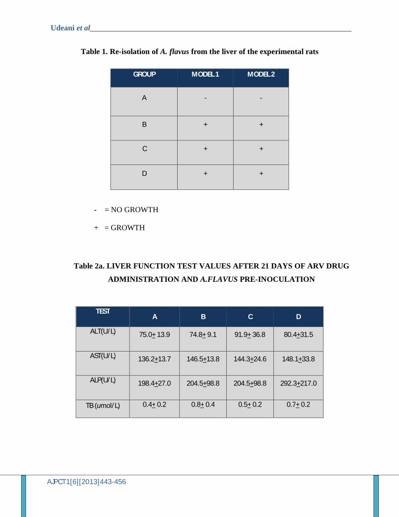
PLATE 1A: H & E PHOTOMICROGRAPH OF GROUP B ADMINISTERED with NEVIRAPINE ONLY SHOWING INFLAMATORY CELLS AROUND SINUSOIDAL
SPACES AND SPOTTY NECROSIS
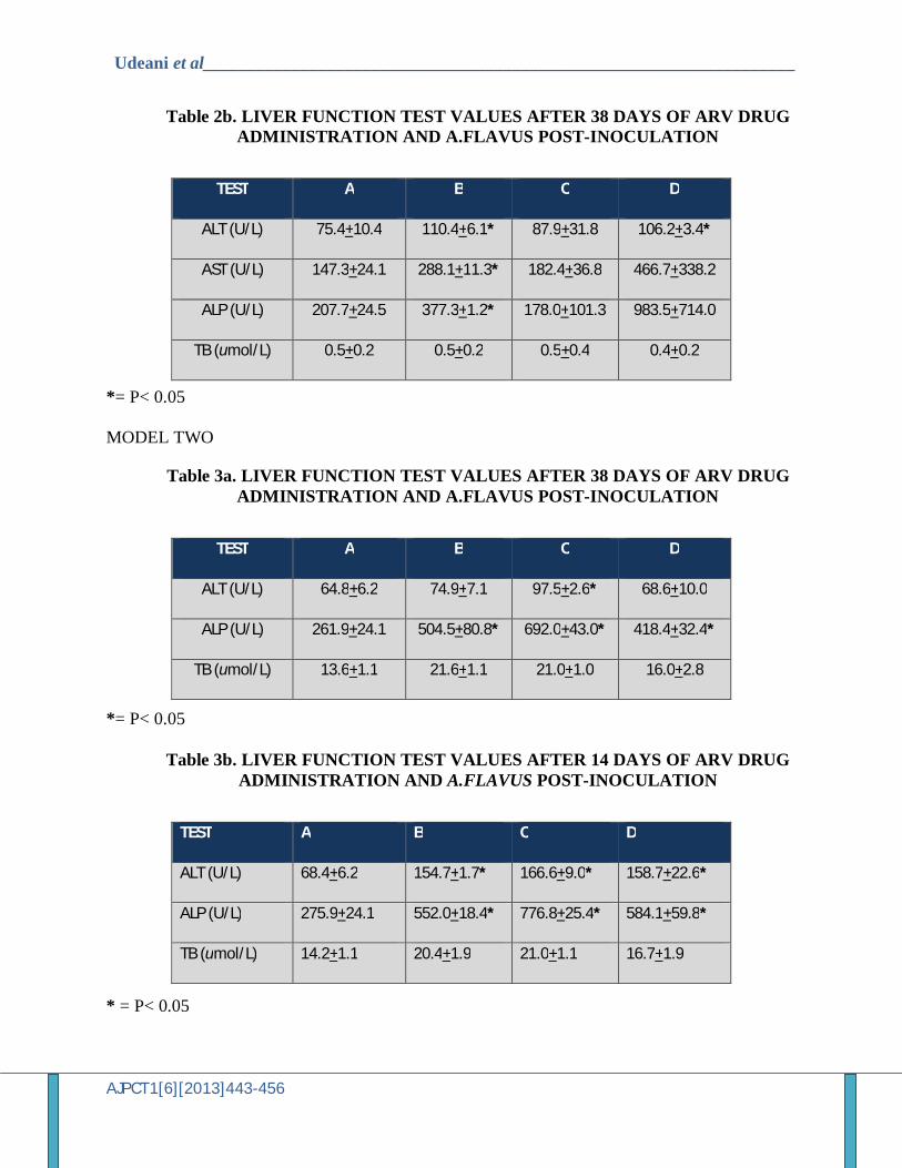
PLATE 1B: H & E PHOTOMICROGRAPH OF GROUP B ADMINISTERED NEVIRAPINE AND A. FLAVUS SHOWING FIBROSIS AROUND PERIPORTAL REGIONS, NECROTIC HEPATOCYTES AND INFLAMATORY CELLS AROUND
PORTAL TRIAD AND WITHIN SINUSOISDAL SPACES
Udeani et al___________________________________________________________________
AJPCT1[6][2013]443-456
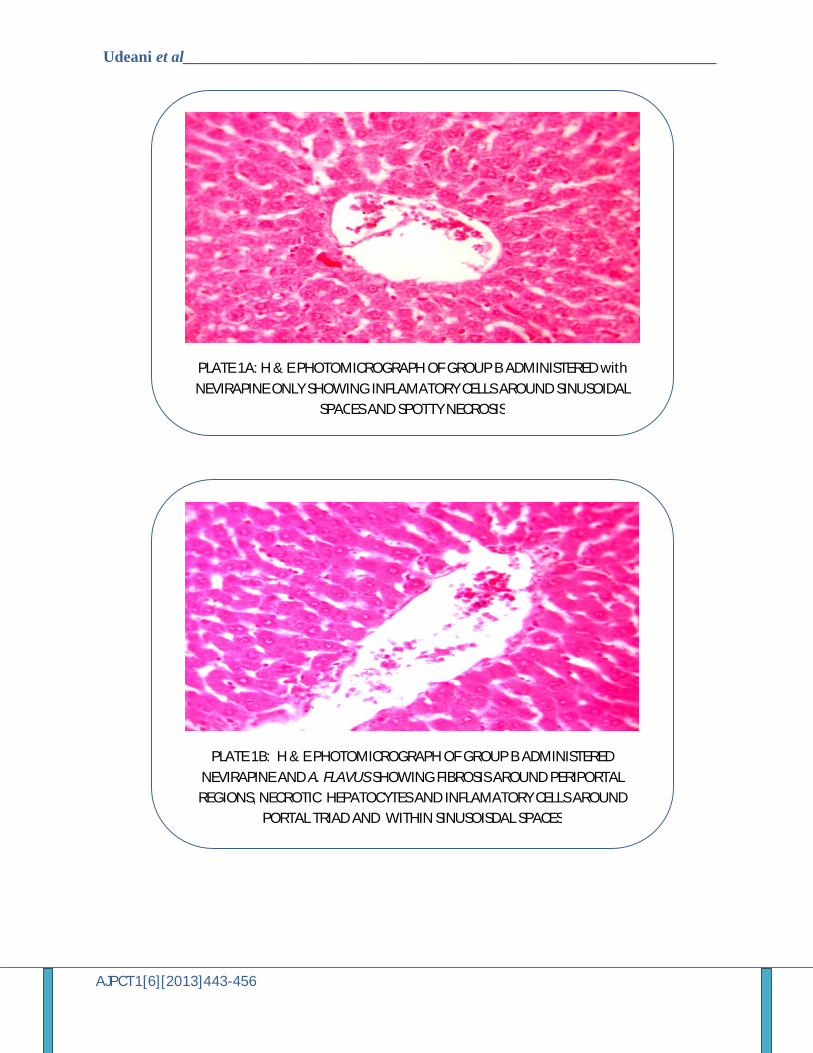
PLATE 2A: H & E PHOTOMICROGRAPH OF GROUP C ADMINISTERED ZIDOVUDINE AND LAMIVUDINE SHOWING FIBROSIS AROUND VESSELS AND
FRANK RED CELLS WITHIN THE CENTRAL CANAL
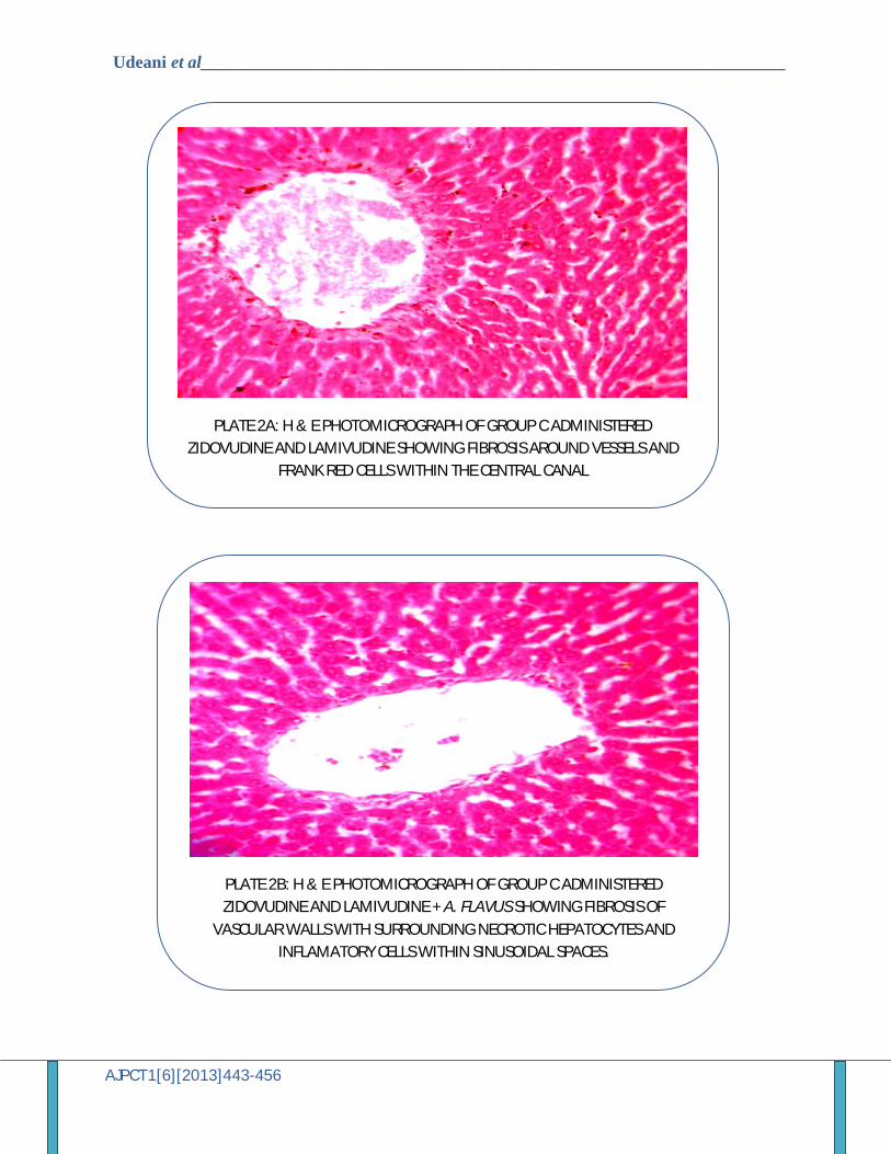
PLATE 2B: H & E PHOTOMICROGRAPH OF GROUP C ADMINISTERED ZIDOVUDINE AND LAMIVUDINE + A. FLAVUS SHOWING FIBROSIS OF
VASCULAR WALLS WITH SURROUNDING NECROTIC HEPATOCYTES AND INFLAMATORY CELLS WITHIN SINUSOIDAL SPACES.
Udeani et al___________________________________________________________________
AJPCT1[6][2013]443-456
PLATE 3A: H & E PHOTOMICROGRAPH OF GROUP D ADMINISTERED HAART SHOWING WIDENED SINUSOIDAL SPACES.
PLATE 3B: H & E PHOTOMICROGRAPH OF GROUP D ADMINISTERED WITH ARV AND A. FLAVUS SHOWING DILATED VESSELS, FIBROSIS AROUND PORTAL
TRIAD, NECROTIC HEPATOCYTES AND INFLAMATORY CELLS.
Udeani et al___________________________________________________________________
AJPCT1[6][2013]443-456
MODEL TWO
PLATE 4A: H & E PHOTOMICROGRAPH OF GROUP B ADMINISTERED ARV + A.FLAVUS SHOWING FIBROUS TISSUE SEPERATING AGGREGATION OF
INFLAMMATORY CELLS AND DEGENERATED HEPATOCYTES.
PLATE 4B: H & E PHOTOMICROGRAPH OF GROUP C ADMINISTERED HAART + CYCLOPHOPHAMIDE + A.FLAVUS SHOWING DEGENERATED HEPATOCYTES,
PANLOBULAR INFLAMMATORY CELLS AT PERIPORTAL REGIONS, SPOTTY NECROSIS AND BINUCLEATED CELLS.
Udeani et al___________________________________________________________________
AJPCT1[6][2013]443-456
PLATE 4C: H & E PHOTOMICROGRAPH OF GROUP D ADMINISTERED CYCLOPHOSPHAMIDE SHOWING MILD BALOONED HEPATOCYTES,
INFLAMMATORY CELLS AND BINUCLEATED CELLS.
PLATE 4D: The liver of rats showing normal liver hepatocytes