Embed Size (px)
DESCRIPTION
Revision Notes for Edexcel A2 BiologyFeel free to use this for your revision. Good luck with your exams.
Citation preview

Edexcel Biology Notes Produced by Tim Filtness
1
A2A2A2A2 Biology Revision NotesBiology Revision NotesBiology Revision NotesBiology Revision Notes
“We are what we repeatedly do. Excellence,
therefore, is not an act but a habit.” - Aristotle
Merchant Taylors’ School

Edexcel Biology Notes Produced by Tim Filtness
2
Understanding the jargon:
1. The 9 Core Practicals are not discussed here. Don’t forget to revise them too!
2. All Key Words are given underlined in red, these are words specifically mentioned on the syllabus!
3. There are many blue “How Science Works” boxes in the text book. In past years these have almost always been the basis of a number of exam questions...
A word of caution
These revision notes are designed to help you, NOT do the job of revision for you. Ultimately, only you can learn this material: you can’t pay, cajole or persuade anyone to do it for you! Additionally, these notes are the bare bones (your text book and class notes are almost certainly better sources of information if you’re aiming for the highest grades). So treat these notes as a minimalist approach for someone aiming for a solid B grade. At this point you might want to get your own notes to cross-reference with the material here. Why not add your own annotations to improve what’s already here?

Edexcel Biology Notes Produced by Tim Filtness
3
Unit 4Unit 4Unit 4Unit 4: : : : The Natural EnvironmentThe Natural EnvironmentThe Natural EnvironmentThe Natural Environment & & & & Species SurvivalSpecies SurvivalSpecies SurvivalSpecies Survival
Topic 5: On the Wild Side 4.5.2. The key point is that the Grana are covered in photosystems, which absorb light. It is important, therefore, that the grana have as big a surface area as possible. 4.5.3.
C6H12O6
The energy required to combine These elements comes from ATP
Edexcel A2 Revision Notes
Stroma: site of light-independent step of p/s Grana: site of light-dependent step of p/s Envelope: (made from two layers of membrane), contains ATP Synthase enzymes
Oxygen comes from CO2 (remember, the O from photolysis is excreted!) C comes from CO2
H comes from reduced NADP (and, therefore, ultimately from water).
Edexcel Biology Notes Produced by Tim Filtness
4
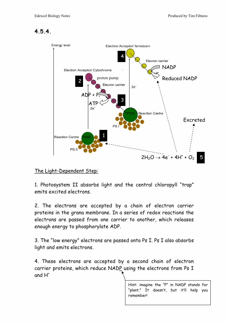
4.5.4. The Light-Dependent Step: 1. Photosystem II absorbs light and the central chloropyll “trap” emits excited electrons. 2. The electrons are accepted by a chain of electron carrier proteins in the grana membrane. In a series of redox reactions the electrons are passed from one carrier to another, which releases enough energy to phosphorylate ADP. 3. The “low energy” electrons are passed onto Ps I. Ps I also absorbs light and emits electrons. 4. These electrons are accepted by a second chain of electron carrier proteins, which reduce NADP using the electrons from Ps I and H+
ADP + Pi
ATP
NADP
Reduced NADP
2H2O → 4e- + 4H+ + O2
Excreted
1
2
3
4
5
Hint: imagine the “P” in NADP stands for “plant.” It doesn’t, but it’ll help you remember!
Edexcel Biology Notes Produced by Tim Filtness
5
5. As the process continues H+ would start to run low and Ps II would begin to run out of electrons. So, using enzymes in the grana, water is split apart (photolysis), producing H+, more electrons and O2, which is excreted. Overall: the entire point of the step is to generate; ATP – energy source for fixing CO2 Reduced NADP – source of H+ and electrons for making glucose 4.5.5.
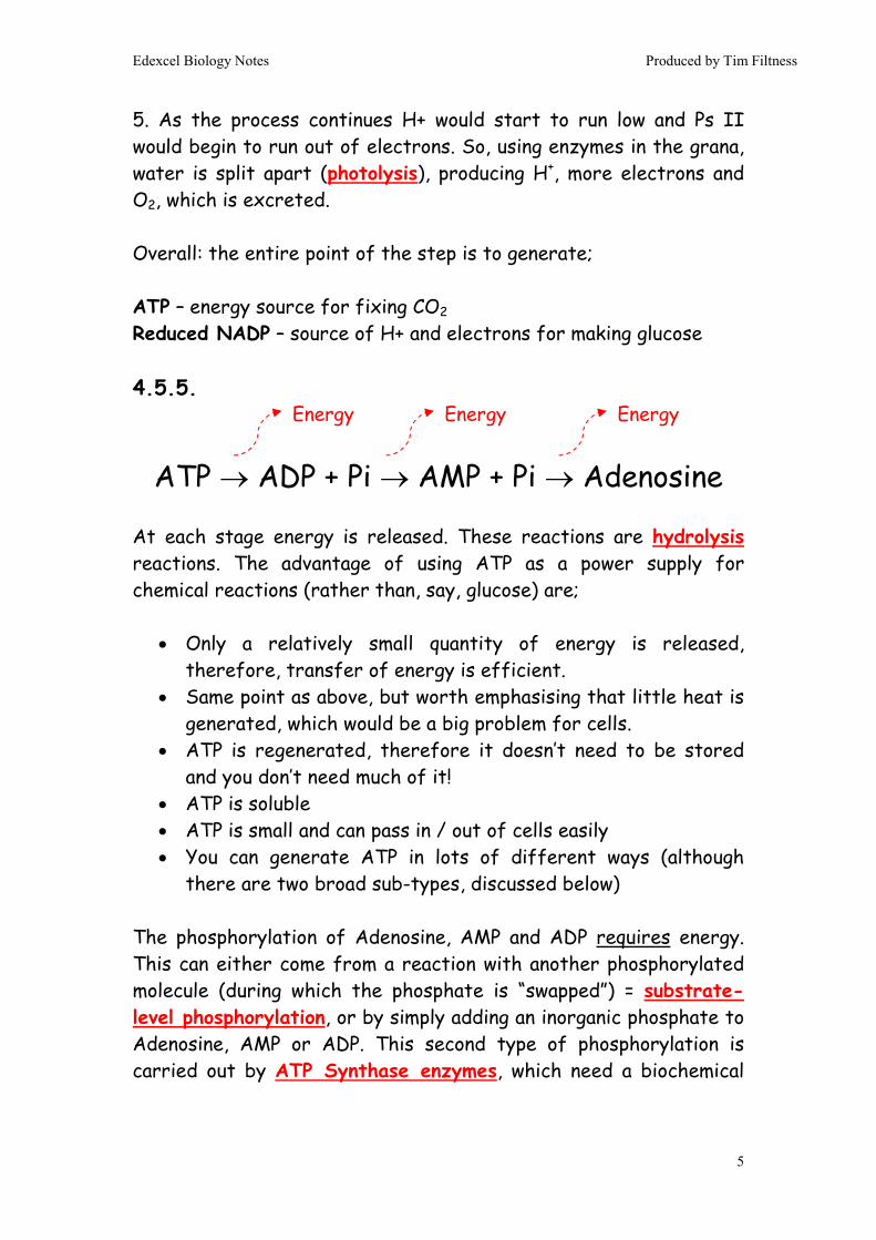
ATP → ADP + Pi → AMP + Pi → Adenosine At each stage energy is released. These reactions are hydrolysis reactions. The advantage of using ATP as a power supply for chemical reactions (rather than, say, glucose) are;
• Only a relatively small quantity of energy is released, therefore, transfer of energy is efficient.
• Same point as above, but worth emphasising that little heat is generated, which would be a big problem for cells.
• ATP is regenerated, therefore it doesn’t need to be stored and you don’t need much of it!
• ATP is soluble • ATP is small and can pass in / out of cells easily • You can generate ATP in lots of different ways (although
there are two broad sub-types, discussed below) The phosphorylation of Adenosine, AMP and ADP requires energy. This can either come from a reaction with another phosphorylated molecule (during which the phosphate is “swapped”) = substrate-level phosphorylation, or by simply adding an inorganic phosphate to Adenosine, AMP or ADP. This second type of phosphorylation is carried out by ATP Synthase enzymes, which need a biochemical
Energy Energy Energy

Edexcel Biology Notes Produced by Tim Filtness
6
gradient of H+ (therefore, a source of potential energy) to work (see 5.7.10). 4.5.6.
Light-Independent Step (or Calvin Cycle)
1. Carboxylation Step (reaction with CO2) – CO2 is “fixed” by RuBP (5C) into 2 x GP (3C). This step is catalysed by the enzyme RUBISCO.
2. Reduction Step – Reduced NADP is oxidised (therefore, GP is reduced, hence the step’s name!) forming 2 x GALP (3C). ATP is also required.
3. Regeneration Step – 5 out of 6 GALP molecules are converted
back into RuBP, which requires energy.
4. 1 out of 6 GALP molecules are turned into Glucose (6C). Therefore, on average a glucose molecule is generated every 6 turns of the cycle.
RuBP GP
CO2
GALP
Glucose
ADP + P
NADP
ATP
NADPH
ATP
ADP + P
1
2
3
4

Edexcel Biology Notes Produced by Tim Filtness
7
Fates of glucose;
• Used in respiration (to make ATP when it’s dark) • Used to make cellulose • Used in conjunction with nitrate to make amino acids
(remember, plants don’t eat like we do – they have to make everything themselves!)
• Used to make nucleotides • Converted into triglycerides and phospholipids
4.5.7.
GPP = NPP + R Hint: do remember that GPP doesn’t include energy that’s lost before p/s occurs i.e. light that is reflected by the leaf, or absorbed as heat energy.
Net Primary Productivity is the balance of glucose a plant has left over to use for growth. NPP is proportional to biomass
Gross Primary Productivity is the amount of glucose a plant generates through p/s.
Respiration is the amount of glucose a plant uses in respiration.
Think of it in terms of a job. You get paid a GROSS salary and you’re reasonably happy with it. However, I come along and take a massive slice of this in TAX (ha ha!), so what you’ve actually got left to spend – your NET salary – is considerably smaller. Unlucky, sucks to be you.

Edexcel Biology Notes Produced by Tim Filtness
8
4.5.8. Energy is lost between trophic levels of a food chain. It is lost in the following ways; 4.5.9.
End of Topic 5End of Topic 5End of Topic 5End of Topic 5
Can you work out the labels for yourself? (I really hope you can, it’s very easy & this is an A2 revision guide after all)
Sun & Producer
• Lost in reflected light • Lost in light energy of a λ
the plant can’t absorb (i.e. green light)
• Passes through the leaf • Lost in heat • Lost in respiration (that’s
where GPP & NPP come in) Overall, only ~1% of solar energy that hits a plant is converted into biomass
Producer & Consumer
• Lost in undigested material • Lost in excreted molecules • Lost in heat • Lost in movement
Overall, only ~10% of energy a consumer eats winds up as new biomass in the consumer. The same points are also valid for consumers eating other consumers.

Edexcel Biology Notes Produced by Tim Filtness
9
You can make the Carbon Cycle hugely complicated, but it’s basically a balance between photosynthesis and respiration. You are supposed to understand how we can use the carbon cycle to come up with strategies for lowering atmospheric PCO2.
1. Reforestation schemes. More trees = more p/s. Therefore, more CO2 “fixed” in the bodies of trees.
2. Use biofuels instead of fossil fuels. Common biofuels are plant oils (e.g. palm oil) and ethanol from fermentation.
Arguably neither of these suggestions actually lead to a permanent decrease in PCO2! Reforestation does temporarily, but eventually the trees will die and decompose, which releases the carbon back into the atmosphere again. Equally, whilst biofuels stop PCO2 climbing higher by slowing our consumption of fossil fuels, the biofuels themselves are, at best, only carbon neutral (also problems with where do you grow the biofuel crop – on farm land? cut down some more rainforest?) The only way to lower PCO2 permanently is to regenerate fossil fuels. That’s hard! The only viable strategies suggested so far involve growing forests, felling them when fully grown and then burying the timber in environments where it won’t decompose (i.e. in a bog or the bottom of the deep sea). Neither scheme is practical or cost effective. 4.5.10. & 4.5.12. Ecosystem: the community in a habitat and the combined biotic & abiotic factors of the habitat. Habitat: the part of an environment where an organism lives Niche: the specific part of a habitat in which a species lives and the adaptations of that species that allow it to survive there

Edexcel Biology Notes Produced by Tim Filtness
10
Biotic Factor: a living factor within a habitat (e.g. intraspecific & interspecific competition, presence of predators and prey) Abiotic factor: a non-living factor within a habitat (e.g. light intensity, heat) Edaphic factor: a factor of the soil. Population: the total number of individuals of one species. Community: the populations of all the species living in a habitat. The numbers and distribution of organisms in a habitat are determined by the abiotic and biotic factors in that environment. If an environmental gradient exists (e.g. sand dune, rocky sea shore) the distribution will occur in zones within the habitat = zonation
Abiotic Factors include; dessication, salinity, wave action, temperature, water availability, substrate, aspect, pH etc
Biotic Factors include; interspecific competition, intraspecific competition, predation, food availability, presence of excreted wastes
An example of a Named Environment Is the British Rocky Seashore
Abiotic Factors have more affect going up the beach
Biotic Factors have more affect going down the beach
Edexcel Biology Notes Produced by Tim Filtness
11
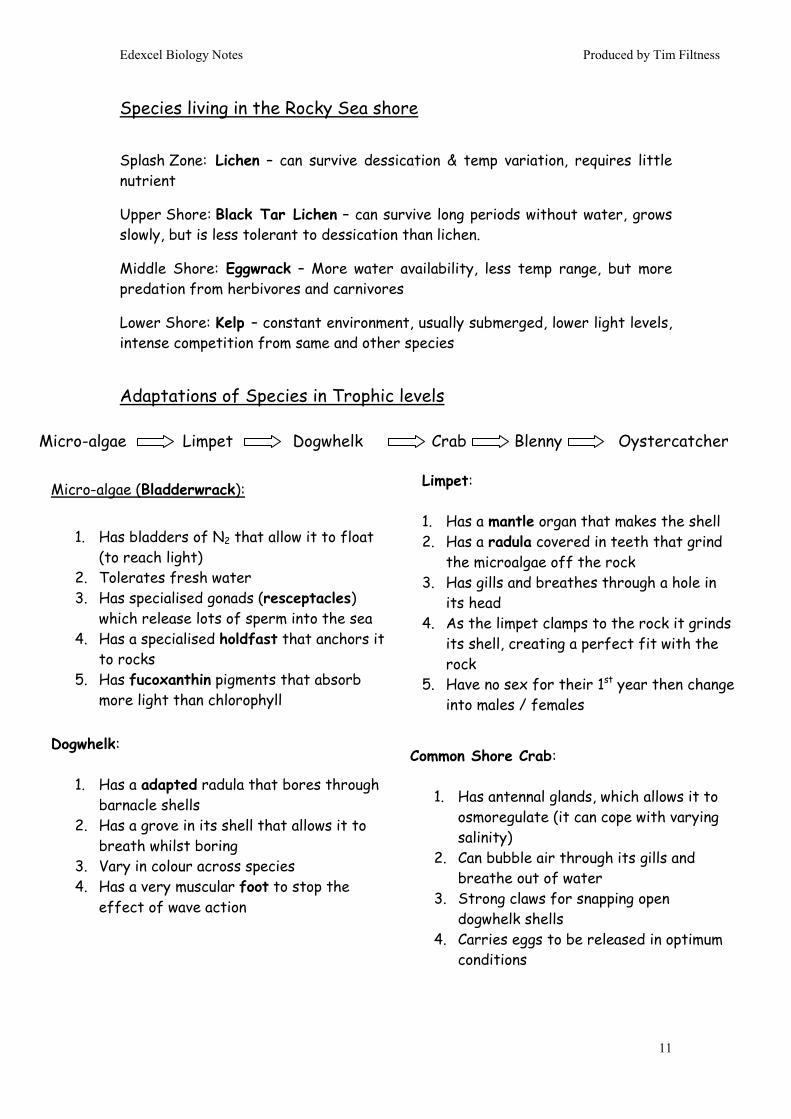
Species living in the Rocky Sea shore
Splash Zone: Lichen – can survive dessication & temp variation, requires little nutrient
Upper Shore: Black Tar Lichen – can survive long periods without water, grows slowly, but is less tolerant to dessication than lichen.
Middle Shore: Eggwrack – More water availability, less temp range, but more predation from herbivores and carnivores
Lower Shore: Kelp – constant environment, usually submerged, lower light levels, intense competition from same and other species
Adaptations of Species in Trophic levels
Micro-algae (Bladderwrack):
1. Has bladders of N2 that allow it to float
(to reach light) 2. Tolerates fresh water 3. Has specialised gonads (resceptacles)
which release lots of sperm into the sea 4. Has a specialised holdfast that anchors it
to rocks 5. Has fucoxanthin pigments that absorb
more light than chlorophyll
Micro-algae Limpet Dogwhelk Crab Blenny Oystercatcher
Limpet: 1. Has a mantle organ that makes the shell 2. Has a radula covered in teeth that grind
the microalgae off the rock 3. Has gills and breathes through a hole in
its head 4. As the limpet clamps to the rock it grinds
its shell, creating a perfect fit with the rock
5. Have no sex for their 1st year then change into males / females
Dogwhelk:
1. Has a adapted radula that bores through barnacle shells
2. Has a grove in its shell that allows it to breath whilst boring
3. Vary in colour across species 4. Has a very muscular foot to stop the
effect of wave action
Common Shore Crab:
1. Has antennal glands, which allows it to osmoregulate (it can cope with varying salinity)
2. Can bubble air through its gills and breathe out of water
3. Strong claws for snapping open dogwhelk shells
4. Carries eggs to be released in optimum conditions
Edexcel Biology Notes Produced by Tim Filtness
12
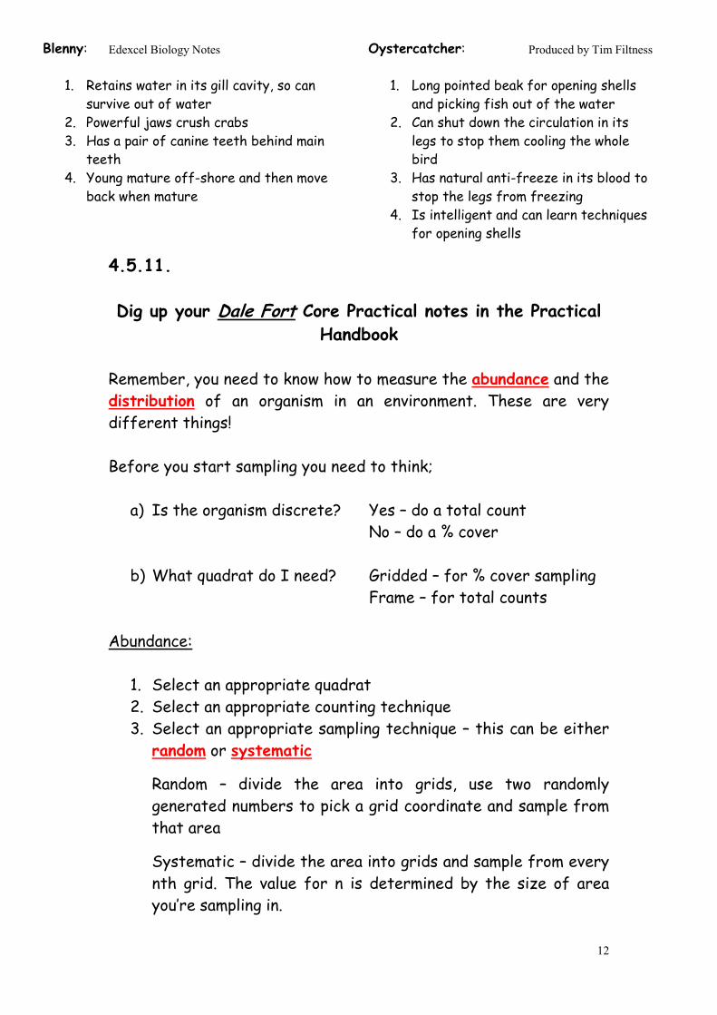
4.5.11. Dig up your Dale Fort Core Practical notes in the Practical
Handbook Remember, you need to know how to measure the abundance and the distribution of an organism in an environment. These are very different things! Before you start sampling you need to think;
a) Is the organism discrete? Yes – do a total count No – do a % cover
b) What quadrat do I need? Gridded – for % cover sampling Frame – for total counts
Abundance:
1. Select an appropriate quadrat 2. Select an appropriate counting technique 3. Select an appropriate sampling technique – this can be either random or systematic
Random – divide the area into grids, use two randomly generated numbers to pick a grid coordinate and sample from that area
Systematic – divide the area into grids and sample from every nth grid. The value for n is determined by the size of area you’re sampling in.
Blenny:
1. Retains water in its gill cavity, so can survive out of water
2. Powerful jaws crush crabs 3. Has a pair of canine teeth behind main
teeth 4. Young mature off-shore and then move
back when mature
Oystercatcher:
1. Long pointed beak for opening shells and picking fish out of the water
2. Can shut down the circulation in its legs to stop them cooling the whole bird
3. Has natural anti-freeze in its blood to stop the legs from freezing
4. Is intelligent and can learn techniques for opening shells
Edexcel Biology Notes Produced by Tim Filtness
13
4. Repeat and take an average (running mean is a good idea too!) 5. Find the total area of the habitat 6. Divide the total area by the area of your quadrat and multiply
by the average. Distribution: 1. Select an appropriate quadrat 2. Select an appropriate counting technique 3. Lay out a transect line along the environmental gradient 4. Select an appropriate sampling technique – this can be either
continuous or interrupted 5. Repeat and take an average by turning your transect into a
belt 6. Plot your results as a kite diagram. 4.5.13.
Primary succession is the first stage of the ecological succession of plant life from abiotic land with no soil to fully support plant ecosystems (e.g., a forest). In primary succession, pioneer species like mosses and lichen, start to "normalize" the habitat, creating rudimentary soil from their dead matter. These pioneer plants create conditions for the start of plant growth and so more complex plants like grasses and shrubs begin to colonise the area. Over time the plants change the area to make it suitable for other species to move in. The biodiversity of the area slowly increases as does the biomass. Eventually, after a few hundred years, the biodiversity and biomass become constant as no further change takes place. This is the climax community because succession stops at this point.
A good example of primary succession takes place after a volcano has erupted. The barren land is first colonised by simple pioneer plants which pave the way for more complex plants, such as hardwood trees by creating soils and other necessities. Unlike secondary succession, which refers to succession after an

Edexcel Biology Notes Produced by Tim Filtness
14
environmental disaster (such as a forest fire) primary succession occurs on the geologic timescale, over thousands of years. Secondary succession is much faster because the soil is already there and also, in the soil, are seeds!
4.5.14.
Greenhouse Effect:
1. Incoming (short λ) solar light hits Earth 2. Absorbed 3. Energy re-emitted as longer λ radiation 4. Longer λ absorbed by greenhouse gases in atmosphere 5. Reflected & re-emitted back towards Earth
Cumulative effect: energy enters atmosphere and ultimately doesn’t leave, therefore, Earth is getting warmer = global warming.
Greenhouse gas Source
CO2 Respiration & Combustion (particularly of fossil fuels). You could also argue deforestation led to an increase in PCO2 due to a decrease in global p/s rates.
Methane (CH4) Decomposition by saphrophytes (bacteria & fungi mostly) in; paddy fields, land fill sites, guts of ruminants (cows)

Edexcel Biology Notes Produced by Tim Filtness
15
Be aware that NOx, water vapour and CFCs are also greenhouse gases. However, the vast global production of CO2 makes the others relatively insignificant. 4.5.15. Some of the potential effects of global warming; 4.5.16.
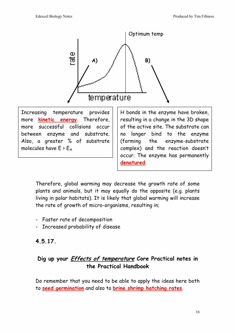
Temperature affects the rate of enzyme controlled reactions.
Rising temperature - causing
• Melting Ice Caps • Sea level rises • Loss of polar ecosystems • Desertification • Costal flooding • Net loss of global arable
land • Polar “migration” of species • Impact on crops • Famine • Disease • Alterations in ♀:♂ ratio in
reptiles & fish • Alterations in insect life
cycles
Changing Weather Cycles - causing
• Change in rainfall patterns • Drought • Flooding • Impact on crops • Impact on housing • Shortage of drinking water
(also impact on farming) Also most of the points on the left as well!
Edexcel Biology Notes Produced by Tim Filtness
16
Therefore, global warming may decrease the growth rate of some plants and animals, but it may equally do the opposite (e.g. plants living in polar habitats). It is likely that global warming will increase the rate of growth of micro-organisms, resulting in; - Faster rate of decomposition - Increased probability of disease 4.5.17.
Dig up your Effects of temperature Core Practical notes in the Practical Handbook
Do remember that you need to be able to apply the ideas here both to seed germination and also to brine shrimp hatching rates.
Increasing temperature provides more kinetic energy. Therefore, more successful collisions occur between enzyme and substrate. Also, a greater % of substrate molecules have E > EA
H bonds in the enzyme have broken, resulting in a change in the 3D shape of the active site. The substrate can no longer bind to the enzyme (forming the enzyme-substrate complex) and the reaction doesn’t occur. The enzyme has permanently denatured
Optimum temp
A) B)

Edexcel Biology Notes Produced by Tim Filtness
17
4.5.18. Evidence for climate change
Evidence Explanation
Direct Measurement
In the UK people first began to record atmospheric temperature directly in 1889. Records show a clear rise in temperature by 0.5 – 1.5°C over that period. However, that’s only 120 years of data.
Dendrochronology
Trees tend to grow more when it’s hotter. Therefore, the thickness of each ring of xylem in a tree trunk is roughly proportional to the temperature of that particular year. As some trees live for over 5000 years we have a year-by-year indirect record of the temperature over that period. Rings are getting thicker ⇒ it’s getting warmer!
Pollen from peat bogs
The shape of the outer exine coat of a pollen grain is species-dependent i.e. you can identify the species that made the pollen by the shape of the pollen grains. Bogs are anoxic environments, so any pollen that falls into a bog won’t decay. The depth of a bog is proportional to age (some bogs are 1000s of years old). This provides a record of the plants that lived around the bog over history. Species that used to live around the bog are now found a lot further north ⇒ it’s got warmer.
CO2 readings from ice cores
Ice cores go back over 50,000 years (therefore the best data record). Glaciers trap microscopic bubbles of atmospheric gas inside their ice, allowing one to measure directly the PCO2 year by year over 50,000 years.
Worth noting the following;
• Coral cores also show the same pattern as dendrochronology (for the same reasons!) except that some reefs are over 10,000 years old, giving a much better record than trees!

Edexcel Biology Notes Produced by Tim Filtness
18
• By looking at the relative ratios of O16 & O18 isotopes in the ice in ice cores, one can estimate global temperature (the cooler it is, the less likely O18 is to evaporate, so the ratio of O18:O16 in the ice is proportional to temp).
Overall the data show a close correlation between PCO2 and global temp. However, this does not necessarily mean that CO2 and global temperature are linked causally (i.e. that one causes the other). It may be that some unidentified 3rd factor causes both? 4.5.19. All predictions of future climate change are based on models. The models give rise to different predictions for climate change. This is a problem because it means our models are unreliable. So, what are the problems with these models?
Limitation How it affects the model
Extrapolation This is where a best fit line is extended to make a prediction. This relies on two factors, 1) that the best fit line is accurate (see next point) and 2) that the trend continues unchanged
Limited data With only a few data points we may well draw our line of best fit in the wrong place. This will mean our extrapolation is wrong.
Some factors not included
We may well not have considered an additional factor in our model that will ALSO affect the Dependent Variable and, therefore, alter the actual outcome from what we had predicted.
Changes in factors
We had assumed all factors in the model were constant. What if one changes?
Edexcel Biology Notes Produced by Tim Filtness
19
4.5.20. All people show bias, even if they are trying not to. This is because the way people interpret results is based on the way they process information, which is in turn based on their training, experience and relative psychology – hence the glass is half-full / half-empty debate. Just remember that a conclusion is always based (at some level) on someone’s interpretation of data, which is subject to bias. 4.5.21 Process of Natural Selection
1. There is variation within a species 2. Not all organisms that are born will survive long enough to
reproduce 3. Natural Selection is the idea that the best adapted (“fitter”)
individuals will be the ones most likely to survive and reproduce.
4. They pass their alleles onto the next generation 5. Over time, the frequency of the “fitter” allele increases
within the population How NS leads to Speciation
1. Two pockets of the same species become isolated (not necessarily geographically!)
2. The habitats within each pocket will be different; therefore, each pocket presents different selective pressures
3. The selective pressures select for different “fitter” phenotypes (and, therefore, alleles) in each pocket
4. NS occurs 5. Over time mutations occur, producing new “even fitter” alleles 6. The populations inbreed / cannot breed with each other 7. Over time the new mutations accumulate within each pocket 8. Eventually, the individuals in the different pockets are so
different that they cannot reproduce with one another to produce viable offspring. This is speciation.

Edexcel Biology Notes Produced by Tim Filtness
20
4.5.22. Types of isolation – remember, it doesn’t have to be geographic! 4.5.23. “New” evidence supporting evolutionary theory has come from;
1. DNA – shows huge similarity of code sequences between similar living organisms. Also, it’s a universal code, so it does exactly the same thing in every living organism.
2. Proteomics – there are lots very similar proteins found in a variety of organisms e.g. all living species share cytochrome proteins, which carry out respiration.
This evidence has been validated by the Scientific Community. This means;
• Scientists publish their findings in a “paper” in a Scientific Journal.
• Each journal paper is checked for validity before publishing by a number of other scientists. They look for obvious flaws in experiment design, statistical errors and bad logic. This is the peer review process.
• Scientists with competing views publish counter-arguments (supported by data / evidence from experiements) in other journals. This establishes a public, chronological record of
Method of isolation Description
Ecological isolation The species occupy different parts of the habitat
Temporal isolation The species exist in the same area, but reproduce at different times
Behavioural isolation The species exist in the same area, but do not respond to each other’s courtship behaviour
Physical incompatibility Species coexist, but there are physical reasons which stop them from copulating

Edexcel Biology Notes Produced by Tim Filtness
21
changes in thinking / theory for the topic. It is an open-access system: anyone can contribute as long as their papers are accepted by the journal.
End of Topic 5End of Topic 5End of Topic 5End of Topic 5

Edexcel Biology Notes Produced by Tim Filtness
22
Unit 4Unit 4Unit 4Unit 4: : : : The Natural EnvironmentThe Natural EnvironmentThe Natural EnvironmentThe Natural Environment & & & & Species SurvivalSpecies SurvivalSpecies SurvivalSpecies Survival
Topic 6: Infection, Immunity & Forensics 4.6.2 Each codon is non-overlapping. Because there are 64 possible codons and only 20 amino acids quite a few amino acids have more than one codon coding for them. Often, the last letter of the codon makes no difference in determining the amino acid (i.e. AAA, AAC, AAG & AAT all code for the amino acid Phenylalanine). This is referred to as the degenerate nature of the genetic code. 4.6.3. Protein Synthesis occurs in two steps
(i) Transcription – in the nucleus (ii) Translation - in the cytoplasm
Edexcel A2 Revision Notes
The genetic code is formed from the sequence of bases along the coding strand of the DNA. We read the genetic code as a series of three bases, which is called a codon.

Edexcel Biology Notes Produced by Tim Filtness
23
Transcription: • Takes place in nucleus • A complementary copy of the gene is made using RNA • Only one side of the DNA is transcribed (the sense strand)
because only this side begins with the start codon (TAC in DNA). The antisense strand isn’t transcribed.
1. A Transcription factor binds to a promoter sequence of DNA
upstream of the gene. 2. Gene opens up. Hydrogen bonds break between bases. 3. RNA nucleotides are attracted to complementary bases and form
hydrogen bonds. 4. RNA nucleotides joined together by enzyme RNA Polymerase. 5. Complementary RNA copy of gene now made. It is called mRNA
(messenger RNA) 6. Single stranded mRNA molecule diffuses out of gene 7. mRNA molecule leaves nucleus through nuclear pore (large holes
in nuclear envelope) 8. Many mRNA strands are made before gene closes. MRNA is complementary, not a copy! Translation: • Takes place in cytoplasm • mRNA code read by ribosome and amino acids are assembled in
correct order to make protein 1. mRNA strand binds to cleft in ribosome. Start AUG codon fits
into bottom of ribosomal P site 2. tRNA diffuses into P site and recognises the mRNA codon using
its specific anticodon 3. A second tRNA diffuses into the A site and recognises the
mRNA codon there. 4. The amino acids between the two tRNAs join together forming a
peptide bond
Edexcel Biology Notes Produced by Tim Filtness
24
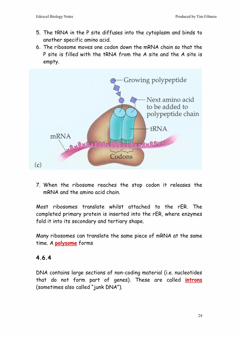
5. The tRNA in the P site diffuses into the cytoplasm and binds to another specific amino acid.
6. The ribosome moves one codon down the mRNA chain so that the P site is filled with the tRNA from the A site and the A site is empty.
7. When the ribosome reaches the stop codon it releases the mRNA and the amino acid chain.
Most ribosomes translate whilst attached to the rER. The completed primary protein is inserted into the rER, where enzymes fold it into its secondary and tertiary shape. Many ribosomes can translate the same piece of mRNA at the same time. A polysome forms 4.6.4 DNA contains large sections of non-coding material (i.e. nucleotides that do not form part of genes). These are called introns (sometimes also called “junk DNA”).

Edexcel Biology Notes Produced by Tim Filtness
25
After DNA has been transcribed the introns are snipped out of the mRNA by enzymes called spliceosomes. These spliceosome enzymes sometimes also change the mRNA code slightly, so it is possible to have many subtly different proteins produced from just one gene. 4.6.5. DNA Profiling (also called Genetic Fingerprinting) is used to;
- Determine identity - Determine genetic relationships between species / people
Process of DNA Profiling:
1. Copy a sample of DNA using PCR. 2. Cut the DNA sample using a Restriction Endonuclease. 3. Run the cut sample of DNA through an Electrophoresis gel. 4. Compare the pattern of DNA bands in the gel (the
“fingerprint” with a known sample to determine similarity / identity.
This process works because, although people have incredibly similar genetic sequences in their genes, their introns are often very different. People have lots of different repeating short sequences

Edexcel Biology Notes Produced by Tim Filtness
26
of code (satellites) in their introns. One person may have 10 – 100 repeats for a particular satellite, whereas another person may have 1000 – 10,000 repeats for that satellite. Across different satellites, the relative differences in numbers of repeats will produce very different banding patterns on the electrophoresis gel and, therefore, a different genetic fingerprint! 4.6.6.
Dig up your PCR Core Practical notes in the Practical Handbook
Polymerase Chain Reaction:
1. Heat sample of DNA to 95°C - H bonds break & it becomes single-stranded.
2. Add free nucleotides – they will H bond with complimentary partners in DNA (step 4)
3. Add RNA Primers – short lengths of single-stranded RNA that bind to the DNA and stop it annealing (going back together) when cooled (step 4)
4. Cool to 37°C – allows H bonds to form between nucleotides 5. Add Taq Polymerase – a DNA Polymerase enzyme from
Thermus aquaticus (a bacterium that lives in hot springs). This sticks the free nucleotides together forming the sugar-phosphate backbone.
6. Heat to 72°C – optimum temp for Taq. We use this enzyme because, next time the cycle repeats, it won’t denature at 95°C.
Number of DNA copies = 2n
n = number of PCR Cycles

Edexcel Biology Notes Produced by Tim Filtness
27
4.6.7.
Dig up your Electrophoresis Core Practical notes in the Practical Handbook
4.6.8. A Prokaryote:
Ribosomes - Same function as eukaryotic cells (protein synthesis), but are smaller (70s rather than 80s).

Edexcel Biology Notes Produced by Tim Filtness
28
Nuclear Zone - The region of the cytoplasm that contains DNA. There is no nuclear membrane. DNA - Circular, and not in chromosome form (i.e. not super-coiled onto histones). Plasmid - Very small circles of DNA, containing non-essential genes. Can be exchanged between different bacterial cells. Cell membrane - Made of phospholipids and proteins, like eukaryotic membranes. Mesosome - Tightly-folded region of the cell membrane containing all the proteins required for respiration and / or photosynthesis. Cell Wall - DIFFERENT from plant cell wall. Made of murein (a protein). Capsule (or Slime Layer) - Thick polysaccharide layer outside of the cell wall. Used for:
1. Sticking cells together 2. As a food reserve 3. As protection against desiccation (drying out) and chemicals 4. Protection against phagocytosis (c.f. TB here).
Flagellum - A rotating tail used for propulsion. HIV (a virus):

Edexcel Biology Notes Produced by Tim Filtness
29
Lipid bilayer – allows viral entry into host cell by endocytosis GP120 Ligands – attach to CD4 receptors on Helper T Cells and facilitate endocytosis Capsid – protein coat, protects the RNA and allows virus to assemble inside host cells RNA – contains viral genes Reverse transcriptase – an enzyme that makes a cDNA copy of the viral RNA, which is then inserted into the host’s DNA. 4.6.9.
See 4.5.9. – it’s exactly the same point. 4.6.10.
The major barrier defence mechanisms are:
- Skin - Epithelial lining of respiratory system
Invasion by pathogens
Phagocytosis Commensual organisms
Interferons Lysozyme Stomach acid and proteolytic enzymes
Cell-mediated immunity
Antibody-mediated immunity
Mucus lining of respiratory tract
Skin
Barrier mechanisms
Non-specific defence mechanisms
Specific defence mechanisms
Other non-specific defence mechanisms

Edexcel Biology Notes Produced by Tim Filtness
30
Skin: Skin is made in two layers;
a) Outer Dermis – dead, compacted cells filled with indigestible & insoluble keratin protein
b) Inner Epidermis – site of rapid mitosis & “living” part of skin. Contains all blood vessels, glands etc.
Barrier adaptations include;
- Keratin - Sebum secreted from sebaceous glands creates acidic pH on
skin (therefore harsh environment for pathogenic bacteria). It also contains lysozyme enzyme.
- Presence of normal flora (also called commensual organisms), which are well adapted to life on the dermis and out-compete pathogenic bacteria.
Epithelium: Barrier adaptations include;
- Mucus & cilia trap bacteria and “waft” to throat for swallowing.
- Lysozyme enzyme is secreted. - Presence of normal flora

Edexcel Biology Notes Produced by Tim Filtness
31
4.6.11. Tuberculosis (TB) is caused by the bacterium Mycobacterium tubercolusis. Key Points:
• TB is transmitted by droplet infection (i.e. a person infected with TB coughs, talks or sneezes and droplets of water and mucus are released into the air from the lungs. These droplets contain the TB bacteria. The droplets are inhaled by a second person, who is then infected with the disease.)
• TB affects the lungs predominantly, but can spread to other
parts of the body e.g. lymph (causing scrofula) or the blood (causing sepsis).
• TB has a thick waxy cell wall, which stops it from dessicating. It can, therefore, survive as dust from dried droplets for weeks.
• TB can survive inside macrophages (cell wall of the bacterium is very thick and waxy and is resistant to the macrophage enzymes). The bacterium reproduces inside the macrophage for many years without causing infection. When the immune system is weakened (by stress, malnutrition, or another disease – HIV is a common cause) the TB bacterium breaks out and re-infects the body. This is a secondary infection NOT a true re-infection.
• TB is characterised by fever, cough, blood in sputum, weight loss (it used to be known as “consumption” for this reason). Also, the presence of granulomas in a lung x-ray, which is often how TB is first diagnosed.
It’s also worth knowing about the BCG, which is the TB vaccine. The vaccine contains live M. bovis, which has very similar antigens to M. tuberculosis, but isn’t anything like as nasty!

Edexcel Biology Notes Produced by Tim Filtness
32
HIV Infection is caused by the Human Immunodeficiency Virus. Key Points:
• HIV does not survive well outside of the body and can only be spread by direct contact i.e. through sexual intercourse, blood-to blood transfer (tattoos, needle sharing, piercing & cut-to-cut transfer).
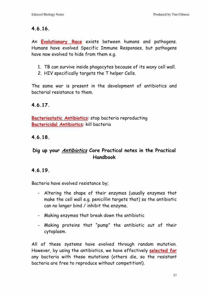
• HIV has specific ligands (called GP120 proteins), which attach to receptors (called CD4 receptors) on the membrane T Helper Cells.
• HIV infection occurs in three stages;
1. The acute phase. HIV virus rapidly infects Helper T cells. The virus population increases quickly & the population of Helper T cells falls rapidly. This phase ends when the immune system begins to respond to the HIV. Killer T cells begin to recognise infected Helper T cells and kill them, which slows the replication of the virus (viral population plateaus). Also, B cells begin to make HIV-specific antibody. The presence of this antibody in the blood can be easily tested for, which is where the term “HIV positive” comes from.
2. The chronic phase. This can last for many years. The virus
continues to replicate, but the Killer T cells keep the numbers in check. New Helper T cells are made continually, but their population stays low as they are continually infected by HIV and then destroyed by Killer T Cells. This fine balance point is affected by the person’s overall health, diet and opportunistic infections.
3. The disease phase. As the numbers of virus increase and the numbers of Helper T cell fall the immune system becomes weaker and weaker. Eventually a second pathogen will infect the person (an opportunistic infection) which cannot be fought

Edexcel Biology Notes Produced by Tim Filtness
33
off. The person will die quickly from the secondary infection. This is the AIDS disease state.
4.6.12. Non-Specific, Non-Barrier Immune Responses: Inflammation: damaged tissue (and special Mast Cells) release histamine into the blood. This causes localised vasodilation, which increases blood flow to the infected area. The increased blood flow brings phagoctyes to the site of infection (which is the desired outcome), but it also increases the rate at which tissue fluid accumulates, resulting in swelling. Lysozyme: a protease enzyme that breaks down bacterial cell walls. Found inside lysosome organelles in phagocytes, also secreted by skin, epithelial cells, lachrymal glands in eyes. Phagocytes (also known as macrophages): Type of WBC that travels in the bloodstream (as opposed to T & B cells, which are usually found in the lymph – therefore called lymphocytes). Phagocytes have antibody receptors on their cell membrane. Every time they meet an antibody stuck to a pathogen they engulf and destroy that pathogen using lysosomes in their cytoplasm. Interferon: a hormone made by all types of WBC. Interferon has lots of functions, but the main one is to block RNA synthesis, which stops viral replication and can play a role suppressing tumour growth. 4.6.13. & 4.6.14. People find the Specific Immune Response difficult. It isn’t! The thing that makes it appear hard is that, unlike other biological processes, it doesn’t have one single specific starting point, but many. This is actually a good thing, because it means that, whatever happens to you, the immune system can be activated quickly and easily.
Edexcel Biology Notes Produced by Tim Filtness
34
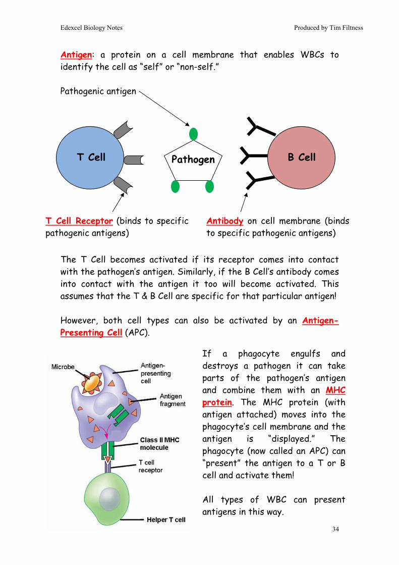
Antigen: a protein on a cell membrane that enables WBCs to identify the cell as “self” or “non-self.” Pathogenic antigen The T Cell becomes activated if its receptor comes into contact with the pathogen’s antigen. Similarly, if the B Cell’s antibody comes into contact with the antigen it too will become activated. This assumes that the T & B Cell are specific for that particular antigen! However, both cell types can also be activated by an Antigen-Presenting Cell (APC).
T Cell B Cell Pathogen
T Cell Receptor (binds to specific pathogenic antigens)
Antibody on cell membrane (binds to specific pathogenic antigens)
If a phagocyte engulfs and destroys a pathogen it can take parts of the pathogen’s antigen and combine them with an MHC protein. The MHC protein (with antigen attached) moves into the phagocyte’s cell membrane and the antigen is “displayed.” The phagocyte (now called an APC) can “present” the antigen to a T or B cell and activate them! All types of WBC can present antigens in this way.
Edexcel Biology Notes Produced by Tim Filtness
35
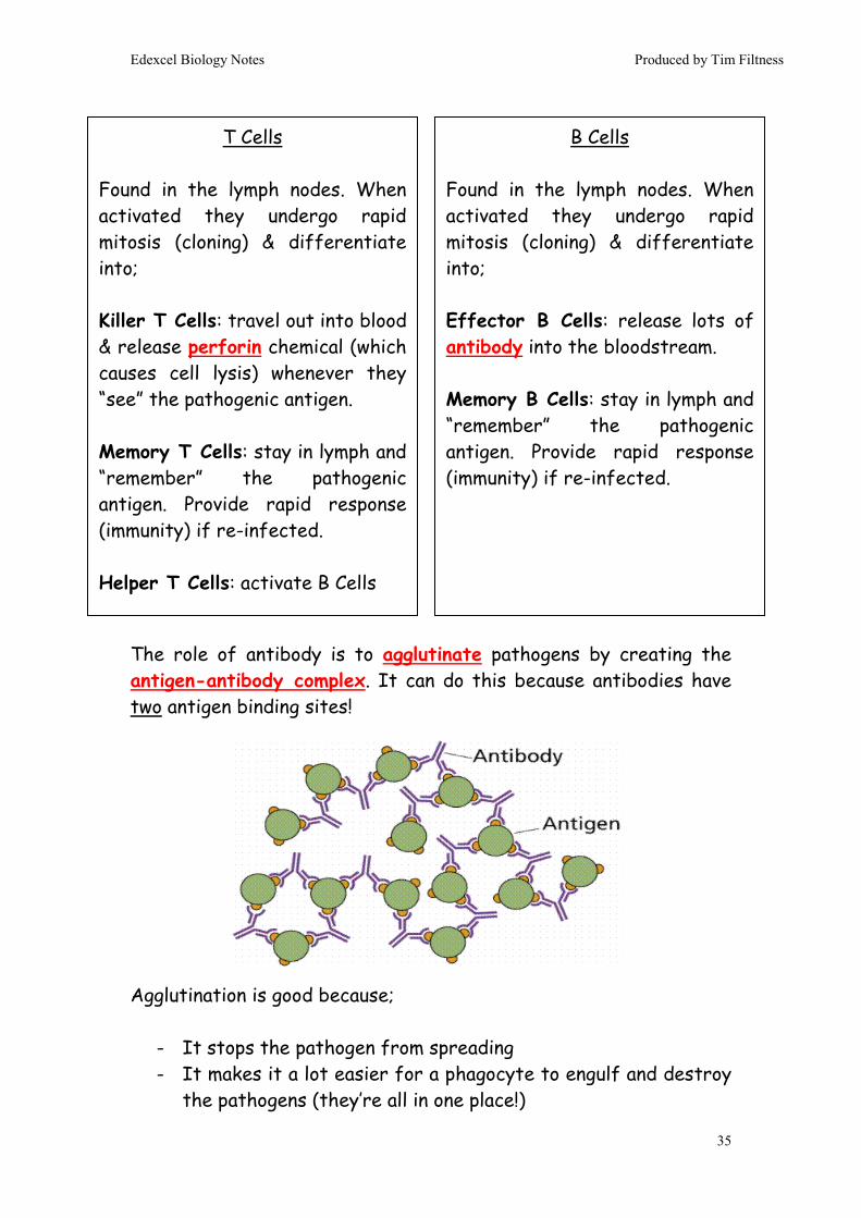
The role of antibody is to agglutinate pathogens by creating the antigen-antibody complex. It can do this because antibodies have two antigen binding sites! Agglutination is good because;
- It stops the pathogen from spreading - It makes it a lot easier for a phagocyte to engulf and destroy
the pathogens (they’re all in one place!)
T Cells Found in the lymph nodes. When activated they undergo rapid mitosis (cloning) & differentiate into; Killer T Cells: travel out into blood & release perforin chemical (which causes cell lysis) whenever they “see” the pathogenic antigen. Memory T Cells: stay in lymph and “remember” the pathogenic antigen. Provide rapid response (immunity) if re-infected. Helper T Cells: activate B Cells
B Cells Found in the lymph nodes. When activated they undergo rapid mitosis (cloning) & differentiate into; Effector B Cells: release lots of antibody into the bloodstream. Memory B Cells: stay in lymph and “remember” the pathogenic antigen. Provide rapid response (immunity) if re-infected.

Edexcel Biology Notes Produced by Tim Filtness
36
4.6.15. Primary Response: no Memory Cells. Antibody production is slow and produced in small quantities (graph doesn’t show this, but there is also often a lag in production). Become ill. Secondary Response: do have Memory Cells so antibody production is rapid and large quantities are made. Don’t become ill (immune).
Immunity Explanation Passive Artificial Antibody injection. (note, doesn’t lead to permanent
immunity) Passive Natural Antibody transferred via breast milk Active Artificial Vaccination Active Natural You come into contact with the pathogen for the
first time. You don’t have Memory Cells, so cannot launch an immediate Specific Immune Response. You make small numbers of T and B Cells with a variety of different shaped receptors / antibody until one of them happens to bind to the pathogenic antigen. When that happens you are able to launch a Specific Immune Response. However, it can take days before this happens, during which you become ill!
Edexcel Biology Notes Produced by Tim Filtness
37
4.6.16. An Evolutionary Race exists between humans and pathogens. Humans have evolved Specific Immune Responses, but pathogens have now evolved to hide from them e.g.
1. TB can survive inside phagocytes because of its waxy cell wall. 2. HIV specifically targets the T helper Cells.
The same war is present in the development of antibiotics and bacterial resistance to them. 4.6.17. Bacteriostatic Antibiotics: stop bacteria reproducting Bactericidal Antibiotics: kill bacteria 4.6.18. Dig up your Antibiotics Core Practical notes in the Practical
Handbook 4.6.19. Bacteria have evolved resistance by;
- Altering the shape of their enzymes (usually enzymes that make the cell wall e.g. penicillin targets that) so the antibiotic can no longer bind / inhibit the enzyme.
- Making enzymes that break down the antibiotic
- Making proteins that “pump” the antibiotic out of their cytoplasm.
All of these systems have evolved through random mutation. However, by using the antibiotics, we have effectively selected for any bacteria with these mutations (others die, so the resistant bacteria are free to reproduce without competition!).
Edexcel Biology Notes Produced by Tim Filtness
38
Bacteria are at an advantage because;
1. They reproduce rapidly (every 20min), which means natural selection occurs quickly (vertical evolution)
2. They can share plasmids, which is where the resistance genes are usually found (horizontal evolution)
3. Their DNA replication process isn’t as good as ours, so mutations occur much more frequently
We haven’t helped things because we have over-used and inappropriately used our antibiotics. Strategies to help “win the war”;
a. Education, so people understand they must complete the course of antibiotics, even if they feel better
b. Change prescription policies so antibiotics are prescribed less and only to people with bacterial diseases (NOT viral ones!).
c. Use narrow spectrum antibiotics when possible.
d. Use a combination of antibiotics, rather than just one.
e. Limit spread of antibiotics by improving sanitary conditions in hospitals. This is also education as well (people need to understand why washing their hands, for example, is important)
4.6.20. Time of death can be measured using the following factors; o Body temperature o Extent of rigor mortis o Level of decomposition o Forensic entomology

Edexcel Biology Notes Produced by Tim Filtness
39
Body temperature: The rate of cooling depends on the situation the body is found in e.g. Clothing – slows cooling Found in water – speeds cooling Found indoors – slows cooling Air movements – speed cooling Extent of rigor mortis: Muscles stiffen because they run out of ATP, causing the actin and myosin muscle fibres to stick permanently to each other. Muscles unstiffen because the muscle fibres begin to break down.
A body cools following an S-shaped (sigmoid) curve. The initial plateau at 37˚C lasts 30 – 60 min, then the body cools quickly to ambient temperature. After 24hrs a body has usually finished cooling and temperature is no longer useful. Temperature is measured using a long thermometer with a wide range. Temperature is usually taken rectally or using an abdominal stab.
Temperature of body Stiffness of body Approx time since death Warm Not stiff No more than 3 hrs Warm Stiff 3 – 8hrs Cold Stiff 8 – 36hrs Cold Not stiff > 36 – 48hrs

Edexcel Biology Notes Produced by Tim Filtness
40
Level of decomposition: Autolysis is the breaking down of body tissues using the body’s own enzymes from the digestive system and from lysosomes After this, bacteria from the gut invade tissues and release more enzymes. This tends to happen in anaerobic conditions, which favours the growth of anaerobic bacteria Autolysis is increased by mild heat and slowed by intense heat. Humidity has a big involvement as well – dry conditions slow autolysis and, in some cases (e.g. mummies) stop it completely. The presence of wounds & the clothing the person was wearing also have an effect. Forensic entomology: The insects found in a dead body can help identify time of death by;
a. Size of maggots (be aware that this is affected by temperature & any drugs in the body)
b. Stage of life cycle c. Stage of succession (i.e. is it a pioneer insect, or one from a
later stage?)
End of Topic 6End of Topic 6End of Topic 6End of Topic 6
Greenish discolouration of abdomen (36hrs) ↓↓↓↓
Spreads across rest of body (36 – 72hrs) ↓↓↓↓
Discolouration darkens to reddish green (36 – 72hrs) ↓↓↓↓
Discolouration darkens to purple-black (72hrs) ↓↓↓↓
Body becomes bloated with gas (one week) ↓↓↓↓
Gas is released, body deflates & shrinks (one week +)

Edexcel Biology Notes Produced by Tim Filtness
41
Unit 5Unit 5Unit 5Unit 5: : : : Energy, ExerciseEnergy, ExerciseEnergy, ExerciseEnergy, Exercise & & & & CoordinationCoordinationCoordinationCoordination
Topic 7: Run for your life 5.7.2. Muscles are made from muscle fibres arranged into bundles. Each fibre is made from bundles of myofibrils, which are extremely long, cylindrical muscle cells. The functional unit of contraction is the sarcomere. Muscle cells contain many sarcomeres arranged in parallel. The muscle cell takes on a characteristic banded appearance because of the regular arrangement of the sarcomeres. This is called striation.
Edexcel A2 Revision Notes
Muscle Fibre
Muscle cells (Myofibrils) Arrangement of myofibrils into a muscle fibre

Edexcel Biology Notes Produced by Tim Filtness
42
Fast & Slow Twitch Muscles 4.7.3. Sarcomeres contract using the Cross-Bridge Cycling process. This is also called the Sliding Filament Theory as the thin filaments are pulled over the thick filaments during the contraction.
A sarcomere. Note the striated appearance of the muscle The sarcomere contains overlapping actin and myosin. The myosin is often called the thick filament because the myosin heads make it appear thick. The actin is, therefore, the thin filament Contraction occurs via Cross-Bridge Cycling (see 5.7.3)
Slow-Twitch Muscles Fast-Twitch Muscles Lots of myoglobin Little myoglobin Appear dark red Appear white Lots of mitochondria Few mitochondria Lots of stored glycogen Little glycogen Dense capillary network Poor capillary network Lactate intolerant Lactate tolerant Weaker force of contraction More powerful Adapted for Aerobic Respiration Adapted for Anaerobic
Respiration

Edexcel Biology Notes Produced by Tim Filtness
43
1. A nerve impulse arrives at the neuromuscular junction
2. The muscle cell is depolarised
3. Ca2+ is released from the sarcoplasmic reticulum inside muscle cells
4. Ca2+ bids to Troponin protein in the thin filament.
5. Troponin protein move position in the thin filament
6. Myosin binding sites are exposed on the thin filament
7. Myosin heads of the thick filament stick to actin
8. ATP (already bound to the myosin head) is hydrolysed causing the myosin head to pivot forwards in the powerstroke
9. As the head pivots the thick filament moves across the thin filament – muscle contraction occurs
10. ADP diffuses away from the myosin head leaving the ATP-binding site empty
11. New ATP binds & the myosin head & causes the myosin head to detach from the actin.
12. The myosin head re-cocks
13. The head rebinds further up the myosin.
14. Repeat stages 7 to 13 until the [Ca2+] falls too low, when contraction stops
Cross-Bridge Cycling:
Key Point: ATP is required to release myosin from actin. If ATP levels drop (assuming Ca2+ is present) the myosin stays attached to the actin and the muscle stays permanently contracted. This is what causes rigor mortis
Edexcel Biology Notes Produced by Tim Filtness
44
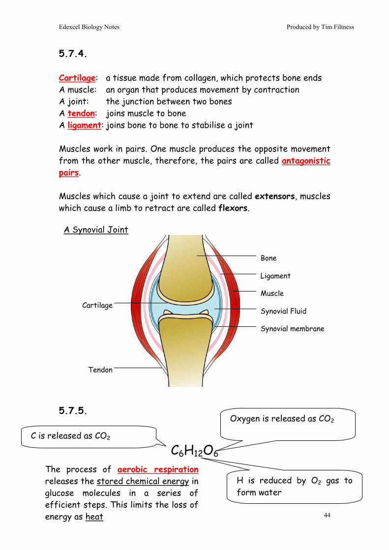
5.7.4. Cartilage: a tissue made from collagen, which protects bone ends A muscle: an organ that produces movement by contraction A joint: the junction between two bones A tendon: joins muscle to bone A ligament: joins bone to bone to stabilise a joint Muscles work in pairs. One muscle produces the opposite movement from the other muscle, therefore, the pairs are called antagonistic pairs. Muscles which cause a joint to extend are called extensors, muscles which cause a limb to retract are called flexors. 5.7.5.
C6H12O6
Cartilage
Tendon
Bone
Ligament
Muscle
Synovial Fluid
Synovial membrane
A Synovial Joint
C is released as CO2
H is reduced by O2 gas to form water
Oxygen is released as CO2
The process of aerobic respiration releases the stored chemical energy in glucose molecules in a series of efficient steps. This limits the loss of energy as heat

Edexcel Biology Notes Produced by Tim Filtness
45
5.7.6.
Dig up your Respirometer Core Practical notes in the Practical Handbook
5.7.7.
Adenosine TriPhosphate (ATP) is made from three components;
- Ribose (the same sugar that forms the basis of DNA).
- A base (a group consisting of linked rings of carbon and nitrogen atoms); in this case the base is adenine.
- Up to 3 phosphate groups. These phosphates are the key to the activity of ATP
The energy used in all cellular reactions comes from ATP. By breaking the 3rd phosphate from the ATP molecule energy is released, which can be used to power intracellular reactions. The ATP is then regenerated by recombining the phosphate and ADP in respiration (or another process e.g. photosynthesis).
The recycling of ATP is crucial for life. For example a runner uses ~84kg of ATP in a marathon (more than their total body weight), yet there are only 50g of ATP in the entire body! This means each that each molecule of ATP has been recycled 1676 times during the race!
Adenine base
Ribose
3 x phosphate
Edexcel Biology Notes Produced by Tim Filtness
46
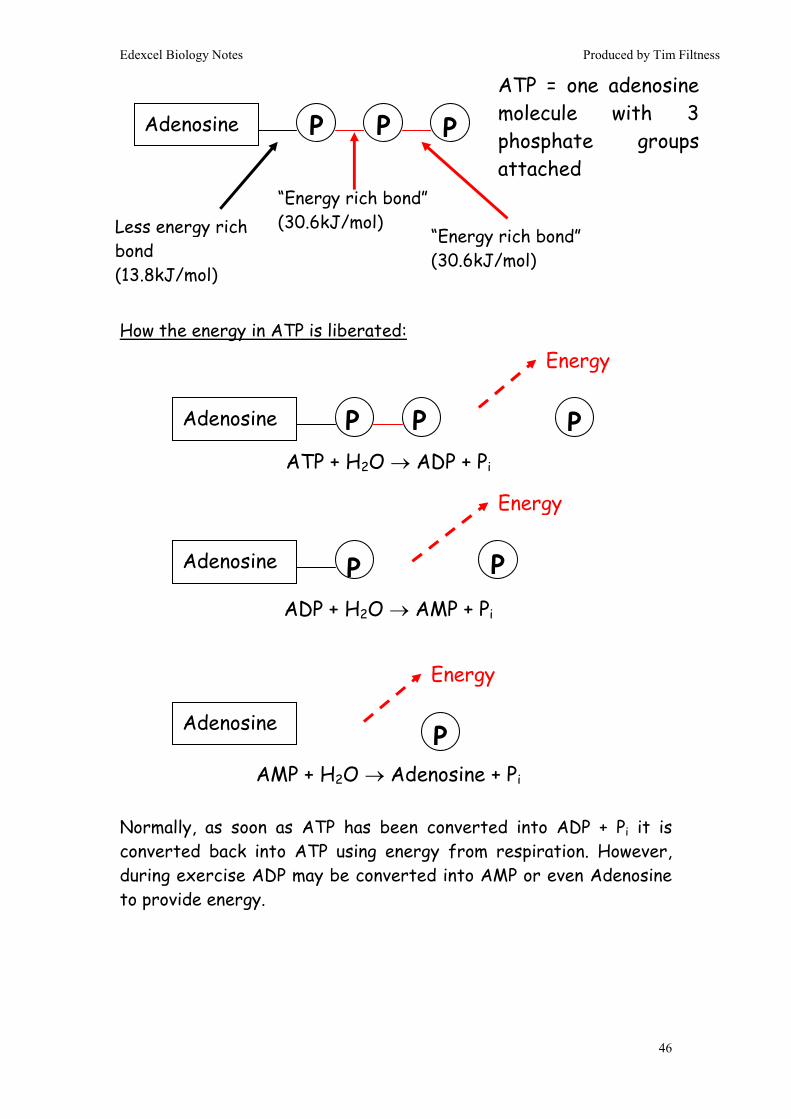
How the energy in ATP is liberated: Normally, as soon as ATP has been converted into ADP + Pi it is converted back into ATP using energy from respiration. However, during exercise ADP may be converted into AMP or even Adenosine to provide energy.
Adenosine P
ATP = one adenosine molecule with 3 phosphate groups attached
“Energy rich bond” (30.6kJ/mol)
“Energy rich bond” (30.6kJ/mol) Less energy rich
bond (13.8kJ/mol)
ATP + H2O → ADP + Pi
ADP + H2O → AMP + Pi
AMP + H2O → Adenosine + Pi
Adenosine P
Adenosine P
Adenosine P
Energy
Energy
Energy
P P
P P
P

Edexcel Biology Notes Produced by Tim Filtness
47
5.7.8, 5.7.9. & 5.7.10. Respiration: a process in which the chemical bond energy in glucose molecules is used to convert 38 ADP molecules into 38 ATP molecules. Oxygen is required and Carbon Dioxide and Water are produced as waste products. Respiration occurs in 4 distinct steps;
Step Reactants Products Summary
1. Glycolysis (cytoplasm)
1 x Glucose 2 x ATP
2 x Pyruvate 4 x ATP 2 x NADH
A 6C glucose molecule is split into two 3C pyruvate molecules. Some ATP is used to split the glucose molecule in the first part of glycolysis
2. Link Reaction (mitochondria matrix)
1 x Pyruvate 1 x CoA
1 x Acetyl CoA 1 x CO2 1 x NADH
3C Pyruvate is split into a 2C molecule, which is attached to a CoA enzyme to form Acetyl CoA. The remaining carbon atom is used to form CO2
3. Krebs’ Cycle (mitochondria matrix)
1 x Acetyl CoA 1 x CoA 1 x ATP 2 x CO2 3 x NADH 1 x FADH 2
CoA enzyme gives its 2C atoms to a 4C molecule to form a temporary 6C molecule. In a series of steps the 6C molecule releases the two C atoms as CO2 eventually re-forming the starting 4C compound. The cycle is then ready to repeat itself. As the cycle turns ATP, NADH & FADH2 are formed
4. Oxidative Phosphorylation (mitochondria christae)
10 x NADH 2 x FADH2 6 x O2
34 x ATP 6 x H2O
The electron transport chain uses the NADH and FADH2 made in previous steps to make lots of ATP

Edexcel Biology Notes Produced by Tim Filtness
48
In Glycolysis a Glucose molecule (6C) is split into 2 molecules of Glyceraldehyde Phosphate (3C). 2ATPs are required for this to happen. Then, each 3C Glyceraldehyde Phosphate molecule is converted into a 3C Pyruvate molecule. In the process of converting one Glyceraldehyde Phosphate to one Pyruvate, enough energy is released to convert one NAD molecules into one NADH molecules and also to make two ATP molecules. Overall; 4ATP are made, 2NADH are made and 2ATPs are used. Net gain: 2ATP and 2NADH
Glucose
Glyceraldehyde Phosphate
Glyceraldehyde Phosphate
Respiration: Step 1 - Glycolysis
Pyruvate Pyruvate
2ATPs are required 2ATPs are made (4 overall) 1 NADH is made (2 overall)
Glycolysis takes place in the cytoplasm of a cell
Edexcel Biology Notes Produced by Tim Filtness
49
Pyruvate Lactate In the liver the lactate is converted back into pyruvate. This requires oxygen, which is the basis of the “Oxygen Debt”
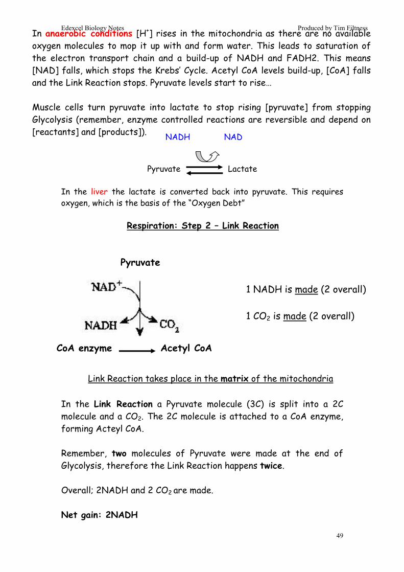
In the Link Reaction a Pyruvate molecule (3C) is split into a 2C molecule and a CO2. The 2C molecule is attached to a CoA enzyme, forming Acteyl CoA. Remember, two molecules of Pyruvate were made at the end of Glycolysis, therefore the Link Reaction happens twice. Overall; 2NADH and 2 CO2 are made. Net gain: 2NADH
In anaerobic conditions [H+] rises in the mitochondria as there are no available oxygen molecules to mop it up with and form water. This leads to saturation of the electron transport chain and a build-up of NADH and FADH2. This means [NAD] falls, which stops the Krebs’ Cycle. Acetyl CoA levels build-up, [CoA] falls and the Link Reaction stops. Pyruvate levels start to rise… Muscle cells turn pyruvate into lactate to stop rising [pyruvate] from stopping Glycolysis (remember, enzyme controlled reactions are reversible and depend on [reactants] and [products]).
Respiration: Step 2 – Link Reaction
1 NADH is made (2 overall) 1 CO2 is made (2 overall)
Link Reaction takes place in the matrix of the mitochondria
Pyruvate
CoA enzyme
Acetyl CoA
NADH NAD

Edexcel Biology Notes Produced by Tim Filtness
50
In the Krebs’ Cycle the Acetyl CoA gives its 2C atoms to a 4C molecule (Oxaloacetate) forming an unstable 6C molecule (Citric Acid). The 6C molecule breaks down into a 4C compound (Succinyl – CoA) releasing enough energy to make one NADH. The two spare C atoms are released as two CO2 molecules. Succinyl – CoA is converted back into Oxaloacetate and this releases enough energy to make one NADH, one FADH2 and one ATP. The Oxaloacetate can then be used in the cycle again. Remember, two molecules of Acetyl CoA were made at the end of the Link Reaction, therefore the Krebs’ Cycle happens twice. Overall; 4NADH, 2FADH2, 2CO2 and 2ATP are made.
Respiration: Step 3 – Krebs’ Cycle
Krebs’ Cycle takes place in the matrix of the mitochondria
3 NADH are made (6 overall) 1 ATP is made (2 overall) 1 FADH2 is made (2 overall) 2 CO2 are made (4 overall)
CoA enzyme
2
2

Edexcel Biology Notes Produced by Tim Filtness
51
Oxidative Phosphorylation uses the NADH and FADH2 produced in the previous steps of respiration to make ATP. Each NADH makes 3ATP and each FADH2 makes 2 ATP. Hydrogen atoms from the NADH and the reduced FADH2 are passed onto 2 the first 2 enzymes of the Electron Transport Chain. These enzymes are Hydrogen Carriers and they accept the H atoms from the NADH and the FADH2. Electrons, which made up the chemical bond between the hydrogen atoms and the NADH / FADH2 are passed onto 3 Electron Carrier enzymes further down the Electron Transport Chain. At the end of the Electron Transport Chain, the electrons are recombined with the H+ atoms and oxygen, to form water. This is the only, but crucial, part of respiration to involve oxygen.
FADH2
ATP FADH
H2O
NAD
NADH
ATP
ATP
½ O2 + 2H+
ATP
ADP ADP
Respiration: Step 4 – Oxidative Phosphorylation
Oxidative Phosphorylation takes place using enzymes embedded in the inner membrane of cristae of the mitochondria
H+ Carrier
H+ Carrier
e- Carrier
e- Carrier
e- Carrier
2e-
Edexcel Biology Notes Produced by Tim Filtness
52
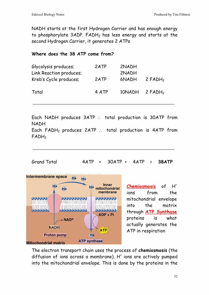
NADH starts at the first Hydrogen Carrier and has enough energy to phosphorylate 3ADP. FADH2 has less energy and starts at the second Hydrogen Carrier, it generates 2 ATPs Where does the 38 ATP come from? Glycolysis produces; 2ATP 2NADH Link Reaction produces; 2NADH Kreb’s Cycle produces; 2ATP 6NADH 2 FADH2 Total 4 ATP 10NADH 2 FADH2 Each NADH produces 3ATP ∴ total production is 30ATP from NADH Each FADH2 produces 2ATP ∴ total production is 4ATP from FADH2
Grand Total 4ATP + 30ATP + 4ATP = 38ATP The electron transport chain uses the process of chemiosmosis (the diffusion of ions across a membrane). H+ ions are actively pumped into the mitochondrial envelope. This is done by the proteins in the
Chemiosmosis of H+ ions from the mitochondrial envelope into the matrix through ATP Synthase proteins is what actually generates the ATP in respiration

Edexcel Biology Notes Produced by Tim Filtness
53
electron transport chain, using the energy stored in NADH and FADH2. The [H+] builds up to very high levels in the envelope. However, H+ cannot escape because it is charged (hydrophilic) and therefore cannot move through the phospholipid bilayer in the envelope membranes. Special proteins called ATP Synthase do allow H+ to pass through them and escape into the mitochondrial matrix. Whenever an H+ ion moves through the ATP Synthase protein an ADP is phosphorylated by the ATP Synthase. In summary;
1. NADH and FADH2 contain stored chemical energy 2. The energy is used to pump H+ into the mitochondrial
membrane against the concentration gradient
3. H+ trapped in one place represents a store of potential energy
4. H+ ions leave the envelope through ATP Synthase proteins.
5. The potential energy of the H+ is used to phosphorylate ATP as the H+ moves out of the envelope
5.7.11. In anaerobic respiration Pyruvate is converted into Lactate. This is good because it oxidises NADH generating NAD, which is required for Glycolysis. However, the lactate is acidic, poisonous & will cause muscle fatigue if it builds up inside cells. So, lactate is taken to the liver and converted back into Pyruvate, which is fed into the link reaction.
Lactate → Pyruvate NAD NADH

Edexcel Biology Notes Produced by Tim Filtness
54
The regeneration of Pyruvate both directly (see equation) and indirectly (via Krebs Cycle) results in more NADH being generated. As this NADH is ultimately oxidised by O2 the liver’s net O2 demand increases temporarily. This is the Oxygen Debt. 5.7.12.
1. SAN sends a wave of electrical activity (depolarization) around the walls of the atria.
2. A ring of insulating tissue blocks the wave from passing into the ventricles.
3. The AVN conducts the wave into the Ventricles slowly, which gives the ventricles time to fill.
4. The Purkinje fibres are fast-conducting and take the wave to the apex of the heart first, so the ventricles contract bottom upwards.
The heart is myogenic – it beats on its own (i.e. doesn’t need the brain to initiate a heartbeat) electrical activity of the heart. We can measure the electrical activity of a heartbeat using an electrocardiogram (or ECG). This enables us to determine whether the heart is healthy or not.
SAN: Sino-Atrial Node AVN: Atro-Ventricular Node Purkinje Fibres (in bundle of His)

Edexcel Biology Notes Produced by Tim Filtness
55
A "typical" ECG tracing is shown below.
The different waves that comprise the ECG represent the sequence of depolarization and repolarization of the atria and ventricles. It appears in 3 distinct segments;
P wave:
The P wave represents the wave of depolarization that spreads from the SA node throughout the atria.
QRS complex:
The QRS complex represents ventricular depolarization.
T wave:
The T wave represents ventricular repolarization and is longer in duration than depolarization (during this stage the ventricles are recovering and the heart is in diastole)
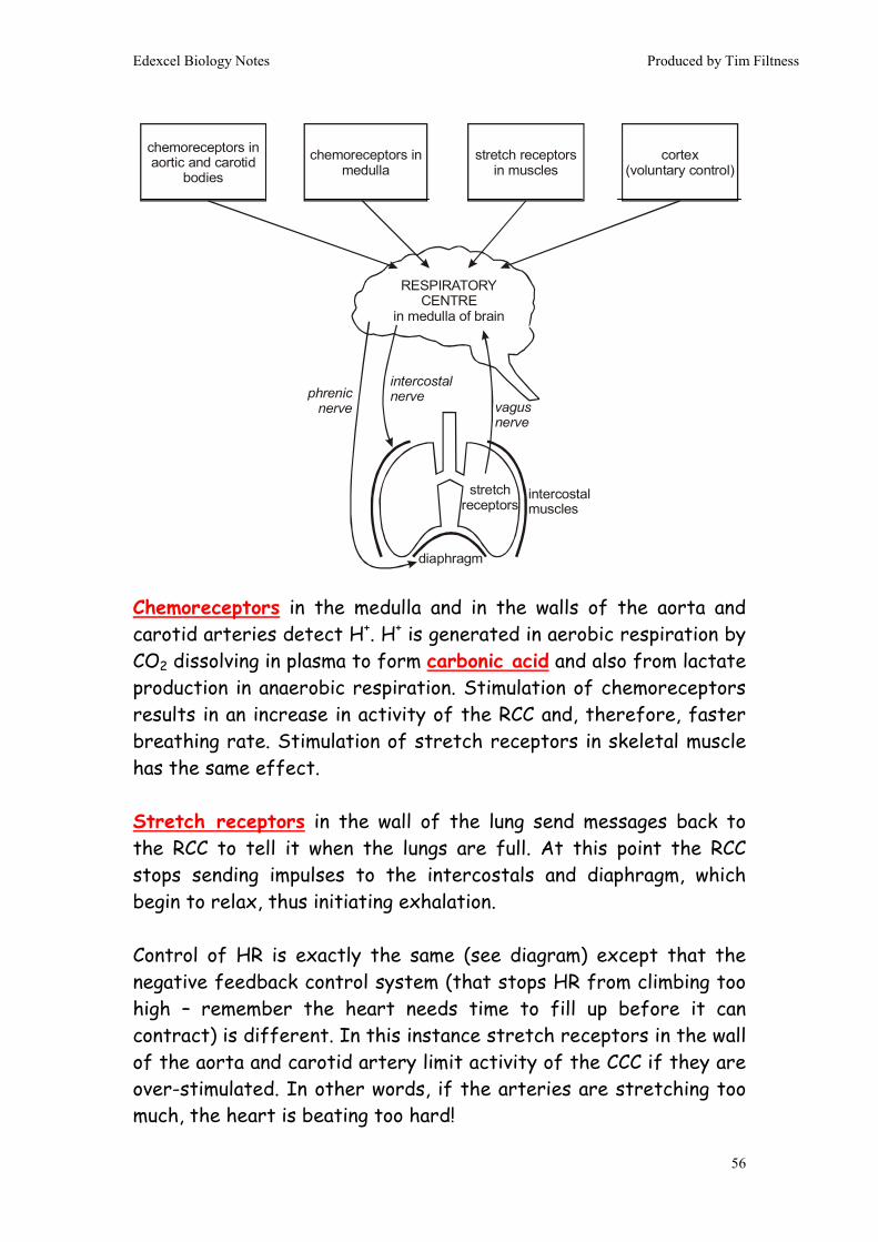
5.7.13. Both Heart Rate and Breathing Rate are controlled by the medulla in the brain. Nerves running towards the SAN and intercostal muscles and diaphragm send impulses which either speed up or reduce the breathing & heart rate.
Edexcel Biology Notes Produced by Tim Filtness
56
Chemoreceptors in the medulla and in the walls of the aorta and carotid arteries detect H+. H+ is generated in aerobic respiration by CO2 dissolving in plasma to form carbonic acid and also from lactate production in anaerobic respiration. Stimulation of chemoreceptors results in an increase in activity of the RCC and, therefore, faster breathing rate. Stimulation of stretch receptors in skeletal muscle has the same effect. Stretch receptors in the wall of the lung send messages back to the RCC to tell it when the lungs are full. At this point the RCC stops sending impulses to the intercostals and diaphragm, which begin to relax, thus initiating exhalation. Control of HR is exactly the same (see diagram) except that the negative feedback control system (that stops HR from climbing too high – remember the heart needs time to fill up before it can contract) is different. In this instance stretch receptors in the wall of the aorta and carotid artery limit activity of the CCC if they are over-stimulated. In other words, if the arteries are stretching too much, the heart is beating too hard!
chemoreceptors in aortic and carotid
bodies
chemoreceptors in medulla
stretch receptors in muscles
cortex(voluntary control)
RESPIRATORYCENTRE
in medulla of brain
diaphragm
intercostalmuscles
stretchreceptors
intercostalnervephrenic
nerve vagusnerve

Edexcel Biology Notes Produced by Tim Filtness
57
5.7.14.
Dig up your Spirometer Core Practical notes in the Practical
Handbook A spirometer is used to plot breathing patterns Vital Capacity: The maximum amount of air a person can
exhale after inhaling the maximum possible volume of air
pressure receptors in aortic
and carotid bodies
chemoreceptors in aortic and carotid
bodies
temperature receptors in muscles
stretch receptors in muscles
vasoconstrictionand
vasodilationsinoatrialnode
parasympatheticnerve
(inhibitor)
CARDIOVASCULARCENTRE
in medulla of brain
sympathetic nerve(accelerator)
TV

Edexcel Biology Notes Produced by Tim Filtness
58
Tidal Volume: The volume of air inhaled & exhaled in one
breath Basal Metabolic Rate: The rate of respiration Minute Volume: The volume of air inhaled in one minute The spirometer can be used to plot VC and TV directly. BMR can be worked out if a CO2 scrubber is used. The spirometer has fixed volume and is filled with 100% O2 before the experiment begins. As the person respires, O2 is replaced proportionally with CO2. The total volume should stay constant. However, if CO2 is removed by a scrubber, the total volume will slowly fall as O2 is used. The rate at which the volume decreases is proportionaly to BMR. You are not expected to know how the spirometer works… although it’s not very difficult to understand. 5.7.15. Homeostasis: the maintenance of a constant internal environment. Homeostasis relies on Negative Feedback systems. In Negative Feedback a stimulus will elict a response. However, the response is specifically designed to remove the stimulus and, thereby, maintain the status quo. E.g. Blood Sugar Level rises → Insulin release Insulin → Lowers blood sugar level
Edexcel Biology Notes Produced by Tim Filtness
59
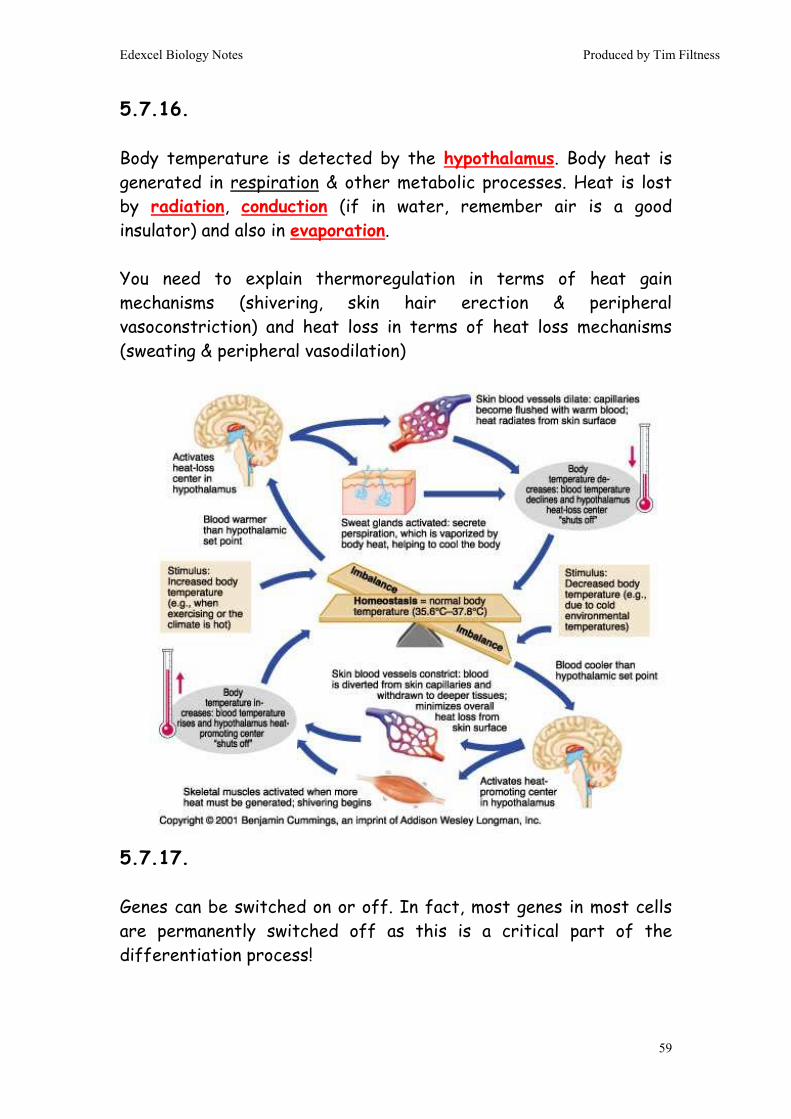
5.7.16. Body temperature is detected by the hypothalamus. Body heat is generated in respiration & other metabolic processes. Heat is lost by radiation, conduction (if in water, remember air is a good insulator) and also in evaporation. You need to explain thermoregulation in terms of heat gain mechanisms (shivering, skin hair erection & peripheral vasoconstriction) and heat loss in terms of heat loss mechanisms (sweating & peripheral vasodilation) 5.7.17. Genes can be switched on or off. In fact, most genes in most cells are permanently switched off as this is a critical part of the differentiation process!

Edexcel Biology Notes Produced by Tim Filtness
60
Genes are switched on by transcription factors binding to promoter regions of DNA “upstream” of the gene.
Genes can also be switched off by inhibitor molecules binding to the promoter region, which stops the gene from being transcribed. A good example of this is the Lac Operon (which is not on your syllabus) found in bacteria. The Lac Operon is normally switched off by an inhibitor. However, when lactose is present it inactivates the inhibitor, thus indirectly activating the gene. As the gene codes for the enzyme β galactosidase (which breaks down lactose) it’s quite clever… the enzyme is only produced when its substrate is around.
Transcription Factor binds to “TATA Box” promoter site. RNA Polymerase binding site revealed, which allows RNA Polymerase to bind to DNA. Gene opens and transcription begins

Edexcel Biology Notes Produced by Tim Filtness
61
5.7.18 Positive effects of exercise include;
1. Increased BMR 2. Decreased blood pressure 3. Increased HDL & Decreased LDL 4. Maintaining healthy BMI 5. Decreased risk of diabetes 6. Increased bone density 7. Improved well being 8. Less stress 9. Decreased risk of CHD 10. Moderate exercise increases levels of Natural Killer cells,
which secrete apoptosis-inducing chemicals in response to non-specific viral or cancerous threat
Negative effects of exercise (over-training) include;
1. Decreased levels of Natural Killer Cells, Phagoctyes and B & T
Cells. This decreses immune response. 2. Increased muscle inflammation 3. Muscle tears and sprains 4. Increased adrenaline levels 5. Increased cortisol levels, which also decreases the immune
response
A moderate level of exercise improves health & well-being. However, over-training can result in the opposite effect. This is the phenomenon known as “burn-out”

Edexcel Biology Notes Produced by Tim Filtness
62
6. Increased stress 7. Damaged cartilage 8. Tendinitis 9. Ligament damage 10. Swollen bursae
5.7.19. Key-hole surgery is a technique which allows doctors to conduct surgery with the minimum possible damage to the patient. The surgeon makes a small incision (a “key-hole”) and uses a fibre-optic camera to view the damaged area. If required, the surgeon can make a second incision and use a number of small, remote operated tools to repair the damage. Because the incisions are small and only the damaged area is targeted, the patient recovers quickly. There is also less chance of infection. Unfortunately, the procedure requires a high degree of training, expensive equipment and can only be used on certain types of surgery. Prosthetics allow people with amputations to participate in many activities, including sports and jobs. 5.7.20. Should athletes use performance enhancing substances?
End of Topic 7End of Topic 7End of Topic 7End of Topic 7
Yes
• Personal Choice • Restores “unfair” genetic
advantages • Athletes capable of performing
at higher level • If we can’t catch them can we
ever stop them? • Potential Revenue source?
No
• Side effects • Illegal / banned if caught • Pressure from coaches • Unfair disadvantage on those
not taking them • Funds manufacture of other
drug? .

Edexcel Biology Notes Produced by Tim Filtness
63
Unit 5Unit 5Unit 5Unit 5: : : : Energy, ExerEnergy, ExerEnergy, ExerEnergy, Exercise & cise & cise & cise & CoordinationCoordinationCoordinationCoordination
Topic 8: Grey Matter 5.8.2. & 5.8.8. Plants are sensitive to environmental stimuli (e.g. gravity, water, temperature, humidity etc). When plants alter their growth in response to a stimulus a tropism has occurred. Most tropisms involve cells in the meristem (the region of elongation behind the shoot tip) stretching. This usually happens in response to an auxin hormone (e.g. IAA – Indolacetic acid), which activates H+ pumps in the cell membrane. The H+ pumps lower the pH of the cytoplasm which has two effects;
- Water enters the cell by osmosis, causing it to swell and stretch
- pH-sensitive enzymes are activated. These enzymes cut the pectin cross-links between cellulose fibrils in the cell wall.
The combined effect of this is to let the cell stretch a lot in one direction, which effectively makes the plant grow. Later on the auxins are broken down, switching off the H+ pumps and allowing the cell wall to become rigid again. We don’t know exactly how auxins cause tropisms, but it is likely that it involves auxins moving to specific parts of the meristem, possibly through plasmodesmata.
Edexcel A2 Revision Notes

Edexcel Biology Notes Produced by Tim Filtness
64
Sensitivity to light Plants make light-sensitive hormones called phytocromes. There are two types; PR (or P660) - stimulates germination PFR (or P730) – inhibits germination & flowering
PR PFR Most plants tend to flower when the ratio of PR:PFR is high (i.e. quite a lot of PR around), which occurs during spring. That’s a great time to flower because it’s warm enough for reactions (like protein synthesis) to occur, but not too warm for the delicate flowers to desiccate. It’s also worth looking up the material on florigen in the text book: technically, it’s not on the syllabus, but we all know how sneaky those examiners like to be...
Daylight contains more red light than far red light. Therefore, during the day only PFR is present.
Low light levels / night causes the conversion of PFR into PR. Therefore, during the night only PR is present, which causes growth. In extreme conditions the low concentrations of PFR causes etiolation

Edexcel Biology Notes Produced by Tim Filtness
65
5.8.3. Sensory Neuron: carries electrical message from receptor to spine
Motor Neuron: carries electrical message from spine to effector
Relay Neuron: connects sensory and motor nerves. Also relays message to the brain.
These three neurones form reflexes, which allow us to respond rapidly to stimuli. They are involuntary (i.e. you can’t control them) Schwann cells: wrap around the axon of the long nerves, creating
a thick layer of membrane (called myelin), which insulates the nerve and allows for much faster conduction speed. The thick layer of membrane has gaps in it between adjacent Schwann cells, these are called Nodes of Ranvier. The nodes speed up conduction (see 5.8.4.)
Edexcel Biology Notes Produced by Tim Filtness
66
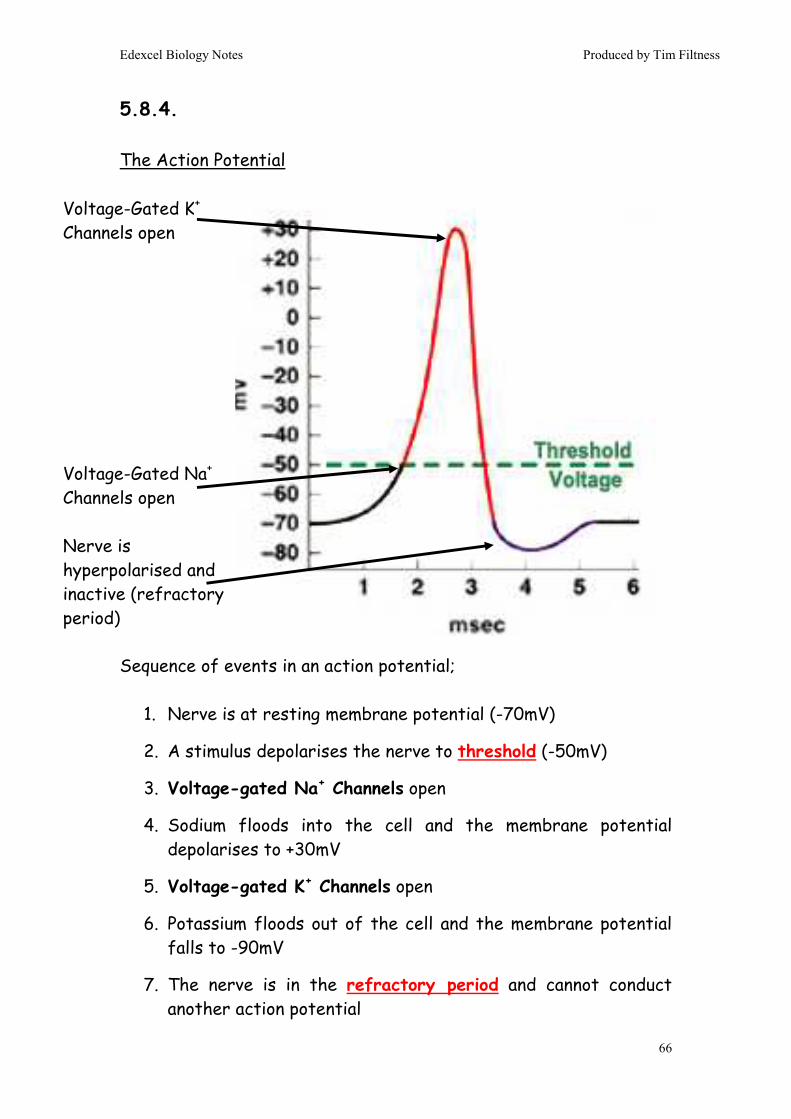
5.8.4. The Action Potential Sequence of events in an action potential;
1. Nerve is at resting membrane potential (-70mV)
2. A stimulus depolarises the nerve to threshold (-50mV)
3. Voltage-gated Na+ Channels open
4. Sodium floods into the cell and the membrane potential depolarises to +30mV
5. Voltage-gated K+ Channels open
6. Potassium floods out of the cell and the membrane potential falls to -90mV
7. The nerve is in the refractory period and cannot conduct another action potential
Voltage-Gated K+ Channels open Voltage-Gated Na+ Channels open Nerve is hyperpolarised and inactive (refractory period)

Edexcel Biology Notes Produced by Tim Filtness
67
8. The 3Na+/2K+ ATPase (Na+/K Pump) restores the ion concentrations
9. The nerve is ready to fire again As one part of the nerve fires off, Na+ diffuses into the next section of the nerve, which depolarises the nerve to threshold. This sequence is repeated like a tiny Mexican wave down the axon of the nerve.
Nodes of Ranvier speed this conduction process up. When one node depolarises it induces the next section of the nerve to depolarise by forming a mini-circuit between nodes. This causes the action potential to “jump” between Nodes of Ranvier, making conduction speed much faster (this is called saltatory conduction). 5.8.5.
3
4
2
1
5
7 6

Edexcel Biology Notes Produced by Tim Filtness
68
A synapse is the junction between two nerves. It is also a verb, i.e. one nerve synapses with another (meaning, passes a message to another). 1. The wave of depolarisation arrives at the synaptic knob. The
membrane in the presynaptic neuron is depolarised to –50mv (threshold potential) and the voltage-gated Na+ channels open, letting Na+ into the cell.
2. The membrane is depolarised to +30mV and voltage-gated K+
channels open. The membrane potential falls to –90mV and the cell goes into its refractory period, where the 3Na+/2K+-ATPase restored the ion concentrations.
3. Unlike axons, presynaptic nerves also contain a Voltage-gated Ca2+ channel. As the presynapstic membrane depolarises these channels open and let Ca2+ into the cell.
4. The Ca2+ causes vesicles in the presynaptic nerve to migrate and
fuse with the presynaptic membrane, where they spill neurotransmitter chemical into the synaptic cleft.
5. The neurotransmitter (Acetyl Choline) diffuses across the cleft
and binds to receptors on the postsynaptic membrane. 6. The receptors let a little Na+ into the postsynaptic neuron, which
is enough to initiate another action potential in the postsynaptic nerve.
7. The ACh is broken down by an enzyme called Acetyl Choline Esterase (AchE), which allows the postsynaptic receptors to be freed ready for a second synapse.
In a neuromuscular junction the sequence of events in the synapse is exactly the same. The only difference is that the posysynaptic nerve is a muscle cell and, instead of being flat, the postsynaptic membrane has deep grooves (t tubules) which allow the

Edexcel Biology Notes Produced by Tim Filtness
69
depolarisation to spread quickly through the muscle so all parts of the muscle contract at the same time. Some neurotransmitters can hyperpolarise postsynaptic nerves, which essentially switches them off. An example of this type of inhibitory neurotransmitter is GABA
5.8.6.
Visual transduction is the process by which light initiates a nerve impulse. The structure of a rod cell is:
The detection of light is carried out on the membrane disks in the outer segment. These disks contain thousands of molecules of rhodopsin, the photoreceptor molecule. Rhodopsin consists of a membrane-bound protein called opsin and a covalently-bound prosthetic group called retinal. Retinal is made from vitamin A, and a dietary deficiency in this vitamin causes night-blindness (poor vision in dim light). Retinal is the light-sensitive part, and it can exists in 2 forms: a cis form and a trans form:
Edexcel Biology Notes Produced by Tim Filtness
70
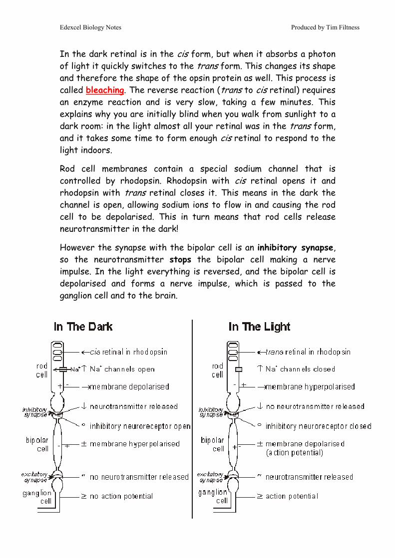
In the dark retinal is in the cis form, but when it absorbs a photon of light it quickly switches to the trans form. This changes its shape and therefore the shape of the opsin protein as well. This process is called bleaching. The reverse reaction (trans to cis retinal) requires an enzyme reaction and is very slow, taking a few minutes. This explains why you are initially blind when you walk from sunlight to a dark room: in the light almost all your retinal was in the trans form, and it takes some time to form enough cis retinal to respond to the light indoors.
Rod cell membranes contain a special sodium channel that is controlled by rhodopsin. Rhodopsin with cis retinal opens it and rhodopsin with trans retinal closes it. This means in the dark the channel is open, allowing sodium ions to flow in and causing the rod cell to be depolarised. This in turn means that rod cells release neurotransmitter in the dark!
However the synapse with the bipolar cell is an inhibitory synapse, so the neurotransmitter stops the bipolar cell making a nerve impulse. In the light everything is reversed, and the bipolar cell is depolarised and forms a nerve impulse, which is passed to the ganglion cell and to the brain.
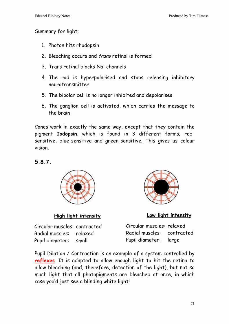
Edexcel Biology Notes Produced by Tim Filtness
71
Summary for light;
1. Photon hits rhodopsin
2. Bleaching occurs and trans retinal is formed
3. Trans retinal blocks Na+ channels
4. The rod is hyperpolarised and stops releasing inhibitory neurotransmitter
5. The bipolar cell is no longer inhibited and depolarises
6. The ganglion cell is activated, which carries the message to the brain
Cones work in exactly the same way, except that they contain the pigment Iodopsin, which is found in 3 different forms; red-sensitive, blue-sensitive and green-sensitive. This gives us colour vision. 5.8.7. Pupil Dilation / Contraction is an example of a system controlled by reflexes. It is adapted to allow enough light to hit the retina to allow bleaching (and, therefore, detection of the light), but not so much light that all photopigments are bleached at once, in which case you’d just see a blinding white light!
High light intensity
Circular muscles: contracted Radial muscles: relaxed Pupil diameter: small
Low light intensity
Circular muscles: relaxed Radial muscles: contracted Pupil diameter: large

Edexcel Biology Notes Produced by Tim Filtness
72
5.8.8. See 5.8.2. 5.8.9.
Cerebellum
Brainstem
Midbrain Cerebrum
Medulla
Forebrain
Midbrain
Hindbrain
Stimuli Commands
Try to think of the brain as a wedding cake in three layers: the bottom layer is the “oldest” part of the brain and controls basic life support systems. The middle part carries out basic processing and is the centre for emotion. The top layer is the bit that “thinks” and stores memory – it’s much bigger in humans than most other mammals.

Edexcel Biology Notes Produced by Tim Filtness
73
Hindbrain Brainstem – Uppermost part of the spine, where the spine joins the brain Medulla - controls vital ‘housekeeping’ functions, such as heartbeat, blood pressure and peristalsis. Cerebellum - controls muscle co-ordination & learns motor programmes (e.g. like how to ride a bike, or write). Midbrain: Thalamus – a relay station that carries sensory information from the sense organs to the correct part of the cortex and hypothalamus. It also carries out basic sensory processing (e.g. identifying faces & some shapes) Hypothalamus – receives sensory information from the thalamus. Contains homeostatic centres, which control factors like body temperature and blood osmolarity. The hypothalamus is connected to the Pituitary gland and therefore the hypothalamus can stimulate the release of a great number of pituitary hormones. Forebrain: Cortex – processes sensory information and controls the body’s voluntary behaviour, i.e. learning, personality and memory. This is the part of the brain that actually “thinks.” The cortex is very large in humans and is folded to increase the surface area further. Other animals have roughly similar size hind- and midbrains. However, their cortex is much, much smaller.

Edexcel Biology Notes Produced by Tim Filtness
74
Occipital lobe - processes & interprets information from the eyes Temporal lobe - processes & interprets information from the ears and processes language and the meaning of words Parietal lobe – processes and interprets information about touch, taste, pressure, pain, heat and cold. Also initiates motor commands. Frontal lobe - plans and organises thought, is involved with short term memory and puts speech together.
(Speech motor area)
(Understanding language)
Auditory association area
Premotor
Somatosensory
Visual association area
The Cortical “Lobes”
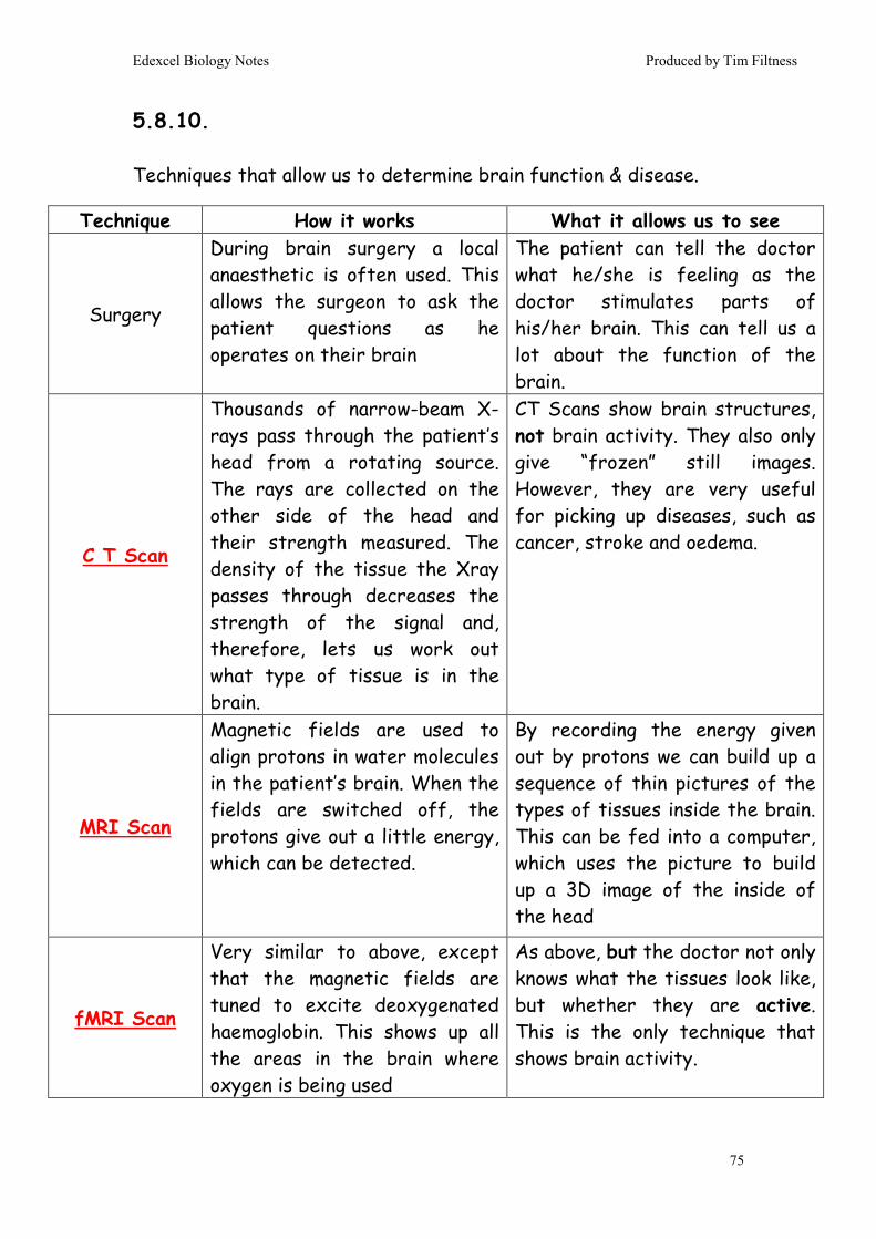
Edexcel Biology Notes Produced by Tim Filtness
75
5.8.10. Techniques that allow us to determine brain function & disease.
Technique How it works What it allows us to see
Surgery
During brain surgery a local anaesthetic is often used. This allows the surgeon to ask the patient questions as he operates on their brain
The patient can tell the doctor what he/she is feeling as the doctor stimulates parts of his/her brain. This can tell us a lot about the function of the brain.
C T Scan
Thousands of narrow-beam X-rays pass through the patient’s head from a rotating source. The rays are collected on the other side of the head and their strength measured. The density of the tissue the Xray passes through decreases the strength of the signal and, therefore, lets us work out what type of tissue is in the brain.
CT Scans show brain structures, not brain activity. They also only give “frozen” still images. However, they are very useful for picking up diseases, such as cancer, stroke and oedema.
MRI Scan
Magnetic fields are used to align protons in water molecules in the patient’s brain. When the fields are switched off, the protons give out a little energy, which can be detected.
By recording the energy given out by protons we can build up a sequence of thin pictures of the types of tissues inside the brain. This can be fed into a computer, which uses the picture to build up a 3D image of the inside of the head
fMRI Scan
Very similar to above, except that the magnetic fields are tuned to excite deoxygenated haemoglobin. This shows up all the areas in the brain where oxygen is being used
As above, but the doctor not only knows what the tissues look like, but whether they are active. This is the only technique that shows brain activity.

Edexcel Biology Notes Produced by Tim Filtness
76
5.8.10. & 5.8.11. How to process stimuli correctly must be learned. The cortex is split into columns of cells. When we are born, the columns overlap and are tangled. As we learn to process stimuli, the cells organise themselves into discrete columns, which no longer overlap – the brain literally wires itself up. There is a “critical window” for this to happen (usually before puberty, younger for visual processing). If we miss the window, our brains will become “fixed” with tangled columns and won’t be able to process stimuli properly. Hubel & Wiesel’s experiments prove this. Hubel & Wiesel’s Method:
1. Raise monkeys from birth in three groups for 6 months
2. Group 1 are the control (no blindfold), Group 2 are blindfolded in both eyes, Group 3 are blindfolded in one eye (monocular deprivation)
3. Test the monkeys to see whether they can see using each eye
4. Test the sensitivity of retinal cells
5. Test the activity of nerves in the visual cortex in response to stimuli
The results:
− Monkeys in Group 2 (both eyes blindfolded) had impaired vision
− Monkeys in Group 3 (monocular deprivation) were blind in the deprived eye
− Retinal cells were responsive in all groups − Cortical activity was reduced in parts of the brain that
process information from the deprived eye − Adults undergoing the same tests showed no difference
between groups. All could see.
Edexcel Biology Notes Produced by Tim Filtness
77
The Conclusion: There is a critical window for visual neural development, which requires stimulus from the eye. If this window is missed the monkey is blind, because of events happening in the brain, not the eye. One or two people objected to this work on ethical grounds. Can’t think why… The utilitarian principle (the “greater good”) is often used as a counter-argument to justify their studies. 5.8.12. Without animal studies we would know literally nothing about the brain. Studies on animal brains usually fall into one of these groups; Dissection – find out where the “wires” go. That’s OK, but it doesn’t tell you what the bits actually DO! Lesion studies – destroy part of the animal’s brain and see what happens. Ethics aside, these kind of studies aren’t great because you’re never sure whether the part you destroyed was actually responsible for the function the animal has lost e.g. if you shout at a spider it runs away. Remove all the spider’s legs and then shout at it again it doesn’t run away. Conclusion: spider’s have their ears in their legs. Poor science… Genetic engineering – engineer an animal so its neurons glow when their active. It lets you see what’s going on without destroying anything, which is perfect! This is used a lot in studies of brain development. 5.8.13. Is brain development a product of nature or nurture? Nature – brain development is controlled by our genes Nurture – brain development occurs as a result of what happens
to us (as children).

Edexcel Biology Notes Produced by Tim Filtness
78
It’s blatantly a bit of both… but which is more important? Overall, then, it’s both. Also be aware that finding an accurate way of measuring “brain development” is itself pretty difficult. For example, the more people practice the better they get at IQ tests, which (if you think about it) shouldn’t happen! 5.8.14. Habituation: learning that a stimulus has no outcome and, therefore, learning to ignore it.
Evidence Nature or Nurture?
Newborn Babies
Lots of studies demonstrating critical windows of development (e.g. babies born with cataracts become blind even if the cataracts are removed in later life). However, babies are also born with the ability to recognise faces and to “imprint” and “attach” onto a maternal / paternal face. This is the same in all babies (therefore genetic). Interestingly, animals also imprint. Evidence supports both nature and nurture
Animal Experiments
Hubel & Wiesel’s experiment supports nurture.
People with brain damage
After the brain has been damaged it is often possible for people to regain function or relearn how to do things using other parts of their brain, or by their brain growing new connections that bypass the damaged area. This is easier if the patient is younger. Supports nurture
Twin studies Identical twins process some information in identical ways (e.g. face recognition). Non-identical twins do it slightly differently, implying a genetic involvement. However, even identical twins learn to read and write in different ways. Supports both nature and nurture.
Cross-cultural studies
There is some evidence that people view optical illusions in different ways (look up the Muller-Lyer illusion & Zulus in your textbook). This supports nurture.
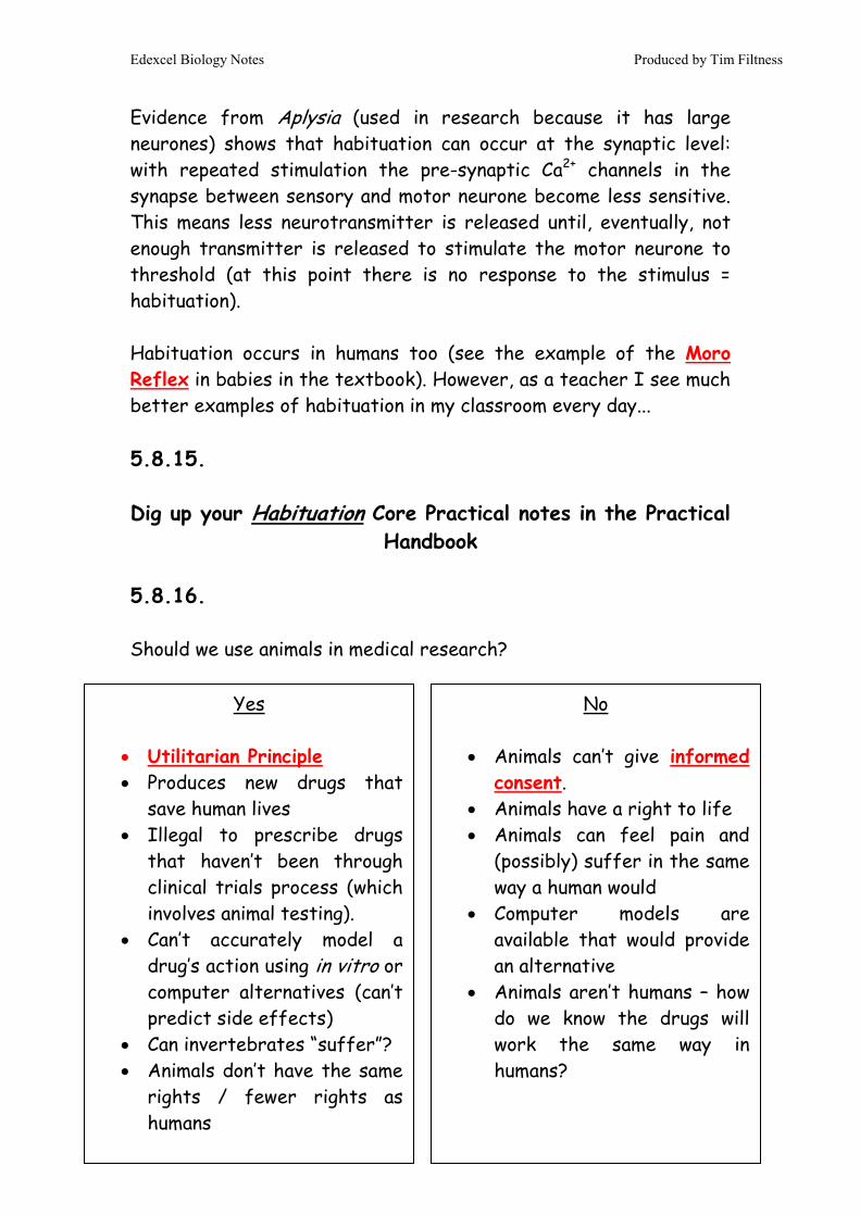
Edexcel Biology Notes Produced by Tim Filtness
79
Evidence from Aplysia (used in research because it has large neurones) shows that habituation can occur at the synaptic level: with repeated stimulation the pre-synaptic Ca2+ channels in the synapse between sensory and motor neurone become less sensitive. This means less neurotransmitter is released until, eventually, not enough transmitter is released to stimulate the motor neurone to threshold (at this point there is no response to the stimulus = habituation). Habituation occurs in humans too (see the example of the Moro Reflex in babies in the textbook). However, as a teacher I see much better examples of habituation in my classroom every day... 5.8.15. Dig up your Habituation Core Practical notes in the Practical
Handbook 5.8.16. Should we use animals in medical research?
Yes
• Utilitarian Principle • Produces new drugs that
save human lives • Illegal to prescribe drugs
that haven’t been through clinical trials process (which involves animal testing).
• Can’t accurately model a drug’s action using in vitro or computer alternatives (can’t predict side effects)
• Can invertebrates “suffer”? • Animals don’t have the same
rights / fewer rights as humans
No
• Animals can’t give informed consent.
• Animals have a right to life • Animals can feel pain and
(possibly) suffer in the same way a human would
• Computer models are available that would provide an alternative
• Animals aren’t humans – how do we know the drugs will work the same way in humans?

Edexcel Biology Notes Produced by Tim Filtness
80
5.8.17. Motor commands are initiated by the Frontal Lobe of the cortex. However, coordination of movement is regulated by the basal ganglia (part of the cerebellum). Nerves in the cerebellum either stimulate or inhibit the basal ganglia, which acts as a fine tuning system for the motor command, i.e. the arm moves enough, but not too much to miss the ball. Some nerves in the cerebellum release the neurotransmitter dopamine, which stimulates the basal ganglia, increasing the motor command. Other nerves in the cerebellum release Acetyl Choline, which has the opposite effect. Therefore, the balance of Dopamine and ACh coordinates the motor command. In Parkinson's disease some of the nerves in the part of the cerebellum that produces dopamine (the substantia nigra) begin to die, decreasing the levels of dopamine. This throws off the normal dopamine/acetylcholine balance, since the level of acetylcholine remains normal. The basal ganglia are, therefore, inhibited too

Edexcel Biology Notes Produced by Tim Filtness
81
much, which results in muscles becoming overly tense, muscle tremor, joint rigidity, and slow movement. In depression neurons in the midbrain (specifically the limbic system, which controls emotion) stop secreting serotonin neurotransmitter / secrete less than they should. We don’t really know why, but there is a strong genetic predisposition (as there is with Parkinson’s) & depression has also been linked to post-viral disease. For both Parkinson’s and Depression treatments that increase the levels of neurotransmitter might prove successful in relieving the symptoms of these diseases. Specific examples include; L-Dopa (see 5.8.15) - for Parkinson’s SSRI Inhibitors (same action as MDMA) - for Depression 5.8.18. Drugs that affect synapses can drastically alter the functioning of the brain; MDMA: Active ingredient in ecstasy. This binds to protein pumps on the pre-synaptic membrane of nerves that secrete serotonin. The pumps would normally take serotonin up after it had been released, therefore reducing firing in post-synaptic nerves. BUT, when these channels are blocked, serotonin builds up in the cleft, giving greater post-synaptic activation and a sense of euphoria. L-Dopa: This is a precursor of dopamine. When given to Parkinson’s sufferers it is turned into dopamine, which helps alleviate some of the symptoms of the disease. We can’t give Dopamine directly because it can’t cross the blood-brain-barier.

Edexcel Biology Notes Produced by Tim Filtness
82
5.8.19. The Human Genome Project (identifying the complete sequence of code in human DNA) has initiated a new field of medicine called pharmacogenetics i.e. the idea that drugs can be targeted at individual people based on their DNA. This would allow;
- Lower dosages to be used (costs less and fewer side-effects) - Ability to choose drugs that work best for individuals - Ability to avoid drugs that won’t work in individuals
Specific examples are given in the textbook e.g. kappa-opioids don’t work as well on men as they do on women. As with every Edexcel topic you need to consider the social, moral and ethical issues of HGP. A few to consider are;
- Can you patent / copyright DNA when you sequence it? - Who should have access to your genetic code? - Should genetic information be kept on record? - What if we identify “genes” for intelligence, sexuality, crime? - Eugenics (i.e. selective breeding in humans)
5.8.20. Genetic Engineering: How to make a transgenic organism (i.e. with DNA from more than one source)
1. Cut required gene out of donor cell DNA. Do this using a restriction enzyme.
2. Separate gene from other fragments of DNA using electrophoresis & a gene probe complementary to the gene
3. Extract the gene from the gel and copy it using PCR 4. Extract plasmids from bacteria (macerate & centrifuge) 5. Cut plasmids open using the same restriction enzyme.

Edexcel Biology Notes Produced by Tim Filtness
83
6. Insert the gene into plasmid and seal with DNA Ligase enzyme
7. Use a vector to get the transgenic plasmid back into a bacterium (vector is usually heat shock)
If you want to introduce the gene into a eukaryotic cell we do things slightly differently. We don’t use a plasmid! Instead the gene itself is inserted into the target cell using a vector. The vectors are different too, for eukaryotes we use viruses, liposomes, microinjection or a gene gun (great name – stick gene to tiny gold ball and fire it into the cell using compressed air) Actually, this process is really, really complicated. For example, how do you know what the “required gene” actually is? How do you know it’s sequence? How do you know the restriction enzyme will cut it out of the donor DNA? All good things to ponder, but none of them are on the syllabus! 5.8.21.
End of Topic 8End of Topic 8End of Topic 8End of Topic 8
Risks of GE
• Side effects • It hardly ever works (so
LOTS of repeats to get one working attempt – bad if we’re engineering animals)
• Costly • In crops the genes often
“escape” into other plants • Opens door to human GE • Accidental release of GE
organisms into the wild is BAD
• GE biological weapons • Religious objection
Benefits of GE
• Can manufacture desired protein easily (e.g. Insulin)
• Most proteins can’t be made synthetically
• Treatment of diseases • Low running costs (after
initial cost of making GE organism
• Renewable • New technology available
(e.g. body armour made from spider’s silk produced by GE goats)