Embed Size (px)
Citation preview
ECHOGRAPHIEPLEURO-PULMONAIREEpanchementspleurauxliquidiens
DrBONNECJ.M.(1)(2)
DrBOBBIAX.(1)–DrCLARETP.G.-PrDELACOUSSAYEJ.E.(1)
(1)PôleAnesthésieRéanimaNonDouleurUrgence-GHUCarémeau-
Nîmes
(2)PôleUrgences–SAMU–SMUR–CHPerpignan
E.A.U.Mars2016
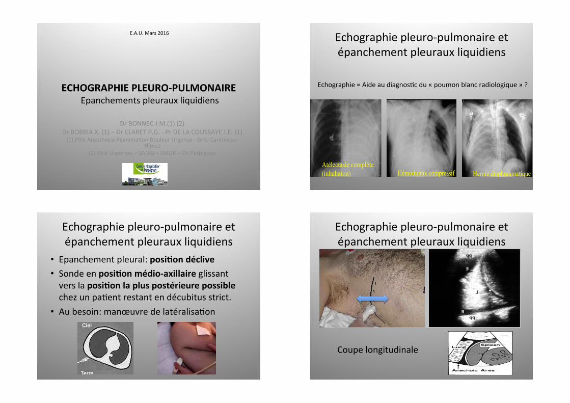
Echographiepleuro-pulmonaireet
épanchementpleurauxliquidiens
Echographie=AideaudiagnosNcdu«poumonblancradiologique»?
Echo = aide au diagnostic radiologique
de « poumon blanc »
Atélectasie complète (inhalation) Hémothorax compressif
Hernie diaphragmatique
Echo = aide au diagnostic radiologique
de « poumon blanc »
Atélectasie complète (inhalation) Hémothorax compressif
Hernie diaphragmatique
Echo = aide au diagnostic radiologique
de « poumon blanc »
Atélectasie complète (inhalation) Hémothorax compressif
Hernie diaphragmatique
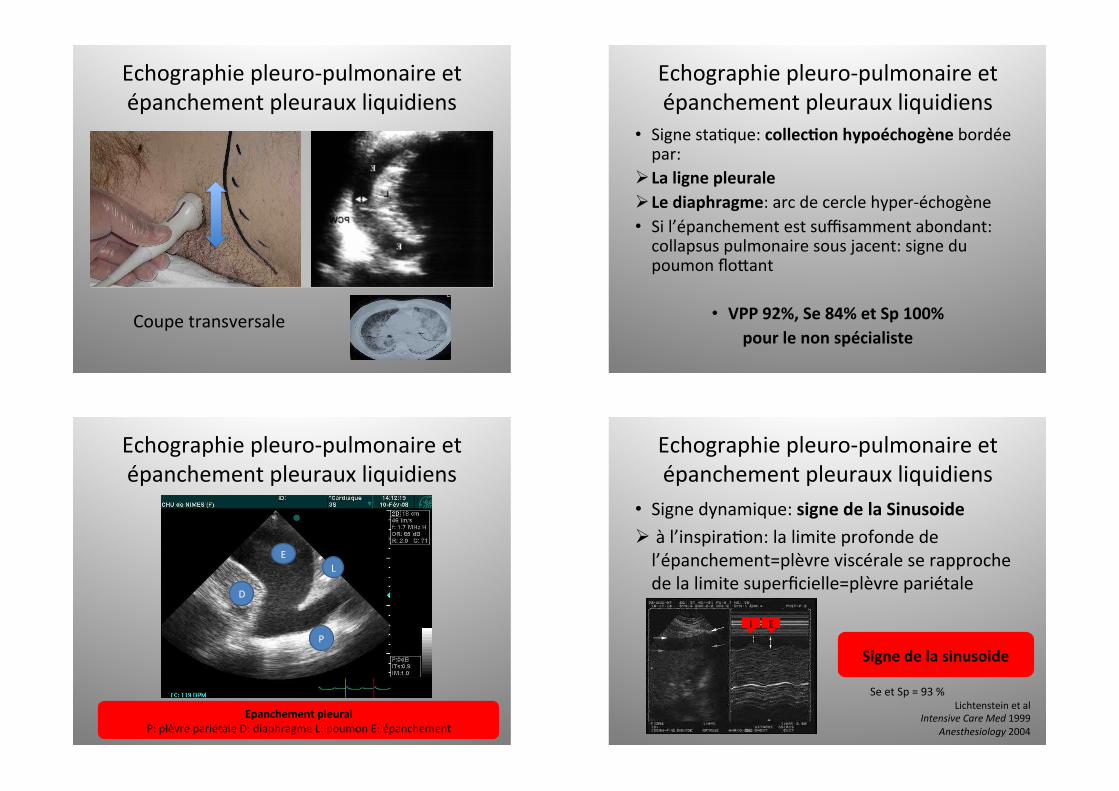
Echographiepleuro-pulmonaireet
épanchementpleurauxliquidiens
• Epanchementpleural:posi4ondéclive• Sondeenposi4onmédio-axillaireglissantverslaposi4onlapluspostérieurepossiblechezunpaNentrestantendécubitusstrict.
• Aubesoin:manœuvredelatéralisaNon
Rate
Repérage du diaphragme en coupe longitudinale
Coupelongitudinale
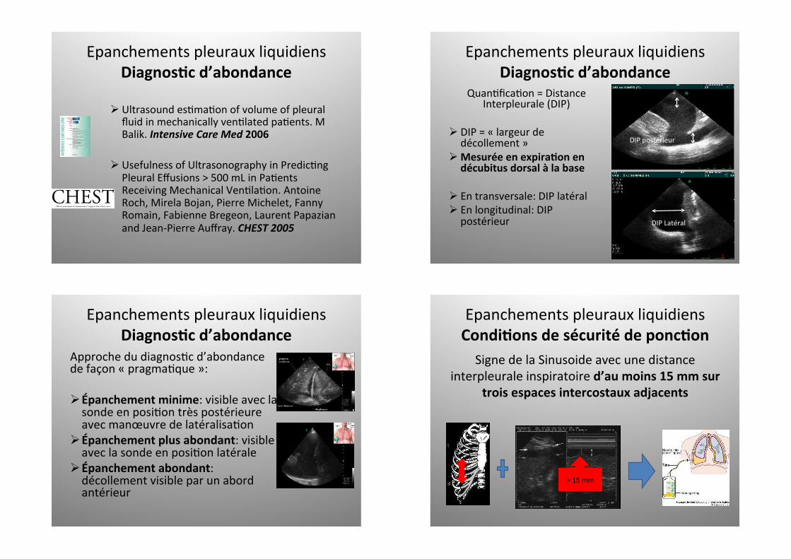
Echographiepleuro-pulmonaireet
épanchementpleurauxliquidiens
Coupe transversale
Comparaison entre la RX au lit et le Scanner pulmonaire dans une série de 71 patients avec SDRA Intensive Care Medicine 26 : 1046-1056 , 2000
30 11 2010
Coupetransversale
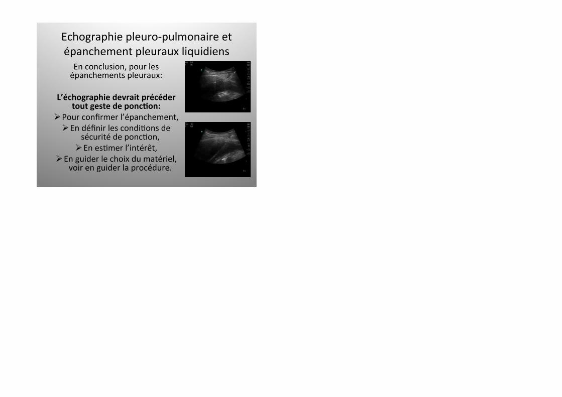
Echographiepleuro-pulmonaireet
épanchementpleurauxliquidiens
Echographiepleuro-pulmonaireet
épanchementpleurauxliquidiens
• SignestaNque:collec4onhypoéchogènebordéepar:
Ø LalignepleuraleØ Lediaphragme:arcdecerclehyper-échogène• Sil’épanchementestsuffisammentabondant:
collapsuspulmonairesousjacent:signedu
poumonflodant
• VPP92%,Se84%etSp100%pourlenonspécialiste
Echographiepleuro-pulmonaireet
épanchementpleurauxliquidiens
EpanchementpleuralP:plèvrepariétaleD:diaphragmeL:poumonE:épanchement
D
E
L
P
Echographiepleuro-pulmonaireet
épanchementpleurauxliquidiens
• Signedynamique:signedelaSinusoideØ àl’inspiraNon:lalimiteprofondede
l’épanchement=plèvreviscéraleserapproche
delalimitesuperficielle=plèvrepariétale
I E
Signedelasinusoide
SeetSp=93%
Lichtensteinetal
IntensiveCareMed1999Anesthesiology2004

• FaiblerentabilitédiagnosNquedelaRT
• Hauterentabilitéducouple«Clinique-US»
Echographiepleuro-pulmonaireet
épanchementpleurauxliquidiens
PerNnenceclinique?
Echographiepleuro-pulmonaireet
épanchementpleurauxliquidiens
ApplicaNonClinique:
AtélectasievsEpanchementvslesdeux?
Echographiepleuro-pulmonaireet
épanchementpleurauxliquidiens
Echographiepleuro-pulmonaireet
épanchementpleurauxliquidiens
DRAINAGE600cc
Epanchementspleurauxliquidiens
Diagnos4cd’abondanceØ UltrasoundesNmaNonofvolumeofpleural
fluidinmechanicallyvenNlatedpaNents.M
Balik.IntensiveCareMed2006Ø UsefulnessofUltrasonographyinPredicNngPleuralEffusions>500mLinPaNents
ReceivingMechanicalVenNlaNon.Antoine
Roch,MirelaBojan,PierreMichelet,Fanny
Romain,FabienneBregeon,LaurentPapazian
andJean-PierreAuffray.CHEST2005
Epanchementspleurauxliquidiens
Diagnos4cd’abondanceQuanNficaNon=Distance
Interpleurale(DIP)
Ø DIP=«largeurdedécollement»
Ø Mesuréeenexpira4onendécubitusdorsalàlabase
Ø Entransversale:DIPlatéralØ Enlongitudinal:DIPpostérieur
Epanchement pleural : quantification – distance interpleurale
Epanchement pleural : quantification – distance interpleurale
DIPLatéral
DIPpostérieur
Epanchementspleurauxliquidiens
Diagnos4cd’abondanceApprochedudiagnosNcd’abondance
defaçon«pragmaNque»:
Ø Épanchementminime:visibleaveclasondeenposiNontrèspostérieure
avecmanœuvredelatéralisaNon
Ø Épanchementplusabondant:visibleaveclasondeenposiNonlatérale
Ø Épanchementabondant:décollementvisibleparunabord
antérieur
Epanchementspleurauxliquidiens
Condi4onsdesécuritédeponc4onSignedelaSinusoideavecunedistance
interpleuraleinspiratoired’aumoins15mmsurtroisespacesintercostauxadjacents
>15mm
3

Epanchementspleurauxliquidiens
Diagnos4cd’abondanceRochetAl,CHEST2005
DistancedelaparoipostérieureàlabaseenexpiraNonPLDbasesurune
coupelongitudinale
Epanchementspleurauxliquidiens
Diagnos4cd’abondanceRochetAl,CHEST2005
Roch et coll Chest 2005
Roch et coll Chest 2005
Epaisseur > 5 cm: VPP = 91% pour épanchement > 500 ml
PLDbase>5cm=>VPPde91%pourunépanchementdrainédeplusde500mL
Epanchementspleurauxliquidiens
Diagnos4cd’abondanceVignonetAl,CritCareMed,2005
Vignon P Crit Care Med 2005
Coupetransversaleenfind’inspira4onetenfind’expira4on:
- Àl’apex
- Àlabase
Epanchementspleurauxliquidiens
Diagnos4cd’abondanceVignonetAl,CritCareMed,2005
Vignon P Crit Care Med 2005
Vignon P Crit Care Med 2005
Echographie pleurale : épanchements liquidiens
Vignon et al Crit Care Med 2005
D
G
DIP > 45 mm à droite DIP > 50 mm à gauche
= volume > 800 ml
DIP>45mmàDroiteSe=94%etSp=76%DIP>50mmàGaucheSe=100%etSp67%
Volumedrainé>800mL

Epanchementspleurauxliquidiens
Diagnos4cd’abondanceUstaetAl,ICTS,2010
Estimation du volume : des formules ?
Usta et al ICTS 2010
D
G
Estimation du volume : des formules ?
Usta et al ICTS 2010
D
G
Mesuredelalongueurdel’anglecosto-diaphragma4que
Epanchementspleurauxliquidiens
Diagnos4cd’abondanceUstaetAl,ICTS,2010
Estimation du volume : des formules ?
Usta et al ICTS 2010
D
G
V = 16 x D
V = (15,06 x D) +
V(mL)=(Dx15,06)+21,45
Epanchementspleurauxliquidiens
Diagnos4cd’abondanceRémérandetAl,IntensiveCareMed,2010
Approcheéchographiquemul:plan
Volumede
l’épanchement
=
Hauteurparavértébrale
X
Surfaceàmi-hauteur
Epanchementspleurauxliquidiens
Diagnos4cd’abondanceRémérandetAl,IntensiveCareMed,2010
Approcheéchographiquemul:planQuantification de tous les épanchements pleuraux :
l’approche échographique multiplan
Figure 5
A
B
C
Figure 5
A
B
C
05 12 2012 JJR 05 12 2012 Rémérand F et al Intensive Care Medicine 2010 36: 656-664

Epanchementspleurauxliquidiens
Aspectqualita4f
Ø Transsudat:collecNonrigoureusementtransonore
Ø Exsudat:peutêtretransonorehomogèneouricheen
septaNons
Ø Pleurésiepurulente:collecNontransonorecontenantdeséchos(«signeduplancton»)etpossiblesfines
septaNons
Ø Hémothorax:collecNontransonorericheenéchos:signeduplancton
Epanchementspleurauxliquidiens
Aspectqualita4f
Transsudat
Pleurésie
purulente
Exsudat
Hémothorax
Epanchementspleurauxliquidiens
Aspectqualita4f
HEMOTHORAX:D:diaphragmeP:ParenchymeE:EpanchementC:Caillot
D
P
E
C
Epanchementspleurauxliquidiens
Aspectqualita4fAppréciaNonqualitaNvedesépanchements:USvsTDM
AJR:193, December 2009 1649
Ultrasound and CT in Evaluation of Pneumonia
admission between December 2006 and January 2009 were included in the study. Nineteen pa-tients (nine boys and 10 girls) with a mean age of 5.4 years (age range, 8 months–17 years) who met the study criteria were identified. The mean time between chest ultrasound and CT was 2.7 days (range, 0–8 days).
Chest Ultrasound TechniqueChest ultrasound was performed by two expe-
rienced staff ultrasound technologists on an iU22 ultrasound system (Philips Healthcare) (n = 15), an HDI 5000 ultrasound system (Philips Health-
care) (n = 3), or an Acuson Sequoia 512 ultra-sound system (Siemens Healthcare) (n = 1). Lin-ear (5–12 MHz), curved linear (2–5, 4–9, or 5–8 MHz), and vector (5–8 MHz) transducers were used. The chest abnormality was localized on the basis of chest radiography findings. Anteri-or, posterior, and midaxillary images were ob-tained using an intercostal approach in transverse and longitudinal planes from the apex to the dia-phragm with the patient in a supine or decubitus position. Color Doppler ultrasound was performed to evaluate the vascularity of regional parenchy-mal abnormalities.
Chest CT TechniqueChest CT was performed on an MX-8000 IDT
16-MDCT scanner (Philips Healthcare) (n = 7), a Brilliance 16-MDCT scanner (Philips Healthcare Electronics) (n = 6), or a LightSpeed VCT 64-MDCT scanner (GE Healthcare) (n = 5). Images were obtained from the level of the thoracic inlet to the diaphragm using a pitch of 1.0, 120 kVp, and a weight-based low-dose tube current. Thirteen of the patients underwent CT with the administration of nonionic IV contrast material (320 mg I/mL io-dixanol, Visipaque, GE Healthcare) at a dose of 1 mL/kg. CT data were reconstructed at a slice thickness of either 3 or 5 mm for image review. For one patient, images from an unenhanced chest CT performed at an outside hospital on the day of transfer to our institution were reviewed.
Image EvaluationThe chest CT and chest ultrasound images were
retrospectively reviewed in consensus by a board-certified pediatric radiologist and a radiology resi-dent. The interpreting radiologists were blinded to the results of the chest CT and chest ultrasound when reviewing either study. Images were exam-ined for the presence of pleural effusion and fibrin strands within the effusion. Pleural effusion was defined as loculated if the collection had a lobulat-ed or lenticular shape with a convex border [4, 5].
Chest CT and chest ultrasound images were also examined for parenchymal consolidation and the presence of lung necrosis or abscess. On chest CT, consolidation was defined as air-space opac-ity with air bronchograms. On chest ultrasound, it was defined as replacement of normal reflections of aerated lung by solid-appearing areas, with
A
Fig. 1—3-year-old boy with pneumonia and loculated effusion.A and B, Axial contrast-enhanced chest CT image (A) and longitudinal image from chest ultrasound (B) show loculated pleural effusion (cursors) with lobulated shape and convex margin. Although pleural fluid appears inhomogeneous on chest CT, numerous fibrin strands are better visualized on chest ultrasound (arrows, B).
AFig. 2—4-year-old boy with pneumonia and empyema.A–C, Axial contrast-enhanced chest CT image (A) and longitudinal image from chest ultrasound (B) show parapneumonic effusion. Fibrin strands within pleural fluid are seen to advantage on chest ultrasound (arrows, B) and correlate with intraoperative findings (C) of empyema and fibrin stranding.
CKurianetal,
AJR2009
BACKGROUND
Unilateral hemithorax opacification is a condition that is not uncommonly encountered in patients with respiratory dis-tress. These patients can be critically-ill and early diagnosis is crucial for management. It can be caused by a paranchy-mal lung disease, pleural disease or chest wall disease.
Multiloculated pleural effusion is one of the causes of uni-lateral hemithorax opacification. It is usually due to an in-fectious process, but occasionally due to chylothorax or he-mothorax. It is considered a complicated parapneumonic effusion by definition. Treatment with antibiotics and drain-age is generally favored over antibiotics alone [1]. Initial attempts for drainage are usually by a chest tube. The im-portance of early recognition of the presence of multi-loculations in patients with unilateral hemithorax opaci-fication lies in the fact that drainage by chest tube alone might not be successful. Therefore, a more invasive meth-od of drainage should be used. These include video-assist-ed thoracoscopic surgery (VATS), open decortication, and open thoracostomy.
The presence of pus in the pleural space or organisms seen on Gram stain defines the multiloculated pleural effusion as empyema. Empyema invariably requires drainage in ad-dition to antibiotic therapy. Many of these patients will re-quire thoracoscopic or open debridement and drainage [2].
While x-ray typically does not show loculations, CT scan might not also identify the loculations. Ultrasound has a high sensitivity in detecting pleural diseases including mul-tiloculated pleural effusion. We present, herein, a case of complicated pleural effusion in which the multiloculations were detected only by a bedside ultrasound and not plain chest radiography or CT scan.
CASE REPORT
A 55-year-old female presented with dyspnea, cough and yellowish sputum for 3 days. She is known to have Crohn’s disease and was started on prednisone and azathioprine one month prior. Her heart rate was 136 bpm, blood pressure 150/103, respiratory rate 37 bpm, O2 saturation 88% and temperature 36.2°C. She had a markedly dimin-ished air sounds over the right lung. Her white cell count was 21,000/mcL, hemoglobin 8.9 g/dL, pH 7.11, pCO2 81 mmHg and HCO3 25 mmol/L. Chest x-ray showed com-plete opacification of right hemithorax (Figure 1).
She was put on BiPAP initially, but then required intuba-tion. A bedside chest tube insertion was unsuccessful. CT scan of the chest (Figure 2) showed a large pleural effu-sion occupying the right hemithorax with collapse of the right lung in a bizarre fashion. Another attempt for drain-age by chest tube yielded only 100 ml. Suspicion of locula-tions at this point was confirmed by a bedside ultrasound (Figure 3) which showed a multiloculated pleural effusion that was not clearly evident on CT scan.
She subsequently underwent thoracotomy which showed multiple, fluid-filled loculations mostly around the right lower lobe with significant adhesions to the diaphragm and along the major and minor fissures. The loculations were
dissected along with decortications of thick a pleural rind. Two chest tubes were inserted. Blood and pleural fluid cul-tures grew Streptococcus pneumoniae and the patient was treat-ed successfully with Penicillin G.
DISCUSSION
Pleural fluid loculations develop secondary to the pres-ence of visceral-to-paraietal adhesions that prevent flu-id from falling to the dependent portion of the pleural
Figure 1. Plain chest x-ray showing a unilateral opacified right hemithorax.
Figure 2A. Coronal chest CT showing an unusual appearance of collapsed right lung without evidence of loculations.
A
Figure 2B. Axial chest CT showing lenticular collection.
B
Case Report
64
cavity. Loculations can develop along any portion of the pleural cavity. The detection of loculations on plain chest radiography depends on the presence of surrounding aer-ated lung tissue that outlines the pleural opacity. A single loculated pocket of pleural effusion can be seen as an oval shape mass-like structure with a longitudinal orientation, a sharply demarcated convex margin at its interface with the lung and an indistinct margin where it contacts the chest wall [3]. However, in the setting of a massive pleural effu-sion and pneumonia with consolidation, multiloclations can not typically be seen on plain radiographs. Therefore, CT scan and/or ultrasound are required to visualize the multiloculations.
CT scan has many advantages over plain radiography for evaluation of patients with unilateral hemithorax opacifi-cation. It can potentially differentiate between pleural and paranchymal lung disease and provide visualization of under-lying lung parenchymal processes that are obscured on the chest radiograph by a large pleural effusion. Loculated effu-sions have a lenticular configuration with smooth margins and relatively homogenous water attenuation. Loculations exert a mass effect, displace the lung and cause atelectasis of the adjacent lung tissue. These features can help differ-entiate empyema from lung abscess, which tend to be round rather than the lenticular shape of empyemas, and also have thick, irregular walls rarely displacing adjacent lung [4].
The attenuation of pleural fluid collections on CT scan does not allow definite distinction between infected and unin-fected effusions, but can provide some diagnostic informa-tion. Infected pleural fluid collections have attenuation sim-ilar to water (i.e., 0 Hounsfield units), whereas collections with high protein content and bloody effusions may have attenuations of soft tissue (i.e., 30–50 Hounsfield units).
Pleural fluid on ultrasound can have one on the following patterns: echo free, complex septated, complex non-sep-tated or hemogenously echogenic [5]. Although transuda-tive pleural effusions are typically anechoic, 55% of proven transudative pleural effusions have a complex nonseptated appearance [6]. Conversely, although most complicated parapneumonic effusions and empyemas contain internal echoes or appear entirely echogenic, up to 27% of exuda-tive effusions are anechoic [5]. Hemogenously echogen-ic collections typically contain blood or debris and almost
invariably suggest the presence of empyema [7]. Large, discrete, primary loculations of pleural fluid indicate the presence of visceral to parietal pleural adhesions and sug-gest empyema [8].
Septae appear as thin, mobile, linear stuctures within a hy-poechoic space [9]. Septae are essential fibrin strands that develop in protein-rich exudative effusions. Septations with-in an infected effusion are less readily imaged as compared with ultrasonography [10].
In a study by Himelman et al. radiographs demonstrated loculated fluid in 12 of the 17 (70%) loculated effusions. There were four cases in which ultrasound demonstrated loculations not seen on chest radiographs. CT scans dem-onstrated loculations not observed on radiograph or ultra-sound in only one case [11].
A study by Kearney et al. showed that there is no relationship between the presence of septated pleural fluid or pleural thickening on imaging, and the failure of aggressive man-agement by chest tube drainage and intrapleural fibrinolyt-ics. Therefore, it may be necessary to treat all patients with empyema by tube drainage in the first instance. The pres-ence of apparent adverse CT and US features should not prevent a trial of drainage by tube as many of these patients will respond to tube drainage [10].
Our patient had a complex septated pleural effusion. This was not evident on CT scan. Bedside ultrasound was cru-cial in making the diagnosis and proceeding with surgical intervention following failure of drainage by a chest tube.
CONCLUSIONS
We advocate bedside ultrasound in patients with complete or near complete opacification of a hemithorax on chest x-ray. It helps identifying the nature of pleural disease, es-pecially when CT does not show features of loculations or empyema. CT scan is less likely to show septations within pleural effusions compared to ultrasounnd. Therefore, CT scan and ultrasound are complementary for the diagnosis of empyema and multiloculated pleural effusion.
REFRENCES:
1. Colice GL, Curtis A, Deslauriers J et al: Medical and surgical treatment of parapneumonic effusions: an evidence-based guideline. Chest, 2000; 118: 1158
2. Wozniak CJ, Paull DE, Moezzi JE et al: Choice of first intervention is related to outcomes in the management of empyema. AU Ann Thorac Surg, 2009; 87: 1525–30; discussion 1530–31
3. Stark P: The pleura. In: Radiology. Diagnosis Imaging, Intervention, Taveras, Ferrucci (eds.), Lippincott, Philadelphia, 2000; 1–29
4. Stark DD, Federle MP, Goodman PC: Differentiating lung abscess and empyema: Radiography and computed tomography. Am J Roentgenol, 1983; 141(1): 163–67
5. Yang P-C, Luh K-T, Chang D-B et al: Value of sonography in determining the nature of pleural effusion: analysis of 320 cases. Am J Roentgenol, 1992; 159(1): 29–33
6. Chen HJ, Tu CY, Ling SJ et al: Sonographic appearances in transuda-tive pleural effusions: not always an anechoic pattern. Ultrasound Med Biol, 2008; 34(3): 362–69
7. Tu CY, Hsu WH, Hsia TC et al: Pleural effusions in febrile medical ICU patients: chest ultrasound study. Chest, 2004; 126(4): 1274–80
Figure 3. Bedside ultrasound of the right chest showing multiple collections with septations of different thickness.
Esmadi M et al – Multiloculated pleural effusion detected by ultrasound only…
65
Esmadietal,
AmJCaseRep2013
Echographiepleuro-pulmonaireet
épanchementpleurauxliquidiens
Enconclusion,pourles
épanchementspleuraux:
L’échographiedevraitprécédertoutgestedeponc4on:
Ø Pourconfirmerl’épanchement,
Ø EndéfinirlescondiNonsdesécuritédeponcNon,
Ø EnesNmerl’intérêt,
Ø Enguiderlechoixdumatériel,
voirenguiderlaprocédure.


![Right congenital pleuro-peritoneal hiatus hernia · Right congenital pleuro-peritoneal hiatus hernia 155 References [1] Adzick NS, Harrison MR, Glick PL, Nakayama DK, Manning FA,](https://img.dokumen.tips/doc/110x75/5b8bb26309d3f231638bd035/right-congenital-pleuro-peritoneal-hiatus-hernia-right-congenital-pleuro-peritoneal.jpg)


