Embed Size (px)
Citation preview

WHO/CDS/CSR/EDC/99.2 Plague ManualEpidemiology, Distribution, Surveillance and Control
135
6PLAGUE SURVEILLANCE
Dr Kenneth L. Gage
Plague pandemics of past centuries illustrate how quickly plague canspread through human populations when medical services and controlmeasures are inadequate. Although no one expects to again see the massivedeaths observed during past pandemics, plague continues to pose a threatto human health in certain regions of the world where natural foci stillexist. Effective plague prevention and control programmes requireup-to-date information on the incidence and distribution of the disease.The best means of gathering this information is through a surveillanceprogramme that collects, analyses, and interprets clinical, epidemiological,and epizootiological data on plague. Surveillance should identify cases andepizootics as quickly as possible so that steps can be taken to controldisease spread. Systematic collection of surveillance information overmany years will provide information that can be used to:
(1) predict areas where future human cases and rodent epizooticsmay occur;
(2) identify the most common zoonotic sources of humaninfection;
(3) identify the most important rodent and flea species maintaininga given focus of Y. pestis;
(4) indicate the hosts and flea species that should be targets forcontrol measures;
(5) assess the effectiveness of plague prevention and controlmeasures;
(6) identify local ecological factors or human activities that mayresult in increased plague exposure risks for humans; and
(7) detect trends in the epidemiology and epizootology of plague ina given region.
Many years may elapse between the occurrence of isolated cases orepidemics. Continuous surveillance of rodent and vector populations istherefore important even during periods when no human cases arereported. This chapter describes a comprehensive plague surveillance

WHO/CDS/CSR/EDC/99.2 Plague ManualEpidemiology, Distribution, Surveillance and Control
136
programme including human, rodent and vector surveillance. The uniqueneeds and resources of each country will determine the actual organizationof national surveillance programmes.
Human surveillance
Reporting human cases
At present, plague is one of only three infectious diseases subject tothe International Health Regulations, which stipulate that all confirmedcases of human plague be investigated and reported through appropriateauthorities to the World Health Organization. Whenever clinicalsymptoms or laboratory results suggest that a patient is infected withY. pestis, the suspect case should be reported immediately. This will allowpublic health authorities to:
(1) advise on treatment and management of human plaguecases;
(2) initiate efforts to identify the source of infection;
(3) determine the extent of any epizootic activity;
(4) assess the potential for additional human cases;
(5) disseminate information on plague to health carepersonnel; and
(6) implement emergency prevention and control measures.
Prompt reporting is especially important for cases of pneumonicplague because this form of the disease can be transmitted directly fromperson to person via infectious aerosols. Emergency procedures asdescribed below must be implemented immediately to prevent furtherhuman infections.
Local physicians and other health care workers must be familiar withthe symptoms of plague and consider it in the differential diagnosis. If apatient's symptoms suggest human plague, samples should be collected fordiagnostic confirmation at a microbiological laboratory. If local laboratoryfacilities are inadequate, health care workers should know where to sendsamples for bacteriological or serological confirmation. The plaguesurveillance programme should be prepared to provide this informationalong with medical and epidemiological assistance.

WHO/CDS/CSR/EDC/99.2 Plague ManualEpidemiology, Distribution, Surveillance and Control
137
Increasing plague awareness and knowledgein the health care community
Because of personnel turnover or lack of prior training, it cannot beassumed that health care workers, laboratory personnel and other publichealth authorities in plague-endemic areas are familiar with plaguediagnosis and treatment. It is therefore important that a plaguesurveillance programme ensure that members of the local health carecommunity are aware of the possibility of cases of plague occurring. Thiscan be accomplished through brief training courses, plague surveillancenewsletters, brief notes in other health-related newsletters or periodiccontact with other health personnel.
Active surveillance
Following identification of a suspect case of human plague,surveillance personnel should immediately determine whether other casesexist or have occurred recently in the same vicinity. Hospital and clinicalrecords from areas near where the case occurred should be reviewed andlocal health care providers should be interviewed to identify otherpotential cases. If possible, blood and other appropriate samples should beobtained from survivors who are considered to be potential cases todetermine whether these persons are infected with or have antibodyagainst Y. pestis. If possible, blood samples should be obtained from otherfamily members or likely contacts. Record reviews and interviews withhealth care personnel should also be done when plague is identified for thefirst time in a region’s animal or flea populations. In such situations,human cases might have occurred recently but may have beenmisdiagnosed or gone unreported (1). While performing the aboveactivities, surveillance personnel should brief local health workers onplague diagnosis, treatment, prevention and control and explain theactivities of the plague surveillance programme (1).
Standardized reports
Human case reports should be standardized so that wheneverpossible the same information is recorded for each case. This will result ina database that can be combined with rodent and vector surveillance datato design better plague prevention and control strategies. The reportingform should include core patient information, clinical observations andtreatment, laboratory results and results from epidemiological andenvironmental investigations.

WHO/CDS/CSR/EDC/99.2 Plague ManualEpidemiology, Distribution, Surveillance and Control
138
Core information
The following core information should be collected for each patient:age; sex; occupation; residence, including country; place of exposure ifknown; source of exposure if known; date of onset; clinical presentation(bubonic, septicaemic, pneumonic); treatment; recovered or fatal; possibleexposure of others in contact with the patient; and preliminaryclassification of the case as suspected, presumptive or confirmed.
Case definintion
Suspect cases are those cases that lack laboratory confirmation butwhere the patient has symptoms consistent with plague. Plague shouldalso be suspected when patient specimens contain Gram-negative bacteriathat exhibit bipolar staining with Wayson or Wright's Giemsa stains.Cases may be considered presumptive when immunofluorescence assays onpatient samples are positive, or when a single serum sample is positive.Cases are classified as confirmed when Y. pestis has been isolated andidentified by cultural characteristics, biochemical characterization andspecific bacteriophage typing, or when there is a four-fold rise in antibodytitres against Y. pestis for paired acute phase and convalescent phase serumsamples. The upgrade of a case from suspect or presumptive to confirmedshould be noted on the report form along with the date of confirmation.
Clinical observations and treatment
Whenever possible, additional information on the clinical courseand treatment of the disease should be recorded, including: antibioticsadministered; dosage given; duration of treatment; elapsed time betweenthe onset of symptoms and initiation of antibiotic therapy; unusualobservations or complications (such as the occurrence of skin ulcers, insectbites, disseminated intravascular coagulation, meningitis, other); presenceof cough; productivity of cough; intensity and duration of fever; andlocation and size of buboes.
The last sign (location of buboes) can provide useful information onthe likely modes of transmission. For example, the presence of an inguinalbubo is strong evidence that the patient was infected by flea bite.Laboratory analyses
The report should document all relevant laboratory work including:types of samples analysed (blood, sputum, bubo aspirate, serum, other);dates of sample collection; light and fluorescence microscopy results; chestX-ray results; haematological findings; bacteriological results; results ofserological tests; and autopsy results for fatal cases.

WHO/CDS/CSR/EDC/99.2 Plague ManualEpidemiology, Distribution, Surveillance and Control
139
Additional epidemiological and environmental information
An epidemiological investigation should be performed for eachhuman case to determine the source of infection and the risk of additionalhuman cases. Reports of these investigations should include: 1) a completehistory of the patients' activities and travel during the incubation period ofthe infection; 2) results of field studies to determine which animal and fleaspecies are likely sources of infection or pose a continuing threat tohumans (surveillance techniques for rodents and fleas can be found in latersections of this chapter); 3) proximity of infected rodents and fleas tohuman dwellings or workplaces; 4) estimated number of people involvedin activities that place them at high risk of plague infection; and5) information on possible exposure to Y. pestis infection of patientcontacts (especially important for pneumonic plague cases).
Epidemiologic follow-up of pneumonic plague cases
When there is clinical evidence of plague pneumonia, it is importantto document the efforts that were made to isolate pneumonic plaguepatients and protect health care personnel (2). The length of time apatient remained in isolation should be recorded, along with the results ofperiodic sputum tests. These tests are done to determine whether Y. pestisis present in the patient=s sputum (patients should remain in isolationuntil test results are negative). Attempts should be made to identify andtreat prophylactically individuals who had contact with the patient duringthe incubation period of the infection. If possible, throat swabs or serumsamples should be collected from known patient contacts. Probablecontacts can be ascertained from interviews with the patient, family andfriends. A history of the patient=s travel and activities will suggest possiblecontacts. Even in the absence of plague pneumonia, it should bedetermined whether other persons with similar exposure histories havecontracted plague. The results of tests performed on samples from patientcontacts should be recorded.
Ecological and environmental observations
A basic understanding of the area's landscape ecology is useful forpredicting the future course of epizootics and identifying areas of high riskfor humans. Information should be collected on predominant vegetationtypes and the amount of local land surface covered by each vegetationtype, roads, railways, airports, and seaports, land use patterns (agricultural,residential, industrial, other), types of dwellings present and whether thesedwellings and associated food storage areas or other man-made sitesprovide food and harbourage for rodents.

WHO/CDS/CSR/EDC/99.2 Plague ManualEpidemiology, Distribution, Surveillance and Control
140
Flea and rodent control programmes implemented as a result ofhuman plague case investigations should be described with an evaluationof their success.
Surveillance of rodent populations
Rodents are the primary vertebrate reservoirs of plague, and nearlyall human cases are associated with rodent epizootics. Surveillanceprogrammes that monitor plague activity in susceptible rodent populationsalert public health authorities to increased human plague risks, thusallowing prevention and control programmes to be implemented beforehuman plague cases occur. Identification of plague in rodent populationsalso serves as a warning that human cases may appear and requiretreatment and follow-up.
Rodent sampling techniques:
The most common techniques for monitoring plague in rodentpopulations (discussed in detail under vector control) include:
(1) collecting and examining dead rodents;
(2) monitoring activity among plague-susceptible rodents;
(3) trapping rodents for population data, serum, tissue samples andectoparasite collections; and
(4) conducting serosurveys of carnivore populations that consumerodents.
Recruitment and training of personnel
The techniques of rodent surveillance are relatively simple, but thequality of samples and data obtained using these methods is likely to behigher if the persons performing them receive adequate training. If there isa shortage of trained personnel, it may be possible to enlist the help ofother local health authorities, biologists, game managers, veterinarians,animal damage control personnel, agricultural officials, nature parkemployees, or other individuals working outdoors in plague-endemic areas.These persons often have some appropriate background training and arelikely to be familiar with the area where sampling is to take place (3). Iflocal surveillance personnel and volunteer assistants have not receivedprior training, they should be taught:
(1) rodent and ectoparasite collection techniques;
(2) methods for collecting, preserving and shipping blood, tissues,carcasses and ectoparasite samples;

WHO/CDS/CSR/EDC/99.2 Plague ManualEpidemiology, Distribution, Surveillance and Control
141
(3) measures for safely handling rodents and collecting specimens;
(4) how to identify local rodent species; and
(5) methods of preparing voucher specimens to verify fieldidentification of rodents.
Each of these issues is discussed below or in the flea surveillancesection of this chapter.
Safety concerns and animal handling techniques
Some collection techniques require surveillance personnel to handlelive rodents or rodent carcasses. Personnel must be taught how to protectthemselves from infection with plague or other rodent-borne zoonoses.Collectors should always wear gloves when handling animals. Beforehandling, animals should be anaesthetized, firmly restrained or humanelykilled to reduce the danger of pathogen transmission via scratches or bites.Animals can be anaesthetized by placing them in a jar containing anabsorbent cotton pad soaked with a suitable anaesthetic, such as halothaneor metofane (Fig. 1). Ether should not be used for field work because ofthe danger of accidental explosions. Chloroform also is not recommendedbecause of its presumed carcinogenicity and the possibility that it mightinterfere with attempts to isolate plague bacteria from sample materials(4). Animals also can be anaesthetized by intramuscular injection of a1:10 mixture of Ketamine and Xylazine, respectively. Dosage will varywith the size and species of animal, but the above Ketamine-to-Xylazineratio used at a dosage of between 10-150 mg of Ketamine per kilogram ofbody weight should adequately anaesthetize most small animals (5).Animals can also be restrained in a thick cloth bag for bleeding by cardiacpuncture; the heart can be located by palpation. The latter technique doesnot require anaesthesia, but care must be taken to maintain control of theanimal. Following bleeding, the animal can be killed by cervical dislocationor other humane means.
It may be appropriate for rodent collectors and animal processors toapply insect repellents or insecticides to clothing as a means of reducingthe risk of flea bites. The most commonly used repellents are thosecontaining N,N-diethyl-m-toluamide (DEET) as the active ingredient.Insecticial sprays, such as those containing permethrin, can also be applieddirectly to clothing and are effective against fleas.


WHO/CDS/CSR/EDC/99.2 Plague ManualEpidemiology, Distribution, Surveillance and Control
143
Collection of dead animals after die-offs and ratfalls
One of the simplest techniques for monitoring plague in rodentpopulations is to collect dead rodents and examine the carcasses forevidence of plague infection. Carcasses of other plague-susceptible animals,such as lagomorphs (hares and rabbits) and domestic cats should also becollected for analysis. Plague surveillance personnel always should be alertfor signs of a rodent die-off or ratfall and the public should be encouragedto report sick or dead rodents observed near their homes or work places.Where poisoning can be ruled out, authorities should report rodentdie-offs as soon as possible to verify local reports and collect any deadrodents for laboratory analysis.
Identification of Y. pestis in tissues of dead animals
Y. pestis can be detected in tissues of dead animals by directimmunofluorescence assay, agglutination, enzyme-linked immunosorbentassays, or by isolating the organism in pure culture. Directimmunofluorescence assays have many advantages over other methods forroutine plague surveillance. When performed by an experienced technicianusing appropriate controls and plague-specific conjugates, the test has highspecificity and sensitivity as well as specimen handling times that are oftenless than two hours (6,7). The rapid specimen handling times of directimmunofluorescence assays make them especially useful for emergencysituations because local officials can be notified of positive test results onthe same day the specimens are received and use the results to maketimely decisions on plague control strategies. Another advantage ofimmunofluorescence assay is that Y. pestis can be detected in carcasses longafter an animal has died. Even when animals have been dead for manydays to weeks, it is possible to detect plague antigen in moist marrowsamples taken from long bones such as the femur. Fraction I-specificfluorescent antibody conjugates can be prepared by hyperimmunizingrabbits with purified Fraction I antigen of Y. pestis. The resulting high titreantibody preparation is then conjugated to a fluorescent label by standardmethods (8).
A definitive diagnosis of plague infection of rodents relies onculturing Y. pestis from tissues, but isolation is more time-consuming thandirect immunofluorescence and may not be necessary in situations wherereliable immunofluorescence assay is available. Samples should beprocessed for isolation of Y. pestis when they are collected from poorly-characterized foci or areas where plague has not been previously identified.Samples from well-characterized areas should also be processedperiodically for isolation in order to verify the accuracy of direct

WHO/CDS/CSR/EDC/99.2 Plague ManualEpidemiology, Distribution, Surveillance and Control
144
immunofluorescence results and to monitor the variability of plaguestrains within the foci. Direct isolation of Y. pestis from the tissues ofdecaying carcasses can be complicated by the presence of othermicroorganisms. For this reason, it is often advisable to first inoculatelaboratory mice or guinea pigs subcutaneously with a suspension of tissuesfrom the dead animal. If the sample suspension contains viable Y. pestis theanimals will become infected and provide a source of Y.pestis-infectedtissues free from most of the original contaminants. Suspensions forinoculation can be prepared in a mortar and pestle using physiologicalsaline (0.85%) and a small amount of sterile sand to aid the grindingprocess. Tissue samples (such as liver or spleen) can be aseptically removedfrom infected laboratory animals and streaked on culture plates forisolation of Y. pestis.
Shipping and labelling specimens
Depending on the materials available and the time required to shipspecimens to the laboratory, rodent carcasses or tissues can be shipped onwet ice, dry ice (frozen CO), freezer packs or in special shipping containersfilled with liquid nitrogen. If these are not available samples (such as liveror spleen) can be taken from carcasses and sent at ambient temperature inCary-Blair transport medium (9,10). All specimens should be clearlylabelled with waterproof labels and indelible inks. Each specimen shouldbe accompanied by a data sheet stating: 1) specimen type; 2) where it wascollected; 3) who collected it; 4) what laboratory tests are being requested;and 5) to whom the results should be reported. If an animal has died onlyrecently, it may also be possible to collect fleas from the carcass asdescribed below.
Observations of rodent colonies and signs of rodent activity
Another useful rodent surveillance technique is to map andperiodically check the area for visible signs of activity amongplague-susceptible rodents, especially in areas where colonies of diurnalburrowing rodents are abundant. If these animals are normally visibleduring fair weather, their disappearance following a plague epizootic isusually obvious. The number of animals observed at each site over a setinterval of time should be recorded. If it is suspected that a plagueepizootic has occurred recently or is still underway in one of thesecolonies, the area should be inspected for dead animals. Other telltalesigns of a rodent die-off include carrion-feeding flies at burrow entrances,bad odours near burrows and poorly-maintained burrows. Potentiallyinfected fleas can also be collected from dead animals or abandonedburrows using techniques described in the vector surveillance section of

WHO/CDS/CSR/EDC/99.2 Plague ManualEpidemiology, Distribution, Surveillance and Control
145
this chapter. Other types of rodents also produce visible signs of activity,including droppings, runways, nests, burrows, gnawed objects, or partially-eaten food. Persons familiar with these signs or structures often are able toestimate the age of these signs or structures with reasonable accuracy.This information can be used to determine the level of current rodentactivity in an area.
Trapping rodents
Systematically trapping and examining rodents is important todetermine: 1) the potential plague hosts in an area; 2) the number andkinds of fleas infesting these animals; 3) whether new rodent species haveentered an area; and 4) whether the abundance of resident rodent specieshas changed significantly since the previous trapping period.
Trapping is also a source of basic population ecology data, including:1) population densities (relative or absolute); 2) age structures andreproductive status of rodent populations; 3) rodent habitat preferences;and 4) local distribution. Estimates of absolute densities of rodentpopulations (number of animals present per unit area) can be made usingmark-recapture techniques but these are not practical for most plaguesurveillance programmes. Percent trap success, a relative density estimateis more easily obtained. This quantity refers to the number of animalscaught per unit effort, and equals the number of rodents caught divided bythe number of trapping periods, divided by the number of traps set perperiod, multiplied by 100 {(no. animals caught/no. trapping periods/no.trap sets per period) x 100 = percent trap success}.
Trap selection and trapping techniques
Many types of traps are available for capturing small mamrnals, butsome designs are more suitable than others for collecting certain kinds ofsamples. Although more expensive, live traps are preferable to snap ordead fall traps for capturing hosts for flea collection because fleas tend toleave a dead host's body as it cools (11). Live traps can also be used tocapture animals for tissue and blood samples. Live traps are typicallyrectangular box-shaped devices with hinged doors with spring mechanismsfor shutting the door once an animal has entered the trap. Most modelshave walls made of either wire mesh or sheets of aluminum or light-gauge(usually galvanized) steel (Figs. 2 and 3). If large numbers of simple trapsare required, they can be constructed locally. Traps can be baited withgrains, peanut butter, canned pet food, fish or other bait attractive to aparticular rodent species.


WHO/CDS/CSR/EDC/99.2 Plague ManualEpidemiology, Distribution, Surveillance and Control
148
Rodent serosurveys
Serosurveys have at least two important advantages over attempts toisolate Y. pestis from tissues of captured rodents. First, the likelihood ofdetecting plague antibodies in rodent sera is many times higher thanrecovering an isolate of Y. pestis from tissues taken from captured animals(12). Second, the results of rodent serosurveys are much less likely to beaffected by seasonal factors than are attempts to isolate Y. pestis fromrodent tissues. Rodent serosurveys are most useful when a significantpercentage of the affected rodent population survives plague infection andlater seroconverts. For example, the percentage of seropositive individualsamong resistant populations of California voles (Microtus californicus) canexceed 90% during the months following an epizootic (13,14). Otherrodent species are poor candidates for serosurveys because few individualssurvive epizootics and later seroconvert. This is true for the NorthAmerican sciurid species, Cynomys gunnisoni, which may experience greaterthan 99% mortality during epizootics (3).Collecting and shipping blood samples forserology and isolation attempts
Blood for serology can be collected from rodents by a variety oftechniques, including cardiac puncture and retro-orbital bleeding from theeye. Blood for isolation attempts can be collected aseptically from animalsby cardiac puncture. Blood samples collected for isolation of Y. pestis canbe shipped directly in sterile, sealed tubes without the addition oftransport media or freezing, provided the temperature and time requiredfor shipping do not become excessive. All tubes should be clearly labelledand accompanied by a data sheet containing information similar to thatlisted in the above section on shipping dead animals.
Rodent sera can be analysed by various techniques, includingcomplement fixation, passive haemagglutination, latex agglutination andenzyme immunoassays (13,15,16,17,18,19,20,21,22,23,24,25,26,27,28).Samples for serological analysis can consist of either whole sera or bloodspread onto filter papers or Nobuto strips (Fig. 5) (29). The latter areespecially useful for field studies because there is no need for refrigeration,centrifuges, removal of sera from cell fractions, nor for other specialequipment or handling. After the blood-soaked strip has dried it is placed inan envelope with the appropriate data and mailed to a laboratory for testing(Fig. 5). The antibodies can then be eluted from the strip into a buffersolution and titrated by passive haemagglutination or other serologictechniques (29).


WHO/CDS/CSR/EDC/99.2 Plague ManualEpidemiology, Distribution, Surveillance and Control
150
Carnivore serosurveys
One of the most powerful techniques for detecting evidence ofplague activity is to collect serum samples from carnivores that consumerodent prey or are likely to scavenge fresh rodent carcasses (3,20,22,31,32,33,34,35,36,37). This technique is much more sensitive than rodentserosurveys or attempts to isolate Y. pestis from rodents. Wheneverplague-susceptible rodents constitute a major portion of a carnivore's diet,sampling sera from a few of these carnivores is roughly equivalent tosampling hundreds of rodents for plague infection. Carnivore serosurveysare especially recommended when vast areas must be sampled, plague hasnot previously been detected in local rodent populations, and epizooticshave not occurred in local rodent populations for many years and it issuspected that plague may have disappeared from the area.
Although some carnivore species, such as those belonging to the catfamily (Felidae), often die from Y. pestis infection, others apparently sufferlittle, if any, illness. Wild and domestic dogs and their relatives (familyCanidae) typically survive plague infection and develop antibodies that canbe detected for as long as six months (3). Seropositivity has also beenreported for members of other carnivore families, including Mustelidae,Procyonidae, Ursidae and Viverridae (3,17,37).
Typically a small percentage of carnivores will be seropositive inplague-enzootic areas at any given time. A sudden increase in thepercentage of seropositive animals indicates that there is ongoing or recentepizootic activity in the area's rodent populations. Such a sudden rise inantibody serves as an early warning of increased human risk of plagueinfection. For example, canine serosurveys conducted on the NavajoIndian Reservation in the southwestern United States demonstrated thatwhen the percentage of seropositive dogs increased significantly there washeightened epizootic plague activity among local rodent populations and acorresponding increase in the number of human cases reported (3).Another advantage of carnivore serosurveys conducted in temperateclimates is that sera can be collected early in the year before rodentepizootic activity reaches its peak. A greater-than-normal number ofpositive carnivore serum titres indicates that the risk of epizootic rodentplague will probably be higher than usual in the months to come andshould serve as a warning of potentially-higher plague risk for humansduring the upcoming plague season.

WHO/CDS/CSR/EDC/99.2 Plague ManualEpidemiology, Distribution, Surveillance and Control
151
Follow-up investigations for carnivore serosurveys
Whenever carnivore serosurvey results suggest the presence ofplague in a particular area, surveillance personnel should perform siteinvestigations within the suspected home range of these carnivores todetermine the location of infected rodent populations and whether theepizootic poses a threat to local human populations. These surveys shouldinclude collection of rodent and flea samples for laboratory analysis andvisual inspection for dead animals and signs of rodent activity.
Sources and collectors of carnivore serum samples
Wild carnivores can be collected by trapping or shooting. Oncethese animals have been collected, blood samples can be obtained bycardiac puncture of recently killed or anesthetized animals, bleeding fromlarge veins, or opening the body cavity to gain access to blood in thiscavity or the heart. Less than 0.2ml of blood are required to coat a Nobutostrip with sufficient blood for serologic testing (Fig.5).Valuable samplescan also be obtained from domestic dogs that roam freely and consumelive rodents or fresh rodent carcasses. Live domestic dogs can be bled fromveins in the forelegs or hindlegs without adverse effect. Dogs should beproperly restrained and muzzled, or anesthetized prior to bleeding toprevent them from biting handlers.Serosurveys using animals other than rodents or carnivores
Large- to-medium-sized mammals other than carnivores can be usedas sentinel hosts under some circumstances (36,38). For example, feralswine have proved to be useful sentinel hosts in some areas of California inthe United States (36).
Surveillance of vector populations
Fleas are the primary vectors of plague and knowledge of local fleaspecies and their hosts is essential for estimating risks of human plagueinfection and designing specific control measures appropriate for localsituations. The relative importance of local flea species as plague vectorscan usually be determined by analysing relevant surveillance data,including the numbers of fleas per host, host preferences and Y. pestisinfection rates for the species of fleas collected. Future surveillance effortscan then concentrate on important vectors and their hosts, therebyreducing costs while providing the most relevant information for controlefforts. Host/flea data also provide indirect clues about which mammalianhosts are involved in local epizootics. For example, mortality among rocksquirrels (Spermophilus variegatus) is high during plague epizootics, and it is

WHO/CDS/CSR/EDC/99.2 Plague ManualEpidemiology, Distribution, Surveillance and Control
152
not unusual at these times to find their usual flea parasite Oropsyllamontana (Diamanus montanus) on other hosts such as other sciurids, rabbits,mice or woodrats. The number of fleas per host also is important. Anincrease in the average number of fleas per host may be of little concernwhen the flea species is a poor vector of plague. However, when thenumbers of Xenopylla cheopis on Rattus species increase above a certainlevel, it may be necessary to initiate control measures to decrease the riskof human cases and plague epizootics (39).Importance of proper taxonomic identification of fleas
More than 1500 species of fleas have been described but less than15% of them have been found to be infected naturally with plague (40).Distinguishing important vector species from those of littleepizootiological or epidemiological significance often requires the skills ofa trained entomologist. However, nonspecialists can learn to recognizecommon fleas present in their area. The importance of proper taxonomicidentification of fleas was demonstrated by studies of the PlagueCommission in India during the early 1900s. Initially X. cheopis wasthought to be the only member of its genus infesting the local Rattusexamined by the commission. It was eventually discovered, however, thatthese rats also were infested with X. astia, which is a relatively poor vectorof plague and presents far less risk to humans than X. cheopis. Once itbecame apparent that two species of flea were present and that these fleasdiffered in seasonal abundance and in their ability to transmit plague,investigators were able to explain the observed seasonal fluctuations inhuman cases (39,41).
Often, trained entomologists can identify flea species directly fromsaline or alcohol without having to prepare permanent slide mounts inCanada balsam or other mounting media. Unfortunately, processing fleasfor permanent slide mounts destroys any plague bacteria present and thusprecludes determination of infection with Y. pestis. Nevertheless, at least afew fleas from each surveillance district should be mounted as permanentspecimens for future taxonomic reference. Standard techniques formounting fleas on slides can be found in a number of references (42,43).Removing fleas from captured animals
Techniques for collecting fleas are relatively simple and can be carriedout simultaneously with existing rodent surveillance programmes. The mostcommon method for collecting fleas is to remove them from captured hostanimals. If hosts are captured alive, they should be anaesthetized asdescribed in the rodent surveillance section before further processing


WHO/CDS/CSR/EDC/99.2 Plague ManualEpidemiology, Distribution, Surveillance and Control
155
Flea indices
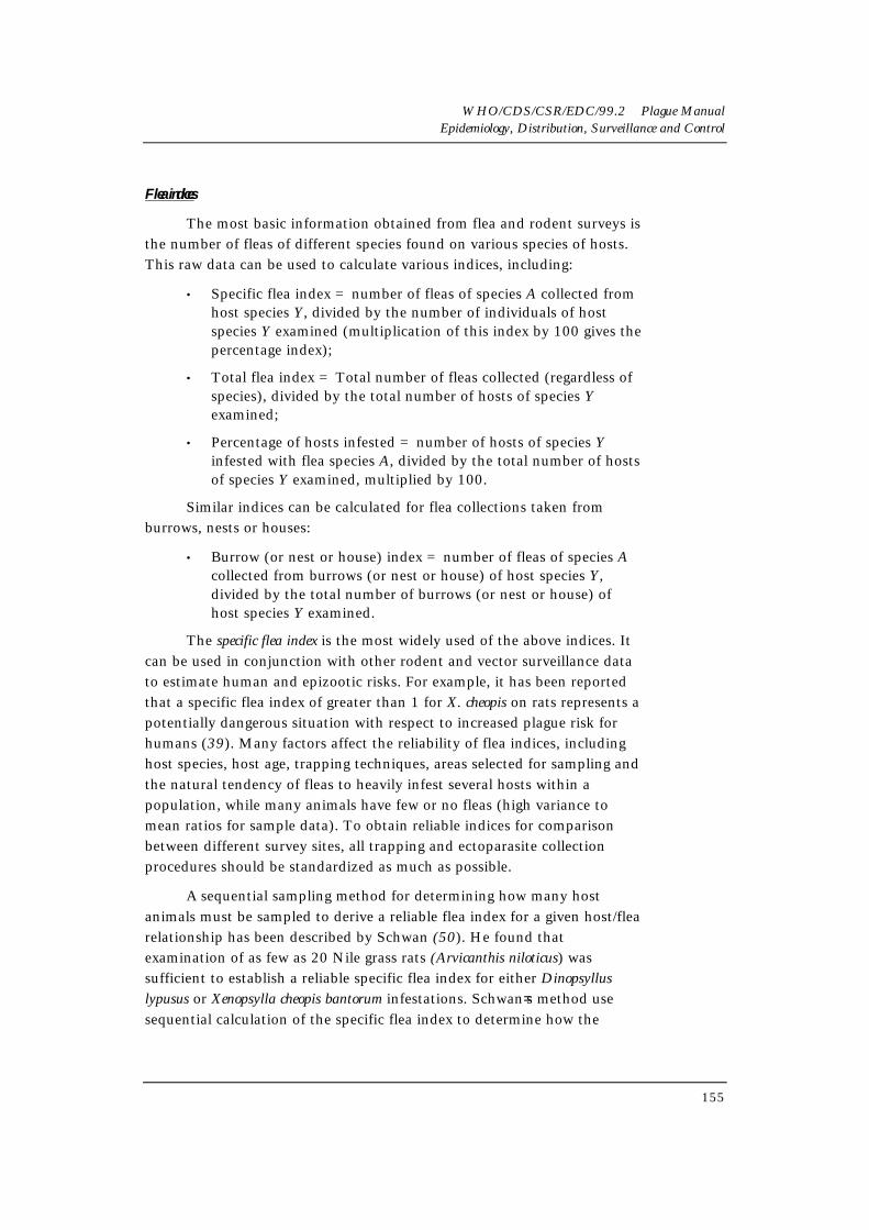
The most basic information obtained from flea and rodent surveys isthe number of fleas of different species found on various species of hosts.This raw data can be used to calculate various indices, including:
• Specific flea index = number of fleas of species A collected fromhost species Y, divided by the number of individuals of hostspecies Y examined (multiplication of this index by 100 gives thepercentage index);
• Total flea index = Total number of fleas collected (regardless ofspecies), divided by the total number of hosts of species Yexamined;
• Percentage of hosts infested = number of hosts of species Yinfested with flea species A, divided by the total number of hostsof species Y examined, multiplied by 100.
Similar indices can be calculated for flea collections taken fromburrows, nests or houses:
• Burrow (or nest or house) index = number of fleas of species Acollected from burrows (or nest or house) of host species Y,divided by the total number of burrows (or nest or house) ofhost species Y examined.
The specific flea index is the most widely used of the above indices. Itcan be used in conjunction with other rodent and vector surveillance datato estimate human and epizootic risks. For example, it has been reportedthat a specific flea index of greater than 1 for X. cheopis on rats represents apotentially dangerous situation with respect to increased plague risk forhumans (39). Many factors affect the reliability of flea indices, includinghost species, host age, trapping techniques, areas selected for sampling andthe natural tendency of fleas to heavily infest several hosts within apopulation, while many animals have few or no fleas (high variance tomean ratios for sample data). To obtain reliable indices for comparisonbetween different survey sites, all trapping and ectoparasite collectionprocedures should be standardized as much as possible.
A sequential sampling method for determining how many hostanimals must be sampled to derive a reliable flea index for a given host/flearelationship has been described by Schwan (50). He found thatexamination of as few as 20 Nile grass rats (Arvicanthis niloticus) wassufficient to establish a reliable specific flea index for either Dinopsylluslypusus or Xenopsylla cheopis bantorum infestations. Schwan=s method usesequential calculation of the specific flea index to determine how the

WHO/CDS/CSR/EDC/99.2 Plague ManualEpidemiology, Distribution, Surveillance and Control
156
inclusion of additional animals and their fleas changes the values for thisindex. As more and more animals and their fleas are included in the indexcalculations, the values begin to approach a particular value of the indexthat remains relatively stable with additional sampling. This can be showngraphically by plotting the number of host animals examined on the X axisand the specific flea index (calculated for X animals and their fleas) on theY axis. Schwan proposed that the point at which the slope of this graphapproaches zero represents the appropriate minimum number of animalsthat must be sampled to obtain a reliable specific flea index usingstandardized sampling techniques.
Identification of Y. pestis in fleas
Determining which flea species are infected with Y. pestis is criticalfor separating locally-important vectors from those which play only aminor role. Probably the most common method for determining whetherfleas are infected with plague is to inoculate susceptible laboratory animalswith ground fleas suspended in physiological saline (0.85%) (44). Materialfor flea suspensions may consist of either individual fleas or pools of fleas;fleas should be pooled by species, type of host, and area where collected.At the United States Centers for Disease Control and Prevention, Atlanta,the standard procedure to prepare fleas for inoculation is to grind fleapools (as many as 25 fleas per pool) in a mortar and pestle and thensuspend the ground material in approximately 2 ml of physiological saline(0.85%). This suspension is then inoculated subcutaneously into mice(0.5ml suspension per mouse). The mice are monitored over the next21 days, and those that die are necropsied to obtain tissues for bacterialisolation. Surviving mice can be sacrificed on day 21 postinoculation forsera and tissues. Y. pestis has also been detected in fleas using immunologictechniques, and PCR, but these procedures have yet to be widely testedunder field conditions (15, 45, 46). Recently, PCR has been demonstratedto be more sensitive and reliable in some situations than mouseinoculation (45). As with all PCR assays, however, care must be taken toavoid false positives due to contamination with amplicons generatedduring previous reactions or as a result of contamination from othersources.
Insecticide sensitivity surveys
After locally-important flea vectors have been identified, theirsensitivity to various insecticides should be determined. Data oninsecticide susceptibility for flea populations in plague-endemic areasshould be retained in the plague surveillance database and periodicallyupdated. Prior knowledge of insecticide resistance among local flea

WHO/CDS/CSR/EDC/99.2 Plague ManualEpidemiology, Distribution, Surveillance and Control
157
populations will enable plague control workers to select an appropriateinsecticide and save valuable time in the event of a plague epizootic. Kitsfor testing fleas for insecticide sensitivity are available through the WorldHealth Organization.
Evaluation of surveillance data
After each collection period, all data from dead animal collections,colony observations and rodent or carnivore serosurveys should beanalysed and mapped to determine the distribution of plague-infectedanimals in the area under study. Information from human and fleasurveillance should be included as well. Mapping this information duringepizootics can help determine the extent of the epizootic and whethercontrol efforts have succeeded in preventing its spread to areas wherehumans would be at high risk of infection. Such mapping helps clarify therisk that plague-infected rodents and their fleas pose to human populationsin the surrounding area. The proximity of infected animals to otherpopulations of the same species or those of other susceptible rodent species,as well as habitat availability, should also be noted. This information isuseful for estimating the likelihood that plague will spread to new areasand other rodent populations.
Whenever possible surveillance personnel should be aware of humanactivities that are likely to affect local rodent populations, such asdevelopment of new agricultural areas, villages or other developmentprojects. Rodents respond quickly to habitat changes that provide themwith new sources of food and harbourage, and existing plague problemswill be exacerbated by human activities that create new rodent habitats.Surveillance by health services
National, regional and local health services should work together todevelop a plague surveillance programme with clearly-definedresponsibilities for routine surveillance tasks and emergency investigations.Responsibilities should be distributed among the different health servicesso that human cases and epizootics can be identified and investigated asquickly as possible by individuals trained to assess human plague risks anddetermine appropriate control measures. Such predictive surveillance andemergency response capabilities require certain personnel, equipment andfacilities. The following section describes these basic requirements andsuggests how responsibilities for various surveillance tasks can be allocatedto local, regional or national services.
Country programmes must have a variety of personnel with trainingin such diverse fields as medicine, epidemiology, bacteriology, serology,

WHO/CDS/CSR/EDC/99.2 Plague ManualEpidemiology, Distribution, Surveillance and Control
158
entomology, mammalogy, health education and environmental sanitation.There should be personnel at the local level trained to inspect areas forevidence of rodent die-offs, collect samples for routine rodent and vectorsurveillance, and conduct educational programmes to promote plagueawareness, prevention and control. If local health services lack adequately-trained staff, experts from the regional or national health services shouldprovide this training. At the local level, health services are also expected tomaintain close contacts with the medical community so that human casesare recognized and reported as soon as possible.
Because of time and travel constraints, local agencies are normallyresponsible for at least the initial stages of human case investigations,including coordinating the collection and shipping of diagnosticspecimens, obtaining exposure histories from patients and performingpreliminary investigations of likely exposure sites.
Following these initial steps, more extensive epidemiological andenvironmental investigations (described earlier) should be instigated.Although local workers might be sufficiently trained to perform theseinvestigations, the national (or regional) health services should beprepared to provide additional expert assistance if necessary. For thisreason, national health services maintain at least one plague teamcomposed of experts whose combined training includes the disciplineslisted above, as well as knowledge of plague prevention and controltechniques (12,51,52). A minimum, but adequate, plague team iscomprised of an epidemiologist, bacteriologist/serologist andentomologist/zoologist.
At least one of these individuals (usually the epidemiologist) shouldhave medical qualifications (12). These experts can participate directly inhuman case investigations and surveillance activities or serve asconsultants for local or regional health officials. They can also train localworkers in techniques of plague diagnosis, treatment, surveillance,prevention and control. If a country lacks the resources and personnel toform such a plague team, or if plague has just recently entered a countryand a team has yet to be formed, it may be necessary to request theassistance of international consultants working under the direction of theWorld Health Organization. The national plague team should havesufficient field equipment and supplies to conduct emergency epidemic orepizootic investigations, as well as adequate transportation to moveequipment and plague team members to the affected area (52).Surveillance programmes should be prepared, if necessary, to hire and

WHO/CDS/CSR/EDC/99.2 Plague ManualEpidemiology, Distribution, Surveillance and Control
159
train temporary workers during emergency situations or seasonal peaks inplague activity.
Surveillance programmes must have adequate laboratory facilitiesfor performing bacteriological and serological analyses on plague-suspectspecimens. While it is preferable to have several laboratories located nearplague foci, at a minimum a central (or national) laboratory that cananalyse surveillance and diagnostic samples is essential (12). The centrallaboratory should be able to confirm the presence of Y. pestis in samples byculture, biochemical characterization and bacteriophage typing. Thelaboratory should also be proficient in using standard serologicaltechniques to detect plague antibodies in serum samples. Wheneverpossible the laboratory should be able to analyse samples by directimmunofluorescence. Personnel at the central laboratory should keepabreast of recent developments in molecular biology and be prepared toadopt new techniques that are cost-effective and useful for surveillancepurposes.

WHO/CDS/CSR/EDC/99.2 Plague ManualEpidemiology, Distribution, Surveillance and Control
161
References
1. Isaacson M, Hallett AF. Serological studies on human plague inSouthern Africa. Part 1: Plague antibody levels in a populationduring a quiescent and a subsequent active period in an endemicregion. South African Medical Journal, 1975, 49:1165-1168.
2. White ME, Gordon D, Poland JD, Barnes AM. Recommendationsfor the control of Yersinia pestis infections. Infection Control, 1980,1:324-329.
3. Barnes AM. Surveillance and control of bubonic plague in theUnited States. Symposium Zoological Society London, 1982,50:237-270.
4. Goldenberg MI, Hudson BN, Kartman L. Pasteurella Infections:Pasteurella pestis. In: Bodily HL, Updyke EL, Mason JO (eds.)Diagnostic Procedures for Bacterial, Mycotic, and Parasitic Infections, NewYork, American Public Health Association, 1970, 422-439.
5. Clark JD, Olfert ED. Rodents (Rodentia); Part 4. Special Medicine:Mammals. In: Fowler ME (ed.) Zoo and Wild Animal Medicine,Philadelphia, W.B. Saunders, 1986, 727-748.
6. Moody MD, Winter CC. Rapid identification of Pasteurella pestiswith fluorescent antibody. III. Staining Pasteurella pestis in tissueimpression smears. Journal of Infectious Diseases, 1959, 104:288-294.
7. Winter CC, Moody MD. A rapid identification of Pasteurella pestiswith fluorescent antibody. I. Production of specific antiserum withwhole cell Pasteurella pestis antigen. Journal of Infecious Diseases,1959, 104:274-280. II. Specific identification of Pasteurella pestis indried smears. Journal of Infectious Diseases, 1959, 104:281-287.
8. Harlow E, Lane D. Antibodies: A Laboratory Manual. Cold SpringHarbor, New York, Cold Spring Harbor Laboratory, 1988.
9. Cary SG, Blair EB. New transport medium for the transport orshipment of clinical specimens. Journal of Bacteriology, 1964,88:99-98.
10. Cavanaugh DC, Vivona S, Do-Van-Quy, Gibson FL, Deuber GL,Rust JH Jr. A transport medium for specimens containing Pasteurellapestis. Bulletin of the World Health Organization, 1967, 37:455-460.
11. Gross B, Bonnet DH. Snap versus cage traps in plague surveillance.Public Health Reports, 1949, 64:1214-1216.
12. Bahmanyar M, Cavanaugh DC. Plague Manual, Geneva, WorldHealth Organization, 1976.

WHO/CDS/CSR/EDC/99.2 Plague ManualEpidemiology, Distribution, Surveillance and Control
162
13. Hudson BW, Quan SF, Goldenberg MI. Serum antibody responsesin a population of Microtus califomicus and associated rodent speciesduring and after Pasteurella pestis epizootics in the San FranciscoBay Area. Zoonoses Research, 1964, 3:15-29.
14. Hudson BW, Goldenberg MI, Quan TJ. Serologic and bacteriologicstudies on the distribution of plague infection in a wild rodentplague pocket in the San Francisco Bay area of California. Journal ofWildlife Diseases, 1972, 8:278-286.
15. Levi MI, Ch.B. LTIukhanov, Kuznetskova KA. [Detection of theplague bacillus in fleas by means of an immunoenzyme assay withmonoclonal antibodies.] Meditsinskaya Parazitologiya ParasitaryneBolezni, 1989, 1:57-60.
16. Cavanaugh DC, Thorpe BD, Bushman JB, Nicholes PS, Rust JH Jr.Detection of an enzootic plague focus by serological methods.Bulletin of the World Health Organization, 1965, 32:197-203.
17. Davis DHS, Heisch, RB, McNeill D, Meyer KF. Serological surveyof plague in rodents and other small mammals in Kenya.Transactions of the Royal Society of Tropical Medicine and Hygiene, 1968,62(2):838-861.
18. Hudson BW, Goldenberg MI, McKluskie JD, Larson HE, McGuireCD, Barnes AM, Poland JD. Serological and bacteriologicalinvestigations of an outbreak of plague in an urban tree squirrelpopulation. American Journal of Tropical Medicine and Hygiene, 1971,20:255-263.
19. Hudson BW, Quan TJ. Serologic observations during an outbreakof rat-borne plague in the San Francisco Bay area of California.Journal of Wildlife Diseases, 1975, 11:431-436.
20. Taylor P, Gordon DH, Isaacson M. The status of plague inZimbabwe. Annals of Tropical Medicine and Parasitology, 1981,75:165-173.
21. Shepherd AJ, Leman PA, Hummutzsch DE, Swanepoil R. Acomparison of serological techniques for plague surveillance.Transactions of the Royal Society of Tropical Medicine and Hygiene,1984, 78:771-773.
22. Isaacson M. Plague and cholera surveillance in southern Africa.South Africa Medical Journal, Supplement 1986:43-46.
23. Gill DE, Shepherd AJ, Leman PA, Erasmus BH. Plague surveillancein the northern Cape Province, South Africa. South African Journal ofScience, 1987, 83:159-162.

WHO/CDS/CSR/EDC/99.2 Plague ManualEpidemiology, Distribution, Surveillance and Control
163
24. Golubinski IE, Rudnik MP, Innokentieva TI, Lesnikov NT,Shmidt AA, Dneprovskaya LG, Brinkman VD, Andreevskaya NM.[Results of enzyme immunoassay for epizootiological investigationsof natural plague foci in Siberia.] Zhurnal Microbiologii, Epidemiologii iImmunobiologii, 1988, 10:34-37.
25. Njunwa KJ, Mwaiko GL, Kilonzo BS, Mhina JI. Seasonal patternsof rodents, fleas and plague status in the Western UsambaraMountains, Tanzania. Medical and Veterinary Entomology, 1989,3:17-22.
26. Williams JE. Use of ELISA to reveal rodent infections in plaguesurveillance and control programmes. Bulletin of the World HealthOrganization, 1990, 68:341-345.
27. Kilonzo BS. Observations on the epidemiology of plague inTanzania during the period 1974-1988. East African Medical Journal,1992, 69:494-499.
28. Kilonzo BS, Makundi RH, Mbise TJ. A decade of plagueepidemiology and control in the western Usambara mountains,north-east Tanzania. Acta Tropica, 1992, 50:323-329.
29. Wolff KL, Hudson BW. Paper-strip blood-sampling technique forthe detection of antibody to the plague organism Yersinia pestis.Applied Microbiology, 1974, 28:323-325.
30. Hall ER. The mammals of North America. Vol. 1 and Vol. 2. 2ndedition. John Wiley and Sons, New York, 1981.
31. Rust JH Jr, Miller BE, Bahmanyar M, Marshall JD Jr, Pumaveja S,Cavanaugh DC, Hia UST. The role of domestic animals in theepidemiology of plague; Part II:Antibody to Yersinia pestis in sera ofdogs and cats. Journal of Infectious Diseases, 1971, 124:527-531.
32. Cruickshank JG, Gordon DH, Taylor P, Naim H. Distribution ofplague in Rhodesia as demonstrated by serological methods. CentralAfrican Journal of Medicine, 1976, 22:127-130.
33. Willeberg PW, Ruppanner R, Behymer DE, Higa HH, Franti CE,Thompson RA. Epidemiologic survey of sylatic plague byserotesting coyote sentinels with enzyme immunoassay. AmericanJournal of Epidemiology, 1979, 110:328-334.
34. Hopkins DD, Gesbrink RA. Surveillance of sylvatic plague inOregon by serotesting carnivores. American Journal of Public Health,1982, 72:1295-1297.
WHO/CDS/CSR/EDC/99.2 Plague ManualEpidemiology, Distribution, Surveillance and Control
164
35. Smith CR, Nelson BC, Barnes AM. The use of wild carnivoreserology in determining patterns of plague activity in rodents inCalifornia. In: Clark DO (ed.) Proceedings of the Eleventh VertebratePest Conference, Sacramento, University of California, Davis, 1984:71-76.
36. Nelson JH, Decker RH, Barnes AM, Nelson BC, Quan TJ,Gillogly AR, Phillips GS. Plague surveillance using wild boars andwild carnivore sentinels. Environmental Health, 1985, 47:306-309.
37. Clover JR, Hofstra TD, Kuluris BG, Schroeder MT, Nelson BC,Barnes AM, Boltzler RG. Serologic evidence of Yersinia pestis infectionin small mammals and bears from a temperate rainforest of NorthCoastal California. Journal of Wildlife Diseases, 1989, 25:52-60.
38. Gordon DH, Isaacson M, Taylor P. Plague antibody in large Africanmammals. Infection and Immunology, 1979, 26:767-769.
39. Pollitzer R. Plague. Geneva, World Health Organization, 1954(Monograph series).
40. Poland J, Bames A. Plague. In: Steele JH, Stoenner H, Kaplan W(eds.) CRC Handbook Series in Zoonoses. Section A:Bacterial, Rickettsial,and Mycotic Diseases, Volume 1, Boca Raton Fla., CRC Press, 1979.
41. Kartman L, Prince FM, Quan SF, Stark HE. New knowledge on theecology of sylvatic plague. Annals of the New York Academy of Science,1958, 70:668-71.
42. Holland GP. The Siphonaptera of Canada. Ottawa, Canada;Dominion of Canada, Department of Agriculture, 1949(Publication 817, Technical Bulletin 70).
43. Furman DP, Catts EP. Manual of Medical Entomology, 4th ed.Cambridge, Cambridge University Press, 1982.
44. Quan TJ, Barnes AM, Poland JD. Yersinioses. In: Balows A,Hausler WJ Jr. (eds). Diagnostic Procedures for Bacterial, Mycotic andParasitic Infections, 6th ed. Washington DC, American Public HealthAssociation, 1981:723-745.
45. Engelthaler DM, Gage KL, Montenieri JA, May Chu , Carter LG.PCR Detection of Yersinia pestis in Fleas: Comparison with MouseInoculation. Journal of Clinical Microbiology, 1999, 37(6)1980-1984.
46. Hinnebusch J, Schwan TG. New method for plague surveillanceusing polymerase chain reaction to detect Yersinia pestis in fleas.Journal of Clinical Microbiology, 1993, 31:1511-1514.

WHO/CDS/CSR/EDC/99.2 Plague ManualEpidemiology, Distribution, Surveillance and Control
165
47. Vakhrousheva ZP, Gorchakov AD, Kolupaeva NA, Chernykh EG.[The use of flannel flags for collecting fleas at the entrance ofburrows of steppe rodents.] Meditsinskaya Parazitologiya iParasitarnye Bolenzi, 1989, 1: 54-57.
48. Beard ML, Rose ST, Barnes AM, Montenieri JA. Control ofOropsylla hirsuta, a plague vector, by treatment of prairie dogburrows with 0.5% permethrin dust. Journal of Medical Entomology,1992, 29:25-29.
49. Borror DJ, DeLong DM, Triplehorn CA. Introduction to the study ofinsects, 5th ed. Philadelphia, Saunders College Publishing, 1981.
50. Schwan TG. Sequential sampling to determine the minimumnumber of host examinations required to provide a reliable flea(Siphonaptera) index:. Journal of Medical Entomology, 1984,21:670-674.
51. Technical guide for a system of plague surveillance. WHO WeeklyEpidemiological Record, 1973, 14:149-160.
52. Plague surveillance and control. WHO Chronicle, 1980, 34:139-143.

WHO/CDS/CSR/EDC/99.2 Plague ManualEpidemiology, Distribution, Surveillance and Control
167
7NATIONAL HEALTH SERVICES IN
PREVENTION & CONTROL
Dr Kenneth L. Gage
Although local or regional health departments might have considerableexpertise and do an excellent job of managing plague within their districts, thepotential for the rapid spread of plague from one region of a country to anotherrequires national prevention and control programmes capable of coordinatingand assisting local and regional efforts. Plague's lack of respect for internationalboundaries also requires that national health services of neighbouring countriescooperate with one another to successfully control this disease. Internationalcontrol activities are best administered by national health services rather thanby local or regional agencies. Surveillance and control of plague in port facilitiesand international airports should also fall under the supervision of the nationalhealth services. As was described under plague surveillance, the organization ofnational, local and regional plague prevention and control programmes mayvary considerably from one country to another, but several important featuresare common to all.
The World Health Organization (3) has recommended a four-phasedsystem of plague prevention and control that can be adapted to therequirements and resources of different countries. This section summarizes thissystem and describes how its implementation will result in a national plagueprevention and control programme that is effectively integrated with local andregional programmes.
The first two phases of the WHO system address emergencymeasures to be implemented whenever a human plague case occurs. Plagueprevention and control programmes in each country should have adequatepersonnel, equipment and laboratory facilities to undertake the phase 1 andphase 2 activities described below. Phases 3 and 4 outline the establishment ofa surveillance system and development of long-term prevention and controlmeasures. These activities require a greater commitment of personnel andresources than in phases 1 and 2, but their successful completion willsignificantly reduce the risk of human plague. It is recommended, therefore,that each country implement phases 3 and 4 to the fullest extent possible.
WHO/CDS/CSR/EDC/99.2 Plague ManualEpidemiology, Distribution, Surveillance and Control
168
Phase 1: Case recognition and medical intervention
National health service officials should verify that local and regionalofficials are trained and prepared to undertake emergency measures whenever ahuman case is suspected. After identifying a suspect plague case, local healthservices should:
(1) notify national and/or regional authorities;
(2) ensure that appropriate specimens are shipped to a qualifiedlaboratory for diagnostic confirmation of Y. pestis infection;
(3) verify that patients have been placed on appropriate antibiotictreatment and that local supplies of antibiotics are adequate tohandle further cases; and
(4) isolate pneumonic plague patients and cooperate with other healthservices to identify, monitor and, if necessary, arrange prophylactictreatment for individuals in contact with cases.
In addition to the above measures, a preliminary epidemiologicalinvestigation should be initiated. The purpose of this investigation is to obtainan exposure history from the patient in order to make an initial assessment oflikely sources of infection and potential risks to others in the area. National andregional health services, including the national plague team described earlier inthis manual, may be dispatched to the area if local skills or resources areinadequate. Plague experts with the national health services can also help localand regional authorities determine whether to recommend vaccination forindividuals in high-risk areas or occupations. If a vaccination programme isapproved and vaccine stocks are not locally available, the national healthservices should be prepared to provide local and regional authorities withinformation on where supplies of vaccine can be obtained.
Phase 2: Epidemiological and epizootical investigation and emergency control
The second phase of the programme should be initiated immediatelyfollowing phase 1. Phase 2 activities include an intensive environmentalinvestigation of potential exposure sites for the human case(s) and initiation ofemergency control measures to prevent additional cases. These investigationsrequire both epidemiologists and persons trained in techniques for surveillanceand control of rodents and fleas. The national plague team, whose services mayhave already been requested during phase 1, can provide this expertise whenlocal or regional personnel lack adequate training. The plague team's centrallaboratory resources should be made available for the investigation.

WHO/CDS/CSR/EDC/99.2 Plague ManualEpidemiology, Distribution, Surveillance and Control
169
The goals of the phase 2 environmental investigation are to:
(1) identify the rodent and flea species most likely to be sources ofinfection in the area where the human case(s) was exposed;
(2) determine the extent of epidemics and/or epizootics associated withthe initial human case; and
(3) identify areas of potential risk to humans.
This information is used to determine emergency control measures to betaken to prevent additional human cases.
Phase 3: Surveillance and control
The goal of phase 3 is to establish a surveillance and control programme.Because of ecological differences between plague foci in different geographicregions, preliminary research is needed to identify which local rodent and fleaspecies should be targeted for extensive surveillance and control.
The research data can also be used in conjunction with information onlocal landscape, human activity and host/vector ecology to design preventionand control strategies appropriate for a particular plague focus. Anyrodenticidal, insecticidal or environmental control measures developed duringthis phase should be tested locally to evaluate their effectiveness in reducing thehuman risk of plague.
Where local or regional health services lack the expertise to perform thisresearch they must be assisted by personnel at the national level. The nationalhealth services should also work with local and regional authorities to developand administer educational programmes to increase awareness and knowledgeamong health care personnel and the general public.
Phase 4: Management
The final phase of the plague prevention and control programme stresseslong-term management of plague foci. Such management calls for continuoussurveillance of the important host and vector species identified during phase 3.Once the surveillance programme identifies a plague epizootic, controlmeasures developed in phase 3 should be implemented as soon as possible.Long-term environmental management of plague foci should also be promoted.Environmental management stresses the elimination or reduction of areas, nearhomes or workplaces, that are attractive to plague-susceptible rodents. Plague

WHO/CDS/CSR/EDC/99.2 Plague ManualEpidemiology, Distribution, Surveillance and Control
170
staff should work with other government officials to regulate and modifyactivities and practices B such as agricultural projects, construction, placementof garbage disposal facilities and so on B that are likely to lead to increased foodand harbourage for locally-important rodent hosts. Health services shouldcontinue the educational programmes developed during phase 3. Finally,research to improve existing surveillance and control techniques shouldcontinue, following the procedures outlined for phase 3.

WHO/CDS/CSR/EDC/99.2 Plague ManualEpidemiology, Distribution, Surveillance and Control
171
References
1. Bahmanyar M, Cavanaugh DC. Plague Manual. Geneva, World HealthOrganization, 1976.
2. Technical guide for a system of plague surveillance. WHO WeeklyEpidemiological Record, 1973, 14:149-160.
3. Plague surveillance and control. World Health Organization Chronicle,1980, 34:139-143.