Embed Size (px)
Citation preview

Dr. Dr. AbdulrahmanAbdulrahman HagrHagr AlAl--GhamdiGhamdi MBBS MBBS FRCS(cFRCS(c))Assistant Professor KingAssistant Professor King SaudSaud University University
Otolaryngology ConsultantOtolaryngology ConsultantOtologistOtologist, , NeurotologistNeurotologist & Skull Base Surgeon& Skull Base Surgeon
King King AbdulazizAbdulaziz HospitalHospital

Ear•• EmbryologyEmbryology•• Congenital anomaliesCongenital anomalies• Anatomy• Physiology• Disease of external ear• Acute Otitis media
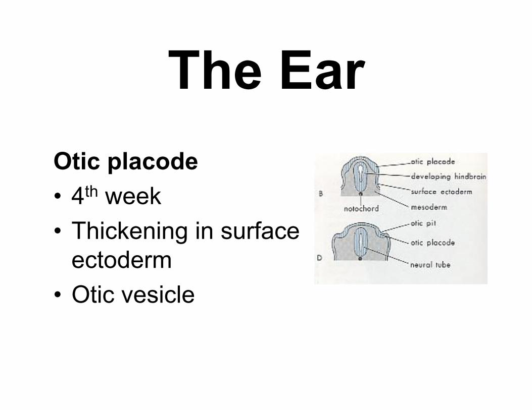
The EarOtic placode• 4th week • Thickening in surface
ectoderm • Otic vesicle

The Ear• Otic vesicle
– Invaginates of mesenchyme– Detaches from ectoderm– divides into 2 regions:
• Utricular portion– Utricle– Semicircular canals
• Saccular portion– Saccule– cochlea

Inner Ear

1st Pharyngeal Pouch• Elongates into the tubotympanic recess
• The tubotympanic recess becomes the tympanic cavity and mastoid antrum
• Distally contacts the 1st pharyngeal cleft TM
• Proximally connects the pharynx eustachiantube

Middle Ear
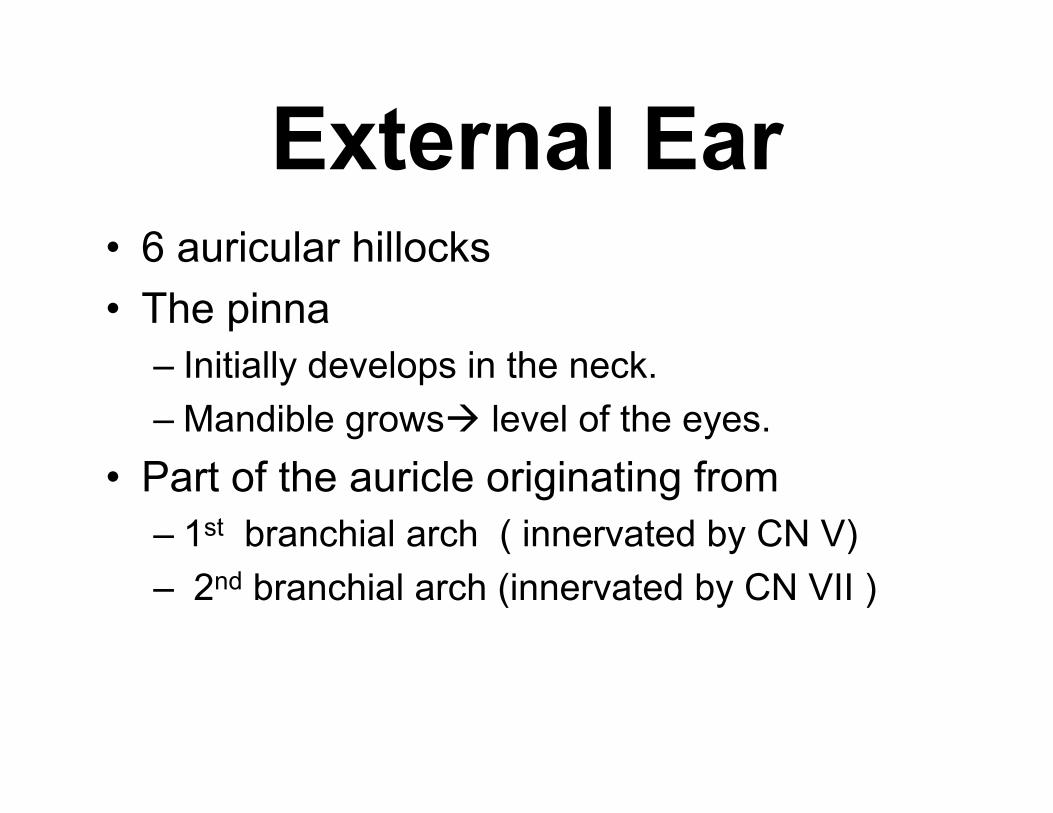
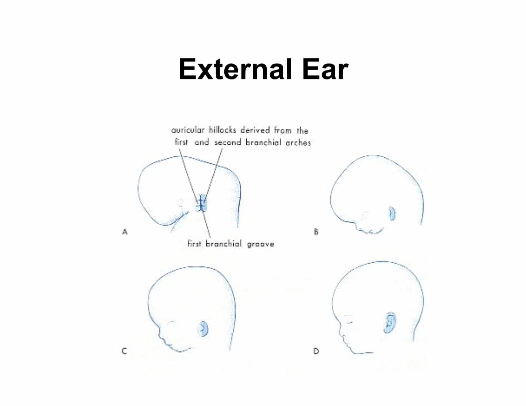
External Ear• 6 auricular hillocks• The pinna
– Initially develops in the neck. – Mandible grows level of the eyes.
• Part of the auricle originating from– 1st branchial arch ( innervated by CN V)– 2nd branchial arch (innervated by CN VII )
External Ear

External Ear

Ear•• EmbryologyEmbryology
•• Congenital anomaliesCongenital anomalies• Anatomy• Physiology• Disease of external ear• Acute Otitis media

Congenital anomaliesCongenital anomalies• Outer ear
– Microtia– Bat Ear
• Middle ear– eustachian tube dysfunction– Otosclerosis
• Inner ear– Aplasia (Michel Aplasia)
– Partial Aplasia (Mondini Aplasia)

Microtia

MicrotiaMicrotia

Bat ear

Down Syndrome• Trisomy 21
• 1 in 700 births
• Maternal age >35
Hearing Concerns• Conductive hearing loss
– more common– small pinna– stenotic EAC– eustachian tube dysfunction– ossicular fixation
• Sensorineural hearing loss– less common
Michel AplasiaMichel Aplasia• 9 weeks gestation Cochlea
fully formed • Complete agenesis of IE• Normal External and
middle ear• Affected ears are anacusic

Mondini Mondini AplasiaAplasia• Only the basal coil can
be identified• Interscalar septum is
absent • enlarged
endolymphatic duct

Ear•• EmbryologyEmbryology•• Congenital anomaliesCongenital anomalies
• Anatomy• Physiology• Disease of external ear• Acute Otitis media

Anatomy• Inner Ear
– Cochlea– Sacule– Utricle– Semicircular canals
• Middle Ear– 3 ossicles– Mastoid– Eustachian Tube

Anatomy• Auricle is mostly skin-
lined cartilage• External auditory
meatus– 2.5 cm long– Cartilage: ~40%– Bony: ~60%– S-shaped– Narrowest portion at
bony-cartilage junction

External Auditory Canal
• Skin– Cartilage
• 1.0 mm• Epidermis with papillae• Dermis• Sub-Q
– Bone• 0.2 mm• No papillae, no sub q

AnatomyEAC is related to various
contiguous structures• Tympanic membrane• Mastoid• Glenoid fossa• Cranial fossa• Infratemporal fossa

AnatomyInnervation: • Cranial nerves
– V (Itching AR)– VII ( Acoustic neuroma sign)– IX (Cough)– X (Vaso-Vagal)
• Greater auricular nerve (Post-Parotidectomy)

Anatomy• Arterial supply
– Superficial temporal– Posterior auricular
• Venous drainage– Superficial temporal – Posterior auricular veins
• Lymphatics– External ear :Parotid, deep cervical– Middle ear : Retropharyngeal


Ear•• EmbryologyEmbryology•• Congenital anomaliesCongenital anomalies• Anatomy
• Physiology• Disease of external ear• Acute Otitis media
ورد في القرآن الكريم مرًة) 19( لفظي السمع و البصر معًا
سبعة عشر لفظة السمع قبل البصر منها ) 17(ذآر في 78: المؤمنون )و هو الذي أنشأ لكم السمع واألبصار و األفئدة : ( قوله تعالى– .36:اإلسراء ) مسؤًالإن السمع و البصر و الفؤاد آل أولئك آان عنه ( قوله –
هماماعدا ايتين اثنين فقطبها ام لهم آذان يسمعون بهاام لهم اعين يبصرون : قال تعالى – الكهف.… واسمع في سورةبهابصر : قال تعالى –
فما السر ؟؟

Hearing: Mechanics
Figure 10-19: Sound transmission through the ear

A Little Vestibular Physiology…….

Why have a VOR?
1. Stabilize retina in space – fast!
On headmovement
2. Posture Control
Do finger test

Types of Spatial Movement
• Rotational – 3 degrees of freedom
• Translational – 3 degrees of freedom
Semicircular Canals
Otolith Organs

Basic Mechanism of Detection of Rotation
• INERTIA• Detects head acceleration – but encodes
head velocity (i.e. integrator)

Velocity Profile vs Signal
VelocityAccn Deaccn
Nerve Firing
Decays with constant velocity

Canals are Paired
AC
PC
AC
PC
HC HC

Push-Pull SystemFiring Rate
0
100s/s
Right HC
Left HC
Differential is Driving VOR

Ear•• EmbryologyEmbryology•• Congenital anomaliesCongenital anomalies• Anatomy• Physiology
• Disease of external ear• Acute Otitis media

Disease of external ear• Wax• Tumor
– Exostosis– Osteoma
• Foreign body• Infection• Trauma

Wax


Tumor• Benign
– Exostosis– Osteoma
• Malignent– Rare– Metastasis

Otitis Externa

Clinical Course• Itching• Progresses to:
– Pain– Decreased hearing– Drainage (usually from bacterial infection)

AOE: Mild to Moderate Stage
• Symptoms– Pain– Increased pruritus
• Signs– Erythema– Increasing edema– Canal debris,
discharge

AOE: Severe Stage
• Severe pain, worse with– Ear movement– Chewing
• Signs– Lumen obliteration– Purulent otorrhea– Involvement of periauricular
soft tissue
Microbiology
• Bacteria 50% of cases– Staph aureus– Pseudomonas– Proteus
• Fungi– Aspergillus – tropical– Candida albicans – temperate

Epidemiology• Warm, humid climate
– “swimmer’s ear”• Poor hygiene• Closed canal
– Hearing aid– Turbans in India
• Composition of cerumen– pH changes from acid to alkaline (D.M)– Softer – washed out– Hard block the canal
• Instrumentation of ear canal

Diagnosis• Persistent disease
– Resistant – Fungal– Dermatological etiologies
Cultures will be helpful

Treatment• meticulous cleaning
• every 2-3 days• weekly
• Topical antibiotic• Water precautions
Furunculosis• Acute localized infection• Lateral 1/3 of posterosuperior canal• Obstructed apopilosebaceous unit• Pathogen: S. aureus

Furunculosis: Symptoms• Localized pain• Pruritus• Hearing loss (if lesion occludes canal)

Furunculosis: Signs• Edema• Erythema• Tenderness• Occasional fluctuance

Furunculosis: Treatment
• Local heat• Analgesics• Oral anti-staphylococcal antibiotics• Incision and drainage reserved for
localized abscess• IV antibiotics for soft tissue extension

Otomycosis• Fungal infection of EAC skin• Primary or secondary (AB)• Most common organisms: Aspergillus and
Candida

Otomycosis: Symptoms
• Often indistinguishable from bacterial OE• Pruritus deep within the ear• Dull pain• Hearing loss (obstructive)• Tinnitus

Physical Exam
• Early – Normal – Canal erythema– Mild edema
• Later – “wet newspaper”– red, tender skin– Fungal hyphae
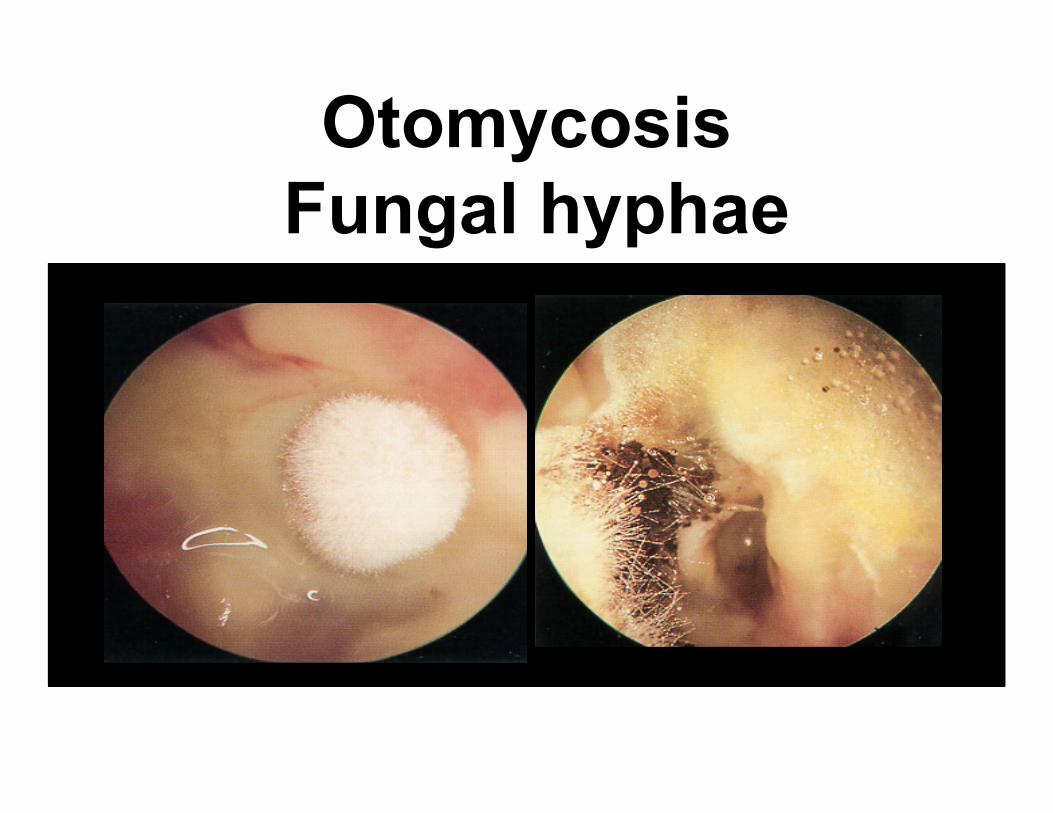
OtomycosisFungal hyphae

Otomycosis: Treatment
• Thorough cleaning • Drying of canal• Topical antifungals

Bullous Myringitis
• Viral infection• Bacteria of OM • Confined to tympanic membrane• Children

Bullous Myringitis: Symptoms
• Sudden onset of severe pain• No fever• No hearing impairment• Bloody otorrhea (significant) if rupture

Bullous Myringitis: Signs• Inflammation limited
to TM & nearby canal• Multiple reddened,
inflamed blebs• Hemorrhagic vesicles

Bullous Myringitis: Treatment
• Self-limiting• Analgesics• Topical antibiotics to prevent secondary
infection• Incision of blebs is unnecessary

Necrotizing External Otitis(NEO)
• Potentially lethal infection • DM and immunocompromised patients• Pseudomonas aeruginosa

Malignant Otitis Externa
4 Ds• Diabetes mellitus • Discharge (Purulent )• Discomfort• Dysfunction Cranial nerve
• Granulation obscured TM

NEO: Imaging
• Plain films• Computerized tomography – most used• Technetium-99 – reveals osteomyelitis • Gallium scan – useful for evaluating Rx• Magnetic Resonance Imaging
NEO: Treatment
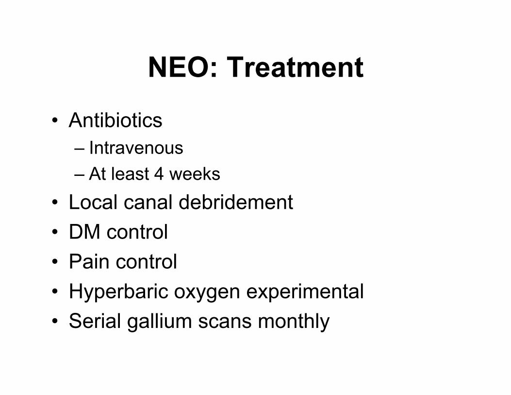
• Antibiotics – Intravenous – At least 4 weeks
• Local canal debridement • DM control• Pain control • Hyperbaric oxygen experimental• Serial gallium scans monthly

NEO: Mortality
• 25 % Death rate • 60% with multiple cranial neuropathies • 25 % Recurrence • May recur up to 12 months after treatment

Perichondritis: Signs• Tender auricle• Induration• Edema• Advanced cases
– Crusting – Involvement of soft
tissues

Herpes Zoster Oticus
• J. Ramsay Hunt • Varicella zoster• Shingles: Infection along one or more
cranial nerve dermatomes • Ramsey Hunt syndrome:
– Herpes zoster of the pinna – Otalgia– Facial paralysis

Herpes Zoster Oticus: Symptoms
• Early: burning pain in one ear, headache, malaise and fever
• Late (3 to 7 days): vesicles, facial paralysis
Herpes Zoster Oticus: Treatment
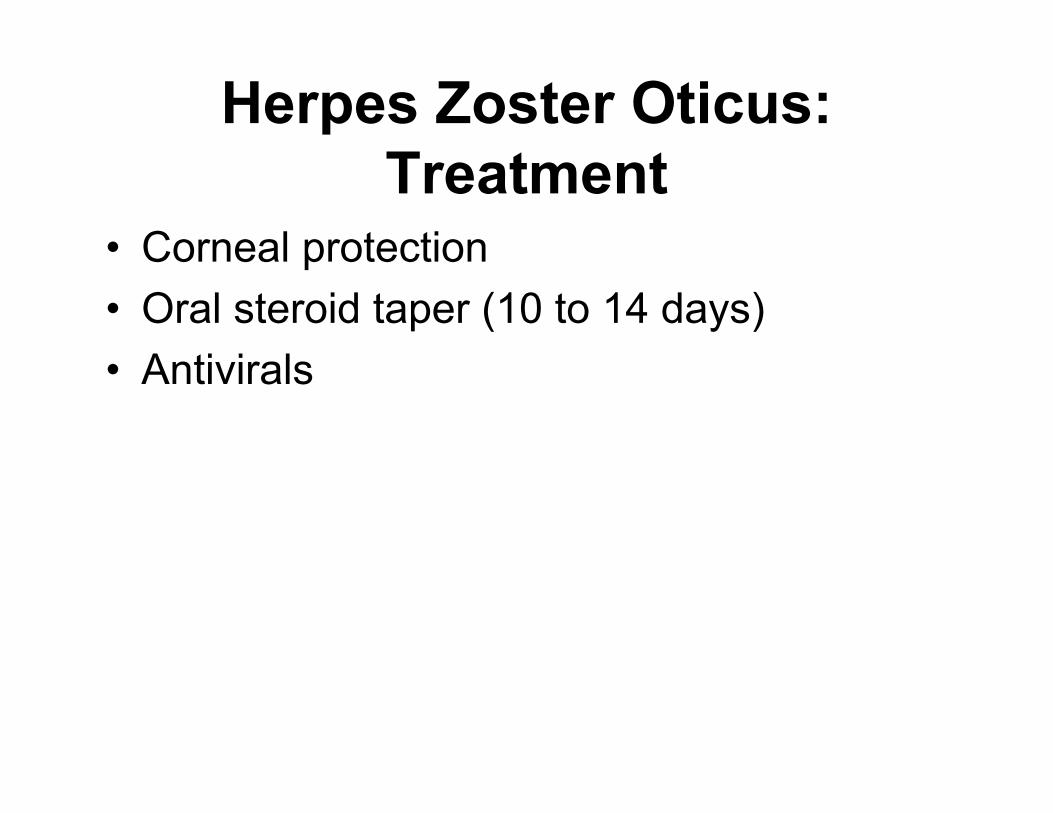
• Corneal protection• Oral steroid taper (10 to 14 days)• Antivirals

Erysipelas• Acute superficial cellulitis• Group A, beta hemolytic
streptococci• Skin: bright red; well-
demarcated, • Rapid treatment with oral
or IV antibiotics if insufficient response

Ear Trauma

Auricle injuriesAuricle injuries• Hematomas
separate the perichondrium (blood supply) from the cartilage
excise fibrous tissue• Apply pressure dressing , drain
• Avulsion: – Reimplantation– Microvascular anastomosis


Cauliflower EarCauliflower Ear

Racoon eyes sign

Battle’s sign

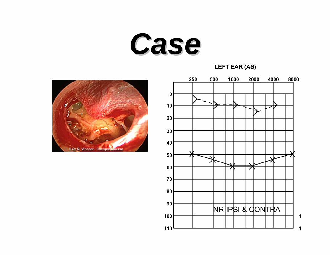
Case Case LEFT EAR (AS)
250 500 1000 2000 4000 8000
100
0
10
20
30
40
50
60
70
80
90
110
1
1
NR IPSI & CONTRA

Fractures• Longitudinal
– 80% of Temporal Bone Fractures– 15-20% Facial Nerve involvement
• Transverse– 20% of Temporal Bone Fractures– 50% Facial Nerve Involvement


R

Ear•• EmbryologyEmbryology•• Congenital anomaliesCongenital anomalies• Anatomy• Physiology• Disease of external ear
• Acute Otitis media

Otitis Media Definition
Inflammation of the middle earMay also involve inflammation of
mastoid, petrous apex, and perilabyrinthine air cells

Otitis Media• Most common reason for visit to
pediatrician• Tympanostomy tube placement is 2nd
most common surgical procedure in children
• Development of multidrug-resistant bacteria

Otitis Media - Classification
• Acute OM < 3 wk• Subacute OM 3 wks to 3 months• Chronic OM > 3 mos

OM - Epidemiology• Age• Sex• Day care• Seasons
• Genetics• Breast-feeding• Smoke exposure• Medical conditions

OM - Medical Conditions• Cleft palate
– decreases after repair• Craniofacial disorders
– Treacher-Collins• Down’s syndrome• Ciliary dysfunction
• Immune dysfunction– AIDS– steroids, chemo– IgG deficiency
• Obstruction– adenoids– NG tubes– NT intubation– malignancy
OM - Epidemiology
• Increasing incidence?• Increases after newborn period• 2/3 with AOM by one year of age• 1/2 with >3 episodes by three years• most common in 6 - 11 mos
OM - Day Care
• Greater risk of AOM in children < 3 years• Home care best• Day care
– Large group – Exposures with wider range of flora– Increased URI’s

OM - Breast-feeding
• Decreases incidence of URI and GI disease
• Decreases duration of OM • Protective factor in breast-milk?

OM - smoke exposure
• Induces changes in respiratory tract• Increased AOM and persistent effusion• Increased chronic and recurrent AOM

التدخين في المملكة ) نسبة استهالك للفرد ( في الترتيب العالمي 23•مليارات ريال 5المدخنين يحرقون اآثر من •الف طن 40اآثر من •القطاع الصحي والتعليمي % 30•
) االطباء ومعلمين والطالب (. الف مدخنة معظمهن من المراهقات 600•
14030 العدد -م 2006 نوفمبر 22 -هـ 1427 ذي القعدة 1الرياض االربعاء

Eustachian Tube• Connects middle ear and nasopharynx• Lumen shaped like two cones • Mucosa
– Mucous producing cells – Ciliated cells

Eustachian tube• Usually closed• Tensor veli palatini active opening• Opens during
– Swallowing– Yawning– Sneezing
• Opening involves cartilaginous portion

Eustachian tubeFunctions• Protection from nasopharyngeal
– Sound – Secretions
• Clearance of middle ear secretions• ME Ventilation (pressure regulation)
Eustachian tube
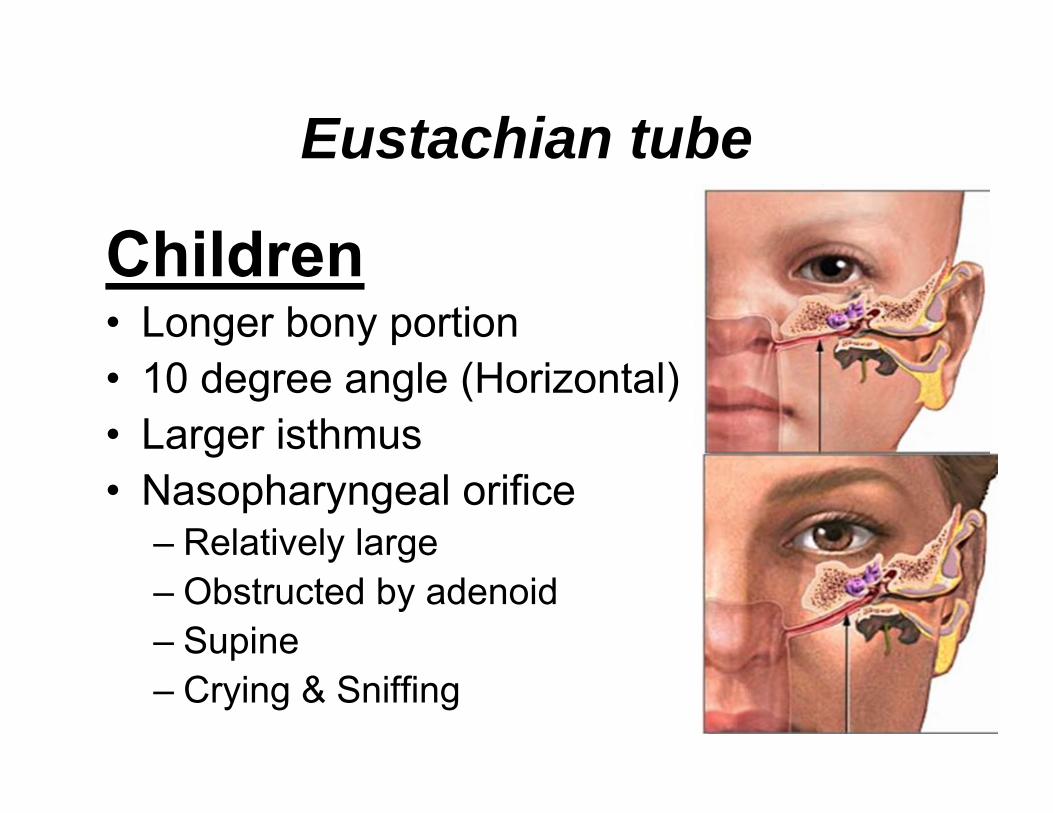
Children• Longer bony portion• 10 degree angle (Horizontal) • Larger isthmus• Nasopharyngeal orifice
– Relatively large – Obstructed by adenoid– Supine – Crying & Sniffing
Middle ear Pathology• Inflammation Edema• PMN infiltration• Epithelial ulceration • Granulation tissue• Fibrosis, • influx of chronic inflammatory cells• Increased columnar and goblet cells• Osteitis

Microbiology• S. pneumoniae - 30-35%• H. influenzae - 20-25%• M. catarrhalis - 10-15%• Group A strep - 2-4%• Infants with higher incidence of gram
negative bacilli

Virology• RSV - 74% of middle ear isolates• Rhinovirus• Parainfluenza virus• Influenza virus

Treatment - AOM
• Adults and older children - observation• Antibiotics - consider drug resistance
patterns

Antibiotics• First line
– Amoxil –– Ceftin - B lactam stable– Bactrim
• Second line– Augmentin– Ceftin– Rocephin– Macrolides - Zithromax, Biaxin

Treatment - Recurrent AOM
• Chemoprophylaxis– Sulfisoxazole, amoxicillin, ampicillin, pcn– less efficacy for intermittent propylaxis
• Myringotomy and tube insertion– decreased # and severity of AOM– otorrhea and other complications
• Adenoidectomy

Tympanostomy tube insertion
• Unresponsive OME > 3 months• Recurrent MEE • Suppurative complication

Ventilating Tubes


Complications• Intratemporal
– hearing loss– TM perforation– CSOM– retraction pockets– cholesteatoma– mastoiditis– petrositis– labyrinthitis– adhesive OM– tympanosclerosis– ossicular dyscontinuity and
fixation – facial paralysis
• Intracranial– meningitis– extradural abscess– subdural empyema– focal encephalitis– brain abscess– lateral sinus thrombosis– otitic hydrocephalus

Drum Retraction (Adhesive OM)

Tympanosclerosis

Cholesteatoma


Malignant (Necrotizing) Otitis Externa
• 55 Y• Left ear
– Pain– Discharge
• Left VII paralysis

Secretory Otitis Media (Glue Ear)
• 3 Y• Recurrent OM• Hearing Loss

Otomycosis• 45 Y• Severe itching• Pain• Hearing loss

Fracture Base of Skull
• MVA• Left earache• Hearing loss

Ramsay Hunt Syndrome• 55 Y• Bilateral Earache• Facial weakness

Mondini
• 4 Y• Normal exam• Rt moderate SNHL

Otosclerosis vs Tympanosclerosis
• 33 y• No hearing loss• Ear exam

Mastoiditis• 3 Y • Fever• Earache• Irritability

Bat ear• 4 Y• Era deformity