Embed Size (px)
Citation preview
Statistical Analysis Plan
Double-bundle versus Single-bundle Anterior Cruciate Ligament reconstruction of the knee, a Randomised Controlled Trial.
Clinical trials ID: NCT01033188
Corresponding author/SAP-author: Cathrine Aga1,2,3
Advisor: Lars Engebretsen2,3
Co-advisors/co-workers: Stig Heir1, May Arna Risberg3,4, Steinar Johansen5
Statistical advisor:
Morten Wang Fagerland2,3
1Martina Hansens Hospital
2Oslo Sports Trauma Research Centre 3Oslo University Hospital 4Norwegian School of Sport Sciences, 5Lovisenberg Diaconal Hospital

Double-bundleversusSingle-bundleRCT
25.09.2017
2
TABLE OF CONTENTS
1. INTRODUCTION………………………………………………………….5
2. STUDY OBJECTIVES………………………………………………….....5
2.1. Primary objective……………………………………………………..5
2.2. Secondary objectives…………………………………………………5
3. STUDY METHODS……………………………………………………….7
3.1. Study design…………………………………………………………..7
3.2. Study settings…………………………………………………………7
3.3. Inclusion and exclusion criteria………………………………………7
3.4. Flow chart…………………………………………………………….8
4. INTERVENTIONS………………………………………………………..8
4.1. Single-bundle reconstruction…………………………………………9
4.2. Double-bundle reconstruction………………………………………..9
5. ANALYSIS SETS…………………………………………………………9
5.1. Full analysis population………………………………………………9
5.2. ITT Population………………………………………………………10
5.3. PP Population………………………………………………………..10
6. SPECIFICATION OF OUTCOME………………………………………10
6.1. Primary Outcome……………………………………………………10
6.2. Secondary Outcomes………………………………………………..10
7. STUDY VARIABLES……………………………………………………11
7.1. Study variables assessments…………………………………………11
7.2. PROMs……………………………………………………………....12
7.2.1. KOOS………………………………………………………......12
7.2.2. IKDC 2000, subjective scale…………………………………...12
7.2.3. Tegner activity scale…………………………………………....13
Double-bundleversusSingle-bundleRCT
25.09.2017
3
7.2.4. Sports Activity Scale……………………………………………..13
7.2.5. Return to previous attended main sports………………………….13
7.3. Clinical testing………………………………………………………....13
7.3.1. Lachman´s test……………………………………………………14
7.3.2. Pivot shift…………………………………………………………14
7.3.3. KT 1000…………………………………………………………..14
7.3.4. Range of motion………………………………………………….15
7.4. Functional performance test…………………………………………...15
7.4.1. One leg hop test…………………………………………………..15
7.5. Radiographic imaging…………………………………………………15
7.6. Adverse events, new injuries and reoperations………………………..15
8. SAMPLE SIZE CALCULATIONS…………………………………………16
9. RANDOMISATION/BLINDING…………………………………………..16
9.1. Randomisation…………………………………………………………16
9.2. Level and methods of blinding…………………………………………17
10. STATISTICAL METHODS………………………………………………...17
10.1. Presentation of observed data…………………………………..17
10.2. Primary hypothesis testing………………………………………17
10.3. Statistical analysis……………………………………………….18
10.3.1. Primary outcome analysis………………………………………18
10.3.2. Secondary outcome analysis……………………………………18
10.3.3. Subgroup analysis……………………………………………….20
10.3.4. Missing data analysis……………………………………………20
10.4. Timing of analysis………………………………………………21
11. SAFETY ANALYSIS……………………………………………………….21
11.1. Associated injuries at operation…………………………………21
11.2. Adverse events…………………………………………………..21
Double-bundleversusSingle-bundleRCT
25.09.2017
4
11.2.1. Events…………………………………………………………..21
11.2.2. New injury………………………………………………………21
11.2.3. Reoperations……………………………………………………22
12. PROTOCOL DEVIATIONS………………………………………………..22
12.1. Study design…………………………………………………….22
12.2. Secondary outcomes……………………………………………22
12.3. Study settings- primary endpoint……………………………….22
12.4. Study settings……………………………………………………23
12.5. Interventions…………………………………………………….23
12.6. Level and method of blinding…………………………………...23
12.7. Clinical trials history of changes………………………………..23
13. OTHERS……………………………………………………………………..24
14. REFERENCES……………………………………………………………….24 15. TABLE 1……………………………………………………………………..27
Double-bundleversusSingle-bundleRCT
25.09.2017
5
1. INTRODUCTION Anterior cruciate ligament (ACL) reconstruction is one of the most common orthopaedic procedures performed among young, active and healthy individuals [9]. The incidence of this operation in Norway is reported to be 85/100,000 in the younger population [9]. However, reports state that there is a subset of patients that remain unstable after reconstruction and almost forty percent of the reconstructed patients are unable to regain their prior function [2]. Long term, more than 50% of ACL reconstructed patients develop radiographic signs of osteoarthritis [21]. The double-bundle ACL reconstruction technique was developed to improve both the anatomical and biomechanical properties of the knee after the reconstruction, due to the restoration of both of the bundles of the ACL: the anteromedial (AM) and the posterolateral (PL) bundle [31]. Their different insertion sites and tension patterns during knee motion, were supposed to resemble the native ACL more closely than the traditional single-bundle reconstruction. Although many laboratory studies support these findings, clinical studies are less convincing[18, 27, 31-33]. It has also been questioned weather these improvements are able to justify the loss in cost-effectiveness by adopting to this technique[5]. Large, high-quality studies, with focus on the patient´s subjective outcome has been requested in order to decide if double-bundle reconstruction technique should continue to be an option for the ACL injured patients [1, 12, 16, 25]. This study was designed to compare the double-bundle to the single-bundle ACL reconstruction with focus on patient reported outcome measurements [30]. 2. STUDY OBJECTIVES 2.1. Primary objective The main objective of this study was to compare the Knee Osteoarthritis Outcome Score (KOOS) Quality of Life (QoL) subscale, two years after the operation between two different surgical techniques for primary ACL reconstruction: the anatomic double-bundle and the anatomic single-bundle ACL reconstruction. We hypothesised that the anatomic double-bundle procedure would result in an improved KOOS QoL subscore, two years postoperatively, compared to the anatomic single-bundle ACL reconstruction technique. 2.2. Secondary objectives Secondary objectives were to compare the other patient reported outcome measurements , functional tests, results from the clinical examination and radiographic imaging (Kellgren-Lawrence standing radiographs) of the double-bundle and single-bundle reconstructions, one and two years postoperatively. Patient reported outcomes : 1) the remaining 4 KOOS subscales: Pain
Double-bundleversusSingle-bundleRCT
25.09.2017
6
Symptoms Activities of daily living (ADL) Sports and Recreation 2) the subjective International Knee Documentation Committee form (IKDC 2000) 3) the Sports activity scales and return to sport questionnaires: Tegner activity scale Sports Activity Scale Return to previous attended main sports Clinical examination: 4) Knee laxity test: Lachmann´s test Pivot shift test KT-1000 arthrometer (Knee Laxity Testing Device) 5) Knee joint range of motion (ROM): Flexion deficit Extension deficit Functional performance test: 8) One leg hop test Radiographic imaging: 9) Kellgren-Lawrence classification grade 1- 4 using standing anterior-posterior (AP) radiographic imaging in a Synaflexer™ X-ray positioning frame (Synarc Inc, San Fransisco, CA, USA). Adverse Events: 10) Postoperative adverse events: Bleeding/hematoma. Deep venous thrombosis (DVT) Nerve or arterial injury Infection
Double-bundleversusSingle-bundleRCT
25.09.2017
7
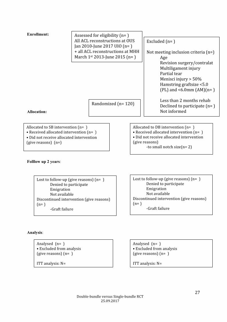
Arthrofibrosis Cyclops Donor site morbidity 11) New injuries: New meniscus injury New cartilage injury Graft re-rupture 12) Reoperations and revision surgery. 3. STUDY METHODS: 3.1. Study design: The study was designed as a prospectively randomised, controlled, superiority study, following two parallel groups with a 1:1 allocation ratio. The intervention group was the anatomic double-bundle ACL reconstruction and the control group, anatomic single-bundle ACL reconstruction. 3.2. Study settings: The protocol was published at ClinicalTrials.gov at 15th Dec 2009 (Clinical trials ID: NCT01033188). The inclusion period was from 01.01.2010 until 18.06.2015. The follow-up was performed at 12 and 24 months (1 and two years) after index surgery. The study was completed with the two-years follow-up of the last patient on 17th June 2017. The patients were recruited from the outpatient clinic at Oslo University Hospital during the entire inclusion period. From March 1st, 2013 Martina Hansens Hospital was added as recruitment hospital. Initially, the interventions were performed at Oslo University Hospital. From March 1st, 2013 until end of inclusion, the site of intervention was changed to Martina Hansens Hospital. Both hospitals perform more than 100 ACL reconstructions yearly and recruit patients from a rural environment. A single surgeon performed the surgery at all study participants except two in both hospitals. The surgeon was experienced and instructed in how to perform the intervention, prior to inclusion of the first patient. Collection of baseline information was conducted after a minimum of two months of rehabilitation and within three months before the operation. The 1 and the two-years follow-up, was performed within three weeks from the examination date. Patients responses obtained outside of these time frames will not be considered in the per protocol analysis. In the intention to treat (ITT) analysis all collected data at each time point will be considered. The average time until the examination was recorded in both groups for one and two years. 3.3. Inclusion and exclusion criteria: Inclusion criteria:
Double-bundleversusSingle-bundleRCT
25.09.2017
8
The participants had to comply with the following at randomization: 1) Age from 18 to 40 years. 2) Symptoms from their knee due to a primary ACL injury - verified by history, -clinical assessments (Lachman >1+ and/or positive Pivot shift test) -and identified under surgery 3) Successfully completed a preoperatively 3 (minimum 2) months of ACL rehabilitation program supervised by a physiotherapist. Exclusion criteria 1) A previous ACL reconstruction in the involved knee. 2) A subtotal (partial) ruptured ACL. 3) PCL, LCL or PLC injury. 4) Increased medial, lateral or posterolateral ligament laxity at the operating table, compared with the uninvolved leg. 5) ACL injury of the uninvolved knee. 6) Less than 50% of one of the menisci preserved after surgical treatment. 7) Established osteoarthritis (Kellgren-Lawrence classification grade 3 or 4) identified at the baseline standing front radiographs of the knee. 8) Hamstring tendons with insufficient graft thicknesses after preparation (defined as less than of 5.0 mm in diameter for the PL, and 6.0 mm for the AM bundle). 9) Patients living outside recruitment area, and patients who could not understand the Norwegian written language. 3.4. Flow chart: Table 1 4. INTERVENTIONS: The surgical technique consisted of the patient in supine position, with the knee at 90 degrees of flexion and with a tourniquet placed around the upper thigh. The establishment of the regular anterior arthroscopic portals was obtained. The ACL lesion was confirmed by visualization and by probing the ACL remnants, and a further debridement of the residual ACL stump and footprints was performed. The femoral and tibial insertion site was visualized, and surrounding soft tissue and bony landmarks were used to identify the centre of the proximal and distal ACL footprint [33]. A 3-5 cm skin incision was performed at the pes anserine insertion site. The semitendinosus and gracilis tendons were identified. A tendon harvester was used to free the tendons, and then they were doubled or tripled according to their length and thicknesses. For the double-bundle operation technique, a minimum graft size of 5.0 mm in diameter for the posterolateral (PL), and 6.0 mm for the anteromedial (AM) bundle was desirable. Both ends of each the grafts were whip stitched with a non-absorbable suture. The drillguide diameters were chosen as close to the graft diameter as possible, in both techniques. 4.1. Single-bundle reconstruction:
Double-bundleversusSingle-bundleRCT
25.09.2017
9
An accessory anteromedial portal was used for the femoral tunnel establishment. A Steadman awl was positioned in a central position of the femoral footprint, targeting to have the tunnel covering both parts of the AM and PL bundle attachment sites. With the knee in hyperflexion, a guide pin was inserted at the same position, and over-reaming of the guide pin was performed, according to measured graft size. With an external tibia guide, the center of the tibial tunnel was positioned according to remaining soft tissue and bony landmarks[33]. The tibia guide pin was aimed towards the tibia guide aimer and over-drilled relative to the distal graft size. The graft was passed through the tibial and then the femoral tunnel, and graft fixation on the femoral side was obtained with a suspension device (Endobutton CL™, Smith & Nephew, London, United Kingdom). The graft was cycled through 20 flexion-extension movements. The tibial fixation was then realized with the knee at 20 degrees of flexion and under manual tensioning, with a non-absorbable PEEK interference screw (Biosure PK™, Smith & Nephew, London, United Kingdom) placed eccentrically to the graft. 4.2. Double-bundle reconstruction: An accessory anteromedial portal was used for the femoral tunnel establishment. The centre of the PL and then the centre of the AM bundle was marked with a Steadman awl. A prefabricated double-bundle femoral drill-guide (Acuflex Pin Point™, Smith & Nephew, London, United Kingdom) was used to drill the femoral tunnels. The posterolateral tunnel was drilled first, then anteromedial tunnel was drilled in sequence, through the accessory anteromedial portal with the knee in a hyperflexion position. On the tibial side, using the prefabricated double-bundle tibia guide (Acuflex Pin Point™, Smith & Nephew, London, United Kingdom) the centre of the AM and then the PL tunnel was aimed at with the two guide pins [33]. The guide pins were overdrilled relative to the distal graft sizes. The grafts were passed through the tibia and then the femoral tunnel and fixation on the femoral side was realized with one suspension device for each tunnel (Endobutton CL™, Smith & Nephew, London, United Kingdom). The knee was cycled through 20 flexion-extension movements, and under manual tension first the AM bundle was fixated at 60 degrees flexion, then the PL bundle was fixated with the knee at full extension. Both tibial fixations were achieved with an eccentric placed PEEK interference screw (Biosure PK ™, Smith & Nephew, London, United Kingdom). Notchplasty was only realized if graft impingement was detected after graft insertion. Measurements of the insertion sites were performed if there were any doubt about the patient having a sufficient notch size, but not as a routine. The wounds were closed and bandaged before the tourniquet was loosened. Free mobilization was achieved from the first postoperative day without brace support or the use of a CPM. 5. ANALYSIS SETS 5.1. Full analysis set (intention to treat; ITT): All subjects randomised to either one of the two treatment arms, who completed the baseline assessment; independent of the actual intervention they received and whether the assessments at all time points were completed. 5.2. Per protocol (PP) analysis set:
Double-bundleversusSingle-bundleRCT
25.09.2017
10
All subjects that received the treatment to which they were randomised, and fulfilled all the required assessments at baseline and at the two-years follow-up. Exclusion criteria from the PP set:
• Patients that did not receive the treatment they were randomised to get. • Patients that did not complete the baseline and two years assessments of the KOOS QoL
subscale. • Patients with an ACL revision procedure performed before the two-years follow-up. (They
were not excluded if the revision surgery was more than two years after index surgery even if they were identified with graft rupture.)
• Patients lost to follow-up. 6. SPECIFICATION OF OUTCOME The primary outcome KOOS QoL subscale and all secondary outcomes will be analysed as the ITT set. Secondary analyses will be performed as PP set for the primary outcome and the remaining 4 KOOS subscales: Pain, Symptoms, Activity of daily living and Sports and recreation. 6.1. Primary outcome: The primary outcome of the study is the difference between the two treatments as mean change of the KOOS QoL subscore from baseline to the two-years follow-up. 6.2. Secondary outcomes: The difference between the two treatments in the following outcomes:
1. The mean change in the KOOS Symptoms subscore from baseline till two years. 2. The mean change in the KOOS Pain subscore from baseline till two years. 3. The mean change in the KOOS Activities of Daily Living subscore from baseline till two
years. 4. The mean change in the KOOS Sports and Recreation subscore from baseline till two years. 5. The mean change in the IKDC 2000 subjective score from baseline till two years. 6. Knee laxity as measured by the Lachman's test at two years. 7. Knee laxity as measured by the Pivot shift test at two years. 8. Knee laxity as measured by the KT-1000 at two years. 9. Range of motion as measured by the mean extension deficit in the involved knee compared to
the uninvolved knee at two years. 10. Range of motion as measured by the mean flexion deficit in the involved knee compared to
the uninvolved knee at two years. 11. The Tegner activity score at two years. 12. The Activity Scale level at two years. 13. Return to sports at two years. 14. The mean change in the One leg hop test from baseline till two years.
Double-bundleversusSingle-bundleRCT
25.09.2017
11
15. The degree of degeneration in the involved and uninvolved knee, classified by the Kellgren-Lawrence classification system, at two years.
16. The number of Adverse events including bleeding, DVT, infection, donor site pain, arthrofibrosis or cyclops within two years after the operation.
17. The number of new menisci injuries at two years. 18. The number of new cartilage injuries at two years. 19. The number of ACL graft re-ruptures at two years. 20. The number of Reoperations within the two-years follow-up.
Secondary outcome derived from the primary outcome
21. The difference between the two treatments in subjective treatment failures, defined as KOOS QoL subscore < 44 point
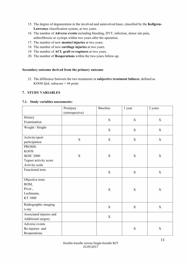
7. STUDY VARIABLES 7.1. Study variables assessments:
Preinjury (retrospective)
Baseline 1 year 2 years
History Examination X X X
Weight / Height X X X
Activity/sport participation
X X X X
PROMS: KOOS IKDC 2000 Tegner activity score Activity scale
X X X X
Functional tests X X X
Objective tests: ROM, Pivot , Lachmann, KT 1000
X X X
Radiographic imaging x-ray
X X X
Associated injuries and Additional surgery
X
Adverse events Re-injuries and Reoperations
X X
Double-bundleversusSingle-bundleRCT
25.09.2017
12
7.2. Patient related outcome measurements: 7.2.1. KOOS: The KOOS was developed to evaluate both short- and long-term outcome after knee injuries in young and active subjects with a knee injury or osteoarthritis in their knee[23]. It is proven as a reliable, valid and responsive score for patients undergoing ACL reconstruction[6, 24]. The KOOS data are obtained from a questionnaire, where five dimensions are rated separately: Pain, Symptoms, Activity of daily living, Sports and recreation and Quality of life. The five different subscores have different effect sizes reflecting knee function, symptoms and expectations on an individual basis. It is therefor desirable to interpret them separately, and an aggregated score of all five subscales will not be calculated [23]. The minimal perceptible clinical improvement (MPCI), has not been formally assessed for the KOOS, all though the 8-10 points change in one subscore has been used as a clinically meaningful change following ACL reconstruction [23]. The primary outcome of this study was the KOOS QoL subscale. This subscale is considered to be the most sensitive and responsive among the five dimensions of the score for ACL injured patients[6]. The four remaining subscales would be considered as additional supportive variables to the primary outcome. The five KOOS scales include 42 items with different numbers of elements within each subscore (4-11). Each item can score on a scale from 0 - 4. The five subscales are separately calculated, ranging from 0-100 points where 100 point is the best score possible.. At least 50% of the items within each subscale must be responded to before the score is calculated. Scale: 0-100 points in each subscale. Assessment: Baseline, one- and two-years follow-up. 7.2.2. IKCD 2000 subjective scale: The IKDC 2000 was developed by the International Knee Documentation Committee (IKDC), as a standardized form, for different knee conditions. The original score contents of both clinical assessments, pathology identified during surgery and the subjective IKDC score. The subjective score contains only the patient administered form of the total score. The score includes 18 different items that cover three domains: Symptoms, sports, and current knee function. Each item is weighted according to its importance on the total score, and the worst score in each category is carried forward. Range 0-100 points. (A total score of 100 points =normal sports participation with absence of symptoms and no limitation in the daily activity [10, 14]). Scale: 0-100 Assessment: Baseline, one- and two-years follow-up. 7.2.3. Tegner activity scale: The score was developed complementary to the Lysholm score, to detect whether loss in function could be masked by the level of activity [28]. The score is graduated in 11 different levels of activity (0-10), from recreational to competitive sports. Level 0 indicates the lowest knee-related activity (sick leave or disability), and 11 the highest knee related activity (competitive sports at a national level).
Double-bundleversusSingle-bundleRCT
25.09.2017
13
Scale: 0-10. Assessment: Before injury, baseline, one- and two-years follow-up. 7.2.4. Sports Activity Scale: The Activity Scale was based on one of the subjective assessments included in the Cincinnati Knee Rating System and is a self-administered score that detects the level of sports activity [4]. The original score contains four different levels of sports frequency. Within each level there is a grading from sports performed with "no running, twisting or jumping"(cycling/swimming), to sports with "hard pivoting, cutting, jumping"(basket, football). In this study, only the frequency of sports participation was recorded in the scale, with four different levels frequency (1=sports performed less than one day per month, 4= sports performed more than 4 days per week). Scale: 1-4. Assessment: Before injury, baseline, one- and two-years follow-up.. 7.2.5. Return to sports: The patients recalled the two main sports they participated in, the year ahead of the ACL injury. Return to sports was defined as the return to one of the two main sports after the ACL reconstruction was performed. If the same sport was recorded in one of the two follow-ups, the patient was defined as having returned to sports. Scale: yes (y) or no (n). Assessed: Before injury, baseline, one- and two-years follow-up.. 7.3. Clinical testing: 7.3.1. Lachman's test: The Lachman's test is a reliable manual laxity test to distinguishing an ACL rupture from an intact ACL. The test has been found to have a higher sensitivity and specificity compared to other manual tests for ACL injury[8, 17]. The test can be graded similarly to the anterior drawer test and is carried out with the patient in supine position and 20 degrees of flexion in the involved leg. One hand is stabilizing the femur and the other hand performing a subluxation of the tibia in the anterior direction. The anterior displacement is recorded in mm and always reported as the difference to the contralateral leg[10]. Grade 3+ = >10mm displacement of the tibia compared to the uninvolved leg Grade 2+ = 5-10mm Grade 1+ = 3-5 mm Grade 0= 0-3mm Scale: 0-3 Assessment: Baseline, one- and two-years follow-up. 7.3.2. Pivot shift test:
Double-bundleversusSingle-bundleRCT
25.09.2017
14
The Pivot shift test is known as a pathognomonic test for the ACL insufficient knee. The phenomenon is described as the reduction of the tibia from a subluxated position as the knee is extended with the tibia internally rotated [11, 20]. The Pivot shift phenomenon can be graded on a scale from 0 to 3+[15, 26] according to the amount of subluxation of the tibia and its the reduction in extension. There has been a discussion among the experts whether a positive test should be recorded as such, or if it should be compared to the contralateral leg. In this study the Pivot shift was detected by the Slocum`s test and not compared to the contralateral leg[26]. Pivot shift: Grade 0 Grade +1= “trace” positive only in medial/internal rotation of the tibia Grade +2= “clunk” subluxation in neutral positioning of the tibia Grade+3= “gross” subluxation in any rotation, laxity due to secondary restraints additional to the ACL injury or in chronic unstable knees. Scale: 0-3. Assessment: Baseline, one- and two-years follow-up. 7.3.3. KT-1000 The KT-1000™ (MEDmetric, San Diego, CA, USA) (Knee Laxity Testing Device), is an instrument detecting knee laxity in the anteroposterior direction[7]. It has two sensor pads that are placed in contact with the patella and the greater tuberosity of the tibia during an instrumented Lachman's test of the knee. The instrument detects the motion between those two sensor pads during anterior translation of the tibia towards the femur. Displacements at loads of 134 N and maximal manual load (MM) are detected. Displacement in the involved compared to the uninvolved knee will be detected[7]. Range: - 20 till +20mm. Assessment: Baseline, one- and two-years follow-up.. 7.3.4. Range of motion (ROM) As the knee joint is a hinge joint, the range of motion can be detected as both the extension/flexion movement and by internal/external rotation[11]. In this study, only the extension/flexion movements are detected. The normal flexion is widely individual from 120-150 degrees, and therefore the flexion deficit was compared to the uninvolved knee. If any extension- or flexion-deficit was detected, a goniometer was used for exact measurement of the deficit and recorded. The extension deficit of the involved knee was compared both to 0(zero) degrees of extension and to contralateral knee extension. Scale: Extension: -20 (hyperextension) to 150 degrees Flexion: 0-150 degrees. Assessment: Baseline, one- and two-years follow-up. 7.4. Functional performance tests:
Double-bundleversusSingle-bundleRCT
25.09.2017
15
The functional tests were performed to evaluate the functional capacity of the knee. The tests reveal both the clinical assessments and the patient´s perception of their knee. 7.4.1. One leg hop test The “one leg hop test” is a functional test often used as part of a performance test for ACL deficient knees[20, 29]. It is known to be highly correlated to the clinically assessed instability of the knee[3]. The test was performed with two attempts at each leg, the best of the two scores were documented and the percentage difference from the uninjured knee presented: Operated knee hop distance/Non operated knee hop distance X 100 Scale: 0-100 %. Assessment: Baseline, one- and two-years follow-up. 7.5. Radiographic imaging: Radiographic imaging was performed with a single, standardised, front standing radiograph of both legs. Both legs were classified according to the Kellgren-Lawrence grading system of osteoarthritis in the joint[19]. Kellgren Lawrence classification[19]: Grade 0: no radiographic features of OA are present Grade 1: doubtful narrowing of joint space and possible osteophytic lipping Grade 2: definite osteophytes and possible narrowing of joint space Grade 3: moderate multiple osteophytes, definite narrowing of joint space and some sclerosis and possible deformity of bone ends Grade 4: large osteophytes, marked narrowing of joint space, severe sclerosis and definite deformity of bone ends Scale 0-4. Assessment: Baseline, one- and two-years follow-up. 7.6. Adverse events, new injuries and reoperations: Adverse Events: Any observed bleeding or excessive hematoma during the first 6-8 weeks after the operation. Deep venous thrombosis (DVT), nerve or arterial injury in the involved leg. Infection including both superficial infection and septic arthritis of the involved joint. Arthrofibrosis, cyclops with or without extension deficit in the involved knee. Donor site morbidity due to fixation device, scar tissue or to the hamstrings tendon harvesting in involved leg. New injuries:
Double-bundleversusSingle-bundleRCT
25.09.2017
16
New menisci injury detected after the intervention either by MRI or verified be second look arthroscopy. The injuries were further differentiated into medial and lateral menisci injuries and into the treatment they accomplished (resection, suture or transplantation). Any new cartilage injury detected after the intervention. ACL Graft reruptures defined as a total rupture of the ACL reconstruction, found at clinical examination and MRI or second look arthroscopy. Reoperations: Reoperations because of hematoma, nerve or arterial injury, infection, arthrofibrosis, cyclops or donor site morbidity. Menisci surgery including suturing, resection or transplantation. Reoperations due to cartilage surgery. Re-arthroscopy due to other reasons. Revision surgery because of graft rupture, both first and second stage revision surgery. 8. SAMPLE SIZE CALCULATION: The sample size was calculated as for a superiority study, based on the null hypothesis that quality of life following double-bundle ACL reconstruction is equal to KOOS quality of life following single-bundle ACL reconstruction. The primary outcome, KOOS QoL subscale, was used for the sample size calculation. A minimal important change (MIC) in QoL of 8 points, has been considered sufficient [23]. With equal allocation in both arms, a standard deviation of 15 points, power of 80%, and assuming a two-sided significance-level of 0.05, the sample size was calculated to be 56 patients in each treatment group. To allow for 5% drop-outs, the final sample size was set to be 60 patients in each treatment arm, and 120 patients in total. 9. RANDOMISATION/BLINDING 9.1. Randomisation A nurse not involved in the research project performed a computer-generated block randomization, ten patients in each block. The allocation sequence was generated by a software program: (http://randomization.com) and was conducted with a 1:1 ratio between the treatment arms. With 60 patients within each intervention group, twelve blocks of ten patients was needed. 120 sequentially numbered, opaque, sealed envelopes, containing a label describing one of the two interventions, were placed in the operating theatre at operation. The envelope was opened only if the patient fulfilled the inclusion criteria and the baseline assessments. One of the assisting nurses would open the envelopes at the request of the surgeon. The randomization was performed only after the ACL rupture was verified by arthroscopy, and at least 50 % of each menisci was left intact after surgical treatment, and if the hamstring graft sizes were sufficient after harvesting (the minimal desired hamstring graft sizes were 5.0 mm for the PL bundle and 6.0 mm in diameter for the AM bundle).
Double-bundleversusSingle-bundleRCT
25.09.2017
17
9.2. Level and method for blinding: KOOS/IKDC and the activity scales: Initially, the trial participants were not intentionally blinded for the intervention, but the outcome assessor was blinded. The outcome assessor ensured that the PRO from the patients were complete. The level of blinding was changed during the enrollment, and the study participants number 62 – 120 were consequently blinded for the intervention. Unblinding was performed after the assessments of the two-years follow-up. Functional tests: The outcome assessor that completed the functional tests, was blinded for the intervention. Clinical assessment: The clinical assessment was performed by the assisting, orthopaedic surgeon (CA). The surgeon was not blinded, as she was also present during the initial surgery. Radiographic imaging: The radiologist was not blinded, as the intervention was visible at the radiographic imaging. Data analysis: The statistical advisor will be blinded when performing the analysis. 10. STATISTICAL METHODS: 10.1. Presentation of observed data All continuous variables including the five subscores will be summarized with means and standard deviations (SD) within each treatment. Categorical data will be summarized with counts and percentages within each category and treatment arm. The observed mean values of the primary outcome (KOOS QoL) will be presented in a figure as two curves (one for each treatment) plotted at three different time points (baseline, 1 and two years). Vertical lines at each time point for each treatment will represent the 95% confidence intervals (CIs). 10.2. Primary hypothesis setup Null hypothesis: Double-bundle ACL reconstruction is equal to single-bundle ACL reconstruction, regarding the change in the KOOS QoL subscale from baseline to the two-years follow-up. Alternative hypothesis: Double-bundle ACL reconstruction is superior to single-bundle ACL reconstruction, regarding the change in the KOOS QoL subscale from baseline to the two-years follow-up. 10.3. STATISTICAL ANALYSIS 10.3.1. Primary outcome analysis:
Double-bundleversusSingle-bundleRCT
25.09.2017
18
The KOOS QoL subscore will be analyzed with a linear mixed model with treatment, time point (baseline, 1 year, 2 years), and treatment x time point interaction as fixed effects. A random intercept will be used. Based on the fitted model, we will estimate the mean baseline, 1 year, and 2 years values (with 95% CIs) for each treatment, and the between-treatment difference in changes from baseline to 2 years (with 95% CI and a P-value for the null hypothesis of no difference). 10.3.2. Secondary outcomes analysis:
1. The KOOS Symptoms subscore will be analyzed with a linear mixed model with treatment, time point (baseline, 1 year, 2 years), and treatment x time point interaction as fixed effects. A random intercept will be used. Based on the fitted model, we will estimate the mean baseline, 1 year, and 2 years values (with 95% CIs) for each treatment, and the between-treatment difference in changes from baseline to 2 years (with 95% CI and a P-value for the null hypothesis of no difference).
2. The KOOS Pain subscore will be analyzed with a linear mixed model with treatment, time point (baseline, 1 year, 2 years), and treatment x time point interaction as fixed effects. A random intercept will be used. Based on the fitted model, we will estimate the mean baseline, 1 year, and 2 years values (with 95% CIs) for each treatment, and the between-treatment difference in changes from baseline to 2 years (with 95% CI and a P-value for the null hypothesis of no difference).
3. The KOOS Activities of Daily Living subscore will be analysed with a linear mixed model with treatment, time point (baseline, 1 year, 2 years), and treatment x time point interaction as fixed effects. A random intercept will be used. Based on the fitted model, we will estimate the mean baseline, 1 year, and 2 years values (with 95% CIs) for each treatment, and the between-treatment difference in changes from baseline to 2 years (with 95% CI and a P-value for the null hypothesis of no difference).
4. The KOOS Sports and Recreation subscore will be analysed with a linear mixed model with treatment, time point (baseline, 1 year, 2 years), and treatment x time point interaction as fixed effects. A random intercept will be used. Based on the fitted model, we will estimate the mean baseline, 1 year, and 2 years values (with 95% CIs) for each treatment, and the between-treatment difference in changes from baseline to 2 years (with 95% CI and a P-value for the null hypothesis of no difference).
5. The IKCD 2000 subjective score will be analysed with a linear mixed model with treatment, time point (baseline, 1 year, 2 years), and treatment x time point interaction as fixed effects. A random intercept will be used. Based on the fitted model, we will estimate the mean baseline, 1 year, and 2 years values (with 95% CIs) for each treatment, and the between-treatment difference in changes from baseline to 2 years (with 95% CI and a P-value for the null hypothesis of no difference).
6. The Lachman’s test at two years will be analysed with the Wilcoxon-Mann-Whitney test for ordered 2xc tables [REF: Chapter 6 of Fagerland MW, Lydersen S, Laake P (2017) Statistical Analysis of Contingency Tables. Chapman & Hall/CRC, Boca Raton, FL.] A P-value for the null hypothesis that the two treatments have equal distributions across the categories will be presented.
7. The Pivot shift test at two years will be analysed with the Wilcoxon-Mann-Whitney test for ordered 2xc tables [REF: Chapter 6 of Fagerland MW, Lydersen S, Laake P (2017) Statistical
Double-bundleversusSingle-bundleRCT
25.09.2017
19
Analysis of Contingency Tables. Chapman & Hall/CRC, Boca Raton, FL.] A P-value for the null hypothesis that the two treatments have equal distributions across the categories will be presented.
8. The KT 1000 measurements, will be analysed with a two-sample T-test, with adjustment for unequal variances (the Welch U test) if the ratio of the largest to the smallest standard deviation is more than 1.5. An estimate of the difference between the mean treatment scores (with a 95% CI), and a P-value for the null hypothesis of no difference, will be presented.
9. The extension deficit (range of motion) will be analysed with a two-sample T-test, with adjustment for unequal variances (the Welch U test) if the ratio of the largest to the smallest standard deviation is more than 1.5. An estimate of the difference between the mean treatment scores (with a 95% CI), and a P-value for the null hypothesis of no difference, will be presented.
10. The flexion deficit (range of motion) will be analysed with a two-sample T-test, with adjustment for unequal variances (the Welch U test) if the ratio of the largest to the smallest standard deviation is more than 1.5. An estimate of the difference between the mean treatment scores (with a 95% CI), and a P-value for the null hypothesis of no difference, will be presented.
11. The Tegner activity score at two years will be analysed with the Wilcoxon-Mann-Whitney test for ordered 2xc tables [REF: Chapter 6 of Fagerland MW, Lydersen S, Laake P (2017) Statistical Analysis of Contingency Tables. Chapman & Hall/CRC, Boca Raton, FL.] A P-value for the null hypothesis that the two treatments have equal distributions across the categories will be presented.
12. The Sports Activity scale at two years will be analysed with the Wilcoxon-Mann-Whitney test for ordered 2xc tables [REF: Chapter 6 of Fagerland MW, Lydersen S, Laake P (2017) Statistical Analysis of Contingency Tables. Chapman & Hall/CRC, Boca Raton, FL.] A P-value for the null hypothesis that the two treatments have equal distributions across the categories will be presented.
13. The difference between the treatment probabilities of Return to sports will be estimated and a 95% Newcombe hybrid score CI will be reported. The null hypothesis of equal probabilities will be analysed with a Fisher mid-P test [REF: Chapter 4 of Fagerland MW, Lydersen S, Laake P (2017) Statistical Analysis of Contingency Tables. Chapman & Hall/CRC, Boca Raton, FL.]
14. The One leg hop test will be analysed with a linear mixed model with treatment, time point (baseline, 1 year, 2 years), and treatment x time point interaction as fixed effects. A random intercept will be used. Based on the fitted model, we will estimate the mean baseline, 1 year, and 2 years values (with 95% CIs) for each treatment, and the between-treatment difference in changes from baseline to 2 years (with 95% CI and a P-value for the null hypothesis of no difference).
15. The Kellgren-Lawrence classification at two years will be analysed with the Wilcoxon-Mann-Whitney test for ordered 2xc tables [REF: Chapter 6 of Fagerland MW, Lydersen S, Laake P (2017) Statistical Analysis of Contingency Tables. Chapman & Hall/CRC, Boca Raton, FL.] A P-value for the null hypothesis that the two treatments have equal distributions across the categories will be presented.
16. Adverse events: the graft failures and new meniscus injuries will be analysed. The null hypothesis of equal probabilities will be analysed with a Fisher mid-P test [REF: Chapter 4 of Fagerland MW, Lydersen S, Laake P (2017) Statistical Analysis of Contingency Tables.
Double-bundleversusSingle-bundleRCT
25.09.2017
20
Chapman & Hall/CRC, Boca Raton, FL.] A P-value for the null hypothesis that the two treatments have equal distributions across the categories will be presented.
17. Reoperations: The null hypothesis of equal probabilities will be analysed with a Fisher mid-P test [REF: Chapter 4 of Fagerland MW, Lydersen S, Laake P (2017) Statistical Analysis of Contingency Tables. Chapman & Hall/CRC, Boca Raton, FL.]
Secondary outcome derived from the primary outcome
18. The difference between the probabilities of treatment failures, defined as KOOS QoL < 44 points, will be estimated and a 95% Newcombe hybrid score CI will be reported. The null hypothesis of equal probabilities will be analyzed with a Fisher mid-P test [REF: Chapter 4 of Fagerland MW, Lydersen S, Laake P (2017) Statistical Analysis of Contingency Tables. Chapman & Hall/CRC, Boca Raton, FL.]
10.3.3. Subgroup analysis: A separate analysis of the primary outcome in the blinded versus not blinded patients will be performed. The subgroup would contain patients with randomization number 1-61 versus 62-120. The subgroup analysis will be performed by adding an interaction term between blinded and treatment and an interaction term between blinded and (treatment x time) to the linear mixed model. The P-values for these interaction terms will indicate whether any difference in treatment effects exists between blinded and not blinded patients. 10.3.4. Missing data analysis: Recording of the reasons for missing data will be done. The KOOS subscales, the IKDC 2000, and the one leg hop test will be analysed with linear mixed models. These models account for missing data on individual time points, thus obviating the need to impute missing values. For the categorical and semi-continuous outcomes, we will use a modified ITT analysis set, where only the observed data will be included, if the amount of missing data is less than 5%. For outcomes with more than 5% missing data, a sensitivity analysis will be performed, wherein the missing data will be imputed according to three scenarios:
1. The two year (missing) measurements will be given the values of the one year measurements (or the baseline measurements if the one year measurements are also missing).
2. The two year (missing) measurements will be imputed as the most favourable score for patients who received the double-bundle treatment, and the least favourable score for patients who received the single-bundle treatment.
3. The two year (missing) measurements will be imputed as the least favourable score for patients who received the double-bundle treatment, and the most favourable score for patients who received the single-bundle treatment.

Double-bundleversusSingle-bundleRCT
25.09.2017
21
10.4 The timing of analysis: The final analysis will be performed after the two-years follow-up of all study candidates and after finalization and approval of the statistical analysis plan by all co-authors (MAR, SJ, SH and LE) and the statistical advisor (MWF). The statistical analysis plan will be published online on the OSTRC websites before the analysis is performed. The data will then be prepared and presented to the statistician as blinded data. The statistical analysis will be performed blinded by the statistical advisor A data collection form will be outlined; the data will have anonymous coding into "treatment 1" and "treatment 2". Analysis of the primary and secondary outcome will be performed blinded and then presented for the other authors. 11. SAFETY ANALYSIS: 11.1. Associated injuries at operation: Any additional chondral or meniscal injury detected during the index operation will be reported and listed in the summary tables of baseline characters. 11.2. Adverse events: “An adverse event refers to an untoward occurrence during the trial, which may or may not be causally related to the intervention or other aspects of trial participation” [13]. The study participants were questioned whether they had observed any adverse events related to the treatment during the last year, at the one and two-years follow-up. Each subject were counted once in each category of AE, but each participant could have more than one AE. Any repetition of the same event in one patient was ignored. Additional information about the event was obtained from the patient`s journal if necessary. 11.2.1. Events: Bleeding/Hematoma Deep venous thrombosis (DVT) Injury larger nerves or vessels Infection/Septic arthritis Arthrofibrosis Cyclops Donor site morbidity 11.2.2. New injury: Meniscal lesion Chondral lesion Graft re-rupture 11.2.3. New operations:
Double-bundleversusSingle-bundleRCT
25.09.2017
22
Because of bleeding/hematoma Because of injury to larger vessels/nerves Because of infection Because of arthrofibrosis Because of cyclops/extensiondeficit Because of donor site morbidity (e.g. removal of fixation device, scar tissue) Because of Menisci surgery Medial menisci resection Lateral menisci resection Medial menisci suture Lateral menisci suture Med menisci transplantation Lateral menisci transplantation Because of Cartilage treatment Because of revision surgery Others reoperations. 12. PROTOCOL DEVIATIONS: 3.1. Study design: The hypothesis in the original study protocol was formulated as in a non-inferiority study. Although further evaluations and sample size calculations of the study were designed as in a superiority study design, with the hypothesis questioning if the double-bundle technique was superior to the single-bundle method regarding the KOOS, QoL subscale. Double-bundle reconstructions are considered more cost demanding, time-consuming and require higher skills of the performing surgeon compared to single-bundle surgery; therefore a superiority study was the preferred study design[22]. 2.2. Secondary outcomes: The Activity scale as one of the PROM`s was added after the protocol was made but before inclusion of the first patient. 3.2. Study settings – primary endpoint: The primary endpoint was changed from five to two-years follow-up, because of a prolonged inclusion period. 3.2. Study settings: Martina Hansens Hospital was in 2013 implemented as an additional recruiting hospital, because of the prolonged inclusion period and because of the first author worked at both hospitals during this time. The interventions were performed at Oslo University Hospital but were changed to Martina
Double-bundleversusSingle-bundleRCT
25.09.2017
23
Hansens Hospital as the operating theatres at Oslo University Hospital were closed down due to rehabilitation, (1st March 2013). The operating surgeon (SJ) continued to perform the intervention under the same conditions, with the same equipment and fixation devices. (The equipment was transported between the hospitals.) 4. Interventions: The inclusion criteria for the hamstring tendon graft sizes were changed from 5.5 mm for both bundles to 5.0 mm for the PL bundle and 6.0 mm for the AM bundle, due to the arising difficulties including patients with a sufficient graft size, during the first year of the study. 9.2. Level and method of blinding: A subgroup of patients, (randomization number 62-120), were blinded for the intervention until they completed the two-years follow-up, to improve the quality of the study. The reason for blinding was to prevent the patients from biasing the results unintentionally, as not blinded studies are known to give larger treatment effects than non-blinded studies. 12.1 Clinical trials, history of changes: Published at https://clinicaltrials.gov 15th of December 2009, (ID: NCT01033188). 5th July 2011: The name of location has been modified from Ullevaal University Hospital to Oslo University Hospital. 2nd June 2014: The surgical procedure: Anatomic ACL reconstruction technique was described in detail, and the name of the fixation devices was changed to the devices that was used on the study participants. The minimum hamstring graft size was adjusted from minimum 5.5 mm to 5.0 mm. The sample-size was changed to 112 patients according to the initial sample size calculation. 12th May 2015: The description of the surgical procedure was further improved to aim the actual anatomic reconstruction that the surgeons performed on all the study participants. The sample size was enlarged to 120 patients because of the block-randomisation, and the anticipated end of study date, changed to 2017. 6th August 2015: Study status was changed from recruiting to active, not recruiting. The minimum hamstring tendon sizes required for inclusion were changed to differentiate between the two bundles: 5.0 mm for the PL bundle and 6.0 mm for the AM bundle. 4th April 2017: An update on recruitment was performed.
Double-bundleversusSingle-bundleRCT
25.09.2017
24
13. OTHERS: Registration numbers:
ClinicalTrials.gov ID: NCT01033188
Ethical approval: REK no: S-09108b
Funded by:
Health South-Eastern Norway Ph.D. research grant: Number 2015049.
Smith & Nephew research grant.
The Norwegian Orthopaedic Association research grant.
14. REFERENCES: 1. AgliettiP,GironF,CuomoP,LoscoM,MondanelliN.Single-anddouble-incision
double-bundleACLreconstruction.ClinOrthopRelatRes.2007;454:108-113.2. ArdernCL,WebsterKE,TaylorNF,FellerJA.Returntosportfollowinganterior
cruciateligamentreconstructionsurgery:asystematicreviewandmeta-analysisofthestateofplay.BrJSportsMed.2011;45:596-606.
3. BarberSD,NoyesFR,MangineRE,McCloskeyJW,HartmanW.Quantitativeassessmentoffunctionallimitationsinnormalandanteriorcruciateligament-deficientknees.ClinOrthopRelatRes.1990:204-214.
4. Barber-WestinSD,NoyesFR,McCloskeyJW.Rigorousstatisticalreliability,validity,andresponsivenesstestingoftheCincinnatikneeratingsystemin350subjectswithuninjured,injured,oranteriorcruciateligament-reconstructedknees.AmJSportsMed.1999;27:402-416.
5. BrophyRH,WrightRW,MatavaMJ.Costanalysisofconvertingfromsingle-bundletodouble-bundleanteriorcruciateligamentreconstruction.AmJSportsMed.2009;37:683-687.
6. CollinsNJ,PrinsenCA,ChristensenR,BartelsEM,TerweeCB,RoosEM.KneeInjuryandOsteoarthritisOutcomeScore():systematicreviewandmeta-analysisofmeasurementproperties.OsteoarthritisCartilage.2016;24:1317-1329.
7. DanielDM,StoneML,SachsR,MalcomL.Instrumentedmeasurementofanteriorkneelaxityinpatientswithacuteanteriorcruciateligamentdisruption.AmJSportsMed.1985;13:401-407.
8. DunnG,TorgersenDM,MandelstamJ.OrderofexpressionofgenesaffectingseptumlocationduringsporulationofBacillussubtilis.JBacteriol.1976;125:776-779.
9. GrananLP,BahrR,SteindalK,FurnesO,EngebretsenL.Developmentofanationalcruciateligamentsurgeryregistry:theNorwegianNationalKneeLigamentRegistry.AmJSportsMed.2008;36:308-315.
Double-bundleversusSingle-bundleRCT
25.09.2017
25
10. HeftiF,MullerW.[Currentstateofevaluationofkneeligamentlesions.ThenewIKDCkneeevaluationform].Orthopade.1993;22:351-362.
11. HirschmannMT,MullerW.Complexfunctionofthekneejoint:thecurrentunderstandingoftheknee.KneeSurgSportsTraumatolArthrosc.2015;23:2780-2788.
12. HusseinM,vanEckCF,CretnikA,DinevskiD,FuFH.Prospectiverandomizedclinicalevaluationofconventionalsingle-bundle,anatomicsingle-bundle,andanatomicdouble-bundleanteriorcruciateligamentreconstruction:281caseswith3-to5-yearfollow-up.AmJSportsMed.2012;40:512-520.
13. IoannidisJP,EvansSJ,GotzschePC,O'NeillRT,AltmanDG,SchulzK,MoherD,GroupC.Betterreportingofharmsinrandomizedtrials:anextensionoftheCONSORTstatement.AnnInternMed.2004;141:781-788.
14. IrrgangJJ,AndersonAF,BolandAL,HarnerCD,KurosakaM,NeyretP,RichmondJC,ShelborneKD.Developmentandvalidationoftheinternationalkneedocumentationcommitteesubjectivekneeform.AmJSportsMed.2001;29:600-613.
15. JakobRP,StaubliHU,DelandJT.Gradingthepivotshift.Objectivetestswithimplicationsfortreatment.JBoneJointSurgBr.1987;69:294-299.
16. JarvelaT,MoisalaAS,SihvonenR,JarvelaS,KannusP,JarvinenM.Double-bundleanteriorcruciateligamentreconstructionusinghamstringautograftsandbioabsorbableinterferencescrewfixation:prospective,randomized,clinicalstudywith2-yearresults.AmJSportsMed.2008;36:290-297.
17. JonssonT,AlthoffB,PetersonL,RenstromP.Clinicaldiagnosisofrupturesoftheanteriorcruciateligament:acomparativestudyoftheLachmantestandtheanteriordrawersign.AmJSportsMed.1982;10:100-102.
18. KarikisI,AhldenM,CasutA,SernertN,KartusJ.Comparisonofoutcomeafteranatomicdouble-bundleandantero-medialportalnon-anatomicsingle-bundlereconstructioninACL-injuredpatients.KneeSurgSportsTraumatolArthrosc.2017;25:1307-1315.
19. KellgrenJH,LawrenceJS.Radiologicalassessmentofosteo-arthrosis.AnnRheumDis.1957;16:494-502.
20. LogerstedtD,GrindemH,LynchA,EitzenI,EngebretsenL,RisbergMA,AxeMJ,Snyder-MacklerL.Single-leggedhoptestsaspredictorsofself-reportedkneefunctionafteranteriorcruciateligamentreconstruction:theDelaware-OsloACLcohortstudy.AmJSportsMed.2012;40:2348-2356.
21. OiestadBE,EngebretsenL,StorheimK,RisbergMA.Kneeosteoarthritisafteranteriorcruciateligamentinjury:asystematicreview.AmJSportsMed.2009;37:1434-1443.
22. PiaggioG,ElbourneDR,AltmanDG,PocockSJ,EvansSJ,GroupC.Reportingofnoninferiorityandequivalencerandomizedtrials:anextensionoftheCONSORTstatement.JAMA.2006;295:1152-1160.
23. RoosEM,LohmanderLS.TheKneeinjuryandOsteoarthritisOutcomeScore(KOOS):fromjointinjurytoosteoarthritis.HealthQualLifeOutcomes.2003;1:64.
24. RoosEM,RoosHP,LohmanderLS,EkdahlC,BeynnonBD.KneeInjuryandOsteoarthritisOutcomeScore(KOOS)--developmentofaself-administeredoutcomemeasure.JOrthopSportsPhysTher.1998;28:88-96.
Double-bundleversusSingle-bundleRCT
25.09.2017
26
25. SieboldR,DehlerC,EllertT.Prospectiverandomizedcomparisonofdouble-bundleversussingle-bundleanteriorcruciateligamentreconstruction.Arthroscopy.2008;24:137-145.
26. SlocumDB,JamesSL,LarsonRL,SingerKM.Clinicaltestforanterolateralrotaryinstabilityoftheknee.ClinOrthopRelatRes.1976:63-69.
27. SuomalainenP,KannusP,JarvelaT.Double-bundleanteriorcruciateligamentreconstruction:areviewofliterature.IntOrthop.2013;37:227-232.
28. TegnerY,LysholmJ.Ratingsystemsintheevaluationofkneeligamentinjuries.ClinOrthopRelatRes.1985:43-49.
29. TegnerY,LysholmJ,LysholmM,GillquistJ.Aperformancetesttomonitorrehabilitationandevaluateanteriorcruciateligamentinjuries.AmJSportsMed.1986;14:156-159.
30. vanEckCF,KopfS,IrrgangJJ,BlankevoortL,BhandariM,FuFH,PoolmanRW.Single-bundleversusdouble-bundlereconstructionforanteriorcruciateligamentrupture:ameta-analysis--doesanatomymatter?Arthroscopy.2012;28:405-424.
31. YagiM,WongEK,KanamoriA,DebskiRE,FuFH,WooSL.Biomechanicalanalysisofananatomicanteriorcruciateligamentreconstruction.AmJSportsMed.2002;30:660-666.
32. YasudaK,vanEckCF,HoshinoY,FuFH,TashmanS.Anatomicsingle-anddouble-bundleanteriorcruciateligamentreconstruction,part1:Basicscience.AmJSportsMed.2011;39:1789-1799.
33. ZieglerCG,PietriniSD,WesterhausBD,AndersonCJ,WijdicksCA,JohansenS,EngebretsenL,LaPradeRF.Arthroscopicallypertinentlandmarksfortunnelpositioninginsingle-bundleanddouble-bundleanteriorcruciateligamentreconstructions.AmJSportsMed.2011;39:743-752.
Table 1, FLOW CHART
Single versus Double-bundle reconstruction
Double-bundleversusSingle-bundleRCT
25.09.2017
27
Enrollment: Allocation:
Folllow up 2 years:
Analysis:
Assessedforeligibility(n=)AllACLreconstructionsatOUSJan2010-June2017UIO(n=)+allACLreconstructionsatMHHMarch1st2013-June2015(n=)
Excluded(n=)Notmeetinginclusioncriteria(n=) Age Revisionsurgery/contralat Multiligamentinjury Partialtear Menisciinjury>50% Hamstringgraftsize<5.0 (PL)and<6.0mm(AM)(n=) Lessthan2monthsrehab Declinedtoparticipate(n=) Notinformed
Randomized(n=120)
AllocatedtoSBintervention(n=)•Receivedallocatedintervention(n=)•Didnotreceiveallocatedintervention(givereasons)(n=)
AllocatedtoDBintervention(n=)•Receivedallocatedintervention(n=)•Didnotreceiveallocatedintervention(givereasons) -tosmallnotchsize(n=2)
Losttofollow-up(givereasons)(n=) Deniedtoparticipate Emigration NotavailableDiscontinuedintervention(givereasons)(n=) -Graftfailure
Losttofollow-up(givereasons)(n=) Deniedtoparticipate Emigration NotavailableDiscontinuedintervention(givereasons)(n=) -Graftfailure
Analysed(n=)•Excludedfromanalysis(givereasons)(n=)ITTanalysis:N=
Analysed(n=)•Excludedfromanalysis(givereasons)(n=)ITTanalysis:N=

Double-bundleversusSingle-bundleRCT
25.09.2017
28