Embed Size (px)
Citation preview

DNA in Nucleus
RNA copy
Protein in cytoplasm
Statistical GeneticsBiological Introduction

Central Dogma
• DNA carries the genetic code and transcribes an RNA copy of the code
• The RNA copy is translated by ribosomes to make protein
DNA RNA Protein
Transcription Translation
1 2

Step 1: RNA SYNTHESIS- TRANSCRIPTION
The process of converting the information contained in a DNA segment into proteins begins with the synthesis of mRNA molecules containing anywhere from several hundred to several thousand nucleotides, depending on the size of the protein to be made.
Each of the 30,000-100,000 proteins in the human body is synthesized from a different mRNA that has been transcribed from a specific gene on DNA.

Transcription• If a protein is required by a cell, that gene is
activated (turned on)..• The gene makes an RNA copy of itself in the form
of a messenger RNA molecule (mRNA)• Enzyme RNA polymerase runs along open DNA
strand and synthesizes RNA complementary to the DNA. A small section of the DNA double helix unwinds, and the bases on the two strands are exposed. RNA nucleotides (ribonucleotides) line up in the proper order to their complementary bases on DNA, the nucleotides are joined together by RNA polymerase, and a piece of mRNA is produced. The DNA strand that is transcribed is called the template strand and is a copy of the DNA informational strand!

RNA Polymerase• Runs along DNA in a 5’ to 3’
direction (adding bases to the 3’ end) and forms mRNA
• Until it hits a STOP signal, falls off and mRNA is released…..DNA reseals…

How does the RNA pol know where to start reading a Gene?

The Genetic CodeIf DNA is a long repeating length of… ACTGAATTGCCCTTCATGGTCATGGCT

The Genetic Code• Every 3 nucleotide
bases in DNA is a Code…
• In RNA, the 3 complementary bases are a Codon…
DNA Code
RNA Codon

The Genetic CodeEvery 3 nucleotides on
mRNA ‘spell’ for one amino acid
ACA spells Threonine
CAC spells Histidine
GUU spells Valine
UUA spells Leucine

The Genetic Code• The specific sequence of 3 nucleotide
bases indicates how a protein is to be constructed
• An mRNA sequence such as:
UUU-UUG-GUA-CCC
Means that a protein of Amino Acids… Phenylalanine-Leucine-Valine-Proline is to be made

How to make that Protein?
Step 2: Translation
• mRNA is produced in nucleus by RNA pol reading the DNA code
• mRNA travels to cytoplasm where proteins are made in a process known as Protein Synthesis or Translation
2

Translation Requires:
• Message in the form of mRNA…
• A Ribosome…..
• Another type of RNA…called Transfer RNA (tRNA)
• A pool of amino acids in cytoplasm
Free Amino Acids
AA1 AA2AA3

Translation Process:Initiation
• Translation is the process of converting the mRNA codon sequences into an amino acid sequence. The initiator codon (AUG) codes for the amino acid N-formylmethionine (f-Met). No transcription occurs without the AUG codon. f-Met is always the first amino acid in a polypeptide chain, although frequently it is removed after translation.
• After the initiation phase the message gets longer during the elongation phase….

Translation: Elongation
• A Ribosome runs along mRNA reading codons, beginning at AUG (Start)
• A tRNA carrying the corresponding AA drops into position and leaves AA off
• New protein emerges from ribosome as a growing peptide chain

Elongation
• Text pg 228, 231

Polysomes…Often many ribosomes will read the same message and a structure known as a polysome forms. In this way a cell may rapidly make many proteins.

Control of Genes:The Operon Model
The operon model of prokaryotic gene regulation was proposed by Fancois Jacob and Jacques Monod…
Groups of genes coding for related proteins are arranged in units known as operons. An operon consists of an operator, promoter, regulator, and structural genes.
The regulator gene codes for a repressor protein that binds to the operator, blocking the promoter and thus blocking transcription of the gene.
If the repressor protein is removed, transcription may again occur.
Such regulatory proteins recognize and bind to specific DNA sequences and can either turn-on or turn-off genes

The Genetic Material-DNA- and it’s Role
• Where is the genetic material stored?
• Before 1930, this was not clear…
• Hammerling in Germany studied the small green algae, Acetabularia to find out

Acetabularia
Cap
Stalk
Foot

Acetabularia• Cut off cap….new one
grows• Cut off foot…no new
growth• Hammerling hypothesized
the information for growth & development resides in the foot.
• AND… the nucleus resides in the foot

Acetabularia…Transplantation Experiments
• In replacing caps and feet between species, Hammerling found that the nucleus-containing foot was the determining factor.

Hershey-Chase Experiment
1952• Worked with viruses
that attacked bacteria…called Bacteriophage
• Composed of only protein coat & DNA
• Look like bizarre spacecraft...
DNA
Protein Coat

Bacteriophage
• Life cycle includes attacking bacteria cell and injecting its genetic material inside…producing thousands of new viruses
• If virus has only protein and DNA, one is the candidate genetic material
• How to determine which?
DNAProtein Coat
Bacteria cell

Hershey-Chase Experiment 1952
In 1952, Alfred Hershey and Martha Chase conducted a series of experiments to determine whether protein or DNA was the hereditary material.
By labeling DNA and protein with different radioisotopes, they would be able to determine which chemical (DNA or protein) was getting into the bacteria. And such material must be the hereditary material.
Since DNA contains Phosphorous (P) but no Sulfur (S), they tagged the DNA with radioactive Phosphorous-32. Conversely, protein lacks P but does have S, thus it could be tagged with radioactive Sulfur-35.
• Alfred Hershey & Martha Chase labeled the Bacteriophage protein with radioactive Sulphur (35S) and the DNA with radioactive Phosphorous (32P)
• Later…The newly formed viruses inside the cell had only 32P label.

Therefore: DNA is the Genetic MaterialStructure of DNA: …• Made of Nucleotide units:
– Ribose sugar (5C sugar)
– Phosphorous group (-PO4)
– Nitrogen-containing base (A,G,C,T)
• Nucleotides are linked together by covalent (phosphodiester) bonds

DNA Structure• Two anti-parallel nucleotide strands
held together by H-bonds• And twisted together to form a helical
structure

DNA Replication
• An enzyme termed DNA Polymerase synthesizes new DNA from original in a 5’ to 3’ direction (the end with a free –OH group)
• DNA replication results in two new complementary strands of DNA….
• One of each is from original and one new…..semi-conservative replication
3’
5’
New nucleotides canOnly add to this end

DNA replication involves a great many building blocks, enzymes and a great deal of ATP energy. DNA replication in humans occurs at a rate of 50 nucleotides per second and ~500/second in prokaryotes.
Nucleotides have to be assembled and available in the nucleus, along with energy to make bonds between nucleotides. DNA helicase enzymes unzip the DNA helix by breaking the H-bonds between bases. Once the polymerases have opened the DNA, an area known as the replication bubble forks (always initiated at a certain set of nucleotides, the origin of replication).
New nucleotides are placed in the fork and link to the corresponding parental nucleotide already there (A with T, C with G). Prokaryotes open a single replication fork, while eukaryotes have multiple forks.

Since the DNA strands are antiparallel, and replication proceeds in the 5' to 3' direction on EACH strand, one strand will form a continuous copy. The top strandhere….

The Lagging Strand
…while the other, lagging strand will form a series of short pieces with gaps. These are called “Okazaki fragments” and require the use of other enzymes to complete the process.

Proofreading
DNA must be faithfully replicated…but mistakes occur:
• DNA polymerase (DNA pol) inserts the wrong nucleotide base in 1/10,000 bases– DNA pol has a proofreading capability and can correct
errors
• Mismatch repair: ‘wrong’ inserted base can be removed
• Excision repair: DNA may be damaged by chemicals, radiation, etc. Mechanism to cut out and replace with correct bases

How Cells Divide: Mitosis

Mitosis
Cells must be able to grow & divide
New cells must contain the entire set of DNA molecules of the parent cell
Cell division involves replication of the DNA molecules stored in chromosomes All the Human chromosomes
must be copied and passed to each new cell during mitosis

Chromosomes:Contain lots of DNA!
DNA must be highly folded to fit...
Chromosome Spread
Some of theDNA from one Chromosome

DNA foldingAn average human cell
measures ~10µm across…. yet contains ~2 meters of DNA!
How to pack it all in???
The DNA double strand is about 2nm across and very long… DNA is highly folded:
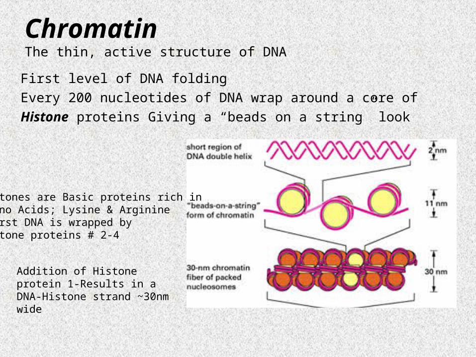
ChromatinThe thin, active structure of DNA
First level of DNA folding
Every 200 nucleotides of DNA wrap around a core of
Histone proteins Giving a “beads on a string” look
Addition of Histone protein 1-Results in a DNA-Histone strand ~30nm wide
Histones are Basic proteins rich in Amino Acids; Lysine & Arginine-First DNA is wrapped by Histone proteins # 2-4

Chromosome
Chromatin lengths of 50,000-100,000 nucleotides are looped together by nonhistone proteins
Chromosomes pack DNA into final structure measuring 5µm long x ~1µm wide
The highly folded DNA structure An inactive form of DNA

Cells must be able to grow & divide
New cells must contain complete copies of the entire set of chromosomes and all their DNA
A Cell’s lifetime of growth & division can be referred to as a Cell Cycle
Cell Cycle

Cell Cycle
Includes not only cell division, but also the intervening time period when cells are not dividing...

Eukaryotic Cell CycleIncludes:
1. Cell growth2. Chromosome replication3. Cell division (2 types)
• Mitosis• Meiosis

Cell Cycle Phases
• Interphase: cell growth & DNA replication (steps 1& 2 from previous slide)
• Mitosis: nuclear & cell Division

InterphaseComposed of G1, S & G2 phases
Interphase includes everything except Mitosis

Interphase
G1- gap phase between Mitosis & S
S phase- DNA replication
G2-gap phase between
S & Mitosis

Mammalian Cell Cycle
G1: Highly variable, Very short in rapidly dividing cells, long in slow-growing cells
S: 3-8 hours
G2: 2-6 hours
M: 1-2 hours

Control of the Cell Cycle
For all living eukaryotic organisms it is essential that the different phases of the cell cycle are precisely coordinated. The phases must follow in correct order, and one phase must be completed before the next phase can begin. Errors in this coordination may lead to chromosomal alterations.
Chromosomes or parts of chromosomes may be lost, rearranged or distributed unequally between the two daughter cells. This type of chromosome alteration is often seen in cancer cells

2001 Nobel Laureates in Physiology or Medicine
Leland Hartwell, Paul Nurse & Timothy Hunt made seminal discoveries concerning the control of the cell cycle. They identified key molecules that regulate the cell cycle in all eukaryotic organisms, including yeasts, plants, animals and human.
Defects in cell cycle control may lead to the type of chromosome alterations seen in cancer cells.

G1 Arrested Cells
• An important control point in cell cycle holds cells in G1
• Cells can remain indefinitely in G1
• Such cells are said to reside in a G0 state, a cell cycle holding point..…
• G0 Cells may re-enter the normal cell cycle if given conditions suitable for growth....

The S PhaseEach Chromosome replicates to form
2 Chromatids.
Replicated chromatids are joined
together at their centromeres
Replication is semi-conservative. Meaning- each DNA strand serves as a template for a new strand

Cytokinesis
• Actual cell division stage
• In animal cells, a constriction furrow forms on the outside to pinch the new cells apart.
• In plant cells, a cell plate forms inside and separates the cell.

Sexual Reproduction:Meiosis

Mitosis vs. Meiosis
• Mitosis: each division gives 2 identical products
2N Cell 2(2N) Cells 4(2N) Cells
• Meiosis: 2 division steps which reduce the number of chromosomes in half
2N Cell 4(1N) Cells

Meiosis• Cell division process in
which the number of chromosomes is cut in half…
• Results in the formation of gametes …such as- eggs and sperm
• Gametes have ½ chromosomes of adult
• Fusion of an egg and sperm results in a zygote • Zygote now has the same number of chromosomes as adult
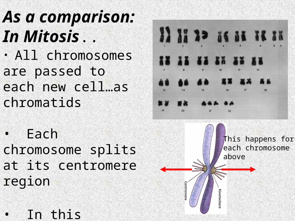
As a comparison: In Mitosis..• All chromosomes are passed to each new cell…as chromatids
• Each chromosome splits at its centromere region
• In this example: 46 chromatids go to each new cell
This happens for each chromosome above

In Meiosis:
During the first stage of Meiosis-Only one of each pair will go to each new cell
This is called the separation ofHomologous chromosomes
Each new cell will then have 23 chromosomes

Haploid vs. Diploid• Typically, each cell of your body has 46
chromosomes..23 from each parent
• So, you have what we call a Diploid value of 46
• Or, referred to as 2N = 46
• Your gametes, however, have 1N values
• 1N = 23….This is a Haploid condition
• All your normal body cells are diploid, only your gametes are haploid

Diploid numbers of some organisms
Homo sapiens (human) 46
Mus musculus (house mouse) 40
Zea mays(corn or maize) 20
Drosophila melanogaster (fruit fly) 8
Xenopus laevis (South African clawed frog) 36
Caenorhabditis elegans (microscopic roundworm) 12
Saccharomyces cerevisiae (budding yeast) 32
Canis familiaris (domestic dog) 78
Arabidopsis thaliana (plant in the mustard family) 10
Muntiacus muntjac (its Indian cousin) 6
Myrmecia pilosula (an ant) 2
Parascaris equorum var. univalens (parasitic roundworm)
2
Cambarus clarkii (a crayfish) 200
Equisetum arvense (field horsetail, a plant) 216
The complete set of chromosomes in the cells of an organism is its karyotype.

The Process of Meiosis: 2 Separate Steps
• Meiosis I: Homologous chromosomes line up and then separate. In Meiosis 1, chromosomes in a diploid cell resegregate, producing four haploid daughter cells. It is this step in Meiosis that generates genetic diversity.
• Meiosis II: Similar process to mitosis

Meiosis I: 4 Stages
• Prophase I: Replicated chromosomes condense and homologs join together in a foursome
• Homologs may exchange entire regions of genes…Crossing over
• Human female eggs remain in Meiosis I until puberty… 12-13 years
Two homologous chromosomes come together…
and may cross and exchange genes

Prophase I in Meiosis has a unique event -- the pairing of homologous chromosomes. Synapsis is the process of linking of the replicated homologous chromosomes. The resulting chromosome is termed a tetrad, being composed of two chromatids from each chromosome, forming a thick (4-strand) structure.
Crossing-over may occur at this point. During crossing-over chromatids break and may be reattached to a different homologous chromosome.
Prophase I in Meiosis

Meiosis II:
• Essentially a mitotic division of the products of Meiosis I that now separates the chromatids
• Meiosis 2 is similar to mitosis. However, there is no "S" phase. The chromatids of each chromosome are no longer identical because of recombination.
• Meiosis II separates the chromatids producing two daughter cells each with 23 chromosomes (haploid), and each chromosome has only one chromatid.

Meiosis II: • Meiosis II: Prophase II• During Prophase II, nuclear envelopes (if re- formed during
Telophase I) dissolve, and spindle fibers reform. • All else is as in Prophase of mitosis. Indeed Meiosis II is very
similar to mitosis. • Meiosis II: Metaphase II• Metaphase II is similar to mitosis metaphase, with spindles
moving chromosomes into the equatorial area and attaching to the opposite sides of the centromeres in the kinetochore region.
• Chromosomes align along center• Meiosis II: Anaphase II• During Anaphase II, the centromeres split and the former
chromatids are segregated into opposite sides of the cell.• Meiosis II: Telophase II• Telophase II is identical to Telophase of mitosis. Cytokinesis
separates the cells.• End up with 4 haploid (1N) products

Meiotic Products
• Final products of meiotic division are:
• 4 cells containing a haploid set (1N) of chromosomes
• These 1N cells become gametes in animals
• But, in plants, they may grow into new 1N individuals.

Meiosis Overview