Embed Size (px)
Citation preview
Chapter 14
DNA cloning
Cloning is the process of moving a gene from the chromosome it occurs in naturallyto an autonomously replicating vector. In the cloning process, the DNA is removedfrom cells, manipulations of the DNA are carried out in a test-tube, and the DNA issubsequently put back into cells. Because E. coli is so well characterized, it is usuallythe cell of choice for manipulating DNA molecules. Once the appropriate combina-tion of vector and cloned DNA or construct has been made in E. coli, the construct canbe put into other cell types. This chapter is concerned with the details of the individ-ual steps in the cloning process:1 How is the DNA removed from the cells?2 How is the DNA cut into pieces?3 How are the pieces of DNA put back together?4 How do we monitor each of these steps?
Isolating DNA from cells
Plasmid DNA isolation
The first step in cloning is to isolate a large amount of the vector and chromosomalDNAs. Isolation of plasmid DNA will be examined first. In the general scheme, cellscontaining the plasmid are grown to a high cell density, gently lysed, and the plasmidDNA is isolated and concentrated. When the cells are growing, the antibiotic corre-sponding to the antibiotic resistance determinant on the plasmid is included in thegrowth media. This ensures that the majority of cells contain plasmid DNA. Withoutthe antibiotic selection, an unstable plasmid (i.e. one without a par function) can belost from the cell population in a few generations.
Cells can be lysed by several different methods depending on the size of the plasmidmolecule, the specific strain of E. coli the plasmid will be isolated from, and how theplasmid DNA will be purified. Most procedures use EDTA to chelate the Mg++ associat-ed the outer membrane and destabilize the outer membrane. Lysozyme is added to digest the peptidoglycan and detergents are frequently used to solubilize the membranes. RNases are added to degrade the large amount of RNA found in activelygrowing E. coli cells. The RNase gains access to the RNA after the EDTA and lysozymetreatments. This mixture is centrifuged to pellet intact cells and large pieces of cell
FYI 14.1
CsCl gradients
In traditional preparations ofhighly purified plasmid DNA,the lysate containing theplasmid DNA was mixed withthe dye, ethidium bromide,that slips between the bases ofthe double helix (intercalation)and unwinds the DNA. Theamount of ethidium bromidethat can intercalate dependson the topology of the DNAmolecule. Covalently closedsupercoiled molecules bindless ethidium bromide thanlinear DNA because the DNA isconstrained. Linear molecules,such as broken chromosomes,can bind more ethidiumbromide because they canunwind as the ethidiumbromide binds. Linearmolecules can be saturatedwith ethidium bromide to thepoint of approximately onemolecule of ethidium bromidefor every two base pairs ofDNA. The ethidium saturatedDNA is centrifuged in a cesiumchloride gradient. Thedifferential binding of the dyeleads to different buoyantdensities for chromosomal andplasmid DNA in the cesiumchloride gradient. This allowsthe plasmid DNA to beseparated from thechromosome. While ethidiumbromide–cesium chloridegradients are time consuming,for extremely pure plasmidDNA, they are still very useful.
PYF14 3/21/05 8:05 PM Page 234
debris. The supernatant contains a mixture of soluble cell components, including theplasmid, and is known as a lysate.
The methods used to purify the plasmid DNA from the cell lysate rely on the smallsize and abundance of the plasmid DNA relative to the chromosome, and the cova-lently closed circular nature of plasmid DNA. Most plasmids exist in the cytoplasm ofthe cell as circular DNA molecules that are highly supercoiled. The lysate is treatedwith sodium hydroxide to denature all of the DNA, and with detergent, SDS. The pHis then abruptly lowered, causing the SDS to precipitate and bring with it denaturedchromosomal DNA, membrane fragments, and other cell debris. Most of the plasmidDNA renneals to form dsDNA because each strand is a covalently closed molecule andthe two strands are not physically separated from each other. The small size of theplasmid allows the plasmid molecules to remain in suspension. The supernatant,which contains plasmid DNA, proteins, and other small molecules can be treated in anumber of different ways to purify the plasmid. The most common protocol relies ona column resin that binds DNA. A small amount of the resin is mixed with the plas-mid-containing supernatant and the plasmid-bound resin is collected in a small col-umn. The remaining cell components are washed away and the plasmid is elutedfrom the resin. This procedure is quick, simple, and reliable and can be easily carriedout on a large number of samples. Many modifications of this procedure have beendevised.
Chromosomal DNA isolation
To isolate chromosomal DNA, cells are lysed in much the same way as for plasmidDNA isolation. The cell lysate is extracted with phenol or otherwise treated to removeall of the proteins. The chromosomal DNA is precipitated as long threads. The chro-mosomal DNA is very fragile and breaks easily. For these reasons, the chromosomalDNA is not usually purified using columns. Rather, the precipitated threads are col-lected by centrifugation.
Cutting DNA molecules
Once DNA has been purified, it must be cut into pieces before the chromosomal DNAand the plasmid DNA can be joined. The problem is to cut the DNA so that it will beeasy to join the cut ends of the chromosomal DNA to the cut ends of the plasmid DNA.A group of enzymes, called restriction enzymes, are used for this purpose. Restric-tion enzymes are isolated from different bacterial species.
Bacteria use restriction enzymes and modification enzymes to identify their ownDNA from any foreign DNA that enters their cytoplasm. The restriction part of thesystem is an enzyme that recognizes a specific DNA sequence or restriction site andcleaves the DNA by catalyzing breaks in specific phosphodiester bonds. The cleavageis on both strands of the DNA so that a double-stranded break is made. The modifica-tion part of the system is a protein that recognizes the same DNA sequence as the re-striction enzyme. The modification enzyme methylates the DNA sequence so that therestriction enzyme no longer recognizes the sequence. Thus, the bacteria can protectits own DNA from the restriction enzyme. Any DNA that enters the bacteria and con-tains the unmethlyated restriction site is cut and degraded. There are three types of re-striction–modification systems (Table 14.1). The types are distinguished based on the
DNA Cloning 235
FYI 14.2
The discovery ofrestriction enzymes
Restriction enzymes werediscovered in E. coli in the1950s by scientists studyingbacteriophage. Bacteriophagel can be grown on an E. coliK12 strain and titered on E. coliK12 to determine the numberof phage per milliliter. A high-titer phage lysate will containapproximately 1010 plaque-forming units per ml (pfu/ml).If this phage lysate is titered onan E. coli B strain, the titer willdrop to 106 pfu/ml. One of thephage that forms a plaque onE. coli B can be used to make ahigh-titer lysate on E. coli B.The lysate grown on E. coli Bwill titer on E. coli B atapproximately 1010 pfu/ml butwill titer on E. coli K12 at 106
pfu/ml. This four-log drop inplating efficiency can be tracedto genes encoded by thebacterial chromosome of eachstrain. The system is known ashost restriction andmodification. Host restriction iscarried out by a restrictionendonuclease andmodification is carried out bythe protein that modifies therestriction site.
PYF14 3/21/05 8:05 PM Page 235
number of proteins in the system, the cofactors for these proteins, and if the proteinsform a complex.
Type I restriction–modification systems
Type I systems are the most intricate and very few of them have been described. Threedifferent proteins form a complex that carries out both restriction and modificationof the DNA. The complex must interact with a cofactor, S-adenosylmethionine, be-fore it is capable of recognizing DNA. The S-adenosylmethionine is the methyl donorfor the modification reaction and all known Type I systems methylate adenineresidues on both strands of the DNA. The restriction reaction requires ATP and Mg++
for cleavage of the DNA. The complex also has topoisomerase activity. The DNA se-quence recognized by Type I enzymes is also complex. The sequence is asymmetricand split into two parts (Fig. 14.1a). The first part is a 3bp sequence, next is a 6–8bpspacer of nonspecific sequence, and finally there is a 4–5bp sequence. Cleavage of theDNA occurs randomly, usually no closer than 400bp from the recognition sequenceand sometimes as far away as 7000bp.
Type II restriction–modification systems
Type II systems are composed of two independent proteins. One protein is responsi-ble for modifying the DNA and one for restricting the DNA. Modification of the DNAuses S- adenosylmethionine as the methyl donor. The Type II modification enzymesmethylate the DNA at one of three places, with each specific modification enzymemethlyating the same residue every time. The modifications that have been found are5-methlycytosine, 4-methylcytosine, or 6-methlyadenosine. The DNA sequence rec-ognized by Type II restriction enzymes is symmetric and usually palindromic (Fig.14.1b). The DNA sequence is between 4 and 8bp in length, with most restriction en-zymes recognizing 4 or 6bp. Both the cleavage of the DNA and modification of theDNA occur symmetrically on both strands of the DNA within the recognition se-quence. Restriction enzymes function as dimers of a single protein so that each pro-tein monomer can interact with one strand of the DNA. Thus, both strands of the DNA are cleaved at the same time, generating a double-stranded break. Several
236 Chapter 14
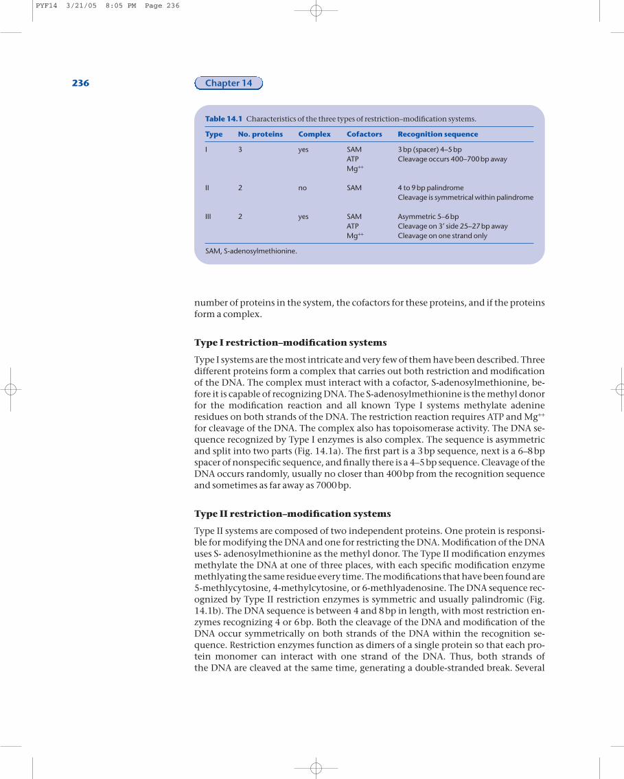
Table 14.1 Characteristics of the three types of restriction–modification systems.
Type No. proteins Complex Cofactors Recognition sequence
I 3 yes SAM 3bp (spacer) 4–5bpATP Cleavage occurs 400–700bp awayMg++
II 2 no SAM 4 to 9bp palindromeCleavage is symmetrical within palindrome
III 2 yes SAM Asymmetric 5–6bpATP Cleavage on 3¢ side 25–27bp awayMg++ Cleavage on one strand only
SAM, S-adenosylmethionine.
PYF14 3/21/05 8:05 PM Page 236
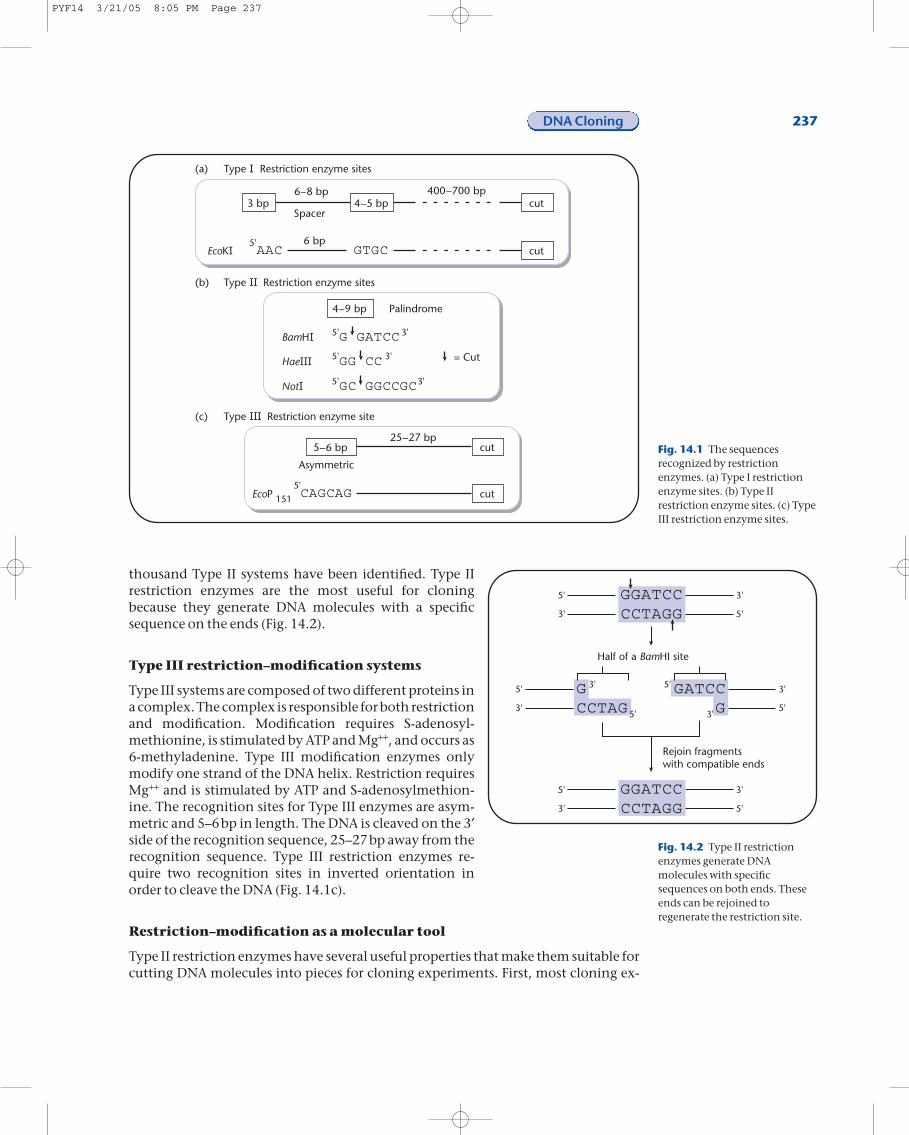
thousand Type II systems have been identified. Type II restriction enzymes are the most useful for cloning because they generate DNA molecules with a specific sequence on the ends (Fig. 14.2).
Type III restriction–modification systems
Type III systems are composed of two different proteins ina complex. The complex is responsible for both restrictionand modification. Modification requires S-adenosyl-methionine, is stimulated by ATP and Mg++, and occurs as 6-methyladenine. Type III modification enzymes onlymodify one strand of the DNA helix. Restriction requiresMg++ and is stimulated by ATP and S-adenosylmethion-ine. The recognition sites for Type III enzymes are asym-metric and 5–6bp in length. The DNA is cleaved on the 3¢side of the recognition sequence, 25–27bp away from therecognition sequence. Type III restriction enzymes re-quire two recognition sites in inverted orientation inorder to cleave the DNA (Fig. 14.1c).
Restriction–modification as a molecular tool
Type II restriction enzymes have several useful properties that make them suitable forcutting DNA molecules into pieces for cloning experiments. First, most cloning ex-
DNA Cloning 237
––
–
–
–– Fig. 14.1 The sequences
recognized by restrictionenzymes. (a) Type I restrictionenzyme sites. (b) Type IIrestriction enzyme sites. (c) TypeIII restriction enzyme sites.
GGATCCCCTAGG
5'
3'
3'
5'
GGATCCCCTAGG
5'
3'
3'
5'
Half of a BamHI site
Rejoin fragmentswith compatible ends
GCCTAG
GATCC G
5'
3'
3'
5'
3'
5'
5'
3'
Fig. 14.2 Type II restrictionenzymes generate DNAmolecules with specificsequences on both ends. Theseends can be rejoined toregenerate the restriction site.
PYF14 3/21/05 8:05 PM Page 237
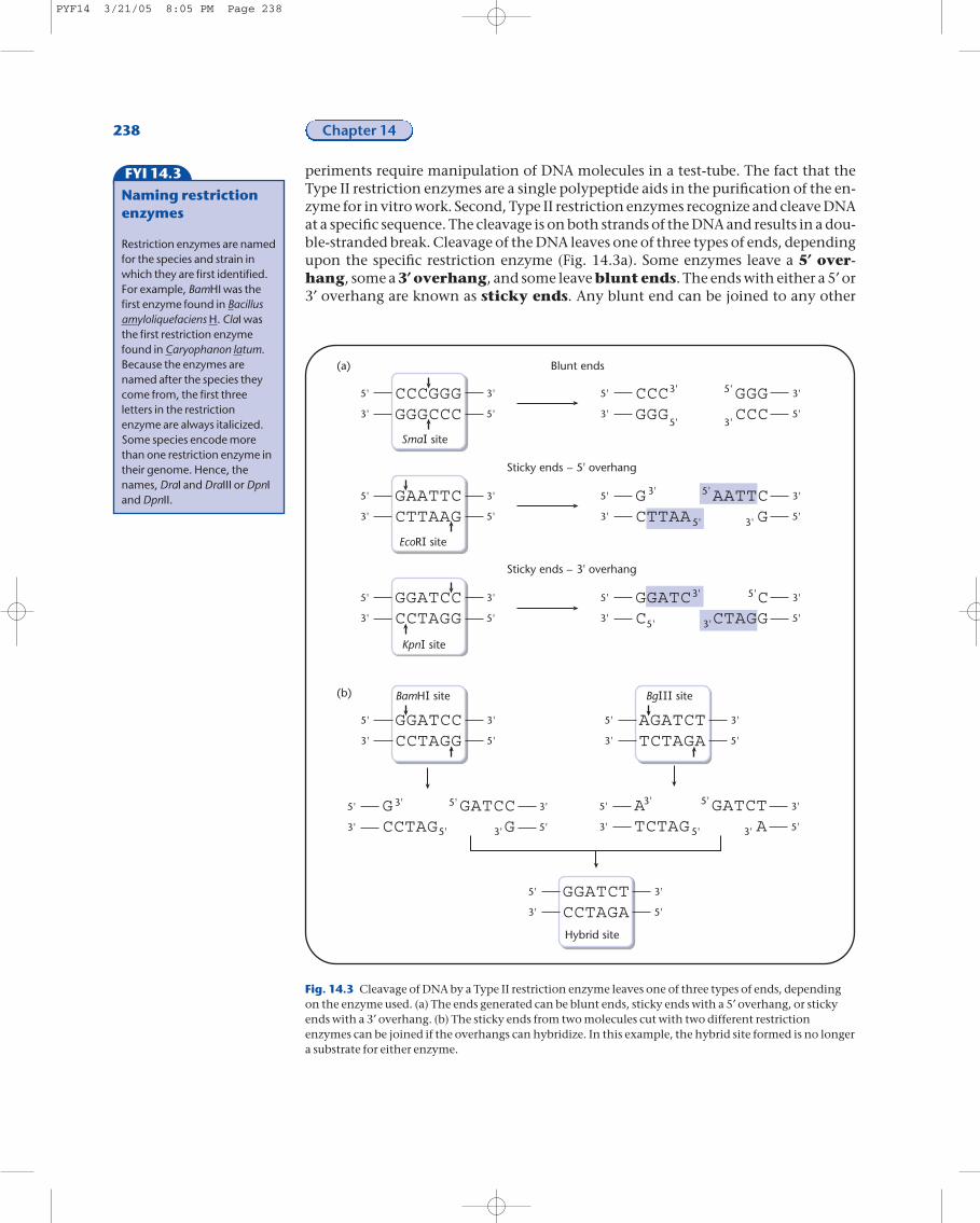
periments require manipulation of DNA molecules in a test-tube. The fact that theType II restriction enzymes are a single polypeptide aids in the purification of the en-zyme for in vitro work. Second, Type II restriction enzymes recognize and cleave DNAat a specific sequence. The cleavage is on both strands of the DNA and results in a dou-ble-stranded break. Cleavage of the DNA leaves one of three types of ends, dependingupon the specific restriction enzyme (Fig. 14.3a). Some enzymes leave a 5¢ over-hang, some a 3¢ overhang, and some leave blunt ends. The ends with either a 5¢ or3¢ overhang are known as sticky ends. Any blunt end can be joined to any other
238 Chapter 14
FYI 14.3
Naming restrictionenzymes
Restriction enzymes are namedfor the species and strain inwhich they are first identified.For example, BamHI was thefirst enzyme found in Bacillusamyloliquefaciens H. ClaI wasthe first restriction enzymefound in Caryophanon latum.Because the enzymes arenamed after the species theycome from, the first threeletters in the restrictionenzyme are always italicized.Some species encode morethan one restriction enzyme intheir genome. Hence, thenames, DraI and DraIII or DpnIand DpnII.
–
–
Fig. 14.3 Cleavage of DNA by a Type II restriction enzyme leaves one of three types of ends, dependingon the enzyme used. (a) The ends generated can be blunt ends, sticky ends with a 5¢ overhang, or stickyends with a 3¢ overhang. (b) The sticky ends from two molecules cut with two different restrictionenzymes can be joined if the overhangs can hybridize. In this example, the hybrid site formed is no longera substrate for either enzyme.
PYF14 3/21/05 8:05 PM Page 238
blunt end regardless of how the blunt end was generated. Sticky ends can be joined toother sticky ends, provided that either the same Type II restriction enzyme was usedto generate both sticky ends that are to be joined or that the bases in the overhang areidentical and have the correct overhang (Fig. 14.3b).
Generate double-stranded breaks in DNA by shearing the DNA
Usually restriction enzymes are used to cut the chromosomal DNA for cloning. Thedrawback of this approach is uncovered when the gene contains a restriction site forthe enzyme being used to construct the library. One way to solve this problem is toonly partially digest the chromosomal DNA with the restriction enzyme. Anotherway is to shear the chromosomal DNA and clone the randomly sheared DNA into ablunt end restriction enzyme site in the vector. One way to shear chromosomal DNAis by passing it quickly through a small needle attached to a syringe.
Joining DNA molecules
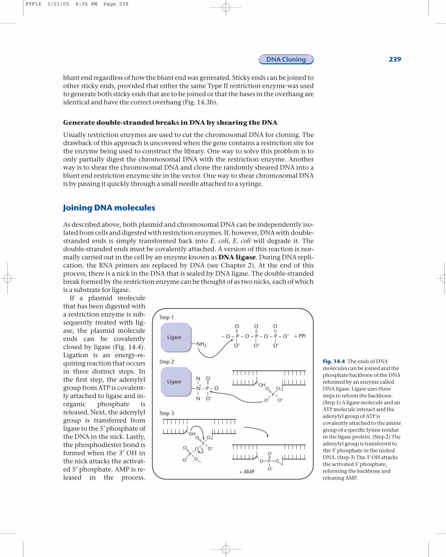
As described above, both plasmid and chromosomal DNA can be independently iso-lated from cells and digested with restriction enzymes. If, however, DNA with double-stranded ends is simply transformed back into E. coli, E. coli will degrade it. Thedouble-stranded ends must be covalently attached. A version of this reaction is nor-mally carried out in the cell by an enzyme known as DNA ligase. During DNA repli-cation, the RNA primers are replaced by DNA (see Chapter 2). At the end of thisprocess, there is a nick in the DNA that is sealed by DNA ligase. The double-strandedbreak formed by the restriction enzyme can be thought of as two nicks, each of whichis a substrate for ligase.
If a plasmid moleculethat has been digested witha restriction enzyme is sub-sequently treated with lig-ase, the plasmid moleculeends can be covalentlyclosed by ligase (Fig. 14.4).Ligation is an energy-re-quiring reaction that occursin three distinct steps. Inthe first step, the adenylylgroup from ATP is covalent-ly attached to ligase and in-organic phosphate isreleased. Next, the adenylylgroup is transferred fromligase to the 5¢ phosphate ofthe DNA in the nick. Lastly,the phosphodiester bond isformed when the 3¢ OH inthe nick attacks the activat-ed 5¢ phosphate. AMP is re-leased in the process.
DNA Cloning 239
– O – P – O – P – O – P – O-
O
O- O- O-
O O
+ PPiLigaseNH2
O
O-N
LigaseN – P – O
N
O-O-
O OP
OH
O-
O-
O
O
O O
OP
P
OH
O-
O
OO P
Step 1
Step 2
Step 3
+ AMP
+
Fig. 14.4 The ends of DNAmolecules can be joined and thephosphate backbone of the DNAreformed by an enzyme calledDNA ligase. Ligase uses threesteps to reform the backbone.(Step 1) A ligase molecule and anATP molecule interact and theadenylyl group of ATP iscovalently attached to the aminegroup of a specific lysine residuein the ligase protein. (Step 2) Theadenylyl group is transferred tothe 5¢ phosphate in the nickedDNA. (Step 3) The 3¢ OH attacksthe activated 5¢ phosphate,reforming the backbone andreleasing AMP.
PYF14 3/21/05 8:05 PM Page 239
Because of this mechanism, ligase requires both a 3¢ OH and a 5¢ phosphate. If the 5¢phosphate is missing (see below), the nick cannot be sealed by ligase.
Manipulating the ends of molecules
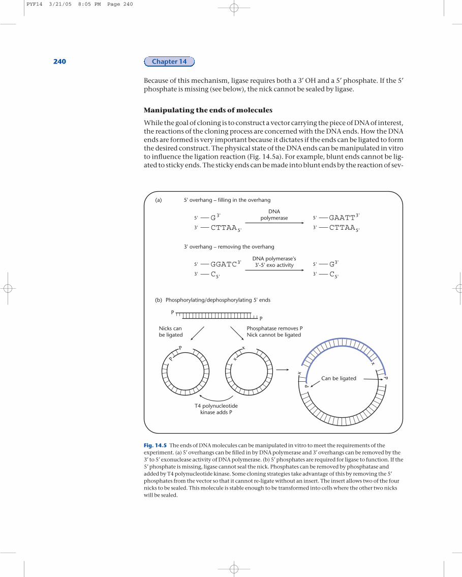
While the goal of cloning is to construct a vector carrying the piece of DNA of interest,the reactions of the cloning process are concerned with the DNA ends. How the DNAends are formed is very important because it dictates if the ends can be ligated to formthe desired construct. The physical state of the DNA ends can be manipulated in vitroto influence the ligation reaction (Fig. 14.5a). For example, blunt ends cannot be lig-ated to sticky ends. The sticky ends can be made into blunt ends by the reaction of sev-
240 Chapter 14
5' overhang – filling in the overhang
3' overhang – removing the overhang
GCTTAA
5'
3'
3'
5'
GAATTCTTAA
5'
3'5'
3'DNA
polymerase
GGATCC
5'
3'
3'
5'
GC
5'
3'
3'
5'
DNA polymerase's3'-5' exo activity
Phosphorylating/dephosphorylating 5' ends
x
x
P
P
PP
x
x P
P
Nicks canbe ligated
Phosphatase removes PNick cannot be ligated
T4 polynucleotidekinase adds P
Can be ligated
(b)
(a)
Fig. 14.5 The ends of DNA molecules can be manipulated in vitro to meet the requirements of theexperiment. (a) 5¢ overhangs can be filled in by DNA polymerase and 3¢ overhangs can be removed by the3¢ to 5¢ exonuclease activity of DNA polymerase. (b) 5¢ phosphates are required for ligase to function. If the5¢ phosphate is missing, ligase cannot seal the nick. Phosphates can be removed by phosphatase andadded by T4 polynucleotide kinase. Some cloning strategies take advantage of this by removing the 5¢phosphates from the vector so that it cannot re-ligate without an insert. The insert allows two of the fournicks to be sealed. This molecule is stable enough to be transformed into cells where the other two nickswill be sealed.
PYF14 3/21/05 8:05 PM Page 240
eral different enzymes. If the sticky end contains a 5¢ overhang, then any one of sev-eral different DNA polymerases can be used to add the missing bases to the 3¢ OHusing the 5¢ overhang as a template. A 3¢ overhang cannot be filled in, rather the over-hang must be removed. Many DNA polymerases have a 3¢ to 5¢ exonuclease activityand this activity can be used to remove 3¢ overhangs.
The 5¢ phosphate can also be manipulated (Fig. 14.5b). If DNA molecules are miss-ing the 5¢ phosphate, the phosphate can be added by an enzyme called T4 polynu-cleotide kinase. T4 polynucleotide kinase is an ATP-requiring enzyme that wasoriginally identified in the bacteriophage T4. Molecules that have been phosphory-lated by T4 polynucleotide kinase can be ligated to other molecules by ligase.
When pieces of chromosomal DNA are mixed with cut vector DNA, ligation of sev-eral different molecules can take place. The vector DNA ends can be ligated to reformthe vector, the ends of a piece of chromosomal DNA can be ligated to each other, theends of several vector or several chromosomal molecules can be ligated, or the ends ofa piece of chromosomal DNA can be ligated to the ends of a piece of vector DNA. Theligation mix is usually put back into cells by transformation (see Chapter 11) and theantibiotic marker on the vector is selected for. Only cells transformed by moleculesthat contain vector DNA will form colonies. Of the molecules in the ligation mix,only religated vector DNA or vector DNA with a chromosomal insert are a possibilityin the transformants. To reduce the number of vector molecules that are religatedwithout a chromosomal insert, the 5¢ phosphates on the vector can be removed by anenzyme known as a phosphatase. The ends of vector molecules that have been de-phosphorylated (the 5¢ phosphate has been removed) can only be ligated to chromo-somal DNA molecules that have 5¢ phosphates (Fig. 14.5b). These molecules still havea nick on each strand but this nick can be sealed inside the cell.
Visualizing the cloning process
At each step of the cloning process, what is happening to the DNA molecules in thetest-tube can be monitored using a technique called gel electrophoresis. In thistechnique, a gel (Fig. 14.6) containing small indentations or wells is cast. The DNA is loaded into the wells and the gel is placed in an electric current. Because DNA is negatively charged, it will move in the gel towards the positive pole. The DNA mi-grates or moves in the electric current based on size and shape. The larger a DNA mol-ecule, the slower it moves. The more compact, or supercoiled a piece of DNA, thefaster it moves.
The gel can be made from several different polymers, depending on the specifics of the experiment. Agarose forms a matrix that will separate DNA molecules from~500bp up to entire chromosomes (several million base pairs). If an electric current isconstantly applied to an agarose gel from only one direction, agarose gels will sepa-rate DNA from ~500bp to ~25,000bp. If the direction and the timing of the currentare varied over the electrophoresis time, then entire chromosomes can be separated inagarose. An alternative polymer, polyacrylamide, can be used to separate molecules afew base pairs in length to approximately 1000bp.
Once the DNA has been separated in the gel, the gel is immersed in a solution con-taining ethidium bromide. If ultraviolet light is used to illuminate the gel, the ethidi-um bromide that is bound to the DNA will fluoresce, indicating the presence of bandsof DNA (Fig. 14.6). Each band is composed of DNA molecules that are similar in sizeand shape. For example, when plasmid DNA is extracted from the cell, the majority of
DNA Cloning 241
FYI14.4
What is agarose?
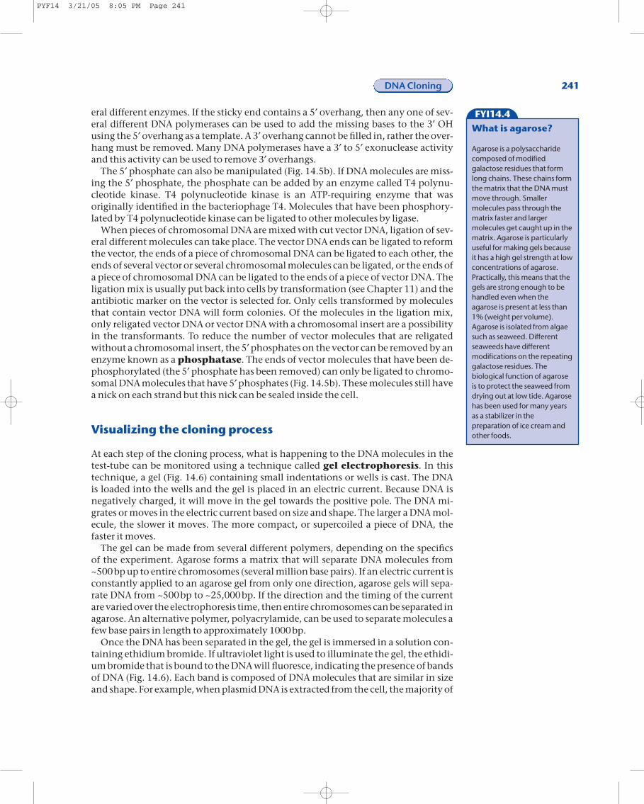
Agarose is a polysaccharidecomposed of modifiedgalactose residues that formlong chains. These chains formthe matrix that the DNA mustmove through. Smallermolecules pass through thematrix faster and largermolecules get caught up in thematrix. Agarose is particularlyuseful for making gels becauseit has a high gel strength at lowconcentrations of agarose.Practically, this means that thegels are strong enough to behandled even when theagarose is present at less than1% (weight per volume).Agarose is isolated from algaesuch as seaweed. Differentseaweeds have differentmodifications on the repeatinggalactose residues. Thebiological function of agaroseis to protect the seaweed fromdrying out at low tide. Agarosehas been used for many yearsas a stabilizer in thepreparation of ice cream andother foods.
PYF14 3/21/05 8:05 PM Page 241
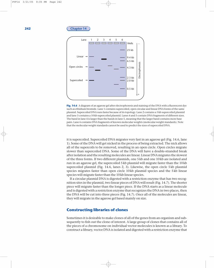
it is supercoiled. Supercoiled DNA migrates very fast in an agarose gel (Fig. 14.6, lane1). Some of the DNA will get nicked in the process of being extracted. The nick allowsall of the supercoils to be removed, resulting in an open circle. Open circles migrateslower than supercoiled DNA. Some of the DNA will have a double-stranded breakafter isolation and the resulting molecules are linear. Linear DNA migrates the slowestof the three forms. If two different plasmids, one 5kb and one 10kb are isolated andrun in an agarose gel, the supercoiled 5kb plasmid will migrate faster than the 10kbsupercoiled plasmid (Fig. 14.6, lanes 2, 3). Likewise, the open circle 5kb plasmidspecies migrates faster than open circle 10kb plasmid species and the 5kb linearspecies will migrate faster than the 10kb linear species.
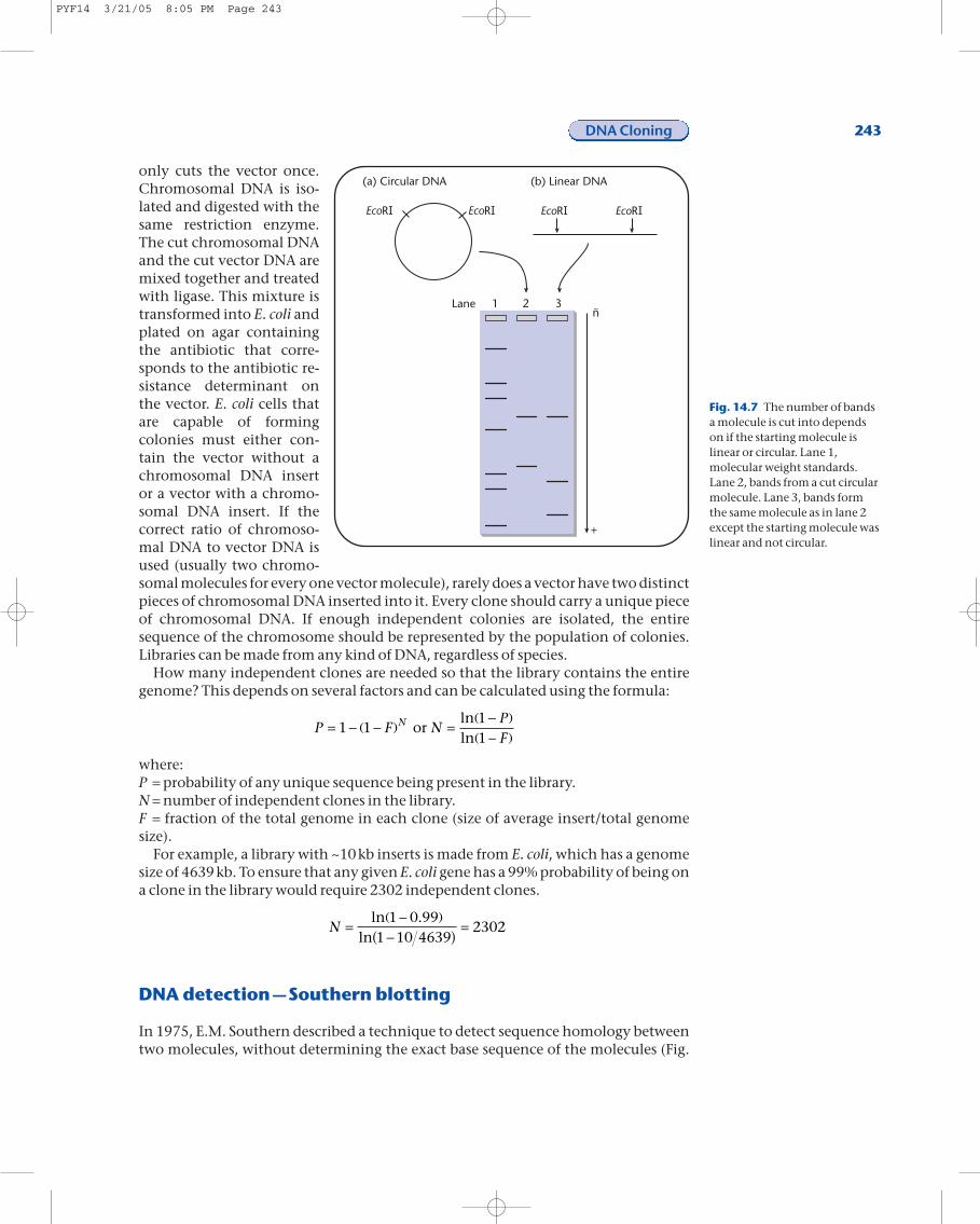
If a circular plasmid DNA is digested with a restriction enzyme that has two recog-nition sites in the plasmid, two linear pieces of DNA will result (Fig. 14.7). The shorterpiece will migrate faster than the longer piece. If the DNA starts as a linear moleculeand is digested with a restriction enzyme that recognizes the DNA in two places, thenthe DNA will be cut into three pieces (Fig. 14.7). Once all of the molecules are linear,they will migrate in the agarose gel based mainly on size.
Constructing libraries of clones
Sometimes it is desirable to make clones of all of the genes from an organism and sub-sequently to fish out the clone of interest. A large group of clones that contains all ofthe pieces of a chromosome on individual vector molecules is known as a library. Toconstruct a library, vector DNA is isolated and digested with a restriction enzyme that
242 Chapter 14
Wells
23 kb
9.4 kb6.5 kb
4.3 kb
2.3 kb
0.5 kb
Elec
trop
hore
sis
1 2 3 4 5 6
Linear
Open circles
Supercoiled 2.0 kb
Lane
–
+
Fig. 14.6 A diagram of an agarose gel after electrophoresis and staining of the DNA with a fluorescent dyesuch as ethidium bromide. Lane 1 contains supercoiled, open circular and linear DNA forms of the sameplasmid. Supercoiled DNA runs faster because of its topology. Lane 2 contains a 5kb supercoiled plasmidand lane 3 contains a 10kb supercoiled plasmid. Lanes 4 and 5 contain DNA fragments of different sizes.The band in lane 4 is larger than the bands in lane 5, meaning that the larger band contains more basepairs. Lane 6 contains DNA fragments of known molecular weights (molecular weight standards). Notethat the molecular weight standards cannot be used to predict the sizes of supercoiled DNA.
PYF14 3/21/05 8:05 PM Page 242
only cuts the vector once.Chromosomal DNA is iso-lated and digested with thesame restriction enzyme.The cut chromosomal DNAand the cut vector DNA aremixed together and treatedwith ligase. This mixture istransformed into E. coli andplated on agar containingthe antibiotic that corre-sponds to the antibiotic re-sistance determinant onthe vector. E. coli cells thatare capable of formingcolonies must either con-tain the vector without achromosomal DNA insertor a vector with a chromo-somal DNA insert. If thecorrect ratio of chromoso-mal DNA to vector DNA isused (usually two chromo-somal molecules for every one vector molecule), rarely does a vector have two distinctpieces of chromosomal DNA inserted into it. Every clone should carry a unique pieceof chromosomal DNA. If enough independent colonies are isolated, the entire sequence of the chromosome should be represented by the population of colonies. Libraries can be made from any kind of DNA, regardless of species.
How many independent clones are needed so that the library contains the entiregenome? This depends on several factors and can be calculated using the formula:
where:P = probability of any unique sequence being present in the library.N = number of independent clones in the library.F = fraction of the total genome in each clone (size of average insert/total genomesize).
For example, a library with ~10kb inserts is made from E. coli, which has a genomesize of 4639kb. To ensure that any given E. coli gene has a 99% probability of being ona clone in the library would require 2302 independent clones.
DNA detection —Southern blotting
In 1975, E.M. Southern described a technique to detect sequence homology betweentwo molecules, without determining the exact base sequence of the molecules (Fig.
N =-( )
-( ) =ln .
ln1 0 99
1 10 46392302
P F NPF
N= - -( ) =-( )
-( )1 1
11
or lnln
DNA Cloning 243
Fig. 14.7 The number of bandsa molecule is cut into dependson if the starting molecule islinear or circular. Lane 1,molecular weight standards.Lane 2, bands from a cut circularmolecule. Lane 3, bands formthe same molecule as in lane 2except the starting molecule waslinear and not circular.
PYF14 3/21/05 8:05 PM Page 243
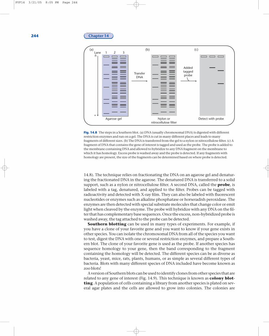
14.8). The technique relies on fractionating the DNA on an agarose gel and denatur-ing the fractionated DNA in the agarose. The denatured DNA is transferred to a solidsupport, such as a nylon or nitrocellulose filter. A second DNA, called the probe, is labeled with a tag, denatured, and applied to the filter. Probes can be tagged with radioactivity and detected with X-ray film. They can also be labeled with fluorescentnucleotides or enzymes such as alkaline phosphatase or horseradish peroxidase. Theenzymes are then detected with special substrate molecules that change color or emitlight when cleaved by the enzyme. The probe will hybridize with any DNA on the fil-ter that has complementary base sequences. Once the excess, non-hybridized probe iswashed away, the tag attached to the probe can be detected.
Southern blotting can be used in many types of experiments. For example, ifyou have a clone of your favorite gene and you want to know if your gene exists inother species. You can isolate the chromosomal DNA from all of the species you wantto test, digest the DNA with one or several restriction enzymes, and prepare a South-ern blot. The clone of your favorite gene is used as the probe. If another species has sequence homology to your gene, then the band corresponding to the fragment containing the homology will be detected. The different species can be as diverse asbacteria, yeast, mice, rats, plants, humans, or as simple as several different types ofbacteria. Blots with many different species of DNA included have become known aszoo blots!
A version of Southern blots can be used to identify clones from other species that arerelated to any gene of interest (Fig. 14.9). This technique is known as colony blot-ting. A population of cells containing a library from another species is plated on sev-eral agar plates and the cells are allowed to grow into colonies. The colonies are
244 Chapter 14
ñ
+
1 2 3Lane
Agarose gel Nylon ornitrocellulose filter
Detect with probe
TransferDNA
Addedtaggedprobe
(a) (b) (c)
Fig. 14.8 The steps in a Southern blot. (a) DNA (usually chromosomal DNA) is digested with differentrestriction enzymes and run on a gel. The DNA is cut in many different places and leads to manyfragments of different sizes. (b) The DNA is transferred from the gel to a nylon or nitrocellulose filter. (c) Afragment of DNA that contains the gene of interest is tagged and used as the probe. The probe is added tothe membrane containing DNA and allowed to hybridize to any DNA fragment on the membrane towhich it has homology. Excess probe is washed away and the probe is detected. If any fragments withhomology are present, the size of the fragments can be determined based on where probe is detected.
PYF14 3/21/05 8:05 PM Page 244
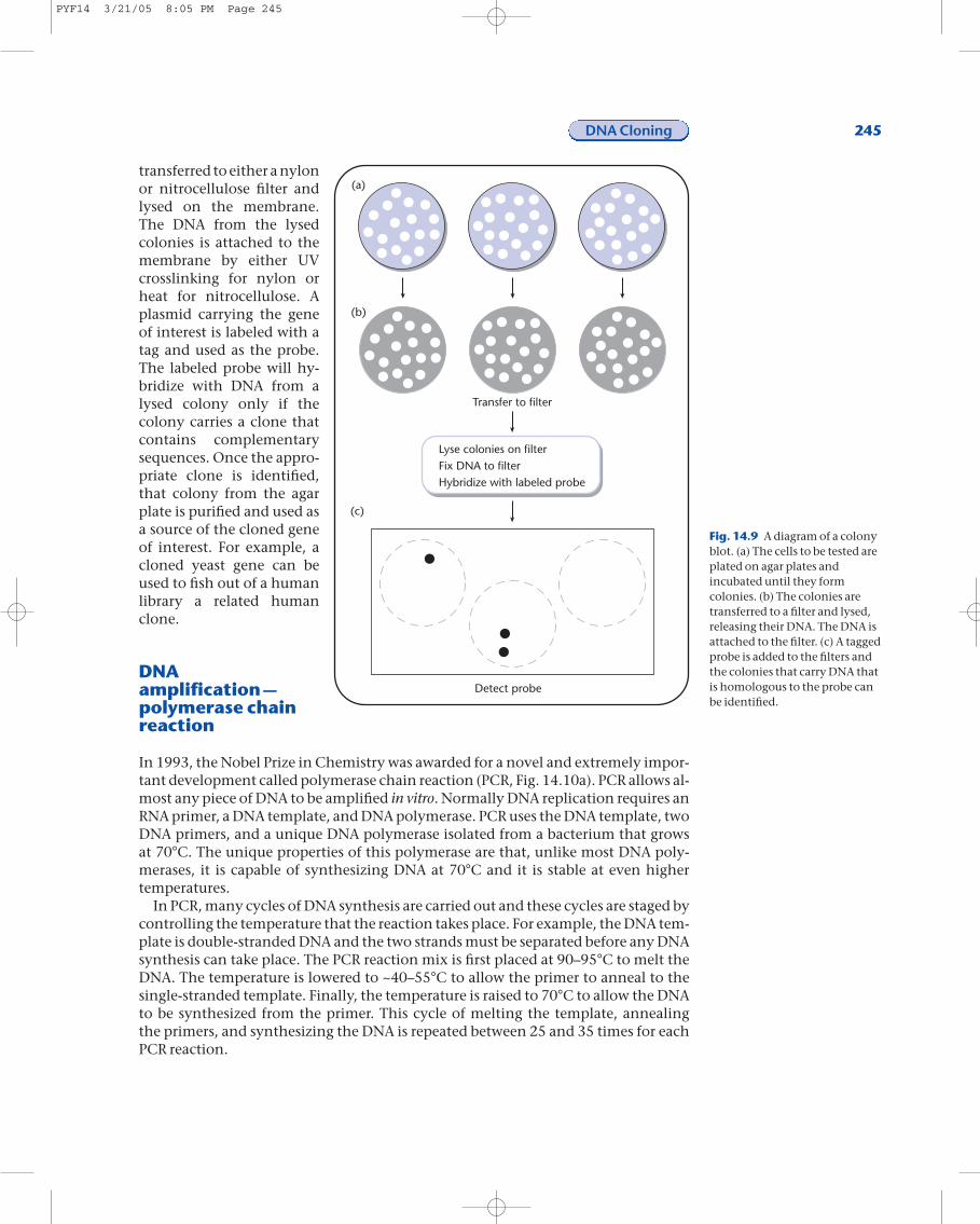
transferred to either a nylonor nitrocellulose filter andlysed on the membrane.The DNA from the lysedcolonies is attached to themembrane by either UVcrosslinking for nylon orheat for nitrocellulose. Aplasmid carrying the geneof interest is labeled with atag and used as the probe.The labeled probe will hy-bridize with DNA from alysed colony only if thecolony carries a clone thatcontains complementarysequences. Once the appro-priate clone is identified,that colony from the agarplate is purified and used asa source of the cloned geneof interest. For example, acloned yeast gene can beused to fish out of a humanlibrary a related humanclone.
DNAamplification —polymerase chainreaction
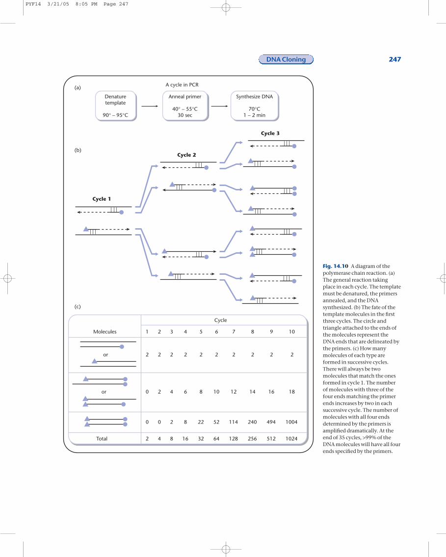
In 1993, the Nobel Prize in Chemistry was awarded for a novel and extremely impor-tant development called polymerase chain reaction (PCR, Fig. 14.10a). PCR allows al-most any piece of DNA to be amplified in vitro. Normally DNA replication requires anRNA primer, a DNA template, and DNA polymerase. PCR uses the DNA template, twoDNA primers, and a unique DNA polymerase isolated from a bacterium that grows at 70°C. The unique properties of this polymerase are that, unlike most DNA poly-merases, it is capable of synthesizing DNA at 70°C and it is stable at even higher temperatures.
In PCR, many cycles of DNA synthesis are carried out and these cycles are staged bycontrolling the temperature that the reaction takes place. For example, the DNA tem-plate is double-stranded DNA and the two strands must be separated before any DNAsynthesis can take place. The PCR reaction mix is first placed at 90–95°C to melt theDNA. The temperature is lowered to ~40–55°C to allow the primer to anneal to the single-stranded template. Finally, the temperature is raised to 70°C to allow the DNAto be synthesized from the primer. This cycle of melting the template, annealing the primers, and synthesizing the DNA is repeated between 25 and 35 times for eachPCR reaction.
DNA Cloning 245
(a)
(b)
Transfer to filter
Lyse colonies on filterFix DNA to filterHybridize with labeled probe
Detect probe
(c)
Fig. 14.9 A diagram of a colonyblot. (a) The cells to be tested areplated on agar plates andincubated until they formcolonies. (b) The colonies aretransferred to a filter and lysed,releasing their DNA. The DNA isattached to the filter. (c) A taggedprobe is added to the filters andthe colonies that carry DNA thatis homologous to the probe canbe identified.
PYF14 3/21/05 8:05 PM Page 245
What happens to a template molecule in each cycle of DNA synthesis? In the firstcycle, the two strands of the template separate, one primer anneals to each strand andtwo dsDNA molecules are produced (Fig. 14.10b). One strand of each dsDNA mole-cule is synthesized only from the primer to the end of the template. In the secondcycle, all four strands are used as templates. Four dsDNA molecules are produced, twoare similar to the dsDNA molecules produced in the first cycle and the other two havethree out of the four DNA ends delineated by the primers. In the third cycle, the eightstrands are used as templates and eight dsDNA molecules are produced. Two of thedsDNA molecules are similar to the products produced in cycle one, four of the
dsDNA molecules havethree out of four ends delin-eated by the primers, andthe other two moleculesnow have all four ends de-lineated by the primer. Inthe remaining cycles, thenumber of dsDNA mole-cules continues to increaselinearly. In cycle 4, 8/16molecules have all four endsdelineated by the twoprimers, in cycle 5, 24/32,cycle 6, 56/64, cycle 7,120/128, and cycle 8,248/256. After 35 cycles, thedsDNA molecule with allfour ends delineated by theprimers is the predominantmolecule in the PCR reac-tion mix.
The DNA primers used inPCR are chosen so that thepiece of DNA of interest isamplified. The DNA primersare synthesized in vitro. Bycarefully choosing primers,the exact base pairs at eitherend of the amplified frag-ment can be predeter-mined. The template DNAcan be from any source.Only a small amount oftemplate is needed. Once afragment of DNA has beenamplified, it can be clonedinto an appropriate vector,used as a probe, restrictionmapped, or used in a num-ber of other techniques.
The DNA replication that
246 Chapter 14
FYI 14.5
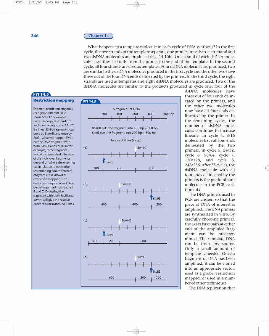
Restriction mapping
Different restriction enzymesrecognize different DNAsequences. For example,BamHI recognizes GGATCCand EcoRI recognizes GAATTC.If a linear DNA fragment is cutonce by BamHI, and once byEcoRI, what will happen if youcut the DNA fragment withboth BamHI and EcoRI? In thisexample, three fragmentswould be generated. The sizesof the individual fragmentsdepend on where the enzymescut in relation to each other.Determining where differentenzymes cut is known asrestriction mapping. Therestriction maps in A and D canbe distinguished from those inB and C. Digesting thefragment with both EcoRI andBamHI will give the relativeorder of BamHI and EcoRI sites.
FYI 14.5
200 400 600 800 1000 bp
A fragment of DNA
BamH cuts the fragment into 400 bp + 600 bp
EcoR cuts the fragment into 200 bp + 800 bp
The possibilities (in bp)
BamH
EcoR
200 400 400
(a)
BamH
EcoR
400 400 200
(b)
BamH
EcoR
200 200 600
(c)
BamH
EcoR
600 200 200
(d)
PYF14 3/21/05 8:05 PM Page 246
DNA Cloning 247
Cycle 1
Cycle 2
Cycle 3
A cycle in PCR
Denaturetemplate
90° – 95°C
Anneal primer
40° – 55°C30 sec
Synthesize DNA
70°C1 – 2 min
Molecules 1
2
0
0
2
2
2
2
0
4
3
2
4
2
8
4
2
6
8
16
5
2
8
22
32
6
2
10
52
64
7
2
12
114
128
8
2
14
240
256
9
2
16
494
512
10
2
18
1004
1024Total
Cycle
or
or
(a)
(b)
(c)
Fig. 14.10 A diagram of thepolymerase chain reaction. (a)The general reaction takingplace in each cycle. The templatemust be denatured, the primersannealed, and the DNAsynthesized. (b) The fate of thetemplate molecules in the firstthree cycles. The circle andtriangle attached to the ends ofthe molecules represent theDNA ends that are delineated bythe primers. (c) How manymolecules of each type areformed in successive cycles.There will always be twomolecules that match the onesformed in cycle 1. The numberof molecules with three of thefour ends matching the primerends increases by two in eachsuccessive cycle. The number ofmolecules with all four endsdetermined by the primers isamplified dramatically. At theend of 35 cycles, >99% of theDNA molecules will have all fourends specified by the primers.
PYF14 3/21/05 8:05 PM Page 247
takes place in PCR, like in vivo DNA replication, is not 100% accurate. Occasionally, amistake is made. If the amplified fragment is to be cloned, the resulting clones must besequenced to ensure that they carry a wild-type copy of the gene. If the amplified frag-ment is to be used as a probe, a few mutant copies in a mixture that contains a largenumber of wild-types copies will not present a problem. Thus, depending on the useof the PCR fragment, these contaminating mutant copies of the fragment must be ac-counted for.
Adding novel DNA sequences to the ends of a PCR amplified sequence
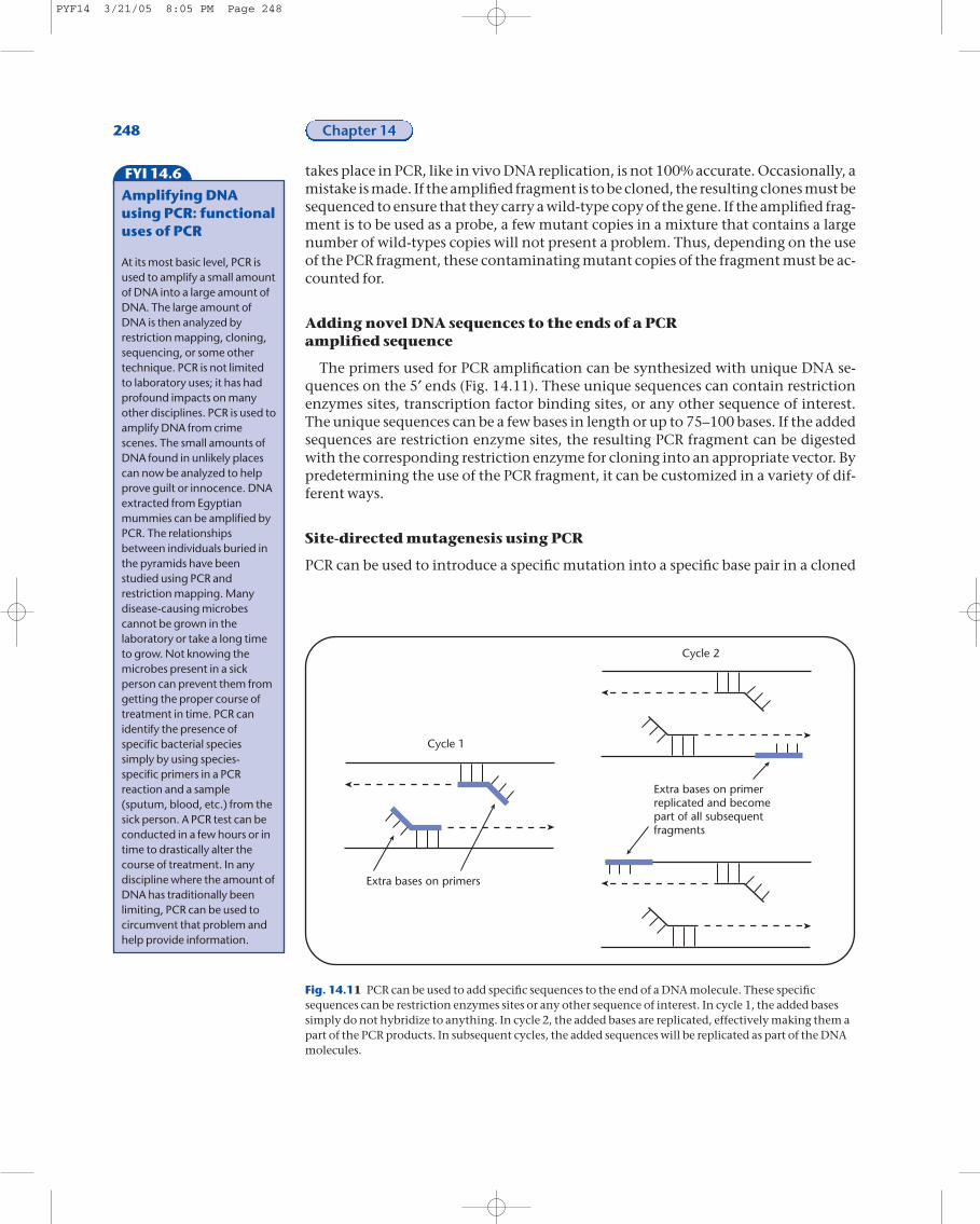
The primers used for PCR amplification can be synthesized with unique DNA se-quences on the 5¢ ends (Fig. 14.11). These unique sequences can contain restrictionenzymes sites, transcription factor binding sites, or any other sequence of interest.The unique sequences can be a few bases in length or up to 75–100 bases. If the addedsequences are restriction enzyme sites, the resulting PCR fragment can be digestedwith the corresponding restriction enzyme for cloning into an appropriate vector. Bypredetermining the use of the PCR fragment, it can be customized in a variety of dif-ferent ways.
Site-directed mutagenesis using PCR
PCR can be used to introduce a specific mutation into a specific base pair in a cloned
248 Chapter 14
FYI 14.6
Amplifying DNAusing PCR: functionaluses of PCR
At its most basic level, PCR isused to amplify a small amountof DNA into a large amount ofDNA. The large amount ofDNA is then analyzed byrestriction mapping, cloning,sequencing, or some othertechnique. PCR is not limitedto laboratory uses; it has hadprofound impacts on manyother disciplines. PCR is used toamplify DNA from crimescenes. The small amounts ofDNA found in unlikely placescan now be analyzed to helpprove guilt or innocence. DNAextracted from Egyptianmummies can be amplified byPCR. The relationshipsbetween individuals buried inthe pyramids have beenstudied using PCR andrestriction mapping. Manydisease-causing microbescannot be grown in thelaboratory or take a long timeto grow. Not knowing themicrobes present in a sickperson can prevent them fromgetting the proper course oftreatment in time. PCR canidentify the presence ofspecific bacterial speciessimply by using species-specific primers in a PCRreaction and a sample(sputum, blood, etc.) from thesick person. A PCR test can beconducted in a few hours or intime to drastically alter thecourse of treatment. In anydiscipline where the amount ofDNA has traditionally beenlimiting, PCR can be used tocircumvent that problem andhelp provide information.
Cycle 1
Cycle 2
Extra bases on primers
Extra bases on primerreplicated and becomepart of all subsequentfragments
Fig. 14.11 PCR can be used to add specific sequences to the end of a DNA molecule. These specificsequences can be restriction enzymes sites or any other sequence of interest. In cycle 1, the added basessimply do not hybridize to anything. In cycle 2, the added bases are replicated, effectively making them apart of the PCR products. In subsequent cycles, the added sequences will be replicated as part of the DNAmolecules.
PYF14 3/21/05 8:05 PM Page 248
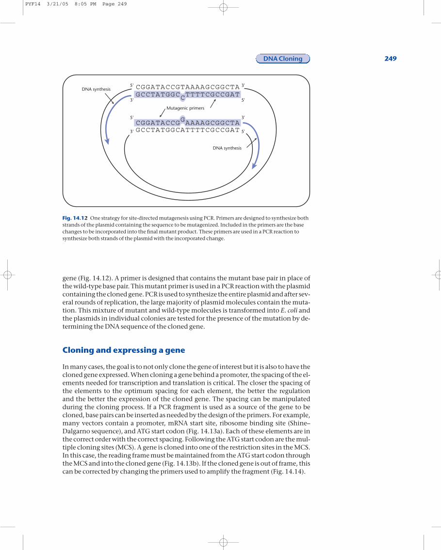
gene (Fig. 14.12). A primer is designed that contains the mutant base pair in place ofthe wild-type base pair. This mutant primer is used in a PCR reaction with the plasmidcontaining the cloned gene. PCR is used to synthesize the entire plasmid and after sev-eral rounds of replication, the large majority of plasmid molecules contain the muta-tion. This mixture of mutant and wild-type molecules is transformed into E. coli andthe plasmids in individual colonies are tested for the presence of the mutation by de-termining the DNA sequence of the cloned gene.
Cloning and expressing a gene
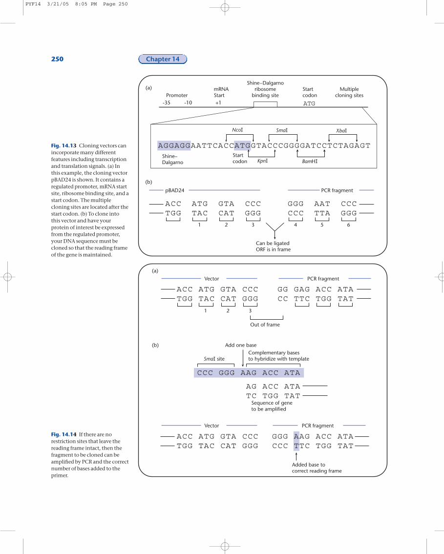
In many cases, the goal is to not only clone the gene of interest but it is also to have thecloned gene expressed. When cloning a gene behind a promoter, the spacing of the el-ements needed for transcription and translation is critical. The closer the spacing ofthe elements to the optimum spacing for each element, the better the regulation and the better the expression of the cloned gene. The spacing can be manipulated during the cloning process. If a PCR fragment is used as a source of the gene to becloned, base pairs can be inserted as needed by the design of the primers. For example,many vectors contain a promoter, mRNA start site, ribosome binding site (Shine–Dalgarno sequence), and ATG start codon (Fig. 14.13a). Each of these elements are inthe correct order with the correct spacing. Following the ATG start codon are the mul-tiple cloning sites (MCS). A gene is cloned into one of the restriction sites in the MCS.In this case, the reading frame must be maintained from the ATG start codon throughthe MCS and into the cloned gene (Fig. 14.13b). If the cloned gene is out of frame, thiscan be corrected by changing the primers used to amplify the fragment (Fig. 14.14).
DNA Cloning 249
DNA synthesis
DNA synthesis
Mutagenic primers
CGGATACCGTAAAAGCGGCTAGCCTATGGCCTTTTCGCCGAT
CGGATACCGGAAAAGCGGCTAGCCTATGGCATTTTCGCCGAT
5'
5'
3'
5'
3'
3'
5'
3'
Fig. 14.12 One strategy for site-directed mutagenesis using PCR. Primers are designed to synthesize bothstrands of the plasmid containing the sequence to be mutagenized. Included in the primers are the basechanges to be incorporated into the final mutant product. These primers are used in a PCR reaction tosynthesize both strands of the plasmid with the incorporated change.
PYF14 3/21/05 8:05 PM Page 249
250 Chapter 14
PromotermRNAStart
Shine–Dalgarnoribosome
binding siteStartcodon
Startcodon
Multiplecloning sites
+1-35 -10
AGGAGGAATTCACCATGGTACCCGGGGATCCTCTAGAGT
ACCTGG
ATGTAC
GTACAT
CCCGGG
GGGCCC
AATTTA
CCCGGG
Shine–Dalgarno
Can be ligatedORF is in frame
PCR fragmentpBAD24
1 2 3 4 5 6
ATG
(a)
(b)
Fig. 14.13 Cloning vectors canincorporate many differentfeatures including transcriptionand translation signals. (a) Inthis example, the cloning vectorpBAD24 is shown. It contains aregulated promoter, mRNA startsite, ribosome binding site, and astart codon. The multiplecloning sites are located after thestart codon. (b) To clone intothis vector and have yourprotein of interest be expressedfrom the regulated promoter,your DNA sequence must becloned so that the reading frameof the gene is maintained.
Added base tocorrect reading frame
Out of frame
1 2 3
Vector PCR fragment
Vector PCR fragment
CCC GGG AAG ACC ATA
AG ACC ATATC TGG TAT
ACC ATG GTA CCCTGG TAC CAT GGG
ACC ATG GTA CCCTGG TAC CAT GGG
GGG AAG ACC ATACCC TTC TGG TAT
GG GAG ACC ATA CC TTC TGG TAT
Add one baseComplementary basesto hybridize with template
Sequence of geneto be amplified
(a)
(b)
Fig. 14.14 If there are norestriction sites that leave thereading frame intact, then thefragment to be cloned can beamplified by PCR and the correctnumber of bases added to theprimer.
PYF14 3/21/05 8:05 PM Page 250
DNA sequencing using dideoxy sequencing
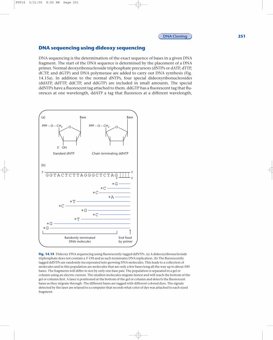
DNA sequencing is the determination of the exact sequence of bases in a given DNAfragment. The start of the DNA sequence is determined by the placement of a DNAprimer. Normal deoxyribonucleoside triphosphate precursors (dNTPs or dATP, dTTP,dCTP, and dGTP) and DNA polymerase are added to carry out DNA synthesis (Fig.14.15a). In addition to the normal dNTPs, four special dideoxyribonucleosides(ddATP, ddTTP, ddCTP, and ddGTP) are included in small amounts. The specialddNTPs have a fluorescent tag attached to them. ddGTP has a fluorescent tag that flu-oresces at one wavelength, ddATP a tag that fluoresces at a different wavelength,
DNA Cloning 251
O
Base
PPP – O – CH2O
Base
OH
PPP – O – CH2
3'
G G T A C T C T T A G G G C T C T A G
G
G
GG
CC
C
C
AT
T
3'
3' 5'
5'
Randomly terminatedDNA molecules
End fixedby primer
Standard dNTP Chain terminating ddNTP
(a)
(b)
Fig. 14.15 Dideoxy DNA sequencing using fluorescently tagged ddNTPs. (a) A dideoxyribonucleosidetriphosphate does not contain a 3¢ OH and as such terminates DNA replication. (b) The fluorescentlytagged ddNTPs are randomly incorporated into growing DNA molecules. This leads to a collection ofmolecules and in this population are molecules that are only a few bases long all the way up to about 500bases. The fragments will differ in size by only one base pair. The population is separated in a gel orcolumn using an electric current. The smallest molecules migrate fastest and will reach the bottom of thegel or column first. A laser is positioned at the bottom of the gel or column and detects the fluorescentbases as they migrate through. The different bases are tagged with different colored dyes. The signalsdetected by the laser are relayed to a computer that records what color of dye was attached to each sizedfragment.
PYF14 3/21/05 8:05 PM Page 251
ddTTP a tag with a third wavelength, and ddCTP a tag with a fourth wavelength.ddNTPs do not have a 3¢ OH and therefore block further synthesis of DNA. The fluo-rescently tagged ddNTPs are randomly incorporated into the growing DNA molecules(Fig. 14.15b). The results of DNA synthesis in the presence of tagged ddNTPs are a col-lection of DNA molecules different from each other by one base.
The fluorescently tagged ddNTP at the end of each molecule is dictated by the se-quence of the template DNA. The tagged fragments are subsequently separated on ei-ther a polyacrylamide gel or on a very thin column. At the base of the column orpolyacrylamide gel is located a laser. As the DNA fragments run off the column or gel,they pass through the laser beam, fluoresce, and the wavelength of the fluorescence isrecorded and sent to a computer. The order of the fluorescently tagged moleculescoming off the column reflects the sequence of the template DNA.
The automation of DNA sequencing has greatly simplified the process and made itmuch faster. Approximately 350 to 500bp of DNA sequence can be read from one
252 Chapter 14
Dashes indicate thatthere is a gap in thissequence.Sequence A has noamino acids thatcorrespond tosequence B
Identical amino acidsare indicated by theone-letter code foramino acids
The similar amino acidsare indicated by a +
SLAVVLQRRDWENPGVTQLNRLAAHPPFASWRNSEEARTDRPSQQLRSLNGEWRFAWFPASL +L RRDWENP +TQ +RL AHPPF SWR+ E A+ DRPS Q ++LNG W F++F SLPQILSRRDWENPQITQYHRLEAHPPFHSWRDVESAQKDRPSPQQQTLNGLWSFSYFTQ
66
73
7
14
PEAVPESWLECDLPEADTVVVPSNWQMHGYDAPIYTNVTYPITVNPPFVPTENPTGCYSLPEAVPE W+ CDL EA + VP+NWQ+HGYDAPIYTN+ YPI VNPP VP NPTGCYS PEAVPEHWVRCDLAEAKPLPVPANWQLHGYDAPIYTNIQYPIPVNPPRVPDLNPTGCYSR
126
133
67
74
TFNVDESWLQEGQTRIIFDGVNSAFHLWCNGRWVGYGQDSRLPSEFDLSAFLRAGENRLA F ++ SWL G+TRIIFDGV+SAF+LWCNG+WVGY QDSRLP+EFDL+ +L+AG NR+ADFTLEPSWLASGKTRIIFDGVSSAFYLWCNGQWVGYSQDSRLPAEFDLTPYLQAGSNRIA
186
193
127
134
VMVLRWSDGSYLEDQDMWRMSGIFRDVSLLHKPTTQISDFHVATRFNDDFSRAVLE--AEV+VLRWSDGSYLEDQDMWRMSGIFRDV LLHKP + D H+ T + +F+ A LE A VLVLRWSDGSYLEDQDMWRMSGIFRDVKLLHKPEIHLRDIHIMTHLSPEFTSANLEVMAA
244
253
187
194
VQMCG------ELRDYLRVTVSLWQGETQVASGTAPFGGEIIDERGGYADRVTLRLNVENV + ++ ++ V LW + VAS P G + IDERG Y DR L L ++ VNIPSLQLNDPQVTGSYQLRVQLWLADKLVASLQQPLGTQAIDERGPYTDRTQLVLRIDQ
298
313
245
254
PKLWSAEIPNLYRAVVELHTADGTLIEAEACDVGFREVRIEXXXXXXXXXXXXIRGVNRHP LWSAE P LYRAVV L LIEAEA DVGFR+V I IRGVNRHPLLWSAEQPTLYRAVVSLLNHQQELIEAEAYDVGFRQVAIHQGLLKINGKAVLIRGVNRH
358
373
299
314
EHHPLHGQVMDEQTMVQDILLMKQNNFNAVRCSHYPHNPLWYTLCDRYCLYVVDEANIETEHHP GQ +DE++++QDILLMKQ+NFNAVRCSHYPNHPLWY LCDRYGLYVVDEANIETEHHPQTGQAIDEESLLQDILLMKQHNFNAVRCSHYPNHPLWYRLCDRYGLYVVDEANIET
418
433
359
374
HGMVPMNRLTDDPRWLPAMSERVTRMVQRDRNHPSVIIWSLGNESGHGANHDALYRWIKSHGM PM+RL+DDP W A SERVTRMVQRDRNHP +IIWSLGNESGHGA HDALYRWIK+HGMQPMSRLSDDPSWFSAFSERVTRMVQRDRNHPCIIIWSLGNESGHGATHDALYRWIKT
478
493
419
434
VDPSRPVQYEGGGADTTATDIICPMYARVD DP+RPVQYEGGGA+T ATDI+CPMYARVDNDPTRPVQYEGGGANTLATDILCPMYARVD
479
494
508
523
A
B
A
B
A
B
A
B
A
B
A
B
A
B
A
B
A
B
Sequence
Fig. 14.16 Sequences that are very similar over the entire length ofthe protein indicate that the two proteins may have a similar
function. The greater the similarity, the greater the chance thefunctions are the same.
PYF14 3/21/05 8:05 PM Page 252
primer. By carrying out separate sequencing reactions using primers located every 350to 450bp on the template, the sequence of the entire template can be determined.
DNA sequence searches
To date, many millions of base pairs of DNA from many species have been sequenced.For example, the chromosomes of at least 50 bacterial species, several yeasts, and thelarge majority of all human chromosomes have been determined. These sequencescontain an incredible amount of information. So much in fact that special computerprograms had to be designed to help interpret just a fraction of the data.
When a DNA sequence is published in a scientific journal, it is also deposited in acomputer database known as GenBank. When a sequence is placed in GenBank, theknown and predicted features of the sequence are also indicated. These include pro-moters, open reading frames, and transcription factor binding sites. Just a listing ofAs, Cs, Gs, and Ts are known as a raw sequence and the sequence with all of the fea-tures indicated is known as an annotated sequence.
It is possible to search the sequences in GenBank using several different programs.You can search by the name of an interesting gene very easily. If you have the se-quence of a gene of interest, it is possible to search for related sequences. GenBank canbe searched using a DNA sequence or using that DNA sequence translated into the
DNA Cloning 253
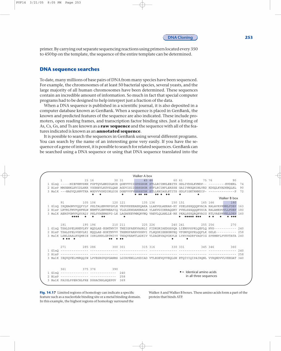
Walker A box
Walker B box
Identical amino acidsin all three sequences
=
1 15 16 30 31 45 46 60 61 75 76 901 GlnQ -----MIEFKNVSKH FGPTQVLHNIDLNIR QGEVVVIIGPSGSGK STLLRCINKLEEITS GDLIVDGLKVNDP-- ---------KVDERL 742 HisP MMSENKLHVIDLHKR YGGHEVLKGVSLQAR AGDVISIIGSSGSGK STFLRCINFLEKSSE GAIIVNGQNINLVRD KDGQLKVADKNQLRL 903 MalK ---MASVQLRNVTKA WGDVVVSKDINLDIH DGEFVVFVGPSGCGK STLLRMIAGLETITS GDLFIGETRMNDIP- --------------P 72
91 105 106 120 121 135 136 150 151 165 166 180 1 GlnQ IRQEAGMVFQQFYLF PHLTALENVMFGPLR VRGVKKEEAEKQAKA LLAKVGLAERAH-HY PSELSGGQQQRVAIA RALAVKPKMMLFDEP 1632 HisP LRTRLTMVFQHFNLW NHMTVLENVMEAPIQ VLGLSKHDARERALK YLAKVGIDERAQGKY PVHLSGGQQQRVSIA RALAMEPDVLLFDEP 1803 MalK AERGVGMVFQSYALY PHLSVAENMSFG-LK LAGAKKEVMNQRVNQ VAEVLQLAHLLE-RK PKALSGGQRQRVAIG RTLVAEPRVELLDEP 160
181 195 196 210 211 225 226 240 241 255 256 2701 GlnQ TSALDPELRHEVLKV MQDLAE-EGMTMVIV THEIGFAEKVASRLI FIDKGRIAEDGSPQA LIENPPSPRLQEFLQ HVS------------ 2402 HisP TSALDPELVGEVLRI MQQLAE-EGKTMVVV THEMGFARHVSSHVI FLHQGKIEEEGNPEQ VFGNPQSPRLQQFLK GSLK----------- 2583 MalK LSNLDAALRVQMRIE ISRLHKRLGRTMIYV THDQVEAMTLADKIV VLDAGRVAQVGKPLE LYHYPADRFVAGFIG SPKMNFLPVKVTATA 240
271 285 286 300 301 315 316 330 331 345 346 3601 GlnQ --------------- --------------- --------------- --------------- --------------- --------------- 2402 HisP --------------- --------------- --------------- --------------- --------------- --------------- 2583 MalK IEQVQVELPNRQQIW LPVESRGVQVGANMS LGIRPEHLLPSDIAD VTLEGEVQVVEQLGH ETQIYIQIPAIRQNL VYRQNDVVLVEEGAT 340
361 375 376 3901 GlnQ --------------- -------------- 2402 HisP --------------- -------------- 2583 MalK FAIGLPPERCHLFRE DGSACRRLHQEPGV 369
Fig. 14.17 Limited regions of homology can indicate a specificfeature such as a nucleotide binding site or a metal binding domain.In this example, the highest regions of homology surround the
Walker A and Walker B boxes. These amino acids form a part of theprotein that binds ATP.
PYF14 3/21/05 8:05 PM Page 253
protein sequence. The programs used for these searches are capable of identifying notonly exact matches but also sequences that have differing degrees of similarity.
What can be learned from sequence searches? First, DNA sequence searches aremore stringent than protein sequences. Two DNA sequences either have an adeninein the same position or they do not. Protein sequences can have the same amino acidin the same place and are, thus, identical at that position. Proteins can also havesimilar amino acids in one position, such as valine in one protein and alanine in theother. Because both amino acids are hydrophobic, they can frequently carry out thesame functions. In this case, the proteins are said to be similar in a given position.
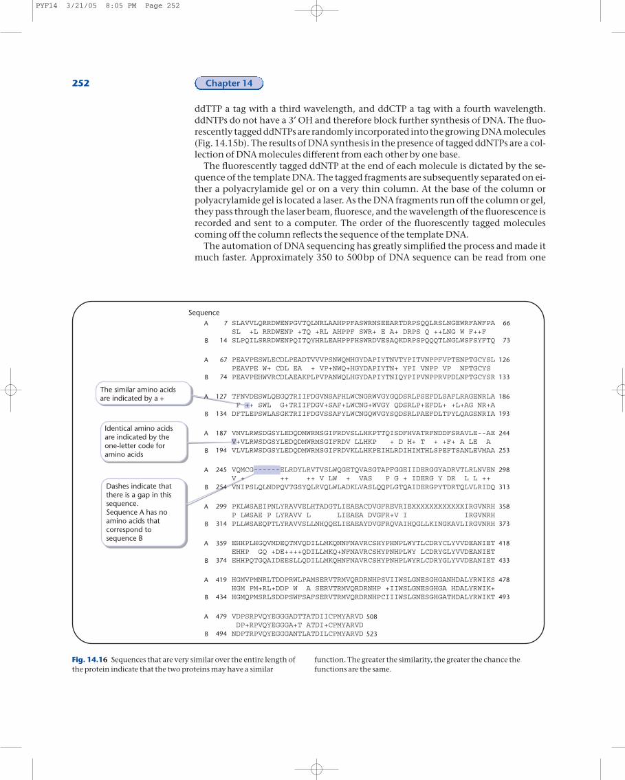
If two proteins have similarity over a large segment of their sequences, they mayhave similar functions (Fig. 14.16). This kind of analysis is especially useful if the func-tion of one of the proteins has been identified. Knowing the function of one of theproteins suggests that the other protein should also be checked for this function.More limited regions of sequence similarity or identity can indicate the presence of acofactor binding site. An example of this is the Walker box, which is an ATP bindingsite (Fig. 14.17). Sequence similarities can provide very valuable information aboutan unknown sequence and dramatically influence the direction of experiments onthe novel gene or protein.
254 Chapter 14
experiments that are now possible but also howscientists think about biological problems. Each of thetechniques described in this chapter allows scientiststo manipulate a novel gene in many different wayswith the goal of uncovering its unique role in the cell.
Summary
In 1962, the Nobel Prize in Medicine and Physiologywas awarded to Watson and Crick for the discovery ofthe structure of DNA. The technology developed inthe 40 years since has revolutionized how biologicalresearch is conducted. The ability to manipulategenes in vitro has greatly increased not only the
PYF14 3/21/05 8:05 PM Page 254
Further reading
Grunstein, M. and Hogness, D. 1975. Colony hybridization: a method for the isolation of cloned DNAs thatcontain a specific gene. Proceedings of the National Academy of Science USA, 72: 3961.
Lobban, P. and Kaiser, A.D. 1973. Enzymatic end-to-end joining of DNA molecules. Journal of Molecular Biology, 78: 453.
Mertz, J. and Davies, R. 1972. Cleavage of DNA: RI restriction enzyme generates cohesive ends. Proceedingsof the National Academy of Science USA, 69: 3370.
Sambrook, J., Fritsch, E.F., and Maniatis, T. 1989. Molecular Cloning: A Laboratory Manual. Cold Spring Harbor, NY: Cold Spring Harbor Laboratory.
DNA Cloning 255St
ud
y q
ues
tio
ns 1 How is plasmid DNA purified away from chromosomal DNA?
2 How does ethidium bromide interact with DNA and how does it help invisualizing DNA on an agarose gel?3 What are the distinguishing features between the three types of restrictionenzymes?4 Which class of enzymes is used for cloning and what characteristic(s) ofthis class make it suitable for the job?5 Could you clone the gene for a restriction enzyme into E. coli in the absenceof the modification enzyme? What would happen to the cellular DNA if youtried this experiment?6 Can a blunt end be ligated to a sticky end?7 Can a clone library be constructed from human DNA? Broccoli DNA? E. coliproteins? Human membranes? Why or why not?8 What kind of gel would you use to separate a 100bp DNA fragment from a35bp DNA fragment? A 750bp fragment of DNA from a 6kb fragment ofDNA?9 How would you determine if a novel bacterium contains a gene fordegrading lactose?10 In a PCR reaction, the reactions are cycled between 90–95°C, 40–55°C and70°C. What biochemical reactions are occurring at each temperature?11 You have the DNA sequence 5¢TGCGCTAGGCTCATGGCCTTATAGACTCAGTCAAACGTCGTAGT 3¢ in yourgene. Design an 18bp primer to change the two Cs to As in the sequence: 5¢GGCCTTA 3¢.12 Two protein sequences are 55% identical and 83% similar. What does thismean? Can two sequences have a greater identity than similarity?
PYF14 3/21/05 8:05 PM Page 255


![kuKeywords: USERTM cloning, Cloning of synthesized nonclonal DNA fragments, Fusion of DNA - fragments, Uracil excision based cloning, uNCDFs, Geneart [Background] For synthesized DNA,](https://img.dokumen.tips/doc/110x75/5fd4e2ff1db7b3255b1a15b8/ku-keywords-usertm-cloning-cloning-of-synthesized-nonclonal-dna-fragments-fusion.jpg)