Embed Size (px)
Citation preview
Proc. Nati. Acad. Sci. USAVol. 85, pp. 5171-5175, July 1988Genetics
Diverse point mutations in the human glucose-6-phosphatedehydrogenase gene cause enzyme deficiency and mildor severe hemolytic anemia
(enzymopathy/genetic variants/human genetics/cloning of mutants)
T. J. VULLIAMY*, M. D'URSOt, G. BATTISTUZZIt, M. ESTRADA§, N. S. FOULKES*, G. MARTINIt,V. CALABROf, V. POGGI*, R. GIORDANOt¶, M. TOWN*, L. LUZZATTO*, AND M. G. PERSICOt*Department of Haematology, Royal Postgraduate Medical School, Du Cane Road, London W12 OHS, Great Britain; International Institute of Genetics andBiophysics, CP 3061, Naples, Italy; tDepartment of Genetics, University of Naples, via Mezzocannone, Naples, Italy; and §Instituto de Hematologia yImmunologia, Havana, Cuba
Communicated by Y. W. Kan, April 4, 1988 (received for review January 14, 1988)
ABSTRACT Glucose-6-phosphate dehydrogenase (G6PD;EC 1.1.1.49) deficiency is a common genetic abnormalityaffecting an estimated 400 million people worldwide. Clinicaland biochemical analyses have identified many variants exhib-iting a range of phenotypes, which have been well characterizedfrom the hematological point of view. However, until now, theirprecise molecular basis has remained unknown. We havecloned and sequenced seven mutant G6PD alleles. In thenondeficient polymorphic African variant G6PD A we havefound a single point mutation. The other six mutants investi-gated were all associated with enzyme deficiency. In one of thecommonest, G6PD Mediterranean, which is associated withfavism among other clinical manifestations, a single amino acidreplacement was found (serine -- phenylalanine): it must beresponsible for the decreased stability and the reduced catalyticefficiency of this enzyme. Single point mutations were alsofound in G6PD Metaponto (Southern Italy) and in G6PD Ilesha(Nigeria), which are asymptomatic, and in G6PD Chatham,which was observed in an Indian boy with neonatal jaundice.In G6PD "Matera," which is now known to be the same asG6PD A-, two separate point mutations were found, one ofwhich is the same as in G6PD A. In G6PD Santiago, a de novomutation (glycine -* arginine) is associated with severe chronichemolytic anemia. The mutations observed show a strikingpredominance of C -* T transitions, with CG doublets involvedin four of seven cases. Thus, diverse point mutations mayaccount largely for the phenotypic heterogeneity of G6PDdeficiency.
Glucose-6-phosphate dehydrogenase (G6PD; EC 1.1.1.49)deficiency is a genetic abnormality associated with a range ofclinical conditions (1). Some subjects are asymptomatic,whereas others suffer from neonatal jaundice, acute hemo-lytic anemia, or severe chronic nonspherocytic hemolyticanemia. Over 300 G6PD variants have been described byusing clinical and biochemical criteria (2) and at least 100 ofthese have polymorphic frequencies in various populations(3). This makes G6PD deficiency probably the most commonhuman enzymopathy, affecting an estimated 400 millionpeople worldwide. There is abundant evidence that -thiswidespread polymorphism results from relative resistance ofheterozygotes to Plasmodiumfalciparum malaria (4-6). Thefact that a large number of different deficient variants havebeen characterized suggests that a considerable variety ofstructural changes can cause abnormal enzyme activity.G6PD is a housekeeping enzyme, present in all species so
far tested (3). It is produced only at low levels in all cells and
therefore purification of sufficient quantities for proteinsequence analysis has been very difficult, or impossible incases of severe deficiency. This has prevented the identifi-cation of the specific amino acid changes responsible foraltered phenotypes. Recently, the gene that encodes humanG6PD, which is located on chromosome Xq28, has beencloned and the coding sequence was determined (7-9),enabling us to study the molecular basis ofG6PD deficiency.In this paper we describe the cloning and sequencing of sevenmutant G6PD alleles, six of them from deficient subjects. Weshow that the wide range of clinical phenotypes associatedwith G6PD deficiency has arisen from different point muta-tions in the coding sequence of the G6PD gene.
MATERIALS AND METHODSBiochemical Characterization. Of the seven G6PD variants
described here, three have been previously published. Forthe other four, the following biochemical properties werestudied by using methods recommended by the World HealthOrganization (WHO) (10): G6PD activity in erythrocytes,electrophoretic mobility in Tris/borate/EDTA buffer (pH8.9), Kr"P, 2-deoxyglucose 6-phosphate % rate, KNADP, andthe thermostability at 560C. KNADPH was determined as inref. 11, and the elution profile from DEAE-Sephadex wasdetermined as in ref. 12. The KGm6P for G6PD Chatham wasmeasured under the conditions described in ref. 13, and thevalue was normalized to WHO conditions by using the ratioof values obtained by the two methods with G6PD B.
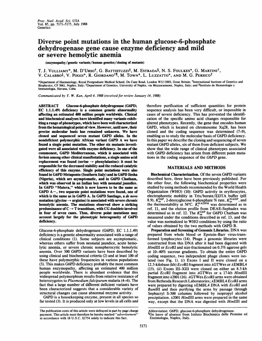
Preparation and Screening of Genomic Libraries. DNA wasprepared from whole blood or Epstein-Barr virus-trans-formed lymphocytes (14). Phage A genomic libraries wereconstructed from this DNA after it had been digested withHindIll or EcoRI and size-fractionated on 0.5% agarose gelsor 10-40% sucrose gradients. To obtain the entire G6PDcoding sequence, two independent phage clones were iso-lated (see Fig. 1). (i) Exons I and II were cloned on a12.5-kilobase (kb) EcoRI fragment into AGTWes or AEMBL4(15). (ii) Exons III-XIII were cloned on either an 8.5-kbpartial EcoRI fragment into AGTWes or a 17-kb HindIIIfragment into A2001 (16). AGTWes EcoRI arms were obtainedfrom Bethesda Research Laboratories; AEMBL4 EcoRI armswere prepared by digesting AEMBL4 DNA with EcoRI andBamHI and then purifying the arms by passage throughSephacryl S-300 columns followed by isopropyl alcoholprecipitation. A2001 HindIII arms were prepared in the sameway, except that the DNA was digested with HindIII and
Abbreviation: G6PD, glucose-6-phosphate dehydrogenase.$On leave of absence from Istituto Biochimica delle Proteine edEnzimologia, Naples, Italy.
5171
The publication costs of this article were defrayed in part by page chargepayment. This article must therefore be hereby marked "advertisement"in accordance with 18 U.S.C. §1734 solely to indicate this fact.
Proc. Natl. Acad. Sci. USA 85 (1988)
EcoRI. Ligated material was packaged by using Gigapackextracts (Northumbria Biologicals, U.K.) or freeze-thawlysates and sonicated extracts (17). The libraries were
screened by standard Benton-Davis filter hybridization (18)using the appropriate G6PD probes labeled by nick-translation (Amersham).
Sequencing. DNA from plaque-purified phage was sub-cloned into M13 phage for Sanger dideoxy sequencing (19) orinto pUC18 for dideoxy sequencing of denatured plasmidDNA (20). Sequencing reactions were primed by using a setof oligonucleotides homologous to intron sequences near theintron-exon junctions (see Fig. 1). Exons I and II were
subcloned on either a 2-kb Pst I fragment or a 6-kb HindIII/EcoRI fragment, whereas for exons III-XIII the 3.5-kb and5-kb EcoRP fragments were subcloned.
Southern Blotting. Analysis ofPst I and Hinfl digestions ofgenomic DNA prepared from various individuals was carriedout by Southern hybridization using conventional methods(17).
RESULTS
The G6PD variants studied are representative ofa wide range
of clinical and biochemical features. G6PD A is a nondefi-cient variant, widely distributed in Africa (21), with poly-morphic gene frequencies reaching 0.25 in several popula-tions (22). G6PD Mediterranean is one of the best-studiedG6PD variants (23); it is polymorphic and is associated withacute hemolytic anemia, including favism. G6PD Ilesha is asporadic variant from West Africa, found in a patient withsickle cell anemia (24). G6PD Metaponto is asymptomatic;since it was encountered in two unrelated people in Lucania(Southern Italy), it may be polymorphic in that region. G6PDChatham was identified in a boy of Indian ancestry living inEngland who has had neonatal jaundice; its frequency in theIndian population as a whole has not been established. G6PD"Matera," which is in fact probably G6PD A- (see Discus-sion), is associated with favism; it was encountered inLucania and it is polymorphic. G6PD Santiago de Cuba is asporadic variant (see below) found in a boy from Cuba withchronic nonspherocytic hemolytic anemia.The biochemical features of the G6PD variants we have
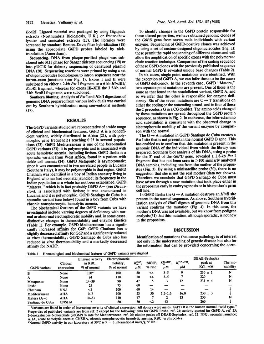
investigated include varying degrees of deficiency with nor-
mal or abnormal electrophoretic mobility and, in some cases,distinctive changes in thermostabiity and enzyme kinetics(Table 1). For example, G6PD Mediterranean has a signifi-cantly increased affinity for G6P; G6PD Chatham has a
slightly decreased affinity for G6P and a significantly reducedin vitro thermostability; G6PD Santiago de Cuba also hasreduced in vitro thermostability and a markedly decreasedaffinity for NADP.
To identify changes in the G6PD protein responsible forthese altered properties, we have obtained genomic clones ofthe G6PD gene from seven male individuals with variantenzyme. Sequencing of G6PD-positive clones was achievedby using a set of custom-designed oligonucleotides (Fig. 1);these permit the rapid sequencing of different clones and willallow the amplification of specific exons with the polymerasechain reaction technique. Comparison ofthe coding sequenceofthese G6PD clones with the previously published sequenceof normal G6PD B revealed unique base changes (Table 2).
In six cases, single point mutations were identified. Withthe exception of G6PD A, we can infer these to be the cause
of G6PD deficiency. In the seventh case, G6PD "Matera,"two separate point mutations are present. One of these is thesame as that found in the nondeficient variant, G6PD A, andso we infer that the other is responsible for enzyme defi-ciency. Six of the seven mutations are C -* T transitions on
either the coding or the noncoding strand, and in four ofthesethe C precedes aG in a CG doublet. The amino acids changedby these mutations are spread throughout the G6PD proteinsequence, as shown in Fig. 2. In each case, the inferred aminoacid substitution is consistent with the observed change inelectrophoretic mobility of the variant enzyme by compari-son with the normal.The G A mutation in G6PD Santiago de Cuba creates a
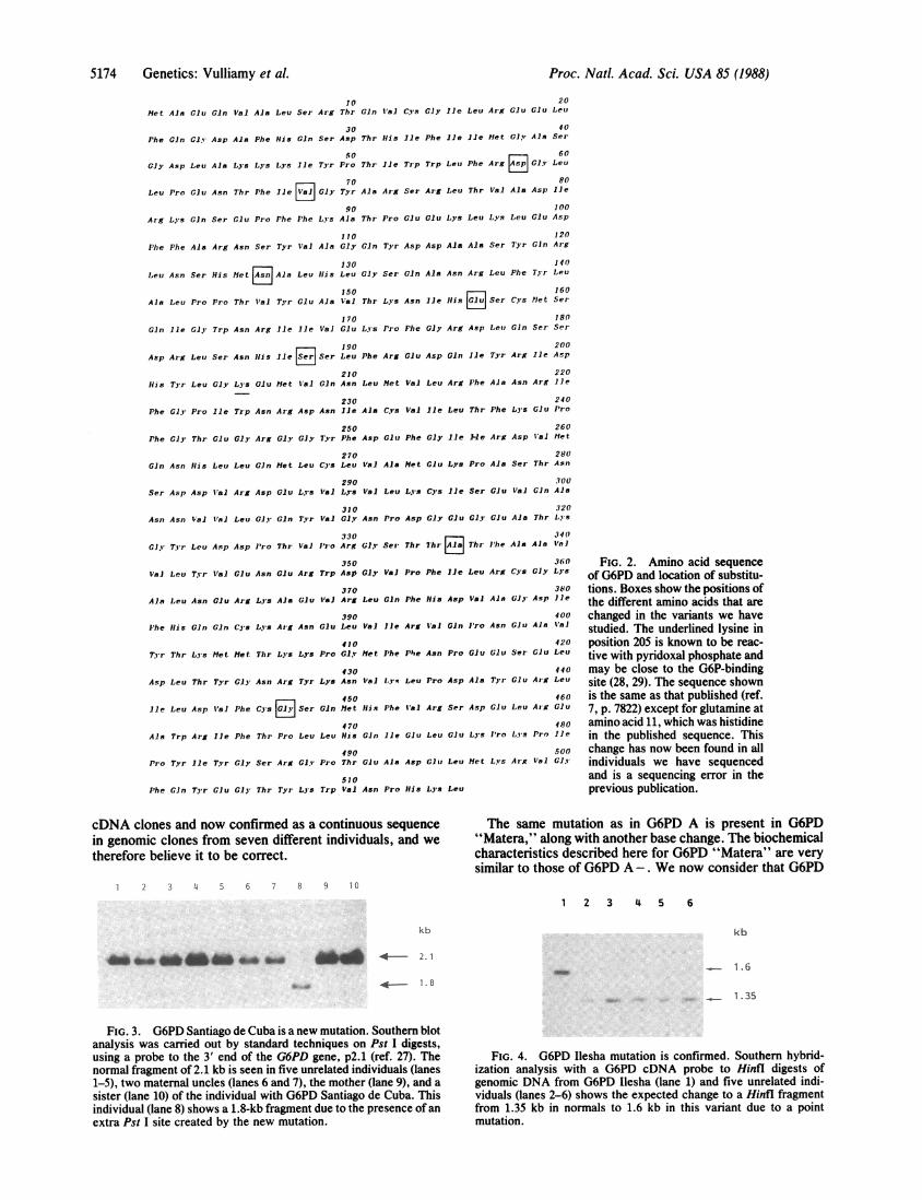
Pst I site that is not present in the normal G6PD B gene. Thishas enabled us to confirm that this mutation is present in thegenomic DNA of the individual from which the library wasprepared. Southern blot analysis of his DNA, using a probefor the 3' end of the G6PD gene, revealed a 1.8-kb Pst Ifragment that has not been seen in >100 similarly analyzedDNA samples, including one from the mother of the propos-itus (Fig. 3). By using a minisatellite probe (30), there is nosuggestion that she is not the real mother (data not shown).Therefore we conclude that G6PD Santiago de Cuba musthave arisen through a new mutation that took place either inthe propositus early in embryogenesis or in his mother's germcell line.
In G6PD Ilesha the G -- A mutation destroys an Hinfl site
present in the normal sequence. As above, Southern hybrid-ization analysis of Hinfl digests of genomic DNA from thisvariant confirms the mutation (Fig. 4). In this case, themother's DNA was not available, but we know from pedigreeanalysis (31) that this mutation, although sporadic, is not newin the propositus.
DISCUSSION
Identification of mutations that cause pathology is of interestnot only in the understanding of genetic disease but also forthe information that can be provided concerning the corre-
Table 1. Hematological and biochemical features of G6PD variants investigatedEnzyme activity Electrophoretic DEAE-Sephadex
Clinical in RBC, mobility, Km", 2dG6P, KNADP, KNADPH peak at Thermo-G6PD variant expression % of normal % of normal uM % rate /AM /AM KCI, mM stability
B None 100* 100 50 <4 3-5 9 230 ± 1 NA None 84 110 50 <4 3-5 7 220 NMetaponto None 14-39 90 47 5 3 12 231 ± 4 NIlesha None 25 75 60Chatham NNJ <2 100 60 14 --lMediterranean AHA 0-7 100 23 50 1.2-1.6 16.0 230 ± 3Matera (A-) AHA 10-23 110 47 7 2 13 230 NSantiago de Cuba CNSHA 5 80 50 <2 43 - 260 l
Variants are listed in order of increasing severity of clinical expression. All donors were males. G6PD B is the human normal "wild type."Properties of published variants are from ref. 2 except for the following: data for G6PD Ilesha, ref. 24; activity quoted for G6PD A, ref. 25;2-deoxyglucose 6-phosphate (2dG6P) % rate for Mediterranean, ref. 26; elution peaks off DEAE-Sephadex, ref. 12. NNJ, neonatal jaundice;AHA, acute hemolytic anemia; CNSHA, chronic nonspherocytic hemolytic anemia; RBC, erythrocytes.*Normal G6PD activity in our laboratory at 300C is 9 ± 3 international units/g of Hb.
5172 Genetics: Vulliamy et al.
Proc. Natl. Acad. Sci. USA 85 (1988) 5173
Metaponto A Mediterranean Santiago
Chatham
200bp
" 6 11 TjQ P A M B K C J L D R E FH Z V C
I I
.1
% I ".%% I"
I I''% , "
lkb-
R H RI IR HR
FIG. 1. Sketch of the human G6PD gene showing the positions of mutations found in seven genetic variants and our cloning and sequencingstrategies. From the bottom upward: the horizontal lines represent the extent of the different phage clones we have used to isolate the G6PDgene from different individuals. Above is a map of the G6PD gene showing EcoRI sites (R) and HindI1 sites (H), and the 13 exons are blackedin (from ref. 8). The two regions of coding sequence are expanded above, again with the exons in black and introns in white. Letters below thesketch show the locations of the 21 custom-synthesized oligonucleotides that we have used to prime sequence reactions into the codingsequences. The vertical bars and names above indicate the positions of the mutations found in the respective variants. bp, Base pairs.
sponding normal function ofthe respective gene product. Thebiochemistry of G6PD and the clinical manifestations of itsdeficiency have been studied extensively (2, 3), yet little isknown about the protein at the molecular level. Recently, theprimary sequence for the protein was determined through theisolation of full-length cDNA clones (7). Here we identifydifferent point mutations in this coding sequence that are thecause of enzyme deficiency in different individuals. They pro-vide direct proof of the genetic heterogeneity of G6PD defi-ciency, previously inferred from biochemical data. We demon-strate that different point mutations give rise to different clinicalphenotypes. In addition these data provide an insight into whichamino acids are important for G6PD enzyme stability andfunction.Of course, assignment of specific function to different
amino acids within the protein awaits elucidation of itsthree-dimensional structure, and this set of variants is toosmall for us to attempt an analysis of how amino acid replace-ments cause a reduction ofG6PD activity by impairing catalyticfunction, making the protein unstable, or both. However, withrespect to catalytic function, the variant with the most abnormalK~'3: (G6PD Mediterranean) has the amino acid replacement
nearest lysine-205, which may be involved in G6P binding (28,29). With respect to protein stability, the two variants withdrastic amino acid changes (serine -- phenylalanine in G6PD
Mediterranean and glycine -b arginine in G6PD Santiago de
Cuba) have very low residual enzyme activity in circulatingerythrocytes, presumably as a result of accelerated G6PDbreakdown (23). They also have decreased in vitro thermosta-bility, as does G6PD Chatham, where the amino acid replace-ment is alanine -* threonine. The glycine -- arginine replace-ment in G6PD Santiago de Cuba is located within a sequence ofhydrophobic amino acids (32), which might make this replace-ment particularly damaging. Also, within our series of variantsthis is the only example that is associated with a severe clinicalcondition and it is a de novo mutation.With respect to G6PD A, our data confirm the asparagineaspartic acid amino acid replacement reported by Yoshida
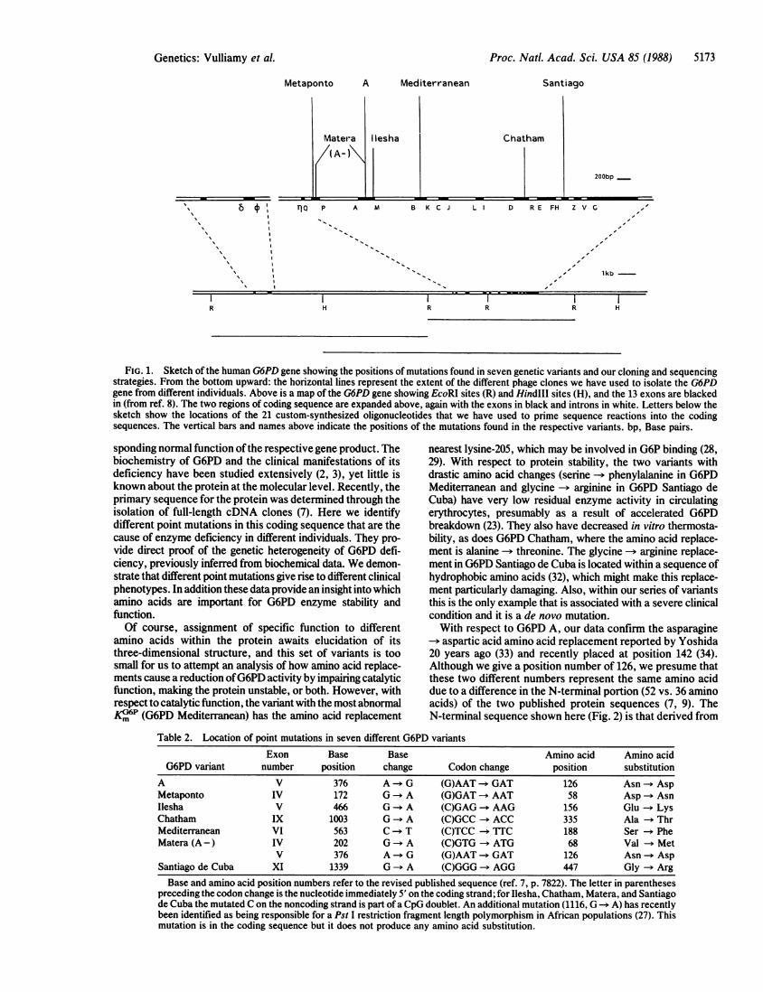
20 years ago (33) and recently placed at position 142 (34).Although we give a position number of 126, we presume thatthese two different numbers represent the same amino aciddue to a difference in the N-terminal portion (52 vs. 36 aminoacids) of the two published protein sequences (7, 9). TheN-terminal sequence shown here (Fig. 2) is that derived from
Table 2. Location of point mutations in seven different G6PD variantsExon Base Base Amino acid Amino acid
G6PD variant number position change Codon change position substitutionA V 376 A->G (G)AAT-GAT 126 Asn AspMetaponto IV 172 G A (G)GAT AAT 58 Asp AsnIlesha V 466 G A (C)GAG - AAG 156 Glu - LysChatham IX 1003 G - A (C)GCC ACC 335 Ala - ThrMediterranean VI 563 C - T (C)TCC TTTC 188 Ser - PheMatera (A-) IV 202 Go A (C)GTG -ATG 68 Val - Met
V 376 A G (G)AAT GAT 126 Asn - AspSantiago de Cuba XI 1339 Go A (C)GGG - AGG 447 Gly - ArgBase and amino acid position numbers refer to the revised published sequence (ref. 7, p. 7822). The letter in parentheses
preceding the codon change is the nucleotide immediately 5' on the coding strand; for Ilesha, Chatham, Matera, and Santiagode Cuba the mutated Con the noncoding strand is part of a CpG doublet. An additional mutation (1116, G -* A) has recentlybeen identified as being responsible for a Pst I restriction fragment length polymorphism in African populations (27). Thismutation is in the coding sequence but it does not produce any amino acid substitution.
I I I
Genetics: Vulliamy et al.
Proc. Natl. Acad. Sci. USA 85 (1988)
1020
Met Aln Giu Gin Val Ala Leu Ser Arg Thr Gin tlal Cys Gly lie Leu Arg Glu Glu Leu
30 40Phe Gin Gly Asp Aia Phe His Gin Ser Asp Thr His Ile Phe lie lie Met Gly Ala Ser
50 60
Giy Asp Leu Ala Lys Lys L's Ile Tyr Pro Thr lie Trp Trp Leu Phe Arg AP Gly Leu
{70 80
Leu Pro Glu Asn Thr Phe Ile Cly Tyr Ala Arg Ser Arg Leu Thr Val Ala Asp Ile
90 100Arg Lys Gin Ser Glu Pro Phe Phe Lys Ala Thr Pro Glu Clu Lys Leu Lys Leu Glu Asp
110 120
Phe Phe Ala Arg Asn Ser Tyr Val Ala Gly Gin Tyr Asp Asp Ala Ala Ser Tyr Gin Arg
130 140
Leu Asn Ser His Met E-J~ Ala Leu Ilis Leu Gly Ser Gin Ala Asn Arg Leu Phe Tyr Leu
150 160Ain Leu Pro Pro Thr Il'a Tr Clu Ala Val Thr Lys Asn Ile His GlSer Cs Met Ser
170 180
Gin lie Gly Trp Asn Arg Ile lie Val Glu Lys Pro Phe Gly Arg Asp Leu Gin Ser Ser
lie Ser190 200
Asp Arg Leu Ser Asn Ills Ile s Ser Leu Phe Arg Glu Asp Gin Ile Tyr Arg Ile Asp
210 220Ills Tyr Leu Gly L's Glu Met Val Gin Asn Leu Met Val Leu Arg Phe Ala Asn Arg lie
230 240
Phe Gl,' Pro Ile Trp Asn Arg Asp Asn Ile Ala C's Val Ile Leu Thr Phe Lys Glu Pro
250 260
Phe Gly Thr Glu Gly Arg Gly Gly Tyr Phe Asp Glu Phe Gly Ile Me Arg Asp Val Met
270 280Gin Asn His Leu Leu Gin Met Leu Cys Leu Val Ala Met Glu LI's Pro Ala Ser Thr Asn
290 300
Ser Asp Asp Val Ar, Asp Clu Lys Val Lys Val Leu Lys Cys Ile Ser Glu Val Gin Ala
310 320Asn Asn Val VA1 Leu Gly Gin Tir Val Gly Asn Pro Asp Gly Glu Gly Glu Ala Thr Lys
330 340
Gly Tyr Leu Asp Asp Pro Thr Val Pro Arg Gly Ser Thr 7hr Ala Thr lhe Ala Ala VolJ
350 360
Val Leu Tr Val 1Gu Asn GCu Arg Trp Asp Gly Va1 Pro Phe lie Leu Arg Cys Gly Lys
370 380Ala Leu Asn Glu Arg Lys Ala Giu V1a Ar, Leu Gin Phe Ills Asp Val AiA GJy Asp lie
390 400
Phe His Gin Gin Ci's Ly's Arg Asn Glu Leu Val Ile Arg Val Gin Pro Asn Glu Ala Val
410 420
Tyr Thr L. s Met Met Thr Lys LIs Pro Gly Met Phe P'he Asn Pro Giu Glu Ser Glu Leu
430 440
Asp Leu Thr Tyr Gl Asn Arg Tyr Lys Asn Ial Lys Leu Pro Asp Ala Tyr Glu Arg Leu
450 460lie Leu Asp I'a] Phe Cys y Ser Gin Met His Phe V'al Arg Ser Asp Glu Leeu Arg Glu
470 480
Ala Trp Arg Ile Phe Thr Pro Leu Leu His Gin lie Glu Leu Glu Lys Pro Lys Pro lie
490 500
Pro Tyr lie Tyr Gly Ser Arg Gil' Pro Thr Glu Ala Asp Glu Leu Met LyPs Ar. Val Gly
510Phe Gin Tyr Glu Gly Thr Tyr L-s Trp Val Asn Pro ills Lys Leu
cDNA clones and now confirmed as a continuous sequence
in genomic clones from seven different individuals, and wetherefore believe it to be correct.
1 2 3 4 5 6 7 8 9 10
FIG. 2. Amino acid sequenceof G6PD and location of substitu-tions. Boxes show the positions ofthe different amino acids that are
changed in the variants we havestudied. The underlined lysine inposition 205 is known to be reac-tive with pyridoxal phosphate andmay be close to the G6P-bindingsite (28, 29). The sequence shownis the same as that published (ref.7, p. 7822) except for glutamine atamino acid 11, which was histidinein the published sequence. Thischange has now been found in allindividuals we have sequencedand is a sequencing error in theprevious publication.
The same mutation as in G6PD A is present in G6PD"Matera," along with another base change. The biochemicalcharacteristics described here for G6PD "Matera" are very
similar to those of G6PD A-. We now consider that G6PD
1 2 3 4 5 6
kb kb
l1 a .o em.
ANAL4 2. 1
M- p. . 1 B
1.6
1.35
FIG. 3. G6PD Santiago de Cuba is a new mutation. Southern blotanalysis was carried out by standard techniques on Pst I digests,using a probe to the 3' end of the G6PD gene, p2.1 (ref. 27). Thenormal fragment of 2.1 kb is seen in five unrelated individuals (lanes1-5), two maternal uncles (lanes 6 and 7), the mother (lane 9), and a
sister (lane 10) of the individual with G6PD Santiago de Cuba. Thisindividual (lane 8) shows a 1.8-kb fragment due to the presence of anextra Pst I site created by the new mutation.
FIG. 4. G6PD Ilesha mutation is confirmed. Southern hybrid-ization analysis with a G6PD cDNA probe to Hinfl digests ofgenomic DNA from G6PD Ilesha (lane 1) and five unrelated indi-viduals (lanes 2-6) shows the expected change to a Hinfl fragmentfrom 1.35 kb in normals to 1.6 kb in this variant due to a pointmutation.
5174 Genetics: Vulliamy et al.
Proc. Natl. Acad. Sci. USA 85 (1988) 5175
"Matera" is in fact A-, as we have learned recently that theamino acid replacements reported here for G6PD "Matera"are the same as those found in G6PD A- (35). The extent towhich biochemically similar variants turn out to be geneti-cally identical will become clear as more are defined at themolecular level. The finding of two mutational differencesbetween G6PD B and G6PD A - did not come as a completesurprise, since this had been hypothesized some time ago onthe grounds of biochemical and genetic considerations (13).We note the striking predominance of C -+ T transitions in
our series of mutations. Although, of course, the sample sizeis small, this finding is entirely consistent with previousobservations on interspecies (36) and intraspecies (37) vari-ation in globins and other genes. In four cases the mutated Cresidue is in a CpG doublet. Methylation and subsequentdeamination of this C are thought to increase the probabilityof a C -- T transition (38, 39), and there is growing evidencethat this is indeed the case in other human genes as well (40).The pattern of mutations causing human pathology has
been extensively explored in a number of genes with cell-specific expression. For instance, with respect to globins,factor VIII, and steroid sulfatase there have been reports ofpoint mutations and sizeable deletions (41-43). The latter arepossible because complete loss of function of such genes iscompatible with development in embryonic life and beyond.The findings reported here begin to define the pattern ofmutations in a highly polymorphic housekeeping gene, inwhich complete loss of function would probably be lethal.
We thank Drs. G. Hayes and M. Cascone for supplying bloodsamples from their patients. We are grateful to Dr. Philip Mason, Dr.Letizia Foroni, and an anonymous reviewer for advice and help.Financial support was received from a Medical Research Council(England) Programme Grant; from the Consiglio Nazionale Ricerche(Italy) Progetti Finalizzati; and from a research contract from theEuropean Economic Community.
1. Beutler, E. (1983) in The Metabolic Basis ofInherited Disease,eds. Stanbury, J. B., Wyngaarden, J. B., Fredrickson, D. J.,Goldstein, J. L. & Brown, M. S. (McGraw-Hill, New York),pp. 1629-1653.
2. Beutler, E. (1978) Haemolytic Anaemia in Disorders of RedCell Metabolism (Plenum, London), pp. 23-167.
3. Luzzatto, L. & Battistuzzi, G. (1985) Adv. Hum. Genet. 14,217-329.
4. Allison, A. C. (1960) Nature (London) 186, 531-532.5. Motulsky, A. G. (1960) Hum. Biol. 32, 28-32.6. Luzzatto, L. (1979) Blood 54, 961-976.7. Persico, M. G., Viglietto, G., Martini, G., Toniolo, D., Pao-
nessa, G., Moscatelli, C., Dono, R., Vulliamy, T., Luzzatto, L.& D'Urso, M. (1986) Nucleic Acids Res. 14, 2511-2522, 7822.
8. Martini, G., Toniolo, D., Vulliamy, T., Luzzatto, L., Dono, R.,Viglietto, G., Paonessa, G., D'Urso, M. & Persico, M. G.(1986) EMBO J. 5, 1849-1855.
9. Takizawa, T., Huang, I.-Y., Ikuta, T. & Yoshida, A. (1986)Proc. Natl. Acad. Sci. USA 83, 4157-4161.
10. Betke, K., Brewer, G. J., Kirkman, H. N., Luzzatto, L.,Motulsky, A. G., Ramot, B. & Siniscalo, M. (1967) W.H.O.Tech. Rep. Ser. 366.
11. Luzzatto, L. (1967) Biochim. Biophys. Acta 146, 18-25.12. D'Urso, M., Battistuzzi, G. & Luzzatto, L. (1980) Anal.
Biochem. 108, 146-150.
13. Babalola, A. 0. G., Beetlestone, J. G. & Luzzatto, L. (1976)J.Biol. Chem. 251, 2992-3002.
14. Foroni, L., Foldi, J., Matutes, E., Catovsky, D., O'Connor,N. J., Baer, R., Forster, A., Rabbitts, T. H. & Luzzatto, L.(1987) Br. J. Haematol. 67, 307-318.
15. Frischauf, A. M., Lehrach, H., Poustka, A. & Murray, N.(1983) J. Mol. Biol. 170, 827-842.
16. Kam, J., Matthes, H., Gait, M. J. & Brenner, S. (1984) Gene32, 217-224.
17. Maniatis, T., Fritsch, E. F. & Sambrook, J. (1982) MolecularCloning:A Laboratory Manual (Cold Spring Harbor Lab., ColdSpring Harbor, NY).
18. Benton, W. D. & Davis, R. W. (1977) Science 196, 180-186.19. Sanger, F., Coulson, A. R., Barrell, B. G., Smith, A. J. H. &
Roe, B. A. (1980) J. Mol. Biol. 143, 161-178.20. Hattori, M., Hidaka, S. & Sakaki, Y. (1985) Nucleic Acids Res.
13, 7813-7827.21. Porter, I. H., Boyer, S. H., Watson-Williams, E. J., Adam, A.,
Szeinberg, A. & Siniscalco, M. (1964) Lancet i, 895-899.22. Luzzatto, L. (1973) Isr. J. Med. Sci. 9, 1181-1194.23. Morelli, A., Benatti, U., Gaetani, G. F. & DeFlora, A. (1978)
Proc. NatI. Acad. Sci. USA 75, 1979-1983.24. Usanga, E. A., Bienzle, U., Cancedda, R., Fasuan, F. A.,
Ajayi, 0. & Luzzatto, L. (1977) Ann. Hum. Genet. 40, 279-286.25. Battistuzzi, G., Esan, G. J. F., Fasuan, F. A., Modiano, G. &
Luzzatto, L. (1977) Am. J. Hum. Genet. 29, 31-36.26. Ferraris, A. M., Giuntini, P., Galiano, S. & Goetani, G. F.
(1981) Am. J. Hum. Genet. 33, 307-313.27. D'Urso, M., Luzzatto, L., Perroni, L., Ciccodicola, A., Gen-
tile, G., Peluso, I., Persico, M. G., Pizella, T., Toniolo, D. &Vulliamy, T. J. (1988) Am. J. Hum. Genet. 42, 735-740.
28. Jeffrey, J., Hobbs, L. & Jornvall, H. (1985) Biochemistry 24,666-671.
29. Camardella, L., Caruso, C., Rutigliano, B., Romano, M.,D'Prisoco, G. & Descalzicancedda, F. (1988) Eur. J. Biochem.171, 485-489.
30. Jeffreys, A. J., Wilson, V. & Thein, S. L. (1985) Nature(London) 314, 67-73.
31. Luzzatto, L., Usanga, E. A., Bienzle, U., Esan, G. F. J. &Fasuan, F. A. (1979) Science 205, 1418-1420.
32. Persico, M. G., Viglietto, G., Martini, G., Dono, R., D'Urso,M., Toniolo, D., Vulliamy, T. & Luzzatto, L. (1986) inGlucose-6-Phosphate-Dehydrogenase, eds. Yoshida, A. &Beutler, E. (Academic, New York), pp. 503-516.
33. Yoshida, A. (1967) Proc. Natl. Acad. Sci. USA 57, 835-841.34. Takizawa, T. & Yoshida, A. (1987) Am. J. Hum. Genet. 41,
A241 (abstr.).35. Hirono, A. & Beutler, E. (1988) Proc. Natl. Acad. Sci. USA 85,
3951-3954.36. Vogel, F. & Kopun, M. J. (1977) Mol. Evol. 9, 159-180.37. Modiano, G., Battistuzzi, G. & Motulsky, A. G. (1981) Proc.
Natl. Acad. Sci. USA 78, 1110-1114.38. Bird, A. P. (1980) Nucleic Acids Res. 8, 4091-4109.39. Barker, D., Schafer, M. & White, R. (1984) Cell 36, 131-138.40. Youssoufian, H., Kazazian, H. H., Phillips, D. G., Aronis, S.,
Tsfitis, G., Brown, V. A. & Antonarakis, S. E. (1986) Nature(London) 324, 380-382.
41. Weatherall, D. J. (1985) The New Genetics and Clinical Prac-tice (Oxford Univ. Press, Oxford), pp. 66-86.
42. Youssoufian, H., Antonarakis, S. E., Aronis, S., Tsiftis, G.,Phillips, D. G. & Kazazian, H. H. (1987) Proc. Natl. Acad. Sci.USA 84, 3772-3776.
43. Ballabio, A., Parenti, G., Carrozzo, R., Sebastio, G., Andria,G., Buckle, V., Fraser, N., Craig, I., Rocchi, M., Romeo, G.,Jobsis, A. C. & Persico, M. G. (1987) Proc. Natl. Acad. Sci.USA 84, 4519-4523.
Genetics: Vulliamy et al.