Embed Size (px)
Citation preview

Cell. Signal. Vol. 10, No. 6, pp. 371–375, 1998 ISSN 0898-6568/98 $19.00 1 .00Copyright 1998 Elsevier Science Inc. PII S0898-6568(97)00178-2
TOPICAL REVIEW
Divergence andConvergence in Regulated Exocytosis:
The Characteristics of cAMP-DependentEnzyme Secretion of Parotid Salivary Acinar Cells
Junko Fujita-Yoshigaki*Department of Physiology, Nihon University
School of Dentistry at Matsudo, Matsudo, Chiba 271, Japan
ABSTRACT. The process of membrane fusion is separated into three steps: docking, priming and fusion. Thelast fusion step of most regulated exocytosis is triggered by cytosolic free calcium (Ca21). However, enzyme secre-tion from the parotid salivary glands is regulated by the accumulation of intracellular cAMP, although Ca21 doesaugment the cAMP-induced secretion. The difference of the regulatory mechanisms is thought to be due to thebrake points that will be passed upon stimulations. Vesicles of Ca21-regulated exocytosis such as neurotransmis-sion and norepinephrine release from chromaffin cells are waiting for the stimulation docked to the plasma mem-brane, and Ca21 triggers the membrane fusion after the priming. In contrast, secretory granules of parotid acinarcells begin exocytosis with the docking step that may be regulated by cAMP. After the start of the docking, theexocytotic process of enzyme release runs a similar course to that of the neurotransmission: the priming and theCa21-enhanced fusion steps. Therefore, there are probably some common mechanisms involving the SNAREproteins both in Ca21-regulated exocytosis and cAMP-dependent secretion. cell signal 10;6:371–375, 1998. 1998 Elsevier Science Inc.
KEY WORDS. Regulated exocytosis, Parotid glands, Amylase secretion, SNARE proteins, VAMP-2, cAMP
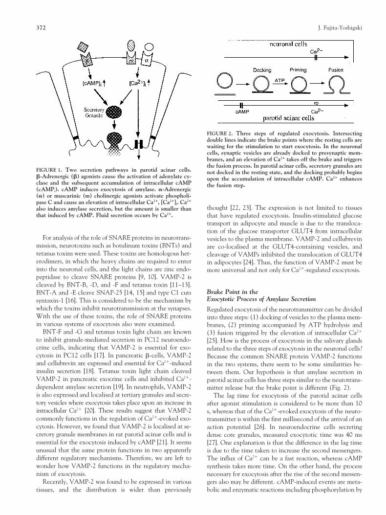
INTRODUCTION causes the secretion of proteins such as amylase and mucin.In contrast, parasympathetic stimulation activates phos-Most regulated exocytoses have been reported to be trig-pholipase C and causes the elevation of intracellular Ca21,gered by the elevation of intracellular Ca21. Neurotransmis-which leads to fluid secretion; that is, water and ion trans-sion at synapse, norepinephrine release from chromaffinport [3]. Whereas other exocytosis systems are mainly regu-cells, insulin release from pancreatic b-cells and amylase se-lated by Ca21 and are sometimes modulated by intracellularcretion from pancreatic exocrine cells are all commonly reg-cAMP [1, 2], protein secretion of the salivary glands can beulated by cytosolic free Ca21. However, cAMP-dependentsolely or mainly induced by cAMP [4]. Therefore, the ques-regulation also is present in some tissues—for example, hor-tion arises, are the salivary glands such special tissues?mone release of pituitary cells [1] and glucagon-induced in-
sulin release from b cells [2]. The exocytosis in the salivaryglands such as the parotid and submandibular glands also is SNARE Proteins in Various Systemsinduced by intracellular cAMP—reviewed in [3].
Various proteins that are essential for exocytosis in the neu-In the salivary glands, there are two secretory pathways:ronal cells have also been found to take part in other sys-protein exocytosis and fluid secretion (Fig. 1). Sympathetictems of vesicle transport. Common proteins have importantstimulation leads to the activation of adenylate cyclase androles for constitutive exocytosis and intracellular vesicleaccumulation of intracellular cAMP. The elevation of cAMPtransport conserved from yeast to mammalian cells [5, 6]. Inparticular, the so-called SNAP receptor (SNARE) proteinsfunction in the recognition between vesicles and target
*Author to whom all correspondence should be addressed. E-mail: yosigaki membranes [7, 8]. For example, the SNARE proteins in [email protected] cells are vesicle-associated membrane protein 2Abbreviations: SNARE–SNAP receptor; VAMP-2–vesicle-associated
membrane protein 2; SNAP-25–synaptosome-associated protein of 25 (VAMP-2) as vesicle SNAREs (v-SNAREs) and are syn-kDa; BNT–botulinum toxin; v-SNARE–vesicle SNARE; t-SNARE–target
taxin-1 and synaptosome-associated protein of 25 kDamembrane SNARE; PKA–cAMP-dependent protein kinase.Received 20 August 1997; and accepted 1 October 1997. (SNAP-25) as target membrane SNAREs (t-SNAREs).
372 J. Fujita-Yoshigaki
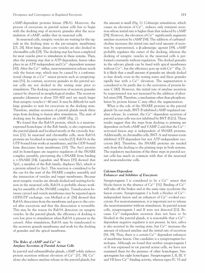
FIGURE 2. Three steps of regulated exocytosis. Intersectingdouble lines indicate the brake points where the resting cells arewaiting for the stimulation to start exocytosis. In the neuronalcells, synaptic vesicles are already docked to presynaptic mem-branes, and an elevation of Ca21 takes off the brake and triggersthe fusion process. In parotid acinar cells, secretory granules are
FIGURE 1. Two secretion pathways in parotid acinar cells. not docked in the resting state, and the docking probably beginsb-Adrenergic (b) agonists cause the activation of adenylate cy- upon the accumulation of intracellular cAMP. Ca21 enhancesclase and the subsequent accumulation of intracellular cAMP the fusion step.(cAMPi). cAMP induces exocytosis of amylase. a-Adrenergic(a) or muscarinic (m) cholinergic agonists activate phospholi-pase C and cause an elevation of intracellular Ca21, [Ca21]i. Ca21
thought [22, 23]. The expression is not limited to tissuesalso induces amylase secretion, but the amount is smaller thanthat induced by cAMP. Fluid secretion occurs by Ca21. that have regulated exocytosis. Insulin-stimulated glucose
transport in adipocyte and muscle is due to the transloca-tion of the glucose transporter GLUT4 from intracellular
For analysis of the role of SNARE proteins in neurotrans- vesicles to the plasma membrane. VAMP-2 and cellubrevinmission, neurotoxins such as botulinum toxins (BNTs) and are co-localised at the GLUT4-containing vesicles, andtetanus toxins were used. These toxins are homologous het- cleavage of VAMPs inhibited the translocation of GLUT4erodimers, in which the heavy chains are required to enter in adipocytes [24]. Thus, the function of VAMP-2 must beinto the neuronal cells, and the light chains are zinc endo- more universal and not only for Ca21-regulated exocytosis.peptidase to cleave SNARE proteins [9, 10]. VAMP-2 iscleaved by BNT-B, -D, and -F and tetanus toxin [11–13].
Brake Point in theBNT-A and -E cleave SNAP-25 [14, 15] and type C1 cutsExocytotic Process of Amylase Secretionsyntaxin-1 [16]. This is considered to be the mechanism by
which the toxins inhibit neurotransmission at the synapses. Regulated exocytosis of the neurotransmitter can be dividedWith the use of these toxins, the role of SNARE proteins into three steps: (1) docking of vesicles to the plasma mem-in various systems of exocytosis also were examined. branes, (2) priming accompanied by ATP hydrolysis and
BNT-F and -G and tetanus toxin light chain are known (3) fusion triggered by the elevation of intracellular Ca21
to inhibit granule-mediated secretion in PC12 neuroendo- [25]. How is the process of exocytosis in the salivary glandscrine cells, indicating that VAMP-2 is essential for exo- related to the three steps of exocytosis in the neuronal cells?cytosis in PC12 cells [17]. In pancreatic b-cells, VAMP-2 Because the common SNARE protein VAMP-2 functionsand cellubrevin are expressed and essential for Ca21-induced in the two systems, there seem to be some similarities be-insulin secretion [18]. Tetanus toxin light chain cleaved tween them. Our hypothesis is that amylase secretion inVAMP-2 in pancreatic exocrine cells and inhibited Ca21- parotid acinar cells has three steps similar to the neurotrans-dependent amylase secretion [19]. In neutrophils, VAMP-2 mitter release but the brake point is different (Fig. 2).is also expressed and localised at tertiary granules and secre- The lag time for exocytosis of the parotid acinar cellstory vesicles where exocytosis takes place upon an increase in after agonist stimulation is considered to be more than 10intracellular Ca21 [20]. These results suggest that VAMP-2 s, whereas that of the Ca21-evoked exocytosis of the neuro-commonly functions in the regulation of Ca21-evoked exo- transmitter is within the first millisecond of the arrival of ancytosis. However, we found that VAMP-2 is localised at se- action potential [26]. In neuroendocrine cells secretingcretory granule membranes in rat parotid acinar cells and is dense core granules, measured exocytotic time was 40 msessential for the exocytosis induced by cAMP [21]. It seems [27]. One explanation is that the difference in the lag timeunusual that the same protein functions in two apparently is due to the time taken to increase the second messengers.different regulatory mechanisms. Therefore, we are left to The influx of Ca21 can be a fast reaction, whereas cAMPwonder how VAMP-2 functions in the regulatory mecha- synthesis takes more time. On the other hand, the processnism of exocytosis. necessary for exocytosis after the rise of the second messen-
Recently, VAMP-2 was found to be expressed in various gers also may be different. cAMP-induced events are meta-bolic and enzymatic reactions including phosphorylation bytissues, and the distribution is wider than previously
Divergence and Convergence in Regulated Exocytosis 373
cAMP-dependent proteins kinase (PKA). Moreover, the the amount is small (Fig. 1). Colinergic stimulation, whichcauses an elevation of Ca21, induces only transient secre-process of exocytosis in parotid acinar cells has to begin
with the docking step of secretory granules after the accu- tion whose initial rate is higher than that induced by cAMP[39]. However, the elevation of Ca21 significantly augmentsmulation of cAMP, unlike that in neuronal cells.
In neuronal cells, synaptic vesicles are waiting for the sig- amylase secretion by cAMP [38]. The addition of carbamyl-choline increases the initial rate and total amount of secre-nals in the docking state to the pre-synaptic membranes
[25, 28]. Most large, dense core vesicles are also docked in tion by isoproterenol, a b-adrenergic agonist [39]. cAMPprobably regulates the outset of the docking, whereas thechromaffin cells [29]. The docking step has been completed
for most vesicles prior to stimulation in these systems. Then, docking of synaptic vesicles in the neuronal cells is per-formed constantly without regulation. The docked granulesafter the priming step that is ATP-dependent, fusion takes
place in an ATP-independent and Ca21-dependent manner in the salivary glands can be fused with apical membraneswithout Ca21, but the efficiency goes up with Ca21 (Fig. 2).[30]. After the Ca21 influx, neurotransmission release needs
only the fusion step, which may be caused by a conforma- It is likely that a small amount of granules are already dockedto fuse slowly even in the resting states and these granulestional change in a Ca21 sensor protein such as synaptotag-
min [31]. In contrast, secretory granules in the parotid aci- rapidly fuse with a Ca21 elevation. The augmentation isconsidered to be partly due to the activation of protein ki-nar cells are not docked in the resting state prior to
stimulation. The docking construction of secretory granules nase C [40]. However, the initial rate of amylase secretionby isoproterenol was not increased by the addition of phor-cannot be observed in morphological studies. The secretory
granules (diameter is about 700–800 nm) are much larger bol esters [39]. Therefore, a mechanism other than phosphory-lation by protein kinase C may effect the augmentation.than synaptic vesicles (z40 nm). It may be difficult for such
large granules to wait for exocytosis in the docking state. What is the role of the SNARE proteins in the parotidglands? In our study, BNT-B inhibited cAMP-regulated am-Therefore, amylase secretion of parotid glands needs three
steps from docking to fusion after stimulation. The start of ylase release. In contrast, the Ca21-dependent secretion ofparotid acinar cells was not inhibited by BNT-B [21]. Thesedocking may be dependent on cAMP (Fig. 2).
We found that the Rab3A protein, which is a neurone- results suggest that the steps from docking to priming aredependent on both cAMP and VAMP-2 and that the Ca21-expressed homologue of the Rab family, is also expressed in
the parotid glands and localised mostly at the cytosolic frac- activated fusion step is independent of SNARE proteins.Additionally, in chromaffin cells, BNT-A and tetanus toxintion [21]. In neuronal and chromaffin cells, most Rab3A
proteins are localised at synaptic vesicles [32]. Rab3A in the inhibited ATP-dependent but not ATP-independent exo-cytosis [41]. Therefore, the SNARE proteins are neededGTP-bound form works at membranes, and the GDP-bound
form dissociates from membranes [33]. The Sec1 protein only from the docking to the priming steps in both systems.The regulatory mechanism of the three steps in parotid aci-and its homologues are negative regulators of the SNARE
complex assembly, preventing the t-SNARE from binding nar cells has much in common with that of the neuronaland neuroendocrine cells.a v-SNARE [34]. Lupashin and Waters [35] showed that
Ypt1, a member of the Rab family, displaces Sly1, which isa protein related to Sec1. This reaction is considered to be
Calcium-Dependentthe cue for the start of the SNARE complex assembly andEnhancer and Inhibitor of Exocytosisthe interaction of vesicles and target membranes. BecauseSynaptotagmin I is considered to be a Ca21 sensor thatmost synaptic vesicles are already docked and waiting for fu-blocks fusion in the absence of Ca21 [31]. Binding of Ca21sion in the neuronal cells, Rab3A is probably always work-will take off the brakes and at the same time accelerate theing for assembly of the SNARE complex. Translocation be-fusion events. Synaptotagmin I is both a blocker of Ca21-tween cytosol and vesicle membranes may be repeated uponindependent fusion and an enhancer of Ca21-evoked exo-GDP/GTP exchange. von Mollard et al. [36] showed thatcytosis. For neurotransmission, it is important not to releaseRab3A dissociates from the membrane and goes to the cyto-the neurotransmitter without stimulation. In parotid acinarsol after exocytosis and that the dissociation is reversible.cells, synaptotagmin I and II were not detected [21]. Be-This may be the reason for Rab3A detection on synapticcause Ca21-independent secretion does not have to bevesicles. In the parotid glands, the efficiency of docking isblocked in the parotid glands, it is reasonable that a Ca21-very low prior to stimulation when Rab3A is present in thedependent negative regulator is not present. In fact, salivacytosol. After stimulation, Rab3A may be translocated tois also secreted in the resting state, but Ca21 increases thethe secretory granule membranes and work for the dockingamount of released amylase and the initial rate of secretionof granules and the apical membrane.[38, 39]. Thus, there is a certain Ca21-dependent enhancerof membrane fusion. One candidate is a synaptotagmin ho-
The Roles of cAMP and Ca21 in mologue. Although we found that neither synaptotagmin IAmylase Secretion of Parotid Acinar Cells or II was expressed in rat parotid acinar cells, we have not
examined them for the presence of other homologues. Syn-In parotid and submandibular glands, cAMP solely inducesprotein secretion without elevation of Ca21 [37, 38]. Ca21 aptotagmin has eight homologues. Synaptotagmin I, II, III, V
and VII have Ca21-binding activity, whereas types IV, VI andalone also induces amylase release in the parotid glands, but
374 J. Fujita-Yoshigaki
VIII do not [42]. The role of each type is not yet clear, but oneof them may function in parotid acinar cells.
Another candidate is the calcium-dependent activator pro-tein for secretion (CAPS, p145). CAPS is reported to be a cy-tosolic protein expressed in brain, pituitary cells, chromaffinand pancreas, all of which have Ca21-dependent secretion sys-tems [43]. Hay and Martin [30] reported that CAPS takes partin the Ca21-dependent fusion of norepinephrine release inchromaffin cells but not in the Mg·ATP-dependent steps.There is a possibility that common CAPS functions in bothchromaffin and parotid acinar cells. In PC12 cells, the in-hibitory efficiency of an antibody against CAPS is about
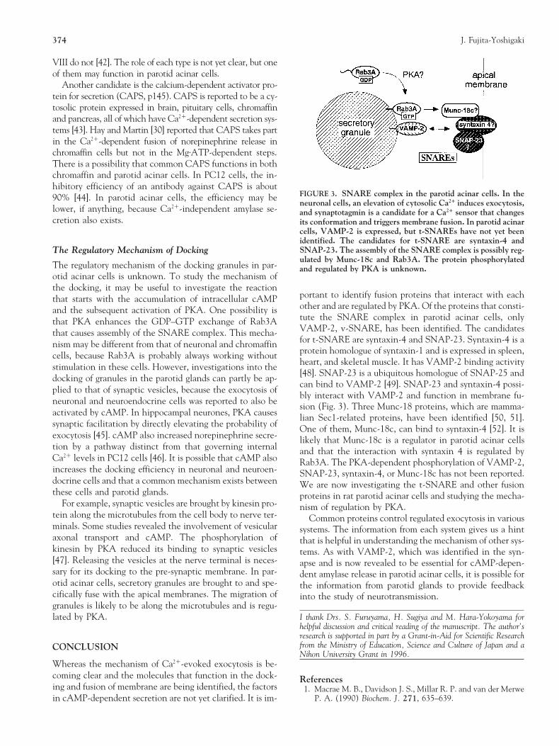
FIGURE 3. SNARE complex in the parotid acinar cells. In the90% [44]. In parotid acinar cells, the efficiency may beneuronal cells, an elevation of cytosolic Ca21 induces exocytosis,
lower, if anything, because Ca21-independent amylase se- and synaptotagmin is a candidate for a Ca21 sensor that changescretion also exists. its conformation and triggers membrane fusion. In parotid acinar
cells, VAMP-2 is expressed, but t-SNAREs have not yet beenidentified. The candidates for t-SNARE are syntaxin-4 andSNAP-23. The assembly of the SNARE complex is possibly reg-The Regulatory Mechanism of Dockingulated by Munc-18c and Rab3A. The protein phosphorylated
The regulatory mechanism of the docking granules in par- and regulated by PKA is unknown.otid acinar cells is unknown. To study the mechanism ofthe docking, it may be useful to investigate the reaction
portant to identify fusion proteins that interact with eachthat starts with the accumulation of intracellular cAMPother and are regulated by PKA. Of the proteins that consti-and the subsequent activation of PKA. One possibility istute the SNARE complex in parotid acinar cells, onlythat PKA enhances the GDP–GTP exchange of Rab3AVAMP-2, v-SNARE, has been identified. The candidatesthat causes assembly of the SNARE complex. This mecha-for t-SNARE are syntaxin-4 and SNAP-23. Syntaxin-4 is anism may be different from that of neuronal and chromaffinprotein homologue of syntaxin-1 and is expressed in spleen,cells, because Rab3A is probably always working withoutheart, and skeletal muscle. It has VAMP-2 binding activitystimulation in these cells. However, investigations into the[48]. SNAP-23 is a ubiquitous homologue of SNAP-25 anddocking of granules in the parotid glands can partly be ap-can bind to VAMP-2 [49]. SNAP-23 and syntaxin-4 possi-plied to that of synaptic vesicles, because the exocytosis ofbly interact with VAMP-2 and function in membrane fu-
neuronal and neuroendocrine cells was reported to also be sion (Fig. 3). Three Munc-18 proteins, which are mamma-activated by cAMP. In hippocampal neurones, PKA causes lian Sec1-related proteins, have been identified [50, 51].synaptic facilitation by directly elevating the probability of One of them, Munc-18c, can bind to syntaxin-4 [52]. It isexocytosis [45]. cAMP also increased norepinephrine secre- likely that Munc-18c is a regulator in parotid acinar cellstion by a pathway distinct from that governing internal and that the interaction with syntaxin 4 is regulated byCa21 levels in PC12 cells [46]. It is possible that cAMP also Rab3A. The PKA-dependent phosphorylation of VAMP-2,increases the docking efficiency in neuronal and neuroen- SNAP-23, syntaxin-4, or Munc-18c has not been reported.docrine cells and that a common mechanism exists between We are now investigating the t-SNARE and other fusionthese cells and parotid glands. proteins in rat parotid acinar cells and studying the mecha-
For example, synaptic vesicles are brought by kinesin pro- nism of regulation by PKA.tein along the microtubules from the cell body to nerve ter- Common proteins control regulated exocytosis in variousminals. Some studies revealed the involvement of vesicular systems. The information from each system gives us a hintaxonal transport and cAMP. The phosphorylation of that is helpful in understanding the mechanism of other sys-kinesin by PKA reduced its binding to synaptic vesicles tems. As with VAMP-2, which was identified in the syn-[47]. Releasing the vesicles at the nerve terminal is neces- apse and is now revealed to be essential for cAMP-depen-sary for its docking to the pre-synaptic membrane. In par- dent amylase release in parotid acinar cells, it is possible forotid acinar cells, secretory granules are brought to and spe- the information from parotid glands to provide feedbackcifically fuse with the apical membranes. The migration of into the study of neurotransmission.granules is likely to be along the microtubules and is regu-
I thank Drs. S. Furuyama, H. Sugiya and M. Hara-Yokoyama forlated by PKA.helpful discussion and critical reading of the manuscript. The author’sresearch is supported in part by a Grant-in-Aid for Scientific Researchfrom the Ministry of Education, Science and Culture of Japan and aCONCLUSIONNihon University Grant in 1996.
Whereas the mechanism of Ca21-evoked exocytosis is be-coming clear and the molecules that function in the dock- Referencesing and fusion of membrane are being identified, the factors 1. Macrae M. B., Davidson J. S., Millar R. P. and van der Merwe
P. A. (1990) Biochem. J. 271, 635–639.in cAMP-dependent secretion are not yet clarified. It is im-
Divergence and Convergence in Regulated Exocytosis 375
2. Ammala C., Aschcroft F. M. and Rorsman P. (1993) Nature 25. Sudhof T. C. (1995) Nature 375, 645–653.26. Almers W. (1994) Nature 367, 682–683.363, 356–358.
3. Quissell D. O. (1993) Stimulus-exocytosis coupling mecha- 27. Thomas P., Wong J. G., Lee A. K. and Almers W. (1993)Neuron 11, 93–104.nism in salivary gland cells. In: Biology of the Salivary Glands
(Dobrosielski-Vergona K., Ed), pp. 181–200. CRC Press, Boca 28. Goda Y. (1997) Proc. Natl. Acad. Sci. USA 94, 769–772.Raton. 29. Banerjee A., Barry V. A., DasGupta B. R. and Martin T. F. J.
4. Takuma T. and Ichida T. (1986) Biochim. Biophys. Acta 887, (1996) J. Biol. Chem. 271, 20223–20226.113–117. 30. Hay J. C. and Martin T.F.J. (1992) J. Cell Biol. 119, 139–151.
5. Bennett M. K. and Scheller R. H. (1993) Proc. Natl. Acad. 31. DeBello W. M., Betz H. and Augustine G. J. (1993) Cell 74,Sci. USA 90, 2559–2563. 947–950.
6. Ferro-Novick S. and Jahn R. (1994) Nature 370, 191–193. 32. von Mollard G. F., Mignery G. A., Baumert M., Perin M. S.,7. Sollner T., Bennet M. K., Whiteheart S. W., Scheller R. H. Hanson T. J., Burger P. M., Jahn R. and Sudhof T. C. (1990)
and Rothman, J. E. (1993) Cell 75, 409–418. Proc. Natl. Acad. Sci. U.S.A. 87, 1988–1992.8. Sollner T., Whiteheart S. W., Brunner M., Erdjument-Bro- 33. Araki S., Kikuchi A., Hata Y., Isomura M. and Takai Y.
mage H., Geromanos S., Tempst P. and Rothman, J. E. (1993) (1990) J. Biol. Chem. 265, 13007–13015.Nature 362, 319–323. 34. Pevsner J., Hsu S. C., Braun J. E., Calakos N., Ting A. E., Be-
9. Sudhof T. C., De Camilli P., Niemann H. and Jahn, R. (1993) nnett M. K. and Scheller R. H. (1994) Neuron 13, 353.Cell 75, 1–4. 35. Lupashin V. V. and Waters M. G. (1997) Nature 276, 1255–
10. Montecucco C. and Schiavo, G. (1995) Q. Rev. Biophys. 28, 1258.423–472. 36. von Mollard G. F., Sudhof T. C. and Jahn R. (1991) Nature
11. Schiavo G., Benfenati F., Poulain B., Rossetto O., de Laureto 349, 79–81.P. P., DasGupta B. R. and Montecucco C. (1992) Nature 359, 37. Takemura H. (1985) Biochem. Biophys. Res. Commun. 131,832–835. 1048–1055.
12. Link E., Edelmann L., Chou J. H., Binz T., Yamasaki S., Eisel 38. Baldys-Waglegorska A., Pour A., Moriarty C. M. and DowdU., Baumert M., Sudhof T. C., Niemann H. and Jahn R. F. (1987) Biochim. Biophys. Acta 929, 190–196.(1992) Biochem. Biophys. Res. Commun. 189, 1017–1023. 39. Yoshimura K. and Nezu E. (1992) Biochem. Pharmacol. 43,
13. Schiavo G., Shone C. C., Rossetto O., Alexander F. C. G. 1031–1041.and Montecucco C. (1993) J. Biol. Chem. 268, 11516–11519. 40. Moller K., Benz D., Perrin D., and Soling H. D. (1996) Bio-14. Blasi J., Chapman E. R., Link E., Binz T., Yamasaki S., De chem. J. 314, 181–187.Camilli P., Sudohof T. C., Niemann H. and Jahn R. (1993) 41. Lawrence G. W., Weller U. and Dolly O. (1994) Eur. J. Bio-Nature 365, 160–163. chem. 222, 325–333.15. Binz T., Blasi J., Yamasaki S., Baumeister A., Link E., Sudhof 42. Li C., Ullrich B., Zhang J. Z., Anderson R. G. W., Brose N.T. C., Jahn R. and Niemann H. (1994) J. Biol. Chem. 269,
and Sudhof T. C. (1995) Nature 375, 594–599.1617–1620.43. Walent J. H., Porter B. W. and Martin T. F. J. (1992) Cell 70,16. Blasi J., Chapman E. R., Yamasaki S., Binz T., Niemann H.
765–775.and Jahn R. (1993) EMBO J. 12, 4821–4828.44. Martin T. F. J. and Kowalchyk J. A. (1997) J. Biol. Chem.17. Papini E., Rossetto O., and Cutler D. F. (1995) J. Biol. Chem.
272, 14447–14453.270, 1332–1336.45. Trudeau L.-E., Emery D. G. and Haydon P. G. (1996) Neuron18. Regazzi R., Wollheim C. B., Lang J., Montecucco C., Sadoul
17, 789–797.K., Weller U., Palmer M., and Thorens B. (1995) EMBO J.46. Martin P. T. and Koshiland J., D. E. (1992) Proc. Natl. Acad.14, 2723–2730.
Sci. USA 89, 10257–10261.19. Gaisano H. Y., Sheu L., Foskett J. K. and Trimble W. S.47. Sato-Yoshitake R., Yorifuji H., Inagaki M. and Hirokawa N.(1994) J. Biol. Chem. 269, 17062–17066.
(1992) J. Biol. Chem. 267, 23930–23936.20. Brumell J. H., Volchuk A., Sengelov H., Borregaard N., Ciu-48. Bennett M. K., Garcia-Arraras J. E., Elferink L. A., Petersontat A.-M., Bainton D. F., Grinstein S. and Klip A. (1995) J.
K., Fleming A. M., Hazuka C. D. and Scheller R. H. (1993)Immunol. 155, 5750–5759.Cell 74, 863–873.21. Fujita-Yoshigaki J., Dohke Y., Hara-Yokoyama M., Kamata Y.,
49. Ravichandran V., Chawla A, and Roche P. A. (1996) J. Biol.Kozaki S., Furuyama S. and Sugiya H. (1996) J. Biol. Chem.Chem. 271, 13300–13303.271, 13130–13134.
50. Hata Y., Slaughter C. A. and Sudhof T. C. (1993) Nature22. Ralston E., Beushausen S. and Ploug T. (1994) J. Biol. Chem.366, 347–351.269, 15403–15406.
51. Tellam J. T., McIntosh S. and James D. E. (1995) J. Biol.23. Rossetto O., Gorza L., Schiavo G., Schiavo N., Scheller R. H.Chem. 270, 5857–5863.and Montecucco C. (1996) J. Cell Biol. 132, 167–179.
52. Tellam J. T., Macaulay S. L., McIntosh S., Hewish D. R.,24. Tamori Y., Hashiramoto M., Araki S., Kamata Y., TakahashiWard C. W. and James D. E. (1997) J. Biol. Chem. 272,M., Kozaki S. and Kasuga M. (1996) Biochem. Biophys. Res.
Commun. 220, 740–745. 6179–6186.